JOURNAL OF THE INDIAN MEDICAL ASSOCIATION, VOL 118, NO 06, JUNE 2020
Case Report An Atypical Manifestation of Post Streptococcal Glomerulonephritis Uddalak Chakraborty1, Tarun Kumar Paria2, Purbasha Biswas2, Tanuka Mandal3, Atanu Chandra4 Acute post streptococcal glomerulonephritis is a relatively common entity encountered by the internist in day to day practice. Aside the relatively common presentation of hematuria, oliguria and facial puffiness, one may present with convulsions and hypertensive encephalopathy. A 19 year old male patient was admitted with generalized tonic clonic seizure and elevated blood pressure, with some papular rash in different stages of healing in both lower limbs, MRI brain revealed bilateral symmetrical hyperintensities in frontal and parieto-occipital areas which resembled posterior reversible encephalopathy syndrome (PRES) like changes. The evaluation of hypertension in the young boy revealed microscopic hematuria and nephritic range proteinuria with decreased serum complements suggestive of acute glomerulonephritis. Symptomatic management ensured a complete recovery without any neurological deficit and resolution of MRI findings over a week. PRES is a rare clinico-radiological entity and may be a presenting symptom in patients with post streptococcal glomerulonephritis. [J Indian Med Assoc 2020; 118(6): 58-9]
Key words : Post-streptococcal glomerulonephritis; PRES; hypertension; convulsions.
A
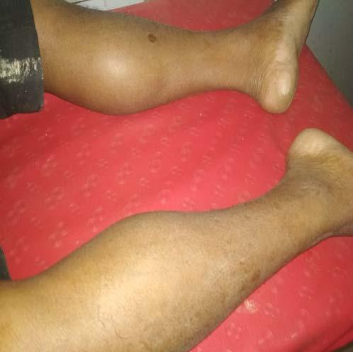
cute post streptococcal glomerulonephritis (PSGN) is a nephritic syndrome which may present commonly with oliguria, cola colored urine, puffiness of face and eyelids, pedal edema, and hypertension. Apart from these classical symptoms, PSGN may present with atypical manifestations like myocardial dysfunction, acute renal failure, etc. Reversible encephalopathy due to dysfunction of cerebral autoregulation has been reported in children and juvenile patients with PSGN1. An adult patient presenting with reversible encephalopathy and posterior reversible encephalopathy syndrome like changes in MRI brain due to PSGN is relatively rare. CASE REPORT A 19 year old male patient presented to us in September, 2019 with his first episode of generalized tonic clonic seizure preceded by headache and nausea. He had a blood pressure of 160/100mmHg and complained of persisting headache and nausea, followed by which he again suffered another episode of generalized tonic clonic convulsion. Clinical examination revealed only a papular rash in both lower limbs in different stages of healing, which was accompanied by some pustular discharge from the rash 2
Editor's Comment :
PSGN may present with hypertensive encephalopathy apart from the common symptoms of nephritic syndrome. PRES is a characteristic finding in brain imaging usually associated with hypertensive emergencies due to failure of cerebral autoregulation. PRES as a presentation of PSGN though seen commonly in children, may also be seen in young adults as in our case.
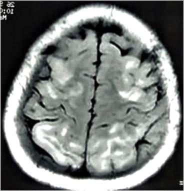
weeks prior to the current episode. Systemic examination did not reveal any abnormality. A subsequent MRI brain revealed bilateral symmetrical hyperintensities in FLAIR (fluid attenuation inversion recovery) sequence in frontal and parieto-occipital areas (Figs 1,2), which predominantly involved the white matter. Further evaluation of hypertension in such a young male patient revealed microscopic hematuria with 8-9 RBC/ hpf with few dysmorphic RBC and pus cells 2-3/hpf with trace albuminuria in routine urinalysis, with negative cultures from urine. Urine output was around 700ml in 24 hours. An urinary albumin-creatinine ratio revealed a nephritic range proteinuria with 1685 mg/mcg (n=30-300mg/mcg). Serum complements revealed a diminished C3 level around 26mg/dl(n= 66-185mg/dl)with a C4 level in the normal range. Ultrasonographic imaging of kidneys did not reveal any abnormality. ASO titre was found to be raised around 600 units/ml (n<200 units/ml) and Anti DNAse B levels were raised as well around 300 units/ml (n<85 units/ml). Renal function tests were unaltered. All other routine investigations were non-contributory. The patient was admitted with a hypertensive emergency and characteristic signal changes in brain imaging. On an
1 MBBS, Postgraduate trainee, Department of General Medicine, RG Kar Medical College and Hospital, Kolkata and Corresponding Author 2 MBBS, Post graduate trainee, Department of General Medicine, RG Kar Medical College and Hospital, Kolkata 3 MD, MACP, Senior Resident, Dept of Medicine, RG Kar Medical College Hospital, Kolkata 4 MBBS, Assistant Professor, Department of General Medicine, RG Kar Medical College and Hospital, Kolkata Received on : 05/06/2020 Accepted on : 15/06/2020
58
