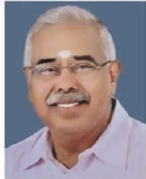
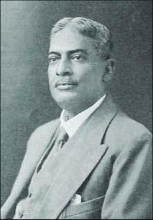
Dr. Gouri Pada Dutta, Past Hony. Editor of Journal of the Indian Medical Association (JIMA) breathed his last at 11.15 pm on 8th June 2020 at the age of 93 years at his Jadavpur residence. He lost his wife in the year 2012 and his elder daughter earlier. He was survived by his younger daughter and grandchildren of both the daughters.
In Memorium
Dr. Gouri Pada Dutta
He was born in Jaipur village of Bankura District of West Bengal on 3rd March, 1928. He passed the matriculation (10th standard) from Jaipur, then he came to Kolkata and studied at St. Paul’s school. He completed his MBBS form Chittaranjan National Medical College and later on completed his MD. At that time Chittaranjan National Medical College used to be a private hospital. He led an agitation to make it a Government Medical College and ultimately in the year 1967 the State Government undertook the hospital. Probably, this was the biggest achievement of his life.
He organized the first ever “Assembly of Editors of Medical Journals’ in August 24-25th, 1985. He was contemporary of another legend of Indian Medical Association, Dr. P.K. Chaudhuri, who was the then the Hony. Secretary of JIMA. He led from the front for the upliftment of Public Health in the State. He was the Chairman of the Permanent Standing Committee for Health & Family Welfare of the West Bengal Legislative Assembly for long. Many political leaders in Kolkata expressed their deep sorrow on his demise.
He was influenced by Marxism and participated the left movement from his very student life. He also led various Farmer, political and social movements. He was jailed for that too. He was associated with various social activities. He served as an Honorary Vice Chancellor in Chittaranjan Seba Sadan for a long period. He was an active member of Indian Medical Association and served the association in various capacities. He was the Editor of the Journal of Indian Medical Association for the year 1985-1988. He was the member of Medical Council of India for a very long period. He was a member of the Rural Health Standing Committee also. He worked as the advisor from time to time in various projects of WHO and UNICEF. He was the pioneer in establishing the Association of Health Service Doctors in the State. He was the founder President of Public Health Kolkata. He was the member of Senate and Syndicate of Calcutta University. He was associated with various institutions including South Calcutta Girls College.
He consecutively fought and won the elections in the years 1987, 1991 and 1996 and became Member of Legislative Assembly of West Bengal. He served as Protem Speaker in the Assembly from time to time.
He published over 550 research papers and books. He was a poet too and published a few books. His autobiography “kabe ami bahir holem” is very much noteworthy.
Prof. (Dr.) Jyotirmoy Pal MD, FRCP, FRCP, FICP, FACP, WHO Fellow, Hony. Editor, JIMA
Pandemics have always had a great influence in shaping human history and politics. From Justinian Plague in sixth century to Influenzae pandemic in twentieth century, pandemics have triggered fall of empires , created social restructuring, changed the demography – leading to huge socio-economic and cultural impact. How Pandemics Changed History :
JustinianPlague (541AD–750AD)— It started in Sixth Century in Egypt and spread to Constantinople. The plague killed 25-100 million people and weakened the Empire Justinian substantially. This destroyed the dream to reunite Roman Empire leading to the beginning of the Dark Age of Europe.
Black Death (1347-1351) — Bubonic Plague spread throughout Europe and killed more than 75 million people. At end of the pandemic, survivors’ standard of living increased, workers had more work, and social mobility increased as resource was more compared to population. Walter Scheidel had narrated in his book “The Great Leveler”- how black death led to improved wages for labourers. People’s faith on Catholic Church was lost, as the Church failed to save people when the pandemic spread like wildfire. Religious dominance on the society was put under question. Jews were blamed for spreading Disease and were burnt alive in many parts of Country. Frank M Snowden in his book- “Epidemics and Society: From the Black Death to the Present” described how a pandemic can change outlook of a society.
Small Pox (15th -17th Century) — The Europeans invaded America in 15th Century. They introduced diseases to this continent against which natives had no immunity, one of these was Smallpox. During this period Small Pox killed approximately 90% of the population of America. In 1520, the Aztec Empire was destroyed by smallpox as it was incapacitated to resist Spanish invasion. The Pandemic helped Europeans to colonize in the newly vacated area thereby altering the history of America.
Cholera(1817-23)— The Cholera pandemic started in Jessore (of Bengal Province), gradually spreading to the entire country. The British Soldiers then carried it to England and further to America. Initially, the pandemic was believed to be a Curse of God, which was thought to be spread by foul air ‘Miasma’. However, John Snow analysed hospital records and found a popular city well to be the cause of outbreak. Thus, the concept of Sanitation and Public health System was born.
SpanishFlu(1918)— 500 million people were infected and 50 million died by the Spanish Flu. One of the major impacts of Pandemic was on the First World War. Germans and Austrians were so badly affected that they lost all their aggressiveness. German General Erich Ludendorff had written in “My War Memories” that the Flu was one of the main reasons for Germany’s Defeat. As the countries devastated in the War had failed to develop a structured protocol, the impact proved to be costly. However, understanding of the Pandemic helped to formulate better Public Health Measures. Use of Masks, Quarantine and Social Distancing were a few key measures adopted during this pandemic. The practices followed presently are basically a product of the experience of Spanish Flu Pandemic.
All pandemics start as biological phenomenon, but keep footprint on Economical, social and political field. Equation of power may be shifted, economy may be remodelled, significant changes in the way we touch, behave and breath.
Response to Pandemic :
Presently, the world is under the siege of Novel Coronavirus, which has declared a War against Humanity. The World Health Organisation has adopted age old measures like masks, lockdown and social distancing, due to lack of effective antiviral drug or vaccine. Social distancing means keeping space between yourself and other people. It is a non-pharmaceutical measure to prevent spread of infection to others. Concept of social
distancing has its origin back to 5th Century BC. It was successfully implemented at St Louis -1918 during the Flu Pandemic which resulted in significantly lesser mortalities than Philadelphia. CDC describes “social distancing as a set of methods for reducing frequency and closeness of contact between people in order to decrease the risk of transmission of disease”.
Socio economic Impact of Pandemic :
Just as in the past, this Pandemic has also brought upon serious Socio economic and Political Impact. The European Union and USA has suffered significantly larger amount of economic losses in comparison to the rest of the countries. Economic losses can be compared with that of Great recession of 2008 or Great depression in 1930. At least thirty million Americans have fled for Unemployment in past few months. Worldwide, the low income group individuals are mostly affected. Overcrowding, lower immunity due to malnutrition and poor elderly care has made these people more vulnerable to infection with a higher mortality. The outbreak of COVID-19 has resulted in an unprecedented number unemployment which reached 20-40% in different countries. Social distancing has decreased the mobility of individuals resulting in negative impact on the production sector.
Besides the Economic impact in India, this Pandemic is going to change the culture and politics in India. According to the beliefs in India, the Sense of Identity never dies in human society; it only mutates like a virus and changes its form. Social distancing may become a long term strategy for human survival and may finally transform into a social mode of Indian Life. Social distancing and Untouchability has been moving in India Horizontal level, if not vertically as was before. This will divide the society based on profession rather than Caste-ism. In India the term “Social distancing” was equivalent to untouchability – century long curse of caste system ‘Varnashram’. Although, social distancing is now being used as a Medical term, but there is a chance of misuse and resurgence of old practice of untouchability again in India.
“Untouchability is a blot on humanity “
— Mahatma Gandhi
Development of Caste System and Untouchability in India :
History of social origin of Caste system is from the period of Aryans. Rigveda was oldest one who described the origin of Varna System. Origin of Brahmin, Kshatriya, Vaishya, Sudra were from different part of God Brahma, none being superior or inferior to
the others. Subsequently origin of self or caste was accepted according to Guna (quality) and Karma (action) not by birth.
(Chaturvarna-Brahmin,Kshatriya,Vaishya,Sudra was classified on the basis of karma and guna.)
In Mahabharata we see the transition. Sri Krishna born as Jadav (baishya) but elevated to Kshatriya. Bhishma being Kshatriya was esteemed by Dronacharya to a Brahmin. On the contrary, the tragic hero Karna was denied of being a Kshatriya, as he was known to be a son of charioteer (sutaputra). But in later part this Caste System in India was perceived as division according to labour. Higher the position in Caste System had lesser role in physical labour and production but had greater control on wealth. So, Nonproductive work became symbol of Purity and Productive work had become the Symbol of Impurity. For better enjoyment of life with less labour, Karma based caste division was gradually converted into caste system based on birth. This was the beginning of pollution of Caste system in India and development of Untouchability.
In Colonial India, the British Government never tried to remove the Caste System, rather patronized to implement its popular divide and rule policy. It was Mahatma Gandhi and Dr B. R. Ambedkar who took up the work of redeeming untouchability. In Independent India, many laws were formulated to safeguard and protect lower castes by empowering them to some extent reduce untouchability - a curse of mankind. COVID-19 Situation in India – response from Society :
COVID-19 pandemic has thrown a new challenge to India. The virus has already spread by aerosol and fomites infecting more than 80 lakh people across the globe.
Doctors, nurses, cleaners, laboratory technicians, police personnel are the frontline warriors. Moreover, labourers who are working in agriculture sector and production sector, keeping our supply lines of food, transport and other demands intact have no less contribution in this war.
Doctors, nurses, care givers and paramedics around the world are facing an unprecedented workload in overstretched health facilities with no end in sight. They are working in stressful and frightening environments, not just because the virus is very little understood, but
because in most settings they are under-protected, overworked and themselves vulnerable to infection. In this connection, I would like to mention about a keen young medico who travelled more than 2200 km by road from Pune to Kolkata, braving fears of infection and restrictions to movement during the country-wide lockdown, only to be with the people of his home stateWest Bengal, in the time of distress.
Tributes to healthcare workers are pouring in from around the world amid the COVID-19 pandemic, as the world gives medical heroes a standing ovation from windows and balconies. Blowing of Conch shells, ringing bells and cheering to show solidarity with the Heath Care Workers for their laudable work to battle COVID-19 was done all around India while on the other side there has also been reports of Physical Violence against doctors and nurses in parts of the same country.
While doing screening work, a team of doctors in a locality in Indore were attacked by a mob .One of the doctors who was injured, Dr. Zakiya Sayed told “We were doing our normal rounds to Screen suspected COVID-19 infected cases. We never thought we would be attacked.. I am injured but not scared.”
In Delhi, doctors working at AIIMS were evicted from their apartments by their landlords and the matter had to be taken up by the Home Ministry and police. In other cases, according to some reports, landlords and neighbours became hostile to the doctors, forcing some to stay back at hospitals or find refuge in friends’ homes. Doctors have been subjected to harassment from various quarters. A young doctor returning home from her night duty was abused and slapped by Telangana police for violating the lockdown, soon after it was imposed on March 24. The stigma does not go even after death, as the healthcare personnels who took bodies of two COVID-19 doctors for cremation were attacked by the local people. Local residents, fearing the spread of the virus, protested and even threw stones at the ambulance. When the fear of infections is high among doctors, the public too will be scared and this is the new pandemic
“While healthcare service personnel are duty bound to serve without discrimination, the cooperation and support from society is a fundamental need for them to perform their duties with confidence,” the ministry had said.
Thanks to leadership of Indian Medical Association who motivated Govt of India to bring Ordinance on Violence of Doctors. Dr R V Ashokan Hony Secretary General, IMA proudly described them as “ Unsung Heros of India’s Corona War .......Write their history
now .”
On the contrary, there is inhibition on part of a section of doctors to be involved in CORONA care programme, even resident Doctors hold agitation against acquisition of hospitals for COVID-19 care. Fear of being infected by the Novel Corona virus became bigger than virus itself.
So we see another face of Pandemic – Fear psychosisanduntouchability.
Migrant workers who constitute 50% of urban population, faced serious job and livelihood crisis owing to COVID 19 pandemic. This Pandemic saw one of the biggest streams of mass return migrant workers in the country. When migrant workers flee from city, they not only lose their livelihood but increase the possibility of carrying the infections to their native places. So initial phase of Lockdown faced food, shelter problem, while returning home battled against stigma and bias in their own village, because residents suspect that they are carrier of Corona virus. Families are singled out, sneered at and harassed by villagers. As most of the workers belongs to lower caste, again caste slurs were hurled at them. Movement of certain ethnic groups are severely restricted.
Xenophobia, racism, hatred, as presumed to be carrier of disease is often directed to certain group worldwide – may be based on religion, race, caste, skin colour or ethnicity. Trump in USA, Orban in Hungary, Savini in Italy all asserted migrant workers’ linkage to spreading of disease. Popular belief that the migrant workers are potential source of infection exacerbate stigmatization and untouchability.
So, we see another face of Pandemic –untouchability
Stigma is hard to undo. Stigmatic suspicion can lead to development of new ethnic profile in country. Steven Vertovec, Director Max Planck Institute for the Study of Religious and Ethnic Diversity called ‘It is a danger to development and calls for countermeasures’.
Another unfortunate angle is tagging the origin and spread of this disease to a particular religious group. San Brownback, the US ambassador for international religious Freedom advised to pull back rising incidents to blame religious minorities. Pandemic does not see race, colour, caste, religion, language or border. Scapegoating, discrimination, repression among minorities exposed many of them in greater crisis than disease itself. Pandemic anxiety has manifested in bigotry and prejudice against minorities who have been blamed for spreading the virus. Few people may be undisciplined, never the whole community. Our response and conduct should be primarily towards
unity and brotherhood.
This is again another face of Pandemic –Untouchability
There are several instances where residents are blocking, protesting against proposed hospitals designated for COVID or suspect treatment. In a densely populated country like India it is difficult to get a place away for human settlement, even if found it would be difficult to prepare logistics and find necessary human resources to maintain the same. Problem is culminated as COVID is basically an urban disease, where overcrowding is always a problem. Lack of awareness regarding disease epidemiology, dynamics is the key issue about this strange attitude, where we forget our beloved one may be infected in next day.
So, again India may heading towards being divided into two groups – Touchable and Untouchable. Touchable sections are those who ‘have ‘, can afford daily living during lockdown period, can maintain social distancing and restrict mobility. Untouchable section are either poorer segment of the society or service provider to this disease. Poorer section cannot afford costly lockdown for prolonged period of time (in spite of getting partial help from Government). They have lesser amenities for segregation and social distancing and thereby being suspected as carriers of the disease. Service providers like Doctors, nurses, laboratory technicians, scavenger and sanitary workers are worst victims, as they presumed to be carriers of COVID-19. This untouchability in India will emerge not by virtue of birth but by virtue of profession. Stigmatization if not uprooted can give birth to a new caste system in India in coming days. Age old definition of Harijan may change. Health care workers may be defined as Harijan in Post Pandemic era. Untouchability – touchability will be determined again on the basis of work of individual like period of Mahabharata. But difference will be that during that era, there was mutual respect for each other while there will only be hatred and avoidance in future.
The term Social Distancing which had been used scientifically, should not be dragged in a dark well to tarnish, to stigmatize a particular section who are only performing duty. Social distancing means physical distancing to keep two person physically at distance so that droplet infection can be prevented. As India have a long tradition of an ugly caste system, it is preferable to change to term social distancing to Physical Distancing. Otherwise there is chance of misinterpretation and misuse the term Social Distancing. We have to act before metastasis of this
social disease occurs or we cannot come out from Bhulbhulaiya ever.
We Doctors are devoted, taken oath of Hippocrates where we have sworn that inspite of all odds we will not stand still just like a soldier in battlefield who never thinks of his life, making victory his bird’s eye.
Doctors here like Arjuna in Bhagawat Gita- caught in moral dilemma. Where on one side ‘karma’ lies in treating so called isolated COVID patients, on other side beloved neighbours, childhood playmates, cousins who shared sorrows and emotions are now pushing you in corner forcing you even to leave the locality, tearing bondage in a fine morning with abusing language as if you are the sole carrier of disease. Although, the Doctors are perplexed but our saviour can be Lord Krishna.
(Wemustdetachourselffromresultofouraction, Karma is only Dharma to us.)
Bhagbat Gita 2nd Chapter (Sankha Jyog), Slok –47
Clinicians may not have complete control over situation, but we have to rise to perform our duties and service with equanimity. COVID 19 have exposed ugly fracture of our society, not only in terms of infrastructure and policy also attitude of society perhaps carrying the virus in latent phase. Pandemic only revitalized the virus from latent to dormant phase.
Though dusk is advancing as a lazy surprise
All musics have paused with signs divine
Though I have no companions in vast skies
Though fatigue is creeping in my chassis Doubtsarereverberatinginsilentpaean. All horizons are covered with obscurities
Still O’ my bird , O’ bird of mine's
Do not fold your wings , do not close eyes.
Dussamay- RabindranathTagore
Our Medical fraternity competent enough to make healthy India, whatever may be the hinderance.
Review Article
Managing COVID-19 in India – STRICT 3C
Shashank R Joshi1,Viraj Suvarna2
The COVID 19 epidemic is gaining momentum in recent times yet prevalence and case fatality rates are lower than many developed countries. Many factors have been postulated for this scenario however preparedness is the need of the hour for future expansion of disease burden.
India needs to continue to be STRICT and follow the 3Cs, viz., Social distancing, Test (screening), Retest (confirmatory), Isolation, Contact tracing and Treatment, with focus on Cleanliness, Containment, and Clusters.
[J Indian Med Assoc 2020; 118(6): 13-6]
Key words : Social distancing, cleanliness, containment, contact tracing.
On December 31, 2019 the WHO was informed of a cluster of cases, of an unusual nature, emanating from the wet (seafood) market of Huanan in Wuhan, Hubei province of China. It is a zoonotic virus, found in bats (who have an altered immune system and hence is not affected by the virus) and it jumped to humans either through a pangolin (scaly anteater) or snake, when the human ate these animals. The WHO decided to name it novel Corona virus 2019 or nCoV 2019, and as it was similar to, but different from the earlier SARS (Severe Acute Respiratory Syndrome) also caused by a Corona virus, they also called it SARS CoV-2. On January 30, 2020 the WHO called it a Public Health Emergency of International Concern (PHEIC), because of the way it was spreading like wild fire, initially in China, and then beyond China to neighbouring countries and eventually countries in Europe, the Middle East, India and also the US. On February 11, 2020 the WHO named the disease as COVID-19 or COronaVIrus Disease. And finally on March 11, 2020, the WHO changed the nomenclature from PHEIC to pandemic. In other words, ‘corona’tion of the epidemic began. As of now more than 7.77 million cases have been reported globally and close to 4.3lac lives have been lost.1,2
2President, Clinical Pharmacology, Eris Lifesciences, India
Received on : 13/06/2020
Accepted on : 18/06/2020
Editor's Comment :
Containment of the COVID 19 pandemic in our country will require strict vigilance and action. STRICT measures involving Social distancing, testing, isolation,contact tracing and treatment are the key to success.
• India is stubbornly refusing to go the way the worldisbeingravagedbyCOVID-19.Indiahasalower prevalence rate and low case fatality rate.When one compares this to a dense population of 1.3 billion, it is a wonder India is doing better than the US (where New York is the epicentre) where over 1.16 million deathshavebeenreportedandtheyhavethehighest number, viz., ~3.6 lakh cases in the world. What is it about India that has resulted in a better outcome? All kindsoftheorieshavebeenputforward.Researchers attheInternationalCenterforGeneticEngineeringand Biotechnology (ICGEB) reported, in a not yet peerreviewed pre-printed publication, that the virus strain in India is a mutant. The micro RNA mutation (hasmiR-27b) of the Spike or S protein apparently makes the virus less lethal in India.3 There is a map going aroundintheWhatsappUniversityshowingthatwhere malaria is endemic, the Corona virus prevalence is low and vice-versa, almost to suggest that somehow the malaria parasite/infection seems to impart a peculiarimmunitytowardsthenovelCoronavirus2019. The fact that India includes BCG vaccination, given onthedayofbirth,aspartofitsUniversalImmunisation Program(UIP)alsoseemstohaveequippeduswitha unique immunity to this virus. We also seem to have
ahighernumberofNaturalKillercells.Indiaisatropical country and the hot Indian summer is expected to be adeterrenttothisviruswhichseemstopreferacooler climate.We currently don’t know if the BCG vaccine and oral polio vaccine to our youth has protected us. Add to this, India is a young country in that most of our population is in the younger, working age-group, arguablylesssusceptibletotheseriouscomplications of COVID-19.
But all the above are conjectural, hypotheses at best that need to be tested. What is however known is that despite the Goldman Sachs employee making a comment (Thank God this happened in China and not India), India is continuing to stun all critics in the way India is managing COVID-19. Is India really a backward and poor country, 50 years behind the US, when in real time we are actually 9.5 to 10.5 hours ahead of the US? In the US, people have run out of Personal Protective Equipment (PPE), they are having to innovate and use one ventilator for 2 or even 4 patients, their doctors are exhausted, petrified and annoyed at the way the administration is still not able to protect them from contracting the virus and then dying of the disease. In comparison, India started implementing tough measures very early. The Janata curfew announced by the Prime Minister for a day (March 22) was only the prelude to a nation-wide complete (except for essential services) lock-down starting March 24,that continued till 8th June and upto 30th June in containment zones. Essentially this was done to break the chain of transmission and flatten the epidemic curve. Surprisingly, many Indians took this seriously, practising social distancing, personal (handwashing with soap and water), and public hygiene (Swach Abhiyan), and this has helped in stemming the spike in cases, alot though some spreader events did occur across India but it was followed by isolation, quarantining those exposed but not showing symptoms, and contact tracing. Relaxations in certain areas was undertaken to revive the sagging economy.
The WHO Director-General, Tedros Adhanom Ghebreyesus, keeps saying, “Test, Test, Test.” But in India, testing is being done only for those who are symptomatic and those who are contacts of cases or
those who have a relevant travel history or are health care workers. It is not cost-effective to test 1.3 billion Indians.. FevIR or infra-red thermal scanners are being used to detect elevated body temperature,but they may have limited utility especially with a large number of asymptomatic cases. But above all, it looks like Indians ‘detest’ being ‘tested’. Why? Because there is a social stigma attached to the fact that people have come to your residence to test you and God forbid, if you are Corona positive, then you will be isolated and your family members quarantined, and all your contacts will be traced, and if positive they will be isolated, but if they are negative they will be quarantined. Testing strategy has evolved in India and is a perfect example of public private partnership.
To understand how best to stop this epidemic in its tracks, one must understand epidemiology. For most medical students, this was a major part of the subject, Preventive & Social Medicine (PSM) or Community Medicine, and a textbook written by Park & Park, but it was a neglected subject and done only because one had to pass MBBS, then go on to specialise, and then super-specialise in the more exciting clinical disciplines. But now all have realised that Community Medicine and epidemiology are extremely important disciplines. Apparently there are four phases of an epidemic; the first phase is when cases are imported, the second phase is when transmission happens locally from one infected person to close contacts, phase 3 is when community transmission happens even without history of contact or relevant travel history, and phase 4 when it becomes a full blown epidemic. In a way it resembles a product lifecycle with a lag phase of slow growth, then the logarithmic or exponential growth, then a plateau, and finally a decline. India seems to be still somewhere between Phase 2 and Phase 3, but the exponential growth or spike in cases that people were fearing has still not happened. Why? Are we that resilient? Or is it that our unhygienic environments have already given us an immunity that makes it easier for us to not succumb to such deadly viruses? Having said this, let us re-examine the triad, viz., agent, host and environment.4,5
Agent :
The novel Corona virus 2019 is a positive sense RNA virus, and it belongs to the family of 7 beta Corona viruses, of which 4 cause a common cold, one caused SARS and one caused MERS (Middle East Respiratory Syndrome). From a case fatality rate (CFR), this virus has the least CFR (it varies from as low as 0.6% to as high as ~4%; much higher among the elderly as we are seeing in Italy), while SARS (bats, palm civet cats) had a CFR of 10% and MERS (bats, dromedary camel) has a CFR of 35%. While SARS started in 2002 and expired on July 5, 2003, MERS is still smouldering and in November 2019 the WHO reported 2494 cases and 858 deaths. It is so named as it looks like a crown (in the Spanish language, crown = Corona) due to the spikes emanating from the body. The virus spreads through droplets (heavy, micro) or through fomites, though airborne transmission (up to 180 cm) has been reported (hence the dictum keep at least 2 meters of 6 feet away from each other), fecooral, urine, and tears are also reported to carry the virus. It enters our upper respiratory tract either when we inhale or touch our face (mouth, eyes, nose – MEN) with droplets that are still alive on some surfaces. Coughing, sneezing and even talking can spread the virus as speech is forced expiration. This Spike or S1 protein is used by the virus to attach itself to the Angiotensin Converting Enzyme (ACE)-2 receptor in respiratory epithelial cells though they are also present in the heart, vascular endothelium, kidneys, endocrine pancreas, testes and liver. Thereafter, another Spike protein S2 fuses with the endosome in the cytoplasm and within this endosome (ERGIC or Endoplasmic Reticulum Golgi Intermediate Compartment) the virus replicates, assembles and gets released. Like any virus it is nothing but a bunch of genes in search of a living cell within which it multiplies. In other words, if it does not get such a viable cell, it will die. The more virulent is the virus, the faster will it die as it will kill humans and then kill itself if it does not get to another living human cell. It is extremely contagious with an R0 of about 2.3 to 3.2 (basic reproductive number), hence the need to break the chain of transmission through social distancing.6
Host & the Environment :
The Indian human being seems to be different, for the reasons alluded to earlier on the first page. Are we genetically resilient? Or has it do with a geneenvironment interaction? Indians are so used to getting infections, because of the unhygienic surroundings, that when confronted by this novel virus, our immune system is better able to manage the virus? Again, conjectural, hypothetical, speculation, and good to have discussions, especially nowadays when we are in this lockdown situation with nothing to do except intellectually masturbate using the electronic medium, be it social media, print media, or television. ICMR has done some mathematical modelling work (Dr. RR Gangakhedkar et al)7 which seems to suggest that port-of-entry-based entry screening of travellers with suggestive clinical features and from COVID-19 affected countries, would achieve modest delays in the introduction of the virus into the community. Once the virus achieves transmission within the community, quarantine of symptomatics may have a meaningful impact on disease burden. As a public health measure, health system and community preparedness would be critical to control any impending spread of COVID19 in the country. A mathematical modelling study done by Kucharski AJ et al,8 published in the Lancet, also concluded that COVID-19 transmission probably declined in Wuhan during late January 2020, coinciding with the introduction of travel control measures. India swung into action swiftly and curtailed all international and then national flights in March which has resulted in a considerable break in the chain of transmission. Our take is that rather than focusing on why Indians may be less susceptible to COVID-19 based on conjecture, let us focus instead on concrete measures taken by intelligent Indians to stem the rot and ensure it did not flare up. Commissioning a nationwide lockdown for months is a big decision, as it will mean a lot of economic loss and untold hardships for the poor migrant labourers/daily wage earners. But to save the country from what’s happening in the US and big five countries in Europe (Italy, Spain, UK, France, Germany), such measures had to be taken. Testing is happening but in a structured focused manner, despite
the fact that at times these workers face the brunt of a community forced to be locked down, and scared of what would happen if they turned out to be positive. Then of course tracing of contacts even though it is so difficult in such a large country where some people actually try to subvert the process and run away from healthcare workers who have come to save their lives and the lives of their families. Quarantining of the contacts is also being done, sometimes selfquarantining at home as well. Social media and other forms of media (print, TV) are being used extensively to promote social distancing, though sometimes it may not be possible for members of a family who stay in a one room tenement to be 6 feet away from each other, simply due to lack of space, e.g., slums in Dharavi. Despite all these problems, it is remarkable that India is continuing to shoulder the problem without giving up or giving into the hopelessness that seems to prevail elsewhere. In fact now doctors are thinking of what can be done with the people who have recovered and have protective antibodies (IgM, IgG) in their plasma. There is a paper published in JAMA on March 27 which showed that 5 serious COVID-19 patients were transfused with plasma of convalescent COVID-19 patients and they recovered.9,10
To conclude, India needs to continue to be STRICT and follow the 3Cs, viz., Social distancing, Test (screening), Re-test (confirmatory), Isolation, Contact tracing and Treatment, with focus on Cleanliness, Contain,and Clusters. Interestingly 80% of cases are being reported in 60% of districts, so it is not following the typical Vinifred Pareto principle (80/20). Clusters or hot spots continue to surface and then reactive measures are put in place. Perhaps we need to be proactive and ahead of the curve, anticipating where such clusters may emerge and then get into action, before it surfaces. Wuhan was locked down for 70 days. Our lockdown is being lifted in a staggered manner. But if we want to get back to work quickly, and prevent the further slide of our already precarious economy, we will need to continue to practise social distancing, personal and public hygiene. This is the new normal
we all will have to get sued to. Technology is being used in a big way to connect without contacting, “touching” (the emotional retina) without actually touching, and bridging distances bringing people closer together. Paradoxically we are all in this together and we need to face it, but we should not come together, face to face, as far as possible. Like the Sherry Turkle book on Social Media, “Alone Together”. To some extent we have now realised what it meant to be an untouchable.
2Phelan AL, Katz R, Gostin LO — Viewpoint. The novel Corona virus originating in Wuhan, China. Challenges for global health governance. JAMA Published online on Jan 30, 2020; E1-E2.
3Sardar R, Satish D, Birla S, Gupta D — Comparative analyses of SARS CoV-2 genomes from different geographical locations and other Corona virus family genomes reveals unique features potentially consequential to host-virus interaction and pathogenesis. https://doi.org/10.1101/ 2020.03.21.001586.
4Surico P, Galeotti A— The economics of a pandemic: the case of COVID-19. London Business School.
5Paules CI, Marston HD, Fauci AS. Corona virus infections –more than just the common cold. Viewpoint. JAMA published online Jan 23, 2020, E1-E2.
6Fineberg HV — Ten weeks to crush the curve. NEJM Editorial published on April 1, 2020 at NEJM.org. DOI: 10.1056/ NEJMe2007263.
7Mandal S, et al — Prudent public health intervention strategies to control the coronavirus disease 2019 transmission in India: a mathematical model-based approach. Ind J Med Res 2020, Epub ahead of print. DOI: 10.4103/ijmr.IJMR_504_20.
8Kucharski AJ et al. Early dynamics of transmission and control of COVID-19: a mathematical modelling study. Lancet Infect Dis 2020; published online March 11, 2020, https://doi.org/ 10.1016/S1473-3099(20)30144-4.
9Parmet WE, Sinha MS — COVID-19 the law and limits of quarantine. Perspective published on March 18, 2020 at NEJM.org.
10Swerdlow DL, Finelli L — Preparation of possible sustained transmission of 2019 novel Corona virus. Lessons from previous epidemics. Viewpoint. Published in JAMA online on Feb 11, 2020, E1-E2.
View of the Expert
The Covid-19 pandemic rages on in the World. With more than eight million people affected and more than 450 thousand dead, the end is still nowhere in sight. India is still in the ascending limb of the epidemic curve and physicians of India are at the forefront of this struggle to quell the scourge. At this juncture, we thought it worthwhile to listen to Prof Manish Soneja, one of the foremost infectious disease experts in India. He is currently working at AIIMS, New Delhi, one of the premier medical institutions of the country. In the first week of June, 2020, members of the Editorial Committee of JIMA held an online interview with Dr Soneja. The transcript of that interview is presented here, exclusively for the readers of JIMA.
Q
U E S T I O N N A I R E
1.AreyouusingsteroidsinCOVID-19patients?
If so, for which indication? What is the regimen, doseanddurationoftherapy?Whatistheresult?
The use of corticosteroids in COVID-19 has generated considerable debate.There was a valid concern based on studies showing lack of benefit and possible harm in SARS-CoV 1 and other viral pneumonia. However, the results of these studies had significant indication bias and the dose of steroids used was high. Although, still in press release, the results of RECOVERY trial, one of the largest trials on COVID-19 management, are encouraging with a significant mortality benefit in both ventilated patients and those who are on oxygen.
At present, our protocol is to use corticosteroids in carefully selected patients with progressive moderate and severe COVID-19 with the premise that a major part of pathophysiology at this stage is mediated by aberrant cytokine response with viral cytopathic effects being predominant in the early stages of illness.
•Dose : Low to moderate dose for short duration without tapering
•Moderate disease : 0.5 to 1 mg/ kg methylprednisolone for 3 days in two divided doses
• Severe disease : 1 to 2 mg/ kg methylprednisolone for 5 to 7 days in two divided doses
Prof Manish Soneja
The decision of whether to shift to dexamethasone from MPS, given the RECOVERY trial partial data released yesterday, needs to be taken after reviewing the details.
Editorial note : The RECOVERY trial is an exciting development in Covid-19 landscape. Readers are requested to go through the results very carefully. Also, be on the lookout for similar trials which will be published in the future.
2.Are you using Anticoagulants in COVID-19 patients? What is the dosage and duration of therapy? What is the anticoagulant regimen advisedinsuchpatientspostdischarge?
Anticoagulation is being used in moderate and severe COVID-19 illness provided there are no contraindications. The regimen currently being used is:
•Moderate disease: Prophylactic dose of UFH or LMWH (e.g., Enoxaparin 40 mg SC OD)
•Severe disease: High dose prophylactic UFH or LMWH (eg, Enoxaparin 40 mg or 0.5mg/kg SC BD), if no contraindications are present
•Weight based dosing is preferred in patients who are overweight
Decision on administering therapeutic anticoagulation (as a form of prophylaxis) is individualized based on patient factors.
At present, we are not using anticoagulation postdischarge in absence of documented thrombotic episode; however, we eagerly await results of studies assessing the need for extended prophylaxis postdischarge which may alter our future protocol.
3.Can you please describe your ventilator strategyforCOVID-19patients?Whatsettingsdo you use in the beginning? Then, how do you escalate?
COVID-19 patients are being managed with conventional low-tidal volume ventilation (ARDSnet protocol) with proning as perexisting guidelines. The subsequent settings are individualized based on patient characteristics and response. In view of the high percentage of dead space fraction seen in these patients (which may worsen with high PEEP levels)close monitoring with subsequent titration remains the key to ventilatory management in these patients.
Editorial Note : Recently, many critical care units of Kolkata have reported spectacular success with Covid-19 patients. All physicians must have a basic understanding of mechanical ventilation in the future.
4.Isthereanytreatmentstrategytoreducethe risk of being put on mechanical ventilation?
Although still preliminary, use of awake prone position has been shown in several small observational studies to delay the need for intubation in a small percentage of patients, and we routinely utilize it in our patients, unless contraindicated (NIH protocol). Additionally, a cautious and monitored trial of NIV (including HFNC) is being used to delay the need for intubation.
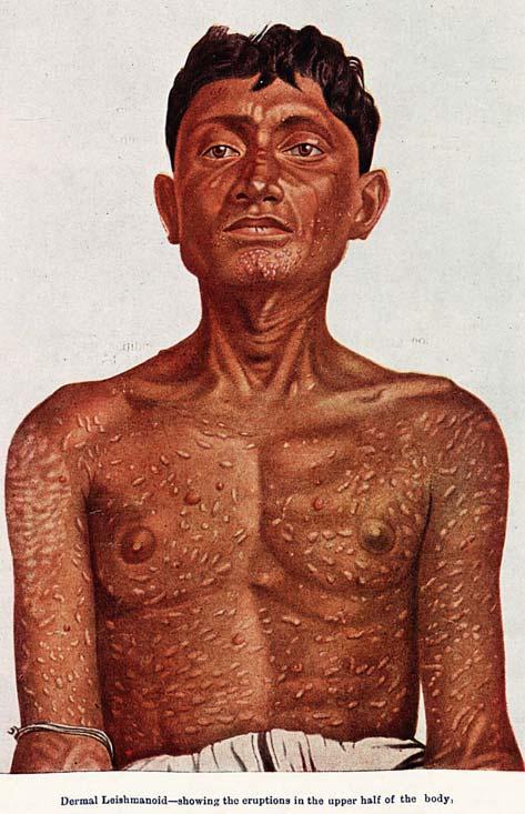
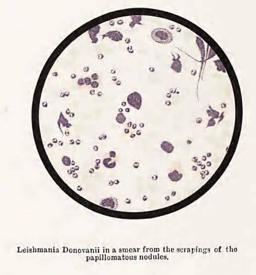
Editorial note : In resource limited countries like India, maneuvers to avoid mechanical ventilation are of utmost importance.
5.How do you identify cytokine storm in COVID-19?
There is no single objective test to diagnose cytokine storm. There is no single biomarker which has been conclusively shown to correlate with the onset of cytokine storm in these patients. Until more studies are available, we use clinical judgement along with the trend of various available inflammatory markers including serum ferritin, CRP, IL-6 and LDH to identify cytokine storm.
6.What cut off value of IL6 in pg/ml do you usetostartthisdrug?
Given the lack of appropriate good quality data and variability of IL-6 levels with available methods of detection, we do not use a single cut-off value for prescribing IL-6 receptor antagonist. We use clinical judgement as mentioned above along with the trend of various inflammatory biomarkers to decide on initiating anti-IL-6 agents.
7.Is there any role of identifying high risk patientstoadministerTocilizumab?
It is of paramount importance to identify patients who are at high risk of disease progression particularly from moderate to severe disease wherein, selective immunomodulation may be considered. Nonetheless, there is unlikely to be a single marker for the same and clinical judgement supported by rising inflammatory markerswill most likely continue to be the criteria to consider selective immunomodulators for interrupting the inflammatory cascade.
8.Any other biomarkers can be done instead ofIL6orinadditiontoittopredictcytokinestorm? (likeCRP,IL10etc.)
As mentioned above, there is no single objective test to diagnose cytokine storm. A host of markers of the pro-inflammatory cascade are being used in addition to the clinical status of the patient to identify whether
the host response has tipped over from protective immune response to an uncontrolled state of hyperinflammation. These include serum ferritin, CRP, CPK, LDH, IL-1, IL-6, TNF-a, etc.
The ongoing research will reveal whether there is ahierarchical pro-inflammatory cytokine which ushers in the cytokine storm in COVID-19 patients. The existence and understanding of the same will be a potential breakthrough in monitoring and management of COVID-19 patients.
9.How early after admission to start this therapy? Can we have a scoring system with multiple variables or single biomarker value?
The administration of immunomodulators late in the COVID-19 clinical course is unlikely to provide large beneficial effects. Identifying the tipping point when the individual immune response goes into the hyperinflammatory drive is key to take the decision on initiating immunomodulators like tocilizumab. As mentioned above, there is no single known biomarker for identifying the same and we rely on a host of biomarkers in addition to the clinical condition of the patient to take a decision. These need to be built into a scoring system in near future as more data emerge.
10.Since it may aggravate tuberculosis, bacterialorfungalinfections;whatinvestigations to precede giving Tocilizumab keeping the time constraint in mind
Active bacterial infection and latent TB should be ruled out prior to administering the drug. The merits and demerits of screening method (TST, IGRA) remains
unknown in these patients. Close monitoring is warranted in view of recent reports of high secondary infection rates among patients receiving tocilizumab. This is of particular relevance to the Indian ICU settings.
No dose modification is required in patients with pre-existing renal impairment. Tocilizumab is to be avoided in pregnancy. Caution is advised in presence of active hepatitis.
12.Whenshouldrepeatdosebeconsidered?
The usual dose is 8mg/kg (maximum: 800 mg/ dose); there are various regimens being used in trials. We repeat the dose between 12 to 24 hours later if no improvement is seen after the first dose.
Editorial note : There are ongoing trials of tocilizumab in Covid-19. The data from those trials will further clarify the utility of this drug.
13.Whatarethecontraindicationsofusingthis drug or in whom to avoid?
This drug should be avoided in patients with any of the following active infections: viral hepatitis, tuberculosis, HIV, bacterial and/ or fungal and/ or viral infections (other than SARS-CoV-2 infection), neutrophil count < 1000/ mm3, platelet counts < 50,000/ mm3.
Dr Soneja, we thank you for the valuable insight into the management of Covid-19 infection. We are sure our readers will benefit a lot from these pearls of knowledge. Please stay safe duringthepandemic.
Original Article
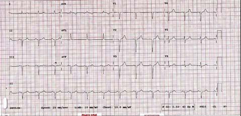
Frequency and Pattern of Primary Headache Disorders at a Tertiary Health Facility in Dhaka, Bangladesh
Aminur Rahman1, Zahed Ali2, Manabendra Bhattacharjee3, Ranajit Sen Chowdhury4, Subash Kanti Dey5, Firoz Ahmed Quraishi6, Uttam Kumar Saha7
Background : Primary headaches are under diagnosed and undertreated, with a significant impact on personal life, social activities and work.
Aim : To determine the frequency and pattern of primary headaches at a tertiary centre in Dhaka, Bangladesh.
Methods : This study was a hospital based cross-sectional descriptive study and conducted at outpatient department (OPD) of neurology in Sir Salimullah Medical College & Mitford Hospital (MH) for duration of one year. A total of 1825 patients were attended to the OPD, of which 551 were diagnosed as primary headache by neurologists were enrolled in this study. Types of primary headache were diagnosed by residents and neurologists according to the criteria of the International Headache Society (2013).
Results : The participants comprised 122 males were 22.1% and 429 females 77.9%. The mean age was 34.78±7.34 years. The overall headache was 30.19% with female predominance (p=0.947). The most common pattern of headache distribution of the study population are migraine (64.4%), then tension-type headache (TTH) (23.4%), chronic daily headache (CDH) (7.6%) and cluster headache (0.6%). Female patients (84.4%) are more suffer in migraine than male (15.6%). In case of TTH female patients (66.7%) are more suffer than male (59.1%). In case of CDH male (60.1%) are more suffering than female (39.9%). In case of cluster male patients (66.7%) are more suffer than Female (33.7%) (p<0.001). The migraine, CDH, and cluster headache are common in age group 30-39 yrs whereas TTH is common in age group 40-49 years (p<0.001). The mean age of onset of migraine was 34.24±7.09 years.TTH was 36.20±7.58 years, CDH was 36.59±8.63 years and cluster headache was 33.91±7.48 years.
Conclusions : The primary headache was common in female among working population, predominantly migraine and tension-type headache. [J Indian Med Assoc 2020; 118(6): 20-5]
Headaches are the most prevalent neurological disorders and among the most frequent symptoms seen in daily practice1. 50% of the general population
Department of Neurology, Sir Salimullah Medical College, Dhaka 1100, Bangladesh
1MBBS, FCPS (Medicine), MD(Neurology), MACP(USA), MAAN(USA), FINR(Switzerland), Assistant Professor and Corresponding Author
2MBBS, FCPS (Medicine), MD (Neurology), Professor
3MBBS, MD(Neurology), Associate Professor, Department of Neurology, Mymensingh Medical College, Mymensingh 2200
4MBBS, FCPS (Medicine), Associate Professor, Department of Medicine, Sir Salimullah Medical College, Dhaka 1100
5MBBS, MD (Neurology), Associate Professor, Department of Neurology, Bangabandhu Sheikh Mujib Medical University, Dhaka 1000
6MBBS, FCPS (Medicine), MD (Neurology), Professor, Department of Neurology, Anwar Khan Modern Medical College, Dhaka 1205
7MBBS, MCPD(Medicine), MD(Neurology), Professor, Department of Neurology, National Institute of Neurosciences & Hospital, Dhaka
Received on : 23/04/2020
Accepted on : 10/06/2020
Editor's Comment :
Primary headaches which are underdiagnosed and undertreated cause significant work inefficiency, quality of life, and lost workdays.
The primary headache was common in female than man among working population in between 30-39 years.
The most common pattern of primary headache distribution is migraine, then tension-type headache.
have headache during any given year, and more than 90% report a lifetime history of headache 2 . It is estimated that 95% of men and 99% of women will have at least one episode throughout their life, provided that about 40% have it quite regularly3
Primary headache disorders constitute the vast majority of headache disorders, with migraine and tension type headache (TTH) being the most prevalent. Primary headaches cause significant disability with reduced efficiency, quality of life, and lost workdays4-6. The global prevalence among adults
is approximately 10% of migraine, 40% for tensiontype headache (TTH) and 3% for chronic daily headache2 .Worldwide; the current global prevalence of primary headache is 47%; migraine headache, 10%; tension-type headache, 38%; and chronic daily headache, 3%5. The lifetime prevalence rates are higher in men,93% for headache of any kind, 8% for migraine, and 69% for tension-type headache. In women, life time prevalence is 99% for headache of any kind, 25% for migraine, and 88% for tension-type headache7
Migraine prevalence during lifespan is also genderdependent8. Migraine occurs most commonly between the ages of 25 and 55 years and is 3 times more common in females5,9. Despite the fact that it causes significant disability, migraine remains under diagnosed and undertreated. Although typically not as severe as migraine, tension-type headache is far more common, with lifetime prevalence in the general population of up to 80%. There is often a degree of associated disability, and this, combined with the high frequency, produces significant socioeconomic impact5
Trigeminal autonomic cephalgias are rare compared with migraine and tension-type headache. The most common trigeminal autonomic cephalgia is cluster headache which is a relatively rare but extremely painful type of headache, usually strictly one- sided, attacks in cyclical pattern and bouts 10 , with a population prevalence of 0.1% and a male/female ratio of 3.5-7:15,9. Cluster headache is a relatively rare but extremely painful type of headache, usually strictly one- sided, attacks in cyclical pattern and bouts10
Chronic Daily Headache (CDH) is a descriptive term and not a diagnosis per se. It is commonly defined as headaches occurring on 15 or more days in a month for at least three months and affects around 4% of the general population11. CDH is widely reported in the literature, yet is not an official diagnosis in the International Classification of Headache Disorders. Chronic daily headaches of long duration include chronic migraine, chronic tension-type headache, hemicrania continua, and new daily persistent headache9. The headache may be disabling not only due to its intensity, but also due to the frequency of attacks, which can be almost daily. This syndrome is known as chronic daily headache (CDH), and its prevalence in the overall population is approximately 5%, while in tertiary care centers it ranges from 30 to 90% of the cases12,13. We aimed to determine the frequency and pattern of primary headaches at a tertiary centre in Dhaka, Bangladesh, using the operational diagnostic criteria of the International
Headache Society (IHS)14. MATERIAL AND METHODS
This study was a hospital based cross-sectional descriptive study and conducted at outpatient department (OPD) of neurology in Sir Salimullah Medical College & Mitford Hospital (MH) during August 2018 to July 2019 for duration of one year. Patients attend to OPD and out of 1825 patients 551 patients were diagnosed as primary headache were enrolled in this study. The inclusion criterion was age 19 and above and patient attending to OPD of the hospital, whilst the exclusion criterion was refusal to participate in the study.
Their informed written consent was taken in a consent form before collecting data. The headache survey was performed by means of an interview based on a detailed pretested structured assessment questionnaire. The interviews were conducted under the supervision of the neurologists. The headache assessment questionnaire contained demographic data included a description of the current features of headache as well as its characteristics. Details of the research were communicated to the consenting participants at the beginning of the exercise. The participants were given the questionnaires to fill out and recorded in the cases for review the following day.
Diagnostic Criteria :
Headache was diagnosed according to the criteria of the International Classification of Headache Disorders (ICHD-3: Beta Version - 2013)14
Migraine was diagnosed in subjects with recurrent, moderate to severe unilateral throbbing headache associated with nausea or vomiting or visual disturbances. The subjects with migraine were not subclassified. Tension-type headache was diagnosed when subjects suffered from bilateral or vertex tightness or pressure-like feeling in the absence of gastrointestinal or visual discomfort.
Details of the diagnostic criteria according to ICHD for migraine and tension-type headaches, cluster headache, hemicrania continua and new daily persistent headache are shown in Appendix.
Proper permission was taken from the concerned departments and local ethical committee.
Exploratory data analysis were carried out to describe the study population where categorical variables were summarized using frequency tables while continuous variables were summarized using measures of central tendency and dispersion such as mean, median, percentiles and standard deviation. All statistical analysis were performed using SPSS 25.0 for Windows (SPSS Inc, Chicago, Illinois, USA) level
of significance was set at 0.05 and p-value <0.05 was considered significant.
OBSERVATIONS AND RESULTS
A total of 1825 patients were attended to the OPD, of which 551 were diagnosed as primary headache and included in this study, giving an enrollment rate of 30.19%. 551 patients with primary headache (429 female and 122 male) were included in the study. The primary headache in males was 22.1% (122/551) and females 77.9% (429/551) (Table 1).
In Table 2 The mean age of the patient group was within the range of 35.07±14.43.
In Table 3 The most common pattern of headache distribution of the study population are migraine (64.4%), then tension-type headache (TTH) (23.4%), Cluster HA (0.6%) and Chronic Daily Headache (CDH) (7.6%).
In Table 4 female patients are more sufferer in primary headache in relation to age than male which are not statistically significant.
In Table 5 Female patients (84.4%) are more suffer in migraine than male (15.6%). In case of TTH female patients (66.7%) are more suffer than male (59.1%). In case of CDH male patients (60.1%) are more suffer than female (39.9%). In case of cluster male patients (66.7%) are more suffer than female (33.3%) which are statistically significant.
Table 1 — Sex distribution of the study patients (n=551)
In Table 3 The most common pattern of CDH distribution are Chronic migraine(47.6%), then Chronic tension-type headache (TTH) (35.7%), Hemicrania continua (2.4%) and New daily persistent headache (NDPH) (11.9%). In Chronic migraine woman (65%) suffered more than man (35%).
In Table 7 migraine, CDH and Cluster HA are common in age group 30-39 yrs whereas TTH in age group 40-49 years which are statistically significant.
In Table 8 The mean age of onset of migraine was 34.24±7.09 years.TTH was 36.20±7.58 years, CDH was 36.59±8.63 years and Cluster HA was 33.91±7.48 years.
DISCUSSION
Among the population the predominant patient profile found in this outpatient department was women (77.9%) compared with men in between the age group from 20 to 49 years old and majority patients were aged between 30 to 39 years (35.4%) & the mean age of all patients was 34.78±7.34 years had higher prevalent rates for primary headache in this present study as has been previously reported15,16. This has been attributed to the effect of female sex hormones specifically oestrogen.
Table 4 — Pattern of headache distribution of the study patients (n=551)
Pattern of headache Frequency Percentage (%) MIG37768.4 TTH12923.4 CDH427.6
Cluster HA30.6 Total551100.0
Table 5 — Pattern of headache relation to sex (n=551)
Table 2 — Age distribution of the study patients (n=551)
Age group Frequency Percentage (%) Mean ±SD (years) 20-29177
Table 3 — Age basis sex distribution (n=551)
Age groupn Male Female Chi-square test (years) No. (%)No. (%)
20-29177 38(21.5%)139(78.5%) χ2= 0.108
30-39195 43(22.1%)152(77.9%) df=2
40-49179 41(22.9%)138(77.1%)p=0.947ns
Total 551122(22.1%) 429(77.9%0
Mean ±SD 35.8±14.734.9±14.3
Chi-square test was done, ns= not significant
Pattern ofn SexChi-square test headache Male Female
Table 7 — Pattern of headache relation to age (n=551)
Pattern ofnAge group Chi-square headache20-29 yrs30-39 yrs40-49 yrs test
MIG377128(34.0%)135(35.8%)114(30.2%) χ2= 27.8
TTH12934(26.4%)45(34.9%)50(38.8%) df=6
CDH4212(28.6%)16(38.1%)14(33.3%) p<0.001*
Cluster HA3 1(33.3%)2(66.7%)0(0.00%)
Total551177(32.1%)195(35.4%)179(32.5%)
Chi-square test was done, *= significant
Table 8 — Mean age of different headache pattern (n=551)
NMean ±SD Range
MIG37734.24±7.0921.00 – 49.00
TTH12936.20±7.5822.00 – 49.00
CDH4236.59±8.6322.00 – 49.00
Cluster HA333.91±7.4822.00 – 49.00
Total55134.78±7.3221.00 – 49.00
We documented a prevalent rate of 68.4 % for migraine in our study at this outpatient clinic. Migraine is the most prevalent type in tertiary care centers, with rates ranging between 35% and 80%17-19. One metaanalysis had indicated that the prevalence of migraine headache varied between different geographical regions, being lower in Europe than in North America but higher than in Asia and Africa20. Diversity of the population studied and racial differences in genetic vulnerability to migraine may also be contributory21
The well-known female preponderance in patients with migraine was also evident in our study. We found a significantly higher proportion of women with migraine headache, 318(84.4%)compared to men, and 59(15.6%). The higher rates in women are thought to be due to factors such as sensitivity to the oestrogen hormone, genetics, and differences in response to stress and pain perception. Premenstrual migraines are known to occur during or after the time when the female hormones, oestrogen and progesterone, decrease to their lowest levels22. We noted that the prevalent rate of migraine increased with age until the 4th decade when it started to decline. Tekle Haimanot23 in Ethiopia had also documented a decline after a peak in the fourth decade of life.
The prevalent rate of tension-type headache (TTH) in our study was 23.4%. TTH, whether episodic or chronic (CTTH), was the second most frequent cause of headache, while in the community it is the most common type, with a prevalence ranging from 30 to 80%14. A Chinese study found a prevalence of 66.9% for TTH in a tertiary care center24. In other studies, the prevalent of TTH was 47.7% in Zimbabwe25, the 25.5%
by Quesada-V´azquez et al in Cuba26, and 11.2% reported in Oman 27 There has been wide variations and differences in the epidemiology of tension-type headache across different cultures12.These variations may result from differences in study design, study population, inclusion or exclusion of cases of infrequent episodic TTH, and overlap with probable migraine, cultural and environmental differences, or even genetic factors 28 . We also found a significantly higher proportion of women with TTH, 86(66.7%) compared to men 43(33.3%) which was consistent with previous study are more common in women than men (23% to 18% respectively)29.
In our present study prevalent rate of cluster headache was 4.2% and male are predominant (66.7%) in comparison to female (33.3%) and male: female ratio is 2:1 age was the respondent in between 19-39 years. Cluster headache affects about 0.1% of the general population at some point in their life and 0.05% in any given year30. The condition usually first occurs between 20 and 40 years of age31. More men are affected than women, with a ratio of 3.5:113
We documented a prevalent rate of 7.6% for CDH in our study at this OPD. CDH was responsible for approximately one-third of the cases, while the prevalence in the community is between 3% and 7%32,33 which was consistent with study.
CONCLUSION
Headache is one of the most common symptoms in the general population. Female are more sufferer than man with primary headache in between 30-39 years. The most common pattern of primary headache distribution of the study population is migraine then tension-type headache. These could be diagnosed and managed in primary care or by general and emergency physicians working in acute medicine. There is a great need for addressing this health problem as the frequency and pattern of primary headache was found to be high among the population. There is an immense need to counsel and treat such individuals, as headache significantly affects an individual, family and society.
Appendix : International Classification of Headache Disorders (ICHD-3: Beta Version-2013)
(a)Nausea and/or vomiting (b)Photophobia and phonophobia
4.Not attributable to any other disorder
Tension-Type Headache :
1. >10 attacks lasting 30 minutes to 7 days
2. >2 of the following 4
(a)Bilateral location (b)Pressing/tightening (non-pulsating) quality (c)Mild or moderate intensity (d)Not aggravated by routine physical activity
3.No nausea or vomiting
4.One or either photophobia or phonophobia
5.Not attributed to another disorder.
Cluster headache:
Diagnostic criteria:
A. At least five attacks fulfilling criteria B–D
B. Severe or very severe unilateral orbital, supraorbital and/or temporal pain lasting 15–180 minutes (when untreated)1
C. Either or both of the following :
1. At least one of the following symptoms or signs, ipsilateral to the headache:
a)Conjunctival injection and/or lacrimation
b)Nasal congestion and/or rhinorrhoea
c)Eyelid oedema
d)Forehead and facial sweating
e)Forehead and facial flushing
f)Sensation of fullness in the ear
g)Miosis and/or ptosis
2. A sense of restlessness or agitation
D. Attacks have a frequency between one every other day and eight per day for more than half of the time when the disorder is active Hemicrania continua (HC):
The ICHD diagnostic criteria for hemicrania continua are:
1.Headache for more than 3 months fulfilling other 3 criteria:
2.All of the following characteristics:
a)Unilateral pain without side-shift
b)Daily and continuous, without pain-free periods
c)Moderate intensity, but with exacerbations of severe pain
3.At least one of the following autonomic features occurs during exacerbations and ipsilateral to the side of pain:
a)Conjunctival injection and/or lacrimation
b)Nasal congestion and/or rhinorrhea
c)Ptosis and/or miosis
4.Complete response to therapeutic doses of indomethacin, although cases of hemicrania continua that do not resolve with indomethacin treatment have been documented.
New daily persistent headache (NDPH) :
The ICHD diagnostic criteria are:
1.Headache that, within 3 days of onset, fulfils criteria 2-4
2.Headache is present daily, and is unremitting, for > 3 months
3.At least two of the following pain characteristics:
a)bilateral location
b)pressing/tightening (non-pulsating) quality
c)mild or moderate intensity
d)not aggravated by routine physical activity such as walking or climbing
4.Both of the following:
a)no more than one of photophobia, phonophobia or mild nausea
b)neither moderate or severe nausea nor vomiting
5.Not attributed to another disorder LIMITATIONS
This study has small sample size and study populations were confined to only one tertiary care hospital which does not reflect the picture of the entire country. The multicentres data should be needed for the actual prevalence of primary headache.. ACKNOWLEDGMENT
The authors are grateful to the staff and management of the department (OPD) of neurology in Sir Salimullah Medical College & Mitford Hospital (MH).
Funding : No funding sources.
Conflict of interest : None declared.
REFERENCES
1P Andlin-Sobocki, B Jönsson, H Wittchen, J Olesen — Cost of disorders of the brain in Europe. European Journal of Neurology 2005; 12(1): 1-27.
2Abu-Arafeh I, Razak S, Sivaraman B, Graham C — Prevalence of headache and migraine in children and adolescents: a systematic review of population-based studies. Dev Med Child Neurol 2010; 52:1088–97.
3Silva Junior, Ariovaldo Alberto da, Tavares, Rafael Mattos, Lara, Rodrigo Pinto, Faleiros, Bruno Engler, Gomez, Rodrigo Santiago, & Teixeira, Antônio Lúcio — Frequency of types of headache in the tertiary care center of the Hospital das Clínicas of the Universidade Federal de Minas Gerais, MG, Brazil. Revista da Associação Médica Brasileira 2012;58(6): 709-13.
4Rasmussen BK, Jensen R, Schroll M, Olesen J — Epidemiology of headache in a general population: a prevalence study. J Clin Epidemiol 1991; 44: 1147-57.
5LJ Stovner, K Hagen, R Jensen, Z Katsarava, RB Lipton, AI Scher, TJ Steiner, J-A Zwart— The global burden of headache: a documentation of headache prevalence and disability worldwide. Cephalalgia 2007; 27: 193-210.
6P Andlin-Sobocki, B Jönsson, H Wittchen, J Olesen — Cost of disorders of the brain in Europe. European Journal of Neurology 2005; 12(1): 1-27,
7J R Saper, S D Silberstein, C D Gordon, R L Hamel, S Swidan — Handbook of Headache Managemented, Lippincott Williams & Wilkins, Philadelphia, Pa, USA, 2ndedition,1999.
8Russell MB, Rasmussen BK, Thorvaldsen P, Olesen J — Prevalence and sex-ratio of the subtypes of migraine. Int J Epidemiol 1995; 24(3): 612-8.
9Robbins MS, Lipton RB. The epidemiology of primary headache disorders. Semin Neurol 2010; 30(2): 107-19.
11Olesen J, Bousser MG, Diener HC, Dodick D, First M, Goadsby PJ, etal— Headache Classification Committee. New appendix criteria open for a broader concept of chronic migraine. Cephalalgia 2006; 26: 742-6.
12Silva-Júnior AA, Faleiros BE, Santos TM, Gómez RS, Teixeira AL — Relative frequency of headache types: a longitudinal study in the tertiary care. Arq Neuropsiquiatr 2010; 68:87881.
13Silva-Júnior AA, Costa EC, Gomes JB, Leite FM, Gomez RS, Vasconcelos LP, et al — Chronic headache and comorbibities: a two-phase, population-based, cross-sectional study. Headache 2010; 50: 1306-12.
14Headache Classification Committee of the International Headache Society (IHS) — The International Classification of Headache Disorders, 3rd edition (beta version). Cephalalgia 2013; 33(9): 629-808.
15E A Mac Gregor, J D Rosenberg, T Kurth — Sex-related differences in epidemiological and clinic-based headache studies. Headache 2011; 51(6): 843-59.
16C.S. Liverman, J.W.Brown, R.Sandhir, R.M.Klein, K.McCarson, and N. E. J. Berman, “Oestrogen increases nociception through ERK activation in the trigeminal ganglion: evidence for a peripheral mechanism of allodynia,”Cephalalgia. 2009; 29(5): 520–531.
17Vasconcelos LPB, Stancioli FG, Leal JC, Costa EAC, SilvaJúnior AA, Gómez RS et al. Cefaleias em serviço especializado. Migrâneas Cefaleias. 2006; 9:4-7.
18Felício AC, Bichuetti DB, Santos WA, Godeiro Junior CO, Marin LF, Carvalho DS. Epidemiology of primary and secondary headaches in a Brazilian tertiary care center. Arq Neuropsiquiatr. 2006;64:41-6
patients in primary care and a tertiary care unit in Zürich, Switzerland. Cephalalgia. 2006; 26:1451-7.
20A. I. Scher, W. F. Stewart, J. Liberman, and R. B. Lipton, “Prevalence of frequent headache in a population sample,” Headache.1998;38(7):497–506.
21W. F. Stewart, R. B. Lipton, and J. Liberman, “Variation in migraine prevalence by race” Neurology. 1996; 47 (1):52–59,
22B. L. Peterlin, S. Gupta, T. N. Ward, and A. MacGregor, “Sex matters: evaluating sex and gender in migraine and headache research,” Headache.2011;51(6): 839–842.
23R. Tekle Haimanot, B. Seraw, L. Forsgren, K. Ekbom, and J. Ekstedt, “Migraine, chronic tension-type headache, and cluster headache in an Ethiopian rural community,” Cephalalgia.1995; 15(6):482–488.
24Li X, Zhou J, Tan G, Wang Y, Ran L, Chen L. Clinical characteristics of tension- type headache in the neurological clinic of a university hospital in China. Neurol Sci. 2012; 33:2837.
25A.J.Quesada-V´azquezandN.Rodr´iguez-Santana, “The prevalence of primary headaches in the working population at a psychiatric hospital in Zimbabwe,” Revista de Neurologia.2006; 43(3):129–131.
26A. J. Quesada-V´azquez, L. J. Contreras-Maure, A. ´Alvarez Aliaga, and E. R. Traba-Tamayo, “Prevalence of primary headaches in a rural population in Cuba,”Revistade Neurologia. 2009; 49(3):131–135.
27D. Deleu, M. A. Khan, and T. A. H. Al Shehab, “Prevalence and clinical characteristics of headache in a rural community inOman,”Headache.2002; vol.42, no.10, pp.963–973.
28K. Sahler, “Epidemiology and cultural differences in tension type headache, ”Current Pain and Headache Reports. ,2012; 16(6):525–532.
29Vos T, Flaxman AD, Naghavi M, Lozano R, Michaud C, Ezzati M, et al. "Years lived with disability (YLDs) for 1160 sequelae of 289 diseases and injuries 1990-2010: a systematic analysis for the Global Burden of Disease Study 2010". Lancet. 2012; 380 (9859): 2163–96. .
30Fischera, M; Marziniak, M; Gralow, I; Evers, S "The Incidence and Prevalence of Cluster Headache: A Meta-Analysis of Population-Based Studies". Cephalalgia. 2008; 28 (6): 614–8.
31Weaver-Agostoni, J "Cluster headache". American Family Physician. 2013; 88 (2): 122–8.
32Schürks M, Kurth T, de Jesus J, Jonjic M, Rosskopf D, Diener HC: Cluster headache: clinical presentation, lifestyle features, and medical treatment. Headache 2006; 46: 1246–54.
33Queiroz LP, Peres MFP, Kowacs F, Piovesan EJ, Ciciarelli MC, Souza JA et al. Chronic daily headache in Brazil: a nationwide population-based study. Cephalalgia. 2008;28:1264-9.
Original Article
Tacrolimus versus Rituximab in adult onset steroid resistant nephrotic syndrome
Introduction : Focal segmental glomerulosclerosis and Minimal change disease are two most important causes of nephrotic syndrome in the adults. Non response with fourmonth therapy in adults with full dose steroid is defined as steroid resistant nephrotic syndrome. Steroid resistance predicts a high risk of progression to end stage renal disease. Calcineurin inhibitors are the first line treatment for steroid resistant disease. Other novel agents like Rituximab is also tried in the disease. This study is done to compare the efficacy of tacrolimus and rituximab in steroid resistant minimal change disease and focal segmental glomerulosclerosis.
Methods : This is an open label prospective randomized parallel group interventional study with a sample size 15, duration of 22 months and conducted in Department of Nephrology, IPGME&R and SSKM hospital Kolkata. Patients of 18 to 60 years of age with kidney biopsy proven minimal change disease and focal segmental glomerulosclerosis who are steroid resistant are randomly assigned in two arms in 2 : 1 distribution for tacrolimus and rituximab.
Results : In tacrolimus arm 70% of patients achieved any form of remission among which 40% achieved complete remission in the study period. In rituximab arm 100% of patients achieved any form of remission among which 40% achieved complete remission. The decrease in proteinuria in both groups from beginning to end of the study are each statistically significant. In tacrolimus group the mean eGFR decreased and in rituximab group mean eGFR increased but each of them is not statistically significant. Two patients did not respond to tacrolimus.
Conclusion : In both groups there was comparable remission without any statistically significant change in eGFR. There is limited serious infection in rituximab group. Recurrent infection is more common in tacrolimus group.
[J Indian Med Assoc 2020; 118(6): 26-30]
Key words : Nephrotic syndrome, steroid resistant, remission, Tacrolimus, Rituximab.
Minimal change disease (MCD ) is a cause of nephrotic syndrome in approximately 10% of adults. Focal segmental glomerulosclerosis (FSGS) accounts for 35% of all adult onset nephrotic syndrome,
1MD (Medicine), DM (Nephrology) Associate Professor, Department of Nephrology, NRS Medical College, Kolkata 700014
2MD (Medicine), DM (Nephrology), Senior resident, Department of Nephrology, NRS Medical College, Kolkata 700014 and Corresponding Author
3MD (Medicine), DM (Nephrology), Tutor, Department of Nephrology, B S Medical College, Bankura 722101
4MD (Medicine), DM (Nephrology), Consultant, Department of Nephrology, Sir Ganga Ram Hospital, New Delhi 110060
5MD (Medicine), DM (Nephrology), Assistant Professor, DKS Postgraduate Institute and Research Centre, Chhattisgarh 492001
6MD (Medicine), DM (Nephrology), Assistant Professor, Department of Nephrology & Rheumatology, IPGME&R, SSKM Hospital, Kolkata 700020
7MD (Medicine), DM (Nephrology), Senior Resident, Department of Nephrology, Burdwan Medical College & Hospital, Burdwan 713101
8MD (Medicine), DM (Nephrology), Post Graduate Institute of Medical Education and Research, Chandigarh 160012
Received on : 18/11/2019
Accepted on : 18/03/2020
Editor's Comment :
Both tacrolimus and rituximab are effective in treating steroid resistant nephrotic syndrome.
Rituximab is not inferior to tacrolimus in treating Steroid resistant nephrotic syndrome due to minimal change disease and focal segmental glomerulosclerosis.
The chance of drug non compliance is lesser with rituximab than tacrolimus.
and over 50% among African Americans1. MCD and FSGS primarily affect the podocytes (podocytopathies) and may be the spectrum of the same disease having same medical management. Adult nephrotic syndrome, if steroid resistant predicts a high risk of progression to end stage renal disease. FSGS may be primary or secondary to adaptive response to glomerular hypertrophy or hyperfiltration. In secondary FSGS, immunosuppression is not indicated. Initial therapy in MCD and FSGS is done with prednisolone 1 mg / kg / day, maximum 80 mg or 2 mg /kg alternate day, maximum 120 mg for minimum 4 weeks and
JOURNAL OF THE INDIAN MEDICAL ASSOCIATION, VOL 118, NO 06, JUNE 2020
maximum 4 months that is 16 weeks. Non response to 4-month therapy with full dose steroid is defined as steroid resistant nephrotic syndrome. Around 10% patient of MCD is steroid resistant, which may be due to undetected FSGS. Calcineurin inhibitors (CNI) ie, cyclosporine and tacrolimus are considered to be first line treatment of steroid resistant disease2,3. In this study we have used tacrolimus. Nephrotoxicity is a major side effect of CNI s, apart from other adverse effects. So, a study with a novel agent is required having equal or better efficacy and favorable side effect profile. Rituximab, a chimeric monoclonal antibody directed against CD 20 bearing cells are tried in treatment of MCD and FSGS. There are some studies which show some benefit of Rituximab in treatment of steroid resistant disease4-6. There is no randomized control study comparing efficacy and safety of CNIs and Rituximab.
MATERIAL AND MEDHODS
This is a single centeropen label prospective randomized control parallel group interventional study conducted in the Department of Nephrology, IPGMER & SSKM Hospital Kolkata from Feb 2016 to Dec 2018. Approvalfrom Ethical Committee IPGME&R was taken prior to study initiation. CTRI Registration number is CTRI/2018/01/011316, Registered on 15/01/2018. All definitions are used as per KDIGO glomerulonephritis guideline published in 2012.
Definition :
Complete Reduction of proteinuria to <0.3 g/d or <300 mg/g remission (<30 mg/mmol) urine creatinine and normal serum creatinine and serum albumin >3.5 g/dl (35 g/l)
Partial Reduction of proteinuria to 0.3–3.5 g/d (300-3500 remission mg/g [30–350 mg/mmol]) urine creatinine and a decrease >50% from baseline, and stable serum creatinine (change in creatinine <25%)
Relapse Proteinuria >3.5 g/d or >3500 mg/g (>350 mg/ mmol) urine creatinine after complete remission has been obtained
SteroidPersistence of proteinuria despite prednisolone resistant1 mg/kg/d or 2 mg/kg every other day for >4 months
Inclusion criteria : (1) Patients of age within 18 to 60 years. (2) Biopsy proven MCD or FSGS who received 16 weeks of oral prednisolone in adequate dose and have not achieved remission. (3) Estimated glomerular filtration rate (eGFRbyMDRD) > 30 ml / min /1.73m2 Body surface area. (4) Tubular atrophy and interstitial fibrosis < 25% of biopsy area. (5) Patient receiving maximum tolerable dose of antiproteinuric
medication. (6) Patients willing to give consent for the study.
Exclusion criteria : (1) Patients with active infection. (2) Any contraindication to any of the medication used in the study. (3) Diabetes Mellitus. (4) Hepatitis B, Hepatitis C or HIV infection. (5) Liver function abnormalities. (6) Active neoplastic condition. (7) Chronic diarrhea. (8) Pregnancy. (9) Secondary FSGS. (10) Collapsing variant of FSGS. (11) Previous therapy within six months with mycophenolate, azathioprine, cyclophosphamide, and cyclosporine. (12) More than one episode of serious infections eg peritonitis, pneumonia, cellulitis in the past twelve months. (13) Current or previous therapy for tuberculosis.
Primary outcome : complete and partial remission
Secondary outcome : (1) change in eGFR at completion of therapy. (2) Doubling of baseline serum creatinine levels. (3) Time required to achieve complete or partial remission. (4) Adverse effects (tremors, nephrotoxicity, gum hypertrophy, impaired glucose tolerance / diabetes mellitus, diarrhea, impaired fasting lipid profile, infection).
Study end point : (1) Completed 12 month follow up. (2) Patient in tacrolimus arm who do not achieve complete or partial remission within 6 months.(3) Death of patient.
Data capture was done in baseline, 1 month, 2 month, 3 month, 6 month, 9 month and 12 month. Steroid resistant FSGS and MCD population was randomly assigned in two arms in 2 : 1 distribution for tacrolimus and rituximab. Arm 1 comprised of tacrolimus and arm 2 of rituximab. Tacrolimus was given in dose of 0.075 mg /kg in two divided doses. Dose was adjusted to maintain tacrolimus trough level (T0 level ) between 5 -10 ng /ml . In patients who achieve complete remission within 6 months, the dose of tacrolimus was reduced to achieve T0 level 3 – 6 ng / ml to be continued till 12 months. The subjects who have not achieved any form of remission at 6 months are declared tacrolimus resistant and their tacrolimus was stopped. All patients were prospectively followed up for 12 months except who reached end point. In Arm 2, patients received inj Rituximab 375 mg / m2 body surface area once weekly IV for total 4 doses with proper premedication as per manufacturer instruction.Rituximab dissolved in normal saline at concentration of 2mg/ml was infused over 3-4 hour. Initial infusion rate was 50 mg/hour, then in next hour the infusion rate was increased. Premedication used were oral acetaminophen 15mg/kg and oral diphenhydramine 0.5 mg/kg 30 minute prior to first
dose of rituximab. Intravenous hydrocortisone 4mg/kg was given prior to first dose of rituximab. Patients were monitored for infusion related reaction and screened for infection in each visit. Rituximab dose was repeated in case of no response after 6 months of last dose. Other therapy including atorvastatin, ACE inhibitors,
Table 1 — Histological types in study group
Total 95 patient of MCD & FSGS
Assessed for eligibility (n=17)
Excluded (n=2)
Did not give consent (n=1) Did not meet inclusion criteria (n=1)
Randomization (n=15)
Allocated to tacrolimus group N=10 Allocated to rituximab group n=5
angiotensin receptor blockers and low dose steroid was continued in both the arms.
RESULTS
Total 95 adult nephrotic syndrome patients due to FSGS and MCD were either diagnosed or referred to our department in the study period. Among them 17 patients developed steroid resistance in the study period. Two patients among the steroid resistant group were excluded as they did not meet the inclusion criteria. The15 steroid resistant MCD and FSGS patients were randomized with random number table in two group in 2:1 distribution in tacrolimus and rituximab group respectively. Steroid resistant disease is 17.89% of study population. In tacrolimus group total number of patients is 10 (male 8 and female 2 ) and in rituximab group total number of patients is 5 (male 3 and female 2). In tacrolimus group the mean age of patient was 34.1±10.14 year and in rituximab group the mean age was 36.2±13.04 years (Table 1).
The baseline characteristics in the both group are comparable and the differences are not clinically significant (Table 2)
The average time of achievement of complete remission in tacrolimus group was 100 days (confidence interval 14.5 to 185.4days) and in rituximab group is 180 days (confidence interval 3.6 to 356.4 days). Both groups are comparable. In tacrolimus group the average time
to achieve first partial remission is 71.143 days (confidence interval 27.1 to 56.86 days) and in rituximab group average time to achieve first partial remission is 42 days (confidence interval 41.1 to 113.1 days). Both groups are comparable.
In tacrolimus arm the mean baseline proteinuria was 7005 mg/24 hour and in the end of study it decreased to 1520.66 mg/day. The decrease is significant statistically. In rituximab group baseline proteinuria was 6310 mg/24 hour which decreased to 1240.85 mg/24 hour. The decrease of proteinuria is also significant in rituximab group.
It is observed that in rituximab arm nadir 24 hour proteinuria was achieved at around 3 month to 6 month and then gradually it showed increasing trend after 6 month.
In tacrolimus group the baseline mean creatinine was 1.14mg/dl (SD±0.43) and at the end of study it was 1.19mg/dl (SD±0.54), which is increase from baseline but not significant. In rituximab arm the baseline mean creatinine was 1.01 mg/dl (SD±0.39) and at the end of study 0.79mg/dl (SD±0.18), which is decrease from baseline but is not statistically significant.
In tacrolimus group baseline eGFR was 80ml/min (SD±31.09) and at the end of study the mean eGFR is 78.28ml/min ( SD±33.8), which is decreasing trend but not statistically significant. In rituximab group the eGFR increase from baseline but it is not statistically significant.
In tacrolimus group three patients did not respond to treatment neither complete nor partial remission. So their study was ended on 6th month due to nonresponse. But in rituximab group all patients achieved any form of remission. But the difference is not statistically significant.
In tacrolimus group there is doubling of creatinine in one patient but in rituximab group no patient has doubling of creatinine. But the finding is not statistically significant.
More than 30 present raises of creatinine occurred in 4 patients in tacrolimus arm but in no patient in rituximab arm but the difference is not significant.
In tacrolimus arm 3 patients relapsed which is 30% of tacrolimus treated patient and in rituximab arm 2 patients relapsed, which is 40% of rituximab treated patient.
In rituximab arm the first relapse occurred in mean duration of 225 days(confidence interval 136.8 to 313.2days) and in tacrolimus group the mean duration of relapse is 180 days (confidence interval 91.2 to 208.8 days). So in tacrolimus group the relapse occurred
earlier than rituximab group. But the difference is not statistically significant.
Adverse effects : Rituximab has infusion related side effect including chill and rigor, back pain and chest pain during infusion. These adverse effectsoccurred in one patient. No second dose infusion reaction occurred. No patient had to discontinue rituximab treatment. In Rituximab group one patient had severe respiratory tract infection which needed hospitalization and intravenous antibiotic. Another patient had multiple upper respiratory tract infection which was treated on outpatient basis. Intacrolimus group 3 patients developed multiple upper respiratory tract infection, which were treated on outpatient basis. A patient who did not respond to tacrolimus developed bacterial peritonitis and requiredhospitalization. In tacrolimus group, altered blood glucose levels were detected in 2 patients. Among them one needed oral antidiabetic and other responded to lifestyle modification. Two patients had sleep disturbance and one patient among them had tremor in hand. One patient developed diarrhea which was non severe and managed conservatively with dose reduction (Table 3).
DISCUSSION
Steroid resistant MCD and FSGS may progress to end stage renal disease. Calcineurin inhibitors are established therapies of the disease. Our study is a non inferiority trial between tacrolimus, which is a standard therapy with rituximab. In our study, in tacrolimus arm 70% patients achieved any form of remission, of which 40% achieved complete remission. 30% patients did not achieve any form of remission. In the study by Ramchandran R et al, total remission is 52.5%7, complete remission being 38.6% and partial remission 13.6%, tacrolimus resistant 47.7%. Our
Table 3 — Adverse event
Tacrolimus Rituximab
Chill and rigor during infusionNA1
Back pain during infusionNA1
Chest pain during infusionNA1
Multiple upper respiratory tract infection31
Oral candidiasis10
Superficial fungal infection20
Hospitalisation11
Anaphylactoid reactionNA0
Bacterial peritonitis10
Altered glucose tolerance20
Difficult controlling blood pressure10
Tremor10
Sleep disturbance20
Diarrhoea10
Total155
study shows better response rates, possibly because we included MCD also. In the study done by Li Fan et al8, total response with tacrolimus was 75%, among response complete remission was 58.3% and partial remission was 16.7%. Our study shows similar result with this group. In rituximab arm, 100% patients achieved any form of remission. Among remission, 40% achieved complete remission and 60% achieved partial remission. No patient was rituximab resistant. Kong et al showed that in rituximab treated patients, MCD patients achieved 100% remission and FSGS patients achieved 75% remission9. Fernandez et al shows rituximab failed to treat 5 out of 8 patients10. The three patients who improved showed remarkable improvement in renal function. El Rashid et al showed that in MCD group 31 out of 31 patients achieved remission11, and in FSGS group 17 out of 18 patients achieved remission. This study shows near similar result to our study.Similar to our study, this study shows increase in proteinuria after 6 to 8 months which may be due to loss of the effect of B lymphocyte suppression. In rituximab group, 80.34% reduction in proteinuria occurs, which is significant p< 0.000. The time to achieve any remission in tacrolimus group is 52 days and in rituximab group 42 days. The difference is not statistically significant. At the end of the study 70% patients in tacrolimus group and 80% patients in rituximab group maintain any form of remission. There is better preservation of kidney function in terms of GFR and serum creatinine in rituximab group than tacrolimus group, though is not statistically significant. In rituximab group, the relapse is more but is not statistically significant. Rituximab had infusion related side effects including chill and rigor, back pain and chest pain in one patient. One patient in our study had severe respiratory tract infection needing hospitalization. In tacrolimus group, three patients developed repeated upper respiratory tract infection which were treated on outdoor basis. One patient developed bacterial peritonitis requiring hospitalization. Altered blood glucose was detected in two patients. Rituximab is a well-tolerated drug. The main problem with this is the requirement of intravenous administration and close monitoring with premedication and the drug cost. No death occurred during the study.
CONCLUSION
This study confirms the benefit of treatment of steroid resistant MCD and FSGS. In our study it is found that both the drugs are effective in treating SRNS. It confirms that rituximab is not inferior to tacrolimus in treating SRNS due to MCD and FSGS. The chance
of drug non compliance is lesser with rituximab than tacrolimus.
LIMITATIONS
The study has multiple limitations. The main limitation is small sample size in study population. It is a single center study which do not include multiethnic population. We used one drug which is intravenous and the other oral. Double dummy model would have been better. In this study CD 19/ CD 20 lymphocyte count was not measured. The number of doses of rituximab was thus based on experience rather than targeting specific CD 19 and CD 20 cell count.
Funding : None
Conflict of Interest : None
REFERENCES
1Haas M, Meehan SM, Karrison TG, Spargo BH — Changing etiologies of unexplained adult nephrotic syndrome: a comparison of renal biopsy findings from 1976-1979 and 1995-1997. Am J Kidney Dis 1997; 30:621.
2Ittel TH, Clasen W, Fuhs M — Long-term ciclosporine A treatment in adults with minimal change nephrotic syndrome or focal segmental glomerulosclerosis. Clin Nephrol 1995; 44: 156-62
3Li X, Li H, Ye H — Tacrolimus therapy in adults with steroidand cyclophosphamide-resistant nephrotic syndrome and normal or mildly reduced GFR. AmJKidneyDis 2009; 54: 518
4Kronbichler A, König P, Busch M, et al — Rituximab in adult patients with multi-relapsing/steroid-dependent minimal change disease and focal segmental glomerulosclerosis: a report of 5 cases. Wien KlinWochenschr 2013; 125: 328.
5Ochi A, Takei T, Nakayama K — Rituximab treatment for adult patients with focal segmental glomerulosclerosis. Intern Med 2012; 51: 759.
6Fernandez-Fresnedo G, Segarra A, González E — Rituximab treatment of adult patients with steroid-resistant focal segmental glomerulosclerosis. Clin J Am Soc Nephrol 2009; 4:1317.
7Ramachandran R, Kumar V, Rathi M, Nada R, Jha V, Gupta KL, et al — Nephrol Dial Transplant. 2014 Oct;29(10):191824. doi: 10.1093/ndt/gfu097. Epub 2014 Apr 24
8Li Fan Qinghua, Liu Yunhua, Liao Zhibin, Li Yulian Ji Zhenhua, Yang Jian, Chen Junzhou, Fu Jinli Zhang Yaozhong Kong Ping Fu Tanqi Lou Zhengrong Liu Xueqing Yu Wei Chen — Tacrolimus is an alternative therapy option for the treatment of adult steroid-resistant nephrotic syndrome: a prospective, multicenter clinical trial. Int UrolNephrol 2013; 45(2):459-68.
9Kong WY, Swaminathan R, Irish A — Our experience with rituximab therapy for adult-onset primary glomerulonephritis and review of literature. Int Urol Nephrol 2013; 45: 795-802.
10Fernandez-Fresnedo G, Segarra A, Gonzalez E, Alexandru S, Delgado R, Ramos N, et al — Rituximab treatment of adult patients with steroid-resistant focal segmental glomerulosclerosis. Clin J Am Soc Nephrol 2009; 4: 131723.
11El-Reshaid K, Sallam HT, Hakim AA, Al-Attiyah R — Rituximab in treatment of idiopathic glomerulopathy. Saudi J Kidney Dis Transpl 2012; 23: 973-8.
Original Article
Frequency of Comorbidities & their Association with Intensive Care Unit Admission in Hospitalised Patients with 2019 Novel Coronavirus infection in Tertiary Care Centres of Three States of India
The coronavirus disease 2019 (COVID-19) has created a substantial burden on healthcare services worldwide. Since its first detection in 30th January, it has rapidly spread throughout India. Severe acute respiratory syndrome coronavirus-2 (SARS-CoV-2) infection results in clusters of severe acute respiratory illness leading to intensive care unit (ICU) admission and considerable mortality. So, there has been an ardent need of data on the frequency of comorbidities in COVID-19 & to assess whether their presence is associated with increased ICU admission. We analysed data from 496 patients with laboratoryconfirmed Covid-19 admitted in tertiary care centers of three states of India from 15th to 30th May, 2020. The mean age was 49.7 years & 41.13% of the patients were female. Hypertension (21.97%) was the most frequent comorbidity followed by diabetes (12.90%) & cardiovascular disease (8.87%). 39.92% of the study population had at least one comorbidity. Patients with comorbidities had higher ICU admission than those without comorbidity (35.35% vs. 20.47%). Associated comorbidity was more frequent among ICU patients in comparison to non-ICU patients (53.43% vs. 35.07%). Our study findings suggest that presence of comorbidity is associated with higher ICU admission thereby indicating more severe disease.
[J Indian Med Assoc 2020; 118(6): 31-4]
Key words : COVID-19, SARS-CoV-2, Comorbidity, Intensive care unit.
Coronavirus disease 2019 (COVID-19), a novel viral infection by severe acute respiratory syndrome coronavirus 2 (SARS-CoV-2) was first detected in Wuhan, China in December 2019 & it has recently become a public health emergency1. COVID-19 has contributed to a massive adverse impact globally due to its high transmission rate. The infection has spread in such a large proportion affecting most of the
1MD (Medicine), DNB (Med), MRCP (UK); Assistant Professor, Department of General Medicine, R.G.Kar Medical College and Hospital, Kolkata 700004
2DCH, MD (Medicine); Assistant Professor, Department of General Medicine, R.G.Kar Medical College and Hospital, Kolkata.
3MD (Medicine), Associate Professor, Department of General Medicine, R.G.Kar Medical College and Hospital, Kolkata
4MD (Medicine), MACP ; Senior Resident, Department of General Medicine, R.G.Kar Medical College and Hospital, Kolkata and Corresponding Author
5MBBS ; Junior Resident, Department of General Medicine, R.G.Kar Medical College and Hospital, Kolkata
6MD(Medicine), FRCP; Associate Professor, Department of General Medicine, Silchar Medical College & Hospital, Silchar 7MD (Medicine) MIP, FCCP, FICP, FCSI, FCGP, FIAMS, FICA, PG Dip (Diabetology), Associate Professor, Department of General Medicine, Chandulal Chandrakar Memorial Hospital, Chattisgarh
Received on : 16/06/2020
Accepted on : 20/06/2020
Editor's Comment :
Hypertension, diabetes, heart disease & CKD are common comorbidities in all studies.
Presence of comorbidity is associated with higher ICU admission thereby indicating more severe disease.
Thorough assessment of comorbidities of patients with COVID-19 & proper management of those chronic ailments during the hospital stay is very important.
Special care should be provided in this subset of patients
countries in the world, that WHO has declared it as a pandemic on March 11, 20202. The first confirmed case of SARS-CoV-2 was reported in India on 30th January 2020. Since then, more than 3 lakh cases of COVID19 have been reported in India, including over 8700 deaths (as on June 12th, 2020)3
Infection by SARS-CoV-2 has myriads of presentations like fever, cough, respiratory distress along with atypical manifestations like diarrhea, neurological, renal dysfunction etc4. In the recently published studies, 20–51% of patients of COVID-19 were reported to have at least one comorbidity.5 COVID-19 patients are predisposed to higher risk of acute respiratory distress syndrome (ARDS) and death if associated with chronic illness5,6. During the last
few months, many studies have been published on SARS-CoV-2 infection describing the comorbidities in patients with COVID-19 & its impact on the outcome. In this study, we performed a comprehensive evaluation of the associated comorbidities in patients with COVID-19 admitted in tertiary care centers of three states of India from 15th to 30th May, 2020. The purpose of our study is to determine the frequency of comorbidities in the laboratory confirmed cases of SARS-CoV-2 infection & whether their presence is associated with increased ICU admission. These findings may help us extend our understanding on these factors associated with COVID-19. To the best of our knowledge, there are no such studies from this area on this topic.
MATERIALS AND METHODS
We had evaluated the medical records of 496 laboratory-confirmed Covid-19 patients admitted to tertiary care centers of three states of India from 15th May to 30th May, 2020. A confirmed case of SARSCoV-2 infection was defined as a positive result on real-time reverse-transcriptase–polymerase-chainreaction (RT-PCR) assay from nasopharyngeal and oropharyngeal swab specimens. Presence of comorbidities was determined on the basis of patient's self-report at the time of admission. Comorbidities were primarily considered as a categorical variable (yes versus no) and then categorized on the basis of the number of comorbidity (single versus multiple). Moreover, comorbidities were classified according to the organ systems involved (ie, respiratory, cardiovascular, renal, central nervous system etc). Continuous variables were presented as mean or median as appropriate, and the categorical variables were expressed as counts and percentages. No imputation was made for missing data. Because the cohort of patients in our study was not derived from random selection, all statistics are deemed to be descriptive only.
OBSERVATIONS
We obtained data regarding comorbidities in 496 patients admitted in tertiary care centers of three states of India from 15th May to 30th May, 2020. Of these 496 cases, mean age was 49.7 years. 292 cases were male (58.87%), while 204 (41.13%) cases were female. Our study revealed that 198 out of 496 patients(39.92%) had at least one comorbidity. Hypertension (21.97%) was the most frequent comorbidity observed in our study. This was followed by diabetes (12.90%), cardiovascular disease (8.87%), chronic kidney disease
(4.43%), obstructive airway disease (4.23%), hypothyroidism (3.43%) & malignancy(3.22%). The ‘others’ category included cerebrovascular accident, parkinsonism, obstructive sleep apnea, post-operative patients etc. & they were responsible for 8.26% of the cases. The summary of the associated comorbidities in our study are shown in Table 1. Summary of the gender wise distribution of comorbidities among COVID-19 patients are shown in Table 2. Bar diagram showing gender wise distribution of comorbidities is depicted in Fig 1.Patients with comorbidity had higher mean age than those without any comorbidity (57.8 years versus 43.2 years). 70 out of 198 cases (35.35%) with comorbidity were admitted in ICCU. On the other
Table 2 — Summary of gender wise distribution of comorbidities
Comorbidity Male Female Hypertension6742
Diabetes Mellitus3727
Cardiovascular Disease2420
Chronic Kidney disease1210
Obstructive airway disease147
Hypothyroidism611
Malignancy106
Chronic Liver Disease94
Others2615
Table 1 — Summary of the Associated Comorbidities in patients with COVID-19
Fig 1 — Bar diagram showing gender wise distribution of comorbidities
hand, 61 out of 298 patients (20.47%) without comorbidity were in ICCU. Among ICCU-patients, 53.43% (70 out of 131patients) had comorbidity ,while in case of non ICCU-patients it was 35.07% (128 out of 365 patients).
DISCUSSION
We analysed data from 496 patients with laboratory-confirmed Covid-19 admitted in tertiary care centers of three states of India. 39.92% of the study population had at least one comorbidity. Hypertension (21.97%) was the most frequent comorbidity followed by diabetes (12.90%) & cardiovascular disease (8.87%). Patients with comorbidity had higher ICU admission than those without comorbidity (35.35% versus 20.47%). Presence of comorbidity was more frequently observed among ICU patients in comparison to non-ICU patients (53.43% versus 35.07%). It was seen in our study that patients with a single comorbidity, or even more so, were associated with increased frequency of ICCU admission thereby indicating more serious illness.
Overall, our study findings are in accordance to the previously published studies in terms of the frequency of comorbidities in patients with COVID-19. There are some variations of our study findings with a few studies. This may be due to difference in the study area, sample size & sample selection. Guan WJ et al. analyzed data from 1590 confirmed hospitalized patients of COVID-19 from 575 hospitals in mainland China.5 In their study, most common comorbidity was hypertension (16.9%), followed by diabetes (8.2%). 16% of the study population had severe disease. 25.1% were reported to have single or multiple comorbidity. They concluded that, patients with any comorbidity had worse clinical outcome than those without comorbidity. Persons with multiple comorbidities also had poorer clinical outcome.
Huang C et al. evaluated the clinical characteristics of 41 patients infected with SARS-CoV-2 in Wuhan,China.4 32% of all the patients had underlying comorbidity. Diabetes mellitus, hypertension & cardiovascular disease were most common among all the comorbidities. Chen N et al studied the epidemiological and clinical features of 99 cases of SARS-CoV-2 pneumonia in Wuhan, China where they found that 51% of all the patients had comorbidities including cardiovascular and cerebrovascular diseases, respiratory ailments, endocrine system disease, choric liver disease, malignancy and nervous system disorders.7 Liu K et al analysed the Clinical & demographic features of 137 patients infected with 2019-nCoV in tertiary hospitals in Hubei Province of
China. In their study, 27 out of 137(19.7%) patients had associated comorbidity.8 Hypertension, diabetes & cardiovascular diseases were most common all the chronic comorbidities.
Singh AK et al retrieved data from ten available Chinese studies (total sample size 2209) by searching the medical database up to March 27, 2020.9 In their review article they found that hypertension was the commonest comorbidity present in nearly 21%.This was followed by diabetes (11%) & cardiovascular disease (7%). Though it was mentioned in most of the studies that there is increase in mortality in patients of COVID-19 with hypertension, diabetes and CVD, they concluded that most of them were not adjusted for confounding factors. They also opined that special attention is definitely required in patients with COVID19 with associated comorbidities including hypertension, diabetes and established CVD. Richardson S et al. studied clinical characteristics, associated comorbidities, and outcomes of 5700 hospitalized patients with COVID-19 in the New York City 10 . This was the first large case series of hospitalized confirmed COVID-19 patients in USA. They found that preexisting hypertension (56.6%) and/ or diabetes mellitus (33.8%) were highly prevalent in their study population. They had not analysed whether the associated comorbidities were related to adverse clinical outcome or not. The higher frequency of hypertension and diabetes as comorbidity in COVID19 patients most probably reflects the overall higher prevalence of those diseases in the general population. Our previous study on clinical characteristics of hospitalized patients of COVID-19 also revealed hypertension & diabetes as the most common comorbidity 11 . The sample size in that study was smaller than the current one. Comparison of comorbidity status in patients with COVID-19 in different studies are shown in Table 3.
In spite of the variations in the frequency of various comorbidities, hypertension and diabetes remained as the most common comorbidities in our study. Among the other comorbidities, the frequency of obstructive airway diseases was relatively lesser. This may be due to under diagnosis of this condition due to lack of awareness or lack of testing like spirometry12. Our findings in terms of lower percentage of chronic kidney disease & malignancy match the results of other recent reports4,6-8,13
The presence of comorbidities has been associated with more adverse clinical outcomes in patients with COVID-19 as seen in different studies 5-7,9 Dysregulated immune response & prolonged
Table 3 — Comparison of comorbidity status in patients with COVID-19 in different studies
Study Sample Frequency of Hyper Diabetes Cardiovascular COPD(%)Malig- CKD(%) Size Comorbidity(%) tension(%)(%)Disease (%) nancy(%)
Guan et al 159025.116.98.23.71.51.11.3
Huang et al 4132152015---
Chen et al 99514013-11(including CVD)
Liu K et al 13719.79.510.27.31.51.5-
Richardson K et al 5700-56.633.8----
Singh AK et al 2209-21117--Our Study496 39.9221.9712.98.874.233.224.43
inflammation is probably responsible for the poorer prognosis in susceptible individuals14. Persons with multiple comorbidities is also seen to have poorer outcome in comparison to those with single comorbidity.
Our study has several limitations. First, there is a chance of under-reporting of comorbidities as the presence of comorbidity was determined on the basis of self-reporting. Second, due to urgency of data extraction, proper random sampling could not be done in our study. Third, the descriptive statistics in our study was not adjusted for multiple confounding factors. Fourth, all the aspects of clinical outcome were not analysed. We only evaluated the relationship of the presence of comorbidity with ICU admission. Fifth, the duration of our study was relatively shorter & followup was not done.
Our findings suggested that patients with comorbidities had higher frequency of ICU admission indicating more severe disease compared to those without comorbidity. Therefore, thorough assessment of comorbidities along with risk stratification of patients with COVID-19 upon hospital admission & proper management of those chronic ailments during the hospital stay is of paramount importance. Special care should be provided in this subset of patients due to susceptibility to more severe illness. As the prevalence of comorbidities among the general population is different among different countries, caution should be taken whenever comparing our study results to the findings in other countries. Further studies with more sample size and longer duration of study period are needed in this aspect for better assessment and analysis.
Funding : None.
Conflict of interest : None.
REFERENCES
1Zhu N, et al. A novel coronavirus from patients with pneumonia in China, 2019. N Engl J Med 2020; 382(8): 727-33.
2World Health Organization. Coronavirus disease (COVID-19) pandemic, 2020.
3COVID-19 Tracker | India [Internet]. Covid19india.org. 2020.https://www.covid19india.org/.
4Huang C, Wang Y, Li X, et al. Clinical features of patients infected with 2019 novel coronavirus in Wuhan, China. Lancet 2020; 395: 497-506.
5Guan WJ, Liang WH, Zhao Y, et al — Comorbidity and its impact on 1590 patients with COVID-19 in China: a nationwide analysis. Eur Respir J. 2020;55(5):2000547. Published 2020 May 14. doi:10.1183/13993003.00547-2020
6Wang D, Hu B, Hu C, et al — Clinical characteristics of 138 hospitalized patients with 2019 novel coronavirus-infected pneumonia in Wuhan, China. JAMA 2020; in press [https:// doi.org/10.1001/jama.2020.1585].
7Chen N, Zhou M, Dong X, et al — Epidemiological and clinical characteristics of 99 cases of 2019 novel coronavirus pneumonia in Wuhan, China: a descriptive study. Lancet 2020; 395: 507-513.
8Liu K, Fang YY, Deng Y, Liu W, Wang MF, Ma JP, Xiao W, Wang YN, Zhong MH, Li CH, Li GC, Liu HG — Clinical characteristics of novel coronavirus cases in tertiary hospitals in Hubei Province. ChinMedJ(Engl)2020 May 5; 133(9):10251031. doi: 10.1097/CM9.0000000000000744. PMID: 32044814; PMCID: PMC7147277.
9Singh AK, Gupta R, Misra A — Comorbidities in COVID-19: outcomes in hypertensive cohort and controversies with renin angiotensin system blockers.Diabetes Metab Syndr, 14 (2020), pp. 283-287, 10.1016/j.dsx.2020.03.016
10Richardson S, Hirsch JS, Narasimhan M, et al — Presenting Characteristics, Comorbidities, and Outcomes Among 5700 Patients Hospitalized With COVID-19 in the New York City Area. JAMA. 2020;323(20):2052–2059. doi:10.1001/ jama.2020.6775
11Chandra A, Chakraborty U, Banik B, Bandopadhyay S, et al Clinical characteristics of hospitalized patients with 2019 novel coronavirus infection in tertiary care centres of three states of India. Journal of the Indian Medical Association. May 2020; 118(05): 31-3.
12Fang L, Gao P, Bao H, et al — Chronic obstructive pulmonary disease in China: a nationwide prevalence study. Lancet Respir Med 2018; 6: 421-30.
13Xu XW, Wu XX, Jiang XG, et al. . Clinical findings in a group of patients infected with the 2019 novel coronavirus (SARSCov-2) outside of Wuhan, China: retrospective case studies. BMJ 2020; 368: m606.
14Moni MA, Lionel P. Network-based analysis of comorbidities risk during an infection: SARS and HIV case studies. BMC Bioinformatics 2014; 15: 333. doi:10.1186/1471-2105-15-333.
Imaging in Medicine
Computed Tomography (CT) Chest imaging in Diagnosis and Management of patients with COVID-19
Vimal Raj1, Jagalpathy Jagdish2
Over the last few weeks, there is an increase in the number of cases of Corona VIrus Disease- 2019 (COVID -19) reported in India. Every clinician is likely to manage one or other form of presentation of the disease in his or her practice. Therefore, every clinician needs to be aware of the imaging appearances of the COVID-19. Chest radiograph is not a good diagnostic tool and higher imaging modality such as Computed Tomography (CT) may be appropriate. The risk of spread of infection amongst patients and to the healthcare workers can be a challenge in performing CT studies for patients with COVID-19. In this review, we describe common CT appearances of COVID-19 infection and its complications. We also discuss the role of CT in assessing disease severity, prognosis and its utility in monitoring of disease progression. National and international guidelines relating to the use of CT imaging in COVID-19 are also highlighted.
Novelcoronavirus was identified, in January 2020, as a cause of pneumonia in many patients since December 2019 in China. The infection spread rapidly within China and in the rest of the world over the next few weeks. The World Health Organisation (WHO) termed the illness as Corona Virus Disease 2019 (COVID-19) and declared it as a global pandemic on 11th of March 20201,2
By 12th of June 2020, more than 74 lakh confirmed cases have been reported to WHO worldwide with a more than 4 lakh patients succumbing to the disease. diseased patients. India with more than 3 lakh cases was the fourth-worst effected country behind the United States of America, Brazil and Russia3. These numbers are likely to further increase in India as travel and lockdown restrictions are eased.
COVID-19 presents with symptoms of lower respiratory tract infection and many patients remain asymptomatic or with mild symptoms4,5.Isolation of virus in the respiratory samples using real-time reverse transcriptase-polymerase chain reaction (RT-PCR) is the testing of choice for detection of COVID-19. RTPCR has variable sensitivity ranging from 37% to 91%6-11. RT-PCR results can be falsely negative initially
1FRCR,CCT (UK), EDM, PGDMLS, Department of Radiology, Narayana Hrudayalaya, Bangalore and Corresponding Author
2Department of Radiology, Al Rahba Hospital, Seha, Abudhabi, UAE
Received on : 16/06/2020
Accepted on : 19/06/2020
Editor's Comment :
CT scans have variable sensitivity in detecting patients with Covid-19 but is specially helpful for areas with limited availability of RT-PCR.
Ground glass opacities, consolidation, reticular changes are most common finding.
Protection of healthcare workers of Radiology Department is of prime importance.
and often require repeating of test. RT-PCR tests are also not easily available in different parts of the country and there can be significant delay in getting the results of these tests.
Chest imaging tests can be useful in the detection and management of patients with suspected or known COVID-19. Chest radiographs (CXR) are widely available and cost-effective but can also have variable sensitivity, with nearly 60% of patients having a normal CXR in one of the recent studies10 12-14. Computed Tomography (CT) imaging of the chest has a better resolution than CXR and may allow improved detection and assessment of patients with known or suspected COVID-19 (Figure 1). In this review, we describe common CT appearances of COVID-19 infection and its complications. We also discuss the role of CT in assessing disease severity, prognosis and its utility in monitoring of disease progression. National and international guidelines relating to the use of CT imaging in COVID-19 are also highlighted.
Figure 1: Chest radiograph (A) and coronal CT (B) images of a 42-year-old man with a one-week history of fever. The true extent of lung involvement is better appreciated on the CT compared to the CXR (red box) in this patient with COVID-19.
CT findings in COVID-19 :
CT chest appearances of COVID-19 can change with the severity of disease and the time course of presentation15-17. Commonly described CT chest findings in COVID-19 include ground-glass opacity, consolidation, reticular pattern and crazy paving. Various other CT findings are also reported in lesser frequency in patients with COVID-19.
GroundGlassOpacities(GGO):
Ground Glass area on CT, appears as an area of hazy increased opacity of lung with preservation of bronchial and vascular margins18. This correlates with post mortem finding of pulmonary oedema and hyaline membrane formation in the lungs19. The finding of GGO has consistently been reported in numerous studies that have described imaging appearances of COVID196 15 20-22.The frequency of GGO seen on imaging varied between 34 to 98% in various studies16 23. The GGO’s are often seen in the periphery of the lungs with lower zone predominance (Figure 2). Unfortunately, GGO is not a highly specific finding and can also be seen in other viral infective and non-infective conditions. GGO can be seen in isolation or in combination with other patterns inCOVID-196 22
Consolidation :
Consolidation is a marker of replacement of alveolar air with exudative tissue or other product of disease at histopathology. It is often considered synonymous with an infective pathology and is seen as an area of homogeneous increase in parenchymal density with obscuration of the vessel and airway walls18. In COVID19, there are multifocal areas of patchy or segmental consolidation distributed in subpleural areas or along the bronchovascular bundles (Figure 3). The frequency of involvement varies between 2-64% in the reported studies 15 16 20 23 . Bilateral involvement is more common, which is in contrast to other bacterial pneumonias which are often unilateral/unilobar in distribution24. Many patients have a mixed pattern with
areas of GGO intermixed with consolidation and other lung patterns (Figure 4).
Reticular Pattern:
Reticular pattern refers to the prominence of the interstitial structures with thickening of inter and intralobular septa. These are seen as increased ‘white lines’ in the chest CT and are a common feature seen in patients with interstitial lung disease. These are often associated with some dilatation/prominence of the adjoining bronchi (Figure 5). This has been reported in multiple studies as the third most common finding after GGO and consolidation15
CrazyPavingPattern:
This pattern refers to thickened inter and intralobular septa superimposed on a background of GGO18 (Figure 6). This pattern has been reported less
Figure 2: Chest CT images in axial (A, B), Coronal (C) and Sagittal (D) planes in a 37-year-old male patient with COVID-19. He presented with a 7-day history of fever and cough and two days of shortness of breath. Bilateral peripheral ground-glass opacities can be seen (red boxed) with lower zone predominance.
Figure 3: Chest CT axial images from three different patients of COVID-19, showing consolidation as the predominant pattern of involvement (red box). Notice the peripheral distribution of disease in A and peribronchial distribution in other cases. In the third patient (C, D) there are large confluent areas of consolidation in the central aspect of the lungs.
frequently in many studies with its presence in COVID19 varying between 10 to 36% 22,23. Although, initially reported in patients with alveolar proteinosis this pattern can be seen in various other diffuse lung diseases and is not specific for COVID-1925,26.
Otherpatterns:
Various other CT patterns and findings have been described in patients with COVID-19. One such pattern is the ‘prominent vessel sign’ reported in few of the published studies 15,27 . In this pattern, there is a prominent/dilated vessel seen within the area of abnormality on lung CT (Figure 7). This may be secondary to damage and swelling of the capillary wall due to pro-inflammatory factors15. ‘Air bronchogram’ is a pattern wherein air-filled bronchi is present in the background of a consolidation lung or GGO. This also has been reported in some cases with COVID-1915 28 (Figure 8). Lung nodules in isolation or associated with surrounding ground-glass change, the so-called ‘Halo’ sign have been reported in varying frequency in multiple studies (Figure 9). Lung nodules are often also seen in patients with other viral pneumonias and are not a very specific feature of COVID-196,15,21,29. A ‘reverse halo sign’ has also been described in patients
4: Chest CT
of a patient with COVID-19,with a
5: CT images of a 53-year-old male, who presented with a history of fever and shortness of breath for 1 week. He had high oxygen requirement at presentation with extensive peripheral ground glass changes (A- black box) and bibasal bronchial dilatation (B&C). The patient went on to be intubated and succumbed to the disease on day 28 of the presentation.
6: CT images of a 53-year-old female with COVID-19. The patient presented with fever and the CT shows areas of crazy paving (A and zoomed image in B) in the periphery with linear opacities overlying the GGO.
with COVID-19. This may represent either progression of disease from GGO to consolidation or resolution of lesion with lower density in the center15 (Figure 10, 11). An ‘air bubble sign’ may also be seen as a dark area containing air in the background of GGO. This could represent early changes of resolution of consolidation/GGO15 (Figure 11). Pleural thickening, pleural effusion, lymphadenopathy and pericardial effusions are other patterns which have been described in patients with COVID-19. These are, however, not very frequent or a predominant feature in these patients. Abnormal coagulation has been increasingly reported in COVID-19 patients with some of them having a pulmonary embolism. The reported incidence of acute pulmonary embolism in COVID-19 patients is as high as 24% especially in patients with severe symptoms and those requiring intensive care therapy30 31. The Ddimer was found to be significantly elevated in these patients.
Figure
axial images
mixture of GGO (red box in A) and consolidation (black box in A& B).
Figure
Figure
Figure 7: CT images of two different patients with COVID-19 with the ‘prominent vessel sign’ (white arrows in A and B) in the background of GGO.
Figure 8: CT images of two different patients with COVID-19 with the ‘air bronchogram sign’ (red box in A and B) in the background of GGO and consolidation.
Learning Points
• Ground glass opacification, consolidation and reticular changes are the most common findingsonCTofpatientswithCOVID-19.
• Multifocal patchy areas of abnormalities involving both lungs are common.
• Peripheral and peri-bronchovascular involvement is common.
• Prominent vessel sign is a new sign describedinpatientswithCOVID-19.
• Acute pulmonary embolism can be seen in ashighas24%ofpatientswithseveresymptoms of COVID-19.
Role of CT in COVID-19 :
CTinDiagnosisandScreeningforCOVID-19: RT-PCR testing of the respiratory sample is considered a gold standard test in the diagnosis of COVID-19. As mentioned previously, there is significant variability in the sensitivity of the test, ranging from 37% to 91%6-11. Questions have been raised on the study design and error in the methodology of some of these studies32. However, with limited availability and long turn around time for RT-PCR reports, another
modality for screening of patients is desirable. During the peak phase of disease in China, CT scans were used in the screening of patients suspected of being infected33. CT scans which were done as part of the ‘fever clinic’ had a significant role in controlling the epidemic in China 34 . A recently published metaanalysis looked at the sensitivity of CT in detecting patients with COVID-19. The sensitivity of CT was extremely high (96-99%) in studies conducted within Wuhan while it varied between 69 to 98% in other regions33. In a different meta-analysis, the pooled sensitivity of chest CT was 94% compared to 89% of RT-PCR35. The sensitivity of CT varied based on the number of asymptomatic patients and the severity of the illness/epidemic33,35. Contradictory results have been shown in some other studies wherein 56% of symptomatic patients had a normal scan in the early phase of the disease despite a positive RT-PCR16 Almost half (46%) of asymptomatic patients in the Diamond Princess cruise ship had a normal CT scan but were positive on RT-PCR testing36. CT findings of COVID-19 are not exclusive and can be seen in other viral pneumonias also. This can reduce the specificity of chest CT studies. However, sensitivity is more important than specificity in the context of emergency disease control33. The negative predictive value of chest CT is more than 95% while the positive predictive value ranges from 1.5% to 30%35
• CT scans have variable sensitivity in detectingpatientswithCOVID-19.
• Higher sensitivity is seen in patients with symptoms and areas with a higher prevalence of disease.
• Poor sensitivity may be seen in young patientsorthosewhoareasymptomatic.
• The use of CT scans in areas with limited availabilityofRT-PCRcanhelpinfasterdiagnosis andbettercontrolofdiseasespread.
• CT findings of COVID-19 can mimic other viralpneumonias.
CTinassessingdiseaseseverityandprogression
Multiple studies have looked at the CT appearances of COVID-19 based on the duration of symptoms 16,17,28 . Most of the patients initially presented with small peripheral GGO and nodules. These progressed into consolidation, reticular changes and crazy paving patterns as the time progressed and
Figure 9: CT images (A-axial, B-coronal) of a 30-year-old male with COVID-19. Notice the well-defined ground glass nodules in both the lungs with some halo around some of the nodules (red box).
Figure 10: Images of a 48-year-old man who presented with cough and fever for 8 days. Multifocal peripheral areas of GGO are seen with central areas of clearing giving the classical ‘reverse halo’ sign (red box in A- coronal image and B/C- axial images).
peaked after 10 days of initial symptom onset (Figure 12). With the progression of time, peripheral involvement with reticular changes and in some cases reverse halo sign was also seen more frequently (Figure 13 and 14).
Assessment of disease severity and its progression can also be objectively estimated with CT. Pan et al, used a simple scoring system to assess the severity of disease on CT28. In this system, each of the 5 lobes was visually scored from 0 to 5 as: 0- no involvement, 1: <5% involvement, 2: 5 to 25% involvement, 3: 2649% involvement, 4: 50-75% and 5: >75% involvement. A minimum score of 0 and a maximum score of 25 is possible. They found an increase in lung involvement and the severity score for the first two weeks after disease onset and before recovery. In a separate study, it was found that patients who had a higher CT severity score at presentation had a significantly lower rate of recovery and discharge37. Patients requiring intensive care unit (ICU) on admission had bilateral multilobular and sub-segmental consolidation in comparison to nonICU patients who presented with GGO and sub-
11: Composite axial CT images of different patients with COVID-19. Various patterns of involvement can be seen in these images. A- peripheral GGO (red box), B- peripheral consolidation/reticular changes (black box), C- Crazy paving (zoomed image) and air bubble sign (black arrow) and DReverse halo (white arrow) and air bubble sign (black arrow).
12: CT
50-year-old man with COVID-19.
segmental consolidation38 (Figure 15). Other CT severity scores have also shown good correlation with clinical severity of disease39,40. The degree of lung inflation at the initial CT can also predict adverse outcomes in patients with COVID-1941.
Learning Points
• InitialpresentationonCTiswithperipheral GGO.
• Expansion of GGO and/or conversion into consolidationisasignofdiseaseprogression.
• PatientsrequiringICUcareonadmissionhad bilateral multiple lobular and sub-segmental consolidation.
• Patients with higher CT severity score at presentation had a significantly lower rate of recoveryanddischarge.
Guidelines on Use of CT in COVID-19 :
National and international guidelines have recommended the use of CT imaging only in patients with moderate to severe symptoms or in those with clinical deterioration 5,42-44. Two of the more recent guidelines have also considered prevalent resource constraints relating to RT-PCR testing in their recommendations8 45. In India, there is a variable availability of RT-PCR testing and the time delay in getting the reports can also be significant. Contrary to most of the guidelines, few units have already started using CT for screening of patients on an ad hoc basis. The main features of the WHO recent recommendations on use of chest imaging are highlighted here45:
Figure 13: CT images of a 37-year-old patient presented with shortness of breath, fever and cough. His CT at presentation (A, B, C) shows mild peripheral GGO (red box). He had a repeat CT after 4 days due to the increasing requirement of oxygen. This showed progression of the GGO bilaterally into organized consolidation with reticular changes (D, E, F).
Figure
images of a
Initial scan on day 6 after initiation of symptoms (A, B, C) showed peripheral GGO (red box) with the progression of the same on follow up scan (D, E, F) when patients symptoms worsened.
Figure
WHO recommendation on use of Chest Imaging
• Chest imaging should not be used for diagnosisofCOVID-19inasymptomaticcontacts.
• For symptomatic patients, chest imaging shouldbeusedforCOVID-19diagnosisonlywhen: RT-PCRtestiseithernotavailable,testresultsare delayedorwhentheinitialtestisnegativebutthere is high clinical suspicion.
• Chest imaging should be used (along with clinical parameters) in non-hospitalized patients with mild symptoms to decide between hospital admissionandhomedischarge.
• Chest imaging should be used (along with clinical parameters) in non-hospitalized patients with moderate to severe symptoms to decide between ICU and ward admission.
• Chest imaging should be used (along with clinical parameters) in patients with moderate to severe symptoms to inform therapeutic management.
• Chest imaging is not recommended in hospitalizedpatientswithoutsymptomstodecide ondecisionsregardingdischarge.
Machine Learning/Artificial Intelligence
Automation is becoming a new norm in all fields. There have been significant technological advances in CT imaging making it faster and improving the resolution of images acquired. CT images are very well suited for machine learning/artificial intelligence (ML/ AI) due to the availability of pixel-level density maps. There have been many initiatives around the world in making COVID-19 detection faster and accurate on imaging by use of ML/AI. These software work independent of the radiologist and can highlight
Figure 14: CT images of a 43-year-old female healthcare worker with COVID-19. CT study at presentation (A,B,C) and after 14 days (D,E,F) from the initial scan are depicted. She presented with 6 days history of cough and fever. Note temporal resolution of parenchymal changes from GGO at presentation to subpleural residual band like opacities. Her symptoms had also resolved (similar to the resolution of CT changes) and she was discharged following the second CT.
abnormal areas on a CT and predict the presence of COVID-19. CT severity scores can also be easily estimated by use of ML/AI algorithms and have shown to be useful in prognosis of patients 39 . Serial examination of patients and assessing disease progression is also easier by using ML/AI software. Some of these products are available commercially and even indigenously built in India (Figure 16).
Learning Points
ArtificialIntelligence/Machinelearning,based algorithms can quickly determine the presence/ absenceofCOVID-19changesonCTandalsohelp inquantifyingthediseaseseverity.
Infection Control
The novel coronavirus is highly contagious and transmission via droplets in radiology departments is a significant concern46. It is important to align local practices to adhere to the national and international guidelines on social distancing and the use of personal protective equipment (PPE)45. Appointment system should be followed to avoid unnecessary rush of patients in the department. The gap between the scans should be extended to allow for cleaning. Where ever possible all positive cases should be scanned in the evening hours after routine hours of working. Hand hygiene and continued education of staff on infection control should be reinforced. All patients coming to the department should be screened as per the local policies for fever, symptoms, travel history and exposure to known patient etc. Patients should wear a facemask and sanitize their hands-on entry into the department. The CT console room entry should be restricted and be treated as a clean zone. The CT gantry room should be treated as contaminated and staff should wear appropriate PPE when dealing with the patient. Appropriate decontamination steps should be taken as per the manufacturers advice after every positive patient45. The workflow of the department should be optimized so that known COVID-19 patients should not be made to wait in the department. Rotational duty of staffs is useful in restricted the risk of exposure.
Figure 15: CT images of two different patients with COVID-19. First patient (A) had minor symptoms at presentation and only had subtle peripheral GGO on CT (red box). He did not require hospital admission and was conservatively managed. Second patient (B) had extensive GGO (red box) and consolidation (black box) at the time of presentation and required prolonged ICU care.
Figure 16: CT images of patients without (A/C) and with annotations (B/D) showing the utility of AI/ML software. The areas of GGO are marked in blue on the annotated images and consolidation as green. Quantification of lung involvement is also possible. Images courtesy of Predible Health.
Learning Points
• Spread of infection from patient to healthcareworkersandotherpatientsispossible in the radiology department.
• All radiology staff should adhere to social distancing norms, hand hygiene and use appropriatePPE.
• Equipment should be regularly decontaminatedaspertheunitpolicy.
Conclusion :
Commonly seen findings of COVID-19 on CT include GGO, consolidation, crazy paving and reticular pattern. In early stages and in those with milder symptoms GGO is more prominent. With disease progression areas of GGO increase or develop into consolidation/ crazy paving and reticular pattern. CT is a highly sensitive modality and it can be used in the diagnosis of COVID-19 when there is limited availability of RT-
PCR testing or delay in getting RT-PCR reports. CT is extremely useful in assessing the severity of disease and in prognosticating patients into those likely to require ICU therapy or not. Spread of infection amongst patients or to health care worker in the radiology department is a concern and appropriate steps should be taken to mitigate this risk.
2World Health Organisation. WHO Timeline- COVID -19 2020 [Available from: https://www.who.int/news-room/detail/2704-2020-who-timeline—covid-19 accessed 12 June 2020.
3World Health Organisation. WHO Coronavirus Disease (COVID-19) Dashboard 2020 [Available from: https:// covid19.who.int/ accessed 12 June 2020.
4Nasir MU, Roberts J, Muller NL, etal— The Role of Emergency Radiology in COVID-19: From Preparedness to Diagnosis. Can Assoc Radiol J 2020:846537120916419. doi: 10.1177/ 0846537120916419
5Rodrigues JCL, Hare SS, Edey A, etal— An update on COVID19 for the radiologist - A British society of Thoracic Imaging statement. Clinical Radiology 2020;75(5):323-25. doi: 10.1016/j.crad.2020.03.003
6Ai T, Yang Z, Hou H, et al — Correlation of Chest CT and RTPCR Testing in Coronavirus Disease 2019 (COVID-19) in China: A Report of 1014 Cases. Radiology 2020:200642. doi: 10.1148/radiol.2020200642
7Li Y, Yao L, Li J, et al — Stability issues of RT-PCR testing of SARS-CoV-2 for hospitalized patients clinically diagnosed with COVID-19. J Med Virol 2020 doi: 10.1002/jmv.25786
8Rubin GD, Ryerson CJ, Haramati LB, et al — The Role of Chest Imaging in Patient Management during the COVID-19 Pandemic: A Multinational Consensus Statement from the Fleischner Society. Radiology;0(0):201365. doi: 10.1148/ radiol.2020201365
9Long C, Xu H, Shen Q, et al — Diagnosis of the Coronavirus disease (COVID-19): rRT-PCR or CT? Eur J Radiol 2020;126:108961. doi: 10.1016/j.ejrad.2020.108961
10Wong HYF, Lam HYS, Fong AH, et al — Frequency and Distribution of Chest Radiographic Findings in COVID-19 Positive Patients. Radiology 2019:201160. doi: 10.1148/ radiol.2020201160
11Liu R, Han H, Liu F, et al — Positive rate of RT-PCR detection of SARS-CoV-2 infection in 4880 cases from one hospital in Wuhan, China, from Jan to Feb 2020. Clin Chim Acta 2020;505:172-75. doi: 10.1016/j.cca.2020.03.009
12Chen N, Zhou M, Dong X, et al — Epidemiological and clinical characteristics of 99 cases of 2019 novel coronavirus pneumonia in Wuhan, China: a descriptive study. The Lancet 2020;395(10223):507-13. doi: 10.1016/s01406736(20)30211-7
13Ng M-Y, Lee EY, Yang J, et al— Imaging Profile of the COVID19 Infection: Radiologic Findings and Literature Review. Radiology: Cardiothoracic Imaging 2020;2(1):e200034. doi: 10.1148/ryct.2020200034
14Weinstock MB, Echenique A, Russell JW, et al — Chest XRay Fidnings in 636 Ambulatory Patients with COVID-19
JOURNAL OF THE INDIAN MEDICAL ASSOCIATION, VOL 118, NO 06, JUNE 2020
Presenting to an Urgent Care Center: A Normal Chest X-Ray Is no Guarantee. The Journal of Urgent Care Medicine 2020:13-18. [published Online First: 13 April 2020]
15Ye Z, Zhang Y, Wang Y, et al — Chest CT manifestations of new coronavirus disease 2019 (COVID-19): a pictorial review. Eur Radiol 2020 doi: 10.1007/s00330-020-06801-0
16Bernheim A, Mei X, Huang M, et al — Chest CT Findings in Coronavirus Disease-19 (COVID-19): Relationship to Duration of Infection. Radiology 2020;295(3):200463. doi: 10.1148/ radiol.2020200463
17Ding X, Xu J, Zhou J, et al — Chest CT findings of COVID-19 pneumonia by duration of symptoms. Eur J Radiol 2020;127:109009. doi: 10.1016/j.ejrad.2020.109009
18Hansell DM, Bankier AA, MacMahon H, et al — Fleischner Society: glossary of terms for thoracic imaging. Radiology 2008;246(3):697-722. doi: 10.1148/radiol.2462070712
19Xu Z, Shi L, Wang Y, et al — Pathological findings of COVID19 associated with acute respiratory distress syndrome. Lancet Respir Med 2020;8(4):420-22. doi: 10.1016/S22132600(20)30076-X
20Wu J, Wu X, Zeng W, et al — Chest CT Findings in Patients With Coronavirus Disease 2019 and Its Relationship With Clinical Features. Invest Radiol 2020;55(5):257-61. doi: 10.1097/RLI.0000000000000670
21Pan Y, Guan H, Zhou S, et al— Initial CT findings and temporal changes in patients with the novel coronavirus pneumonia (2019-nCoV): a study of 63 patients in Wuhan, China. Eur Radiol 2020;30(6):3306-09. doi: 10.1007/s00330-020-06731x
22Shi H, Han X, Jiang N, et al — Radiological findings from 81 patients with COVID-19 pneumonia in Wuhan, China: a descriptive study. Lancet Infect Dis 2020;20(4):425-34. doi: 10.1016/S1473-3099(20)30086-4
23Li K, Wu J, Wu F, et al —. The Clinical and Chest CT Features Associated With Severe and Critical COVID-19 Pneumonia. Invest Radiol 2020;55(6):327-31. doi: 10.1097/ RLI.0000000000000672
24Vilar J, Domingo ML, Soto C, et al — Radiology of bacterial pneumonia. Eur J Radiol 2004;51(2):102-13. doi: 10.1016/ j.ejrad.2004.03.010
26Rossi SE, Erasmus JJ, Volpacchio M, et al — “Crazy-paving” pattern at thin-section CT of the lungs: radiologic-pathologic overview. Radiographics 2003;23(6):1509-19. doi: 10.1148/ rg.236035101
27Xie X, Zhong Z, Zhao W, et al — Chest CT for Typical 2019nCoV Pneumonia: Relationship to Negative RT-PCR Testing. Radiology 2020:200343. doi: 10.1148/radiol.2020200343
28Pan F, Ye T, Sun P, et al — Time Course of Lung Changes On Chest CT During Recovery From 2019 Novel Coronavirus (COVID-19) Pneumonia. Radiology2020:200370. doi: 10.1148/ radiol.2020200370
29Franquet T — Imaging of pulmonary viral pneumonia. Radiology 2011;260(1):18-39. doi: 10.1148/radiol.11092149
30Grillet F, Behr J, Calame P, et al — Acute Pulmonary Embolism Associated with COVID-19 Pneumonia Detected by Pulmonary
31Bompard F, Monnier H, Saab I, et al — Pulmonary embolism in patients with Covid-19 pneumonia. Eur Respir J 2020 doi: 10.1183/13993003.01365-2020
32Waller JV, Kaur P, Tucker A, et al — Diagnostic Tools for Coronavirus Disease (COVID-19): Comparing CT and RT-PCR Viral Nucleic Acid Testing. AJR Am J Roentgenol 2020:1-5. doi: 10.2214/AJR.20.23418
33Xu B, Xing Y, Peng J, et al — Chest CT for detecting COVID19: a systematic review and meta-analysis of diagnostic accuracy. Eur Radiol 2020 doi: 10.1007/s00330-020-069342
34Zu ZY, Jiang MD, Xu PP, et al — Coronavirus Disease 2019 (COVID-19): A Perspective from China. Radiology 2020:200490. doi: 10.1148/radiol.2020200490
35Kim H, Hong H, Yoon SH— Diagnostic Performance of CT and Reverse Transcriptase-Polymerase Chain Reaction for Coronavirus Disease 2019: A Meta-Analysis. Radiology 2020:201343. doi: 10.1148/radiol.2020201343
36Inui S, Fujikawa A, Jitsu M, et al — Erratum: Chest CT Findings in Cases from the Cruise Ship “Diamond Princess” with Coronavirus Disease 2019 (COVID-19). Radiology: Cardiothoracic Imaging 2020;2(2):e204002. doi: 10.1148/ ryct.2020204002
37Liu Z, Jin C, Wu CC, et al — Association between Initial Chest CT or Clinical Features and Clinical Course in Patients with Coronavirus Disease 2019 Pneumonia. Korean J Radiol 2020;21(6):736-45. doi: 10.3348/kjr.2020.0171
38Huang C, Wang Y, Li X, et al — Clinical features of patients infected with 2019 novel coronavirus in Wuhan, China. Lancet 2020;395(10223):497-506. doi: 10.1016/S01406736(20)30183-5
39Lyu P, Liu X, Zhang R, et al — The performance of chest CT in evaluating the clinical severity of COVID-19 pneumonia: identifying critical cases based on CT characteristics. Invest Radiol 2020 doi: 10.1097/RLI.0000000000000689
40Zhao W, Zhong Z, Xie X, et al — Relation Between Chest CT Findings and Clinical Conditions of Coronavirus Disease (COVID-19) Pneumonia: A Multicenter Study. AJR Am J Roentgenol 2020;214(5):1072-77. doi: 10.2214/AJR.20.22976
41Colombi D, Bodini FC, Petrini M, et al — Well-aerated Lung on Admitting Chest CT to Predict Adverse Outcome in COVID-19 Pneumonia. Radiology 2020:201433. doi: 10.1148/ radiol.2020201433
42ACR Recommendations for the use of Chest Radiography and Computed Tomography (CT) for Suspected COVID-19 Infection, 2020.
43ACEP COVID-19 Field Guide- CXR and CT Imaging, 2020.
44Indian College of Radiology and Imaging. Imaging recommendations for COVID-19 patients. In: Chest Radiology Subspecialty Group, ed., 2020.
45World Health Organisation — Use of chest imaging in COVID19. A Rapid advice guide. 2020
46Kooraki S, Hosseiny M, Myers L, et al — Coronavirus (COVID19) Outbreak: What the Department of Radiology Should Know. Journal of the American College of Radiology 2020;17(4):447-51. doi: 10.1016/j.jacr.2020.02.008
Student's Corner
ECG : The Other Face
M Chenniappan1, A Kader Sahib2
Since its discovery in 1902 by William Einthoven, the electrocardiogram (ECG) has served as the most cost-effective investigation. Its usefulness in cardiac conditions, both in coronary and non coronary heart disease is well established. However, most often it is believed that the ECG is a cardiac investigation, utilised only for diagnosing cardiac condition. The beauty of ECG is that it can provide valuable information in variety of non-cardiac conditions also. In this article we explore the usefulness of ECG in many non cardiac situations.
1. CHRONIC OBSTRUCTIVE PULMONARY DISEASE (COPD) :
This ECG is very useful in COPD to assess the prognosis. A peculiar ECG sign in COPD is ‘Lead I sign’ or ‘Schamroth sign’ which is low voltage P, QRS, T in L I because of vertical axis of all the vectors1 (Fig.1). The ECG may also show Right Ventricular hypertrophy and Right Axis Deviation which are the signs of Cor Pulmonale where the prognosis is bad. In addition to this, the symmetrical T inversion in chest leads may be due to Right Ventricular Ischemia rather than Coronary Artery Disease which also indicates a bad prognosis.
2. PULMONARY THROMBO EMBOLISM
There are many ECG signs described in pulmonary thromboembolism. The most important and common ECG sign is symmetrical inversion of T wave in anterior chest leads2. (Fig.2) This is due to Right Ventricular Ischemia and dilatation where Right ventricle occupies region of V1-V3. This ECG sign in appropriate clinical setting not only establishes the diagnosis but also indicates poor response to treatment as well as poor prognosis.
3. SKELETAL ABNORMALITIES
ECG may be abnormal due to skeletal abnormalities such as kyphoscoliosis. The common ECG sign is non progression of R wave in chest leads due to shifting
1Adjunct Professor,Tamilnadu Dr MGR Medical University, Tamil Nadu; Senior consultant cardiologist, Ramakrishna Medical Centre,Apollo Speciality Hospital, Trichy
2Chief Interventional Consultant cardiologist, Apollo Speciality hospital, Trichy
Received on : 09/06/2020
Accepted on : 15/06/2020
of the heart (Fig.3) Non progression of R wave is defined as R wave less than 3mm in V3 when chest electrodes are correctly placed. In this situation, taking ECG one space below or above may increase the R wave voltage in V3 in which case anterior MI as the cause of non-progression of R wave is unlikely
Fig 1. ECG showing Schamroth sign
Fig.2 ECG in acute pulmonary embolism showing symmetrical T inversion in V1 –V4
Fig 3. ECG mimicking anterior MI in a patient with skeletal abnormalities
4. CENTRAL NERVOUS SYSTEM DISORDERS (CNS)
:
ECG can be abnormal in certain CNS disorders. Sub arachnoid haemorrhage (SAH) and some cases of stroke usually produce deep, broad T inversion (Fig.4). CAD also produces deep T inversion in chest leads. But in SAH, T inversion is deep, broad and splayed with prolonged QT interval3 (Fig.4).Rarely in SAH, ECG may show ST elevation mimicking acute ST elevation MI (Fig.4a). This is due to excessive catecholamines released from brain producing extensive myocardial injury. Thrombolysis here is disastrous. In some patients with vertebro basilar insufficiency, atrial fibrillation can occur.
5. GASTRO INTESTINAL DISORDERS (GID)
Some GID may also produce ECG changes. Acute pancreatitis can sometimes produce ECG changes mimicking acute coronary syndrome (Fig.5) The ECG changes in pancreatitis are due to proteolytic enzymes released by pancreas injuring the myocardium. The clinical correlation with ECG interpretation in this situation is crucial as the treatment given for Acute Coronary Syndrome will worsen pancreatitis. In some patients, oesophageal disorders not only mimic CAD but can also produce ECG changes4 due to associated coronary spasm known as ‘Linked Angina’ (Fig 6)
6. ELECTROLYTE DISTURBANCES
Electrolyte disturbance can cause significant ECG changes. The relationship between active potential and ECG is shown Fig.7. The QRS corresponds to sodium entry, calcium to ST segment and potassium to T wave.
A. POTASSIUM :
Hyperkalemia initially produces Tall T waves (Fig 8), with increasing levels producing P and QRS changes5. The ECG changes appear beyond 6meq/L. When hyperkalemia produces tall T waves, it may be mistaken for acute subendocardial ischemia (Fig9). Hyperkalemia produces Tall T with narrow base and sharp apex; acute ischemia produces Tall T waves with wide base and blunt apex.
Hypokalemia: Hypokalemia produces low voltage T waves with prominent U waves. Usually the ECG changes occur when potassium is <2.7meg/l. Whenever there is a low voltage T wave, one should look for ‘u’ wave to rule out hypokalemia. When K is less than 1.7 meq./L, it produces significant ST depression, low voltage T and prominent U mimicking acute coronary syndrome5(Fig.10).The apparent QT prolongation in hypokalemia differentiates it from acute myocardial injury.
Fig 3a.The patient showing skeletal abnormality whose ECG is shown in Fig 3
Fig 4: Deep broad splayed T inversion with prolonged QT interval in SAH
Fig 4a.ST elevation in a patient with SAH (Courtesy : Life in The Fast lane)
Fig 5. ECG in a patient with acute pancreatitis showing ST coving mimicking acute coronary syndrome
JOURNAL OF THE INDIAN MEDICAL ASSOCIATION, VOL 118, NO 06, JUNE 2020
B.CALCIUM :
The abnormalities in calcium produce ST changes. Hypercalcemia produces short QT interval due to a short ST segment and hypocalcemia produces prolonged QT interval due to a prolonged ST segment5 (Fig.11,12).
Digoxin produces short QT interval due to shortening of ST segment because of intracellular hypercalcemia
7. HYPOTHERMIA
Hypothermia is defined as core body temperature below 95 degrees Fahrenheit. ECG changes appear below 90 deg. F and when the temperature approximates 86 deg. F, 80% of patients show an extra deflection at the end of QRS which is known as Osborn wave6 (Fig 13). This change which was described by Dr.John Osborn is due to the gradient of potassium current between epicardial and endocardial surfaces.
8.PNEUMOTHORAX
Diagnosis of pneumothorax is purely clinical. ECG changes are due to shifting of the heart which gets normalised immediately after the relief of pneumothorax.(Fig 14, 15). Comparison of the previous ECGs is very crucial. Although Acute Pulmonary Embolism can present with acute dyspnea with clear lungs, clinical differentiation and rapid reversal of ecg changes would help to arrive at a correct diagnosis.
Fig 6 .A patient with esophageal spasm showing transient ST elevation due to transient coronay spasm (Linked Angina)
Fig.7 Relationship between action potential, movement of ions and ECG.
Fig 8. ECG showing hyperkalemia. Tall T with a narrow base and sharp apex. (Courtesy Tutor health)
Fig.9. ECG showing tall T waves due to sub endocardial ischemia. (broad base with blunt apex)
Fig 10: ECG in severe hypokalemia showing down sloping ST depression, low voltage T wave and prominent U extending into next P wave.
9.DRUG TOXICITY :
Many non-cardiac drugs produce ECG changes at their toxic levels. Tricyclic antidepressant toxicity typically produces wide QRS, sinus tachycardia and terminal R in avR. These ECG changes are due to depressed Na entry during phase 0 of action potential. Terminal R wave in avR more than 3mm, QRS duration more than 100m.sec and sinus tachycardia are bad prognostic signs7 (Fig.16). Many chemotherapeutic drugs especially anthracyclines cause cardiac dysfunction and induce changes of myocardial ischemia
10. POISONING
Cardiac toxicity is a common finding in patients who have been poisoned with wide variety of chemical agents. Carbon monoxide (CO) poisoning typically produces ischemic changes in ECG due to inhibition of cellular respiration8. (Fig.17)
Fig.11. ECG showing short QT due to shortened ST segment interval due to hypercalcemia.(Incidental Technical Dextrocardia)
Fig.12. ECG showing Prolonged ST segment due to hypocalcemia.
Fig.13. Hypothermia showing Osborn Wave.(arrow).This hypothermia was due to paracetamol poisoning.
Organo phosphorous poisoning, cyanide poisoning and heavy metal poisoning produce arrhythmias and ECG changes. One of the common insecticides which is used in South India is Aluminium Phosphide (ALP). ALP poisoning produces cellular hypoxia due to inhibition of cytochrome oxidase in mitochondria. This may produce diffuse ST elevation mimicking Acute Myocardial Infarction (Fig.18).
11. TREMORS
Tremors due to various reasons especially Parkinsonism produce somatic tremor artefacts (STA). This STA will mimic arrhythmias such as atrial flutter, Torsade de pointes and may be wrongly treated with powerful antiarrhythmic agents and DC shock9. The
Fig.14. ECG showing sinus tachycardia, low voltage and nonprogression of R wave in chest leads due to Pneumothorax on left side shifting the heart to right side.
Fig.15. ECG after the relief of Pneumothorax .Please note progression and good voltage of R wave in left sided leads.
Fig.16. ECG showing sinus tachycardia, wide QRS and tall R in avR due to tricyclic antidepressant toxicity.
JOURNAL OF THE INDIAN MEDICAL ASSOCIATION, VOL 118, NO 06, JUNE 2020
clinical examination during the arrhythmia will show disparity between pulse and ECG. The ECG in Parkinsonism is shown in Fig.19, which exactly looks like Torsade de pointes. Careful examination of L II which is simultaneously recorded with other leads did not show the arrhythmia, confirming the diagnosis of tremors. Further careful examination of limb leads confirms that the leads using left arm such as L1, L III, avL show the ECG changes and not L II which is not using left upper limb indicating the tremor is maximum in left upper limb. So, the ECG can be utilized not only to diagnose tremors but also the limb of tremors!
12. LEAD MISPLACEMENT
Upper arm lead reversal is well known to cause technical dextrocardia where limb leads show the evidence of dextrocardia (P,QRS negative in L I and positive in avR) but chest leads show normal R wave progression (fig 20).
Less well known is the reversal of electrodes between upper and lower limbs10. In Fig.20a and Fig. 20b upper, lower limb lead reversal actually changes site of infarction. The actual inferior wall MI is shown as High lateral MI due to upper, lower limb lead reversal
13. PREGNANCY
Pregnancy produces a lot of ECG changes such
ECG showing Parkinson tremor mimicking Torsade de Pointes. Note that LII which is simultaneously recorded with LI and LIII does not show same ecg changes confirming STA
20: Right arm and left arm lead reversal leading to positive complexes in lead aVR and negative complexes in lead I
Fig.17. ECG showing diffuse T inversion due to CO monoxide poisoning which is an indication for hyperbaric therapy.
Fig, 18.ECG showing diffuse ST elevation due to ALP poisoning.(see text)
Fig.19.
Fig 20 a. ECG showing High lateral MI like picture because of upper limb, lower limb lead reversal.
Fig
JOURNAL OF THE
ASSOCIATION, VOL 118, NO 06, JUNE 2020
b. Correctly recorded ECG showing actual inferior MI
Fig 21: ECG changes in pregnancy
Fig. 22: ECG in erect posture showing right axis deviation;Compare the ECG in lying posture (fig.22a)
as Sinus tachycardia, nonspecific ST T changes, short PR, rare premature beats and minor axis deviation towards left due to elevation of diaphragm11 (Fig.21). The pathological changes in ECG during pregnancy are listed in Table 1.
14. POSTURE:
Changes in posture itself can produce significant ECG changes. Standing may produce T wave changes and axis shift (Fig.22); so, when interpreting ECG, it is important to know in which position the ECG has been taken.
Table 1 — Pathologic changes in ECG in pregnancy
Sinus Bradycardia
A.V.Blocks (New onset)
Complex Premature beats
Atrial Fibrillation
Significant chamber enlargements (LA,LV,RV)
Ischemic changes(deep T inversion, ST elevation or depression)
Fig.22a: ECG of the same pt. in fig .22 in lying posture showing normal axis.
15. RENAL DISEASE :
ECG in chronic kidney disease (CKD) usually shows LVH, Left Atrial Enlargement and most often hyperkalemia12. Sometimes combination of electrolyte abnormalities may produce some typical ECG changes which are diagnostic of chronic renal diseases. The combination of hypocalcemia and hyperkalemia shows prolonged ST segment (Hypocalcemia) and peak T waves (hyperkalemia) (Fig.23). Although in this ECG, T wave is not typical of hyperkalemia because of decreased amplitude, one must suspect associated hyperkalemia because of T waves with a sharp apex.
Fig 23: ECG in a CKD patient with hypocalcemia and hyperkalemia
Fig.20
JOURNAL OF THE INDIAN MEDICAL ASSOCIATION, VOL 118, NO 06, JUNE 2020
CONCLUSION :
Most often, whenever there are ECG changes it is presumed, it is due to cardiac disease. It should be realised that many non cardiac conditions can produce significant ECG changes which are mistaken for cardiac disease and wrongly treated especially in critical care settings. The clinical correlation, careful study of ECG and awareness of ECG changes in non cardiac conditions will prevent many such therapeutic disasters.
REFERENCES
1Harrigan RA, Jones K — ABC of clinical electrocardiography. Conditions affecting the right side of the heart. BMJ 2002 May 18;324(7347):1201-4.
2Panos RJ, Barish RA, Depriest WW, et al — The electrocardiographic manifestations of pulmonary embolism. J Emerg Med 1988; 6:301-7.
3Di Pasquale G, Andreoli A, Lusa M, et al. — Cardiologic complications of subarachnoid hemorrhage. J Neurosurg Sci 1998; 42(suppl 1): 33-6.
4Baldi F, Ferrarini F — Non-cardiac chest pain: a real clinical problem. Eur J Gastroenterol Hepatol 1995; 7:1136-40.
5Surawicz B. Electrolytes, hormones, temperature, and miscellaneous factors: electrophysiologic basis of ECG and
cardiac arrhythmias. Baltimore, MD: Williams & Wilkins, 1995; 426-53.
6Gussak I, Bjerregaard P, Egan T, et al — ECG phenomenon called the J wave. J Electrocardiol 1995; 28:49–58.
7Groleau G, Jotte R, Barish R — The electrocardiographic manifestations of cyclic antidepressant therapy and overdose: a review. J Emerg Med 1990; 8:597–605.
8Marius-Nunez AL — Myocardial infarction with normal coronary arteries after acute exposure to carbon monoxide. Chest 1990; 97:491-4
9Knight BP, Pelosi F, Michaud GF, etal— Clinical consequences of electrocardiographic artifact mimicking ventricular tachycardia. N Engl J Med 1999; 341:1270-4
10Peberdy MA, Ornato JP — Recognition of electrocardiographic lead misplacements. Am J Emerg Med 1993; 11: 403-5
11L.Feldman, Harold H Hill — Electrocardiogram of the normal heart in pregnancy. American Heart Journal Oct 1934. Volume 10, Issue 1: 110-17
12Chijioke A, Makusidi AM — Electrocardiographic abnormalities among dialysis naïve chronic kidney disease patients in Ilorin Nigeria. Ann Afr Med 2012 Jan-Mar; 11(1):21-6.
JOURNAL
Become a Sherlock Homes in ECG
M Chenniappan1
Series 1 :
ECG
Clue : ”Clock shows 30 seconds past six thirty”
This is the ECG of 60 years Male smoker.
Questions :
1.What is the diagnosis?
2.Why is this clue?
3.What are the other names of this sign?
4.What is the practical implication?
Answers :
1.This ECG shows very small P, QRS, T complexes in Lead I. This usually happens in patients with COPD.
2.The clue is sixty seconds past six thirty sign is given because, p axis, QRS axis and T axis all in the same line towards lead avF. Because all the three complexes’ axis is going towards aVF , LI becomes equiphasic zone of these complexes and because of this, P QRS and T waves are very small in LI. If you imagine axis diagnosis as a clock, when all waves axis is towards avF, it is like the time of sixty seconds past six thirty in the clock. That is why clue is given
3.Other names for this ECG are “Schamroth sign” or “Lead I sign”
4.The practical implication is that whenever ECG in a patient with COPD shows Right Axis deviation and RA,RV involvement indicates bad prognosis due to associated pulmonary hypertension and corpulmonale.
This sign indicates vertical heart due to compressed heart due hyperinflated lungs.
1Adjunct Professor,Tamilnadu Dr MGR Medical University, Tamil Nadu; Senior consultant cardiologist, Ramakrishna Medical Centre,Apollo Speciality Hospital, Trichy
Fig. 1 Sixty seconds past six thirty LI Avf (P,QRS,T Axis)
Case Discussion in Medicine
COVID-19 : A Gastroenterologist’s
Perspective
Sanjay Bandyopadhyay1
The ongoing novel coronavirus pandemic poses significant challenge to mankind in terms of healthcare resources and economic slowdown. Though mainly manifested as pneumonia, the virus can affect the digestive system producing a gamut of symptoms. The main clinical issues related to gastroenterology include possibility of faeco-oral route of transmission, occurrence of isolated digestive symptoms, abnormalities in liver function tests that correlate with disease severity, and effective infection control measures for endoscopy procedures.
The outbreak of coronavirus disease 2019 (COVID19), caused by severe acute respiratory syndrome coronavirus 2 (SARS-CoV-2), first reported in China, in December, 2019, has posed a critical threat to global public health. The World Health Organization (WHO) has declared the outbreak of COVID-19 a pendemic and the global fatality rate is estimated to be 5.7%1 Most common symptoms are fever (98%), cough (76%), myalgia or fatigue (44%)2. Approximately 80% of patients demonstrate mild symptoms; 20% have severe disease; about 5% exhibit critical disease symptoms such as respiratory arrest, septic shock, or multiple organ failure3. As time passes different case series have demonstrated systemic involvement other than Lung – ie gastrointestinal manifestations, anosmia, hepatic and neurological manifestations.
CASE STUDY
A 45-year-old man, without any history of diabetes and hypertension, presented to us with a 5-day history of colicky abdominal pain associated with watery diarrhoea of four to five times per day. He did not give history of any fever, sore throat or shortness of breath. He only had mild dry cough for 3 days apart from diarrhoea. There was no recent history of sick contacts or travel. On initial presentation, he was afebrile,no evidence of tachycardia or tachypnea was found.There was mild generalised abdominal tenderness, but no, rigidity,guarding or rebound tenderness. Patient was diagnosed as having Tropical Diarrhoea and Intravenous Fluid, Doxycycline, ciprofloxacin, pre and Probiotics
Compared with adult patients, paediatric Covid-19 patients seem to have clinically milder GI symptoms, although vomiting may be more prominent.
Compared with Covid-19 patients without GI symptoms, those presenting with digestive symptoms have a longer time from onset to admission, more severe / critical disease, higher rates of liver injury, and a worse prognosis.
Potential mechanisms of Liver Injury in Covid-19 include binding of viral particles to the Angiotensin converting Enzyme2 enzyme expressed on cholangiocytes, interaction of virus with mitochondrial proteins in liver and cytokine mediated hepatitis.
On entry of SARS-CoV2 into the intestinal mucosal cells through theAngiotensin converting Enzyme 2, the virus alters the permeability of these cells, leading to diarrhoea.
given.Patient partially improved but on 5th day of admission patient develop mild Shortness of Breath with saturation 90% in room air.The chest examination showed a few bi-basal crackles near lung bases. The rest of the physical examination was unremarkable. Initial work-up revealed mild anaemia, thrombocytopaenia and non-elevated inflammatory markers. Two repeated samples revealed asymptomatic hyponatraemia (Sodium values of 124 & 128 meq/L on Day 1 & Day 3). Liver enzymes, renal function and the endocrine panel were unremarkable. Chest Xray revealed a few patchy opacities near lung bases. Observing lung opacities Oropharyngeal & nasopharyngeal swab for COVID-19 was sent which came as positive. Patient was started therapy and Diarrhoea& pain abdomen subsided within 3 days. Patient was discharged after 10 days of admission in a clinically stable condition.
In world literature respiratory symptoms are
considered as principal pathognomic of COVID-19 patients. Gastrointestinal symptoms are not given due attention. Under recognition of symptoms or knowledge gap can lead to delay in diagnosis or missing of cases in community and can lead to viral transmission in community. Understanding of pathophysiology of COVID19 infection justify gastrointestinal manifestation can be a presenting feature.
Pathophysiology of liver involvement in COVID (Fig 1) :
The exact pathophysiology of hepatic injury in Covid-19 is not known. Potential mechanisms include binding of Covid-19 viral particles to the ACE2 enzyme expressed on cholangiocytes, interaction of virus with mitochondrial proteins in liver and cytokine mediated hepatitis. Other external causes of liver injury are drug induced, hypoxia induced and venous congestion induced (secondary to myocarditis). The immune response in Covid-19 involves a rise in IL-6, which has hepatotoxic potential. Sepsis induced liver damage is also a possible pathology.
Pathophysiology
of Intestinal injury in Covid-19 (Fig 2) :
A significant proportion of patients of Covid-19 have diarrhoea. In some cases, as the case study below will demonstrates, diarrhoea can even be the presenting symptom of Covid-19 infection. Again, the exact pathophysiology of diarrhoea is not known. Angiotensin converting enzyme 2 is the receptor for SARSCoV2 in Human body. The intestinal mucosal cells express this protein in large quantities. This may help in entry of the virus into intestinal cells. Once the virus enters, there is altered permeability of these cells, leading to diarrhoea. Also, amino acid transport in intestinal mucosa is hampered. This alters the metabolism of gut microbiota and leads to intestinal inflammation. This can also cause diarrhoea.
DISCUSSION
In one series in Singapore Diarrhoea accounted for 17% cases. In another study in Hubei, China, among 204 patients 20% presented with either gastrointestinal symptoms like diarrhoea, vomiting, nausea or abdominal pain. But most of them also had respiratory problems. Only 6 out of 204 patients had diarrhoea and fever without respiratory symptoms. Only one patient had diarrhoea but no respiratory problems or
Fig 2 — Pathophysiology of Diarrhoea in Covid-19 infection
Fig 1 — Schematic representation of pathophysiology of hepatic injury in Covid-19
JOURNAL OF THE INDIAN MEDICAL ASSOCIATION, VOL 118, NO 06, JUNE 2020
fever. S Sultan et al reported nausea and/ or vomiting (5.2-14.9%), abdominal pain (2.7- 5.3%), and diarrhoea (5.8-18.3%) cases 4 . Other symptoms include anorexia, anosmia, and dysgeusia. The mutual interaction between SARS-CoV-2 and angiotensin converting enzyme 2 (ACE2) receptors, highly expressed on proximal and distal enterocytes, might disrupt the function of absorptive mucosa and results in diarrhoea 5 . In addition, SARS-CoV-2–induced diarrhoea (mostly low volume and non-dehydrating) could be the onset symptom in patient with COVID196. Compared with patients without gastrointestinal symptoms, those presenting with digestive symptoms have a longer time from onset to admission, more severe / critical disease, higher rates of liver injury, and a worse prognosis. Notably, in 3.4% cases, digestive symptoms may be the only presenting symptom of COVID-19 with potential for delay in diagnosis6 However, no significant difference is seen when considering pooled rates of discharge, length of hospital stay, and rates of death between patients with and without gastrointestinal symptoms6. Compared with adult patients, paediatric patients seem to have clinically milder symptoms, with less severe alterations in laboratory parameters, although vomiting may be more prominent7. In MERS though respiratory symptoms was presentation at initial period, diarrhoea subsequently established as additional presenting feature
disease course, severity, and disease recurrence remains unclear. Interestingly, this may provide an opportunity to develop stool-based less invasive diagnostic tests in those with digestive symptoms. The possibility of fecal–oral transmission emphasizes the importance of proper hygienic precautions, especially for healthcare workers and close household contacts. Strict precautions must be observed when handling the stools of patients and sewage from hospitals should also be properly disinfected. Similarly, the fecal microbiota transplant procedure in current circumstances may face various challenges in ensuring that stool samples from donors are not infected with SARS-CoV-2 virus.
COVID-19 current update (18/06/2020)
Total cases (World)8.59 million
Total Death (World) 4,56,650
Total cases (India) 3,81,093
Total Death (India) 12,605
Isolation of viral ribonucleic acid (RNA) from intestinal epithelium and positive intracellular staining for viral nucleocapsid protein lend support for gastrointestinal involvement8 . The RNA could be detected in the stool of patients with COVID-19 (up to 53%), implying that SARS-CoV-2 may be transmitted by the fecal–oral route8. The duration of positive stool ranges from 1 to 12 days, and 23% patients remain positive after showing negative in respiratory samples8 However, the clinical implications of prolonged viral excretion in faeces, including the association with
Between 2% to 11% of patients with COVID-19 have liver co-morbidities and 16% to 53% cases report abnormal levels of alanine aminotransferase (ALT) (19.8% in non-severe disease versus 28.1% in severe disease) and aspartate aminotransferase (AST) (18.2% in non-severe disease versus 39.4% in severe disease)4,9,10 Elevation of AST is even higher (62%) in patients in the intensive care unit (ICU) suggesting that the liver injury is more prevalent in severe cases11. However, most patients only have mild elevation, which resolves with clinical improvement. Putative mechanisms for liver affection are: direct viral cytopathic effect (which is unlikely as hepatocytes do not express ACE2), drug-induced hepatotoxicity, systemic inflammation induced by cytokine storm or pneumonia-associated hypoxia10.
Nearly 17% of patients experience pancreatic injury defined by any abnormality in amylase or lipase with majority developing acute hyperglycemia, though none exhibit clinical symptoms of severe pancreatitis12
The risk of severe disease is not increased among patients with COVID-19 with existing gastrointestinal or liver-related co-morbidities compared with patients without such co-morbidities. SECURE-Cirrhosis and COVIDHEP registries have been initiated internationally to collect data on SERS COV-2 infected patients with
JOURNAL OF THE INDIAN MEDICAL ASSOCIATION, VOL 118, NO 06, JUNE 2020
cirrhosis, and liver transplantation candidates, respectively. Similarly open access SECURE-IBD database is accumulating data on inflammatory bowel disease (IBD) from 32 countries.
The gastrointestinal endoscopy departments face significant risk for transmissions of SARS-CoV-2 during endoscopy13. Possible routes of transmission include respiratory droplets, aerosols generated during endoscopy, and contact with contaminated surroundings, body fluids, and fecal material. It has been strongly advised to reschedule nonurgent endoscopic procedures and perform only in emergency cases14. All personnel involved with endoscopy should wear appropriate personal protective equipment (PPE) (gloves, mask, eye shield/goggles, face shields and gown).
There are currently no approved treatment recommendations for COVID-19 or its gastrointestinal manifestations except for symptomatic and supportive care. Patients of IBD receiving immunosuppressive therapy or autoimmune liver disease pose special challenges. Patients on >20 mg prednisolone, Infliximab, adalimumab, or ustekinumab should discontinue/ delay therapy or consider alternatives if positive for SARS-CoV-215
CONCLUSION
Overall, gastrointestinal symptoms are reported in 15% of patients with COVID-19 and liver injury in 19% (ref 4). With increased recognition of gastrointestinal symptoms in SARS-CoV-2 infection, it is important to keep its risks in perspective and stay up to date on current research and recommendations in order to provide our patients with the most accurate advice.
REFERENCES
1Baud D, Qi X, Nielsen-Saines K, Musso D, Pomar L, Favre G — Real estimates of mortality following COVID-19 infection. Lancet Infect Dis 2020; Mar 12;S1473-3099(20)30195-X.
2Wu D, Wu T, Liu Q, Yang Z — The SARS-CoV-2 outbreak: what we know. Int J Infect Dis 2020; 94(5): 44-8.
3Guan WJ, Ni ZY, Hu Y, Liang WH, Ou CQ, He JX, et al — Clinical characteristics of coronavirus disease 2019 in China. N Engl J Med 2020; 382(18): 1708-20.
4Sultan S, Altayar O, Siddique SM, Davitkov P, Feuerstein JD, Lim JK, et al —AGA Institute Rapid Review of the GI and Liver Manifestations of COVID-19, Meta-Analysis of International Data, and Recommendations for the Consultative Management of Patients With COVID-19. Gastroenterology 2020; S00165085(20)30593-X.
5Hoffmann M, Kleine-Weber H, Schroeder S, Kruger N, Herrler T, Erichsen S, et al— SARS-CoV-2 cell entry depends on ACE2 and TMPRSS2 and is blocked by a clinically proven protease inhibitor. Cell 2020; 181(2): 271-80.
6Pan L, Mu M, Yang PC, Sun Y, Wang RS, Yan J, etal— Clinical characteristics of COVID-19 patients with digestive symptoms in Hubei, China: a descriptive, cross-sectional, multicenter study. Am J Gastroenterol 2020; 115(5): 766-73.
7Xu Y, Li X, Zhu B, Liang H, Fang C, Gong Y, et al — Characteristics of pediatric SARS-CoV-2 infection and potential evidence for persistent fecal viral shedding. Nat Med 2020; 26(4): 502–5.
8Xiao F, Tang M, Zheng X, Liu Y, Li X, Shan H — Evidence for gastrointestinal infection of SARS-CoV-2. Gastroenterology 2020; 158(6): 1831-3.
9Zhang C, Shi L, Wang FS — Liver injury in COVID-19: management and challenges. Lancet Gastroenterol Hepatol 2020; 5(5): 428-30.
10Mao R, Qiu Y, He J-S, Tan J-Y, Li X-H, Liang J, et al — Manifestations and prognosis of gastrointestinal and liver involvement in patients with COVID-19: a systematic review and meta-analysis. Lancet Gastroenterol Hepatol. 2020 May 12. doi: 10.1016/S2468-1253(20)30126-6.
11Huang C, Wang Y, Li X, et al — Clinical features of patients infected with 2019 novel coronavirus in Wuhan, China. Lancet 2020; 395(10223): 497–506.
13Bezerra JA, Pochapin MB, El-Serag HB, Vargo JJ — COVID19 Clinical Insights for Our Community of Gastroenterologists and Gastroenterology Care Providers. Bethesda: American College of Gastroenterology; 2020.
14Repici A, Maselli R, Colombo M, Gabbiadini R, Spadaccini M, Anderloni A, et al — Coronavirus (COVID-19) outbreak: what the department of endoscopy should know. GastrointestEndosc. 2020 Mar 14:S0016-5107(20)30245-5. doi: 10.1016/j.gie.2020.03.019.
15Cha MH, Regueiro M, Sandhu DS — Gastrointestinal and hepatic manifestations of COVID-19: A comprehensive review. World J Gastroenterol 2020; 26(19): 2323-32.
Case Report
Hoffmans’s
Syndrome : A Rare and Reversible Neurological Presentation of Hypothyroidism
Aswinraj1, Bhaskar Kanti Nath2, P Bhattacharjee3
Thyroid is a vital gland that regulates metabolism growth and many other functions of body. Abnormalities in the functioning of thyroid gland is found in 5-10% of the population of which hypothyroidism is more prevalent. The prevalence of hypothyroidism in India is estimated to be 11% whereas the rate in UK and USA are 2% and 4-6% respectively.1 The muscular symptoms are quite common in hypothyroidism but they being the predominant symptom possess a major diagnostic challenge as we have to differentiate it from other causes of myopathy. The rare muscular manifestations of hypothyroidism includes – rhabdomyolysis, Hoffman’s syndrome, acute compartment syndrome and Kocher-Debre-Semelaigne syndrome.2 Hoffman’s syndrome is a pseudo hypertrophic hypothyroid myopathy that is found to be associated with autoimmune thyroiditis. The hallmark presentation is focal or generalized muscle hypertrophy with progressive proximal muscle weakness and muscle pain.
We report the case of a 45 year old male who presented in our Medicine OPD with fatigue, progressive proximal muscle weakness and hypertrophy of bilateral calf muscles. On further work up raised creatine phosphokinase and severe hypothyroidism was found and patient then started on Lthyroxine(100microgram/day) and was kept on regular follow up. After a month of initiation of therapy the patient improved symptomatically and the size of calf muscles reduced and the level of muscle enzyme fell.
Not many cases have been reported in India with pseudo hypertrophic hypothyroid myopathy being the predominant presenting symptom of hypothyroidism. We are reporting the case due to its rarity.
Hoffman’s syndrome is a rare muscular manifestation of hypothyroidism presenting with progressive proximal muscle weakness, pseudo hypertrophic myopathy, muscle pain, fatigue. The presentation makes it difficult to distinguish from other causes of myopathy. The pathogenesis is not yet fully understood but the proposed mechanisms include alteration in oxidative and glycogenolytic metabolism, neuromediated damage and alterations in the expression of contractile proteins. The case being reported is that of a hypothyroid male who had this rare presentation.
CASE REPORT
We report the case of a 45 year old male from a remote village in Karimganj, Assam presented to Medicine Outpatient Department with complaints of weakness of bilateral lower limbs, fatigue gradually progressive over a
1MBBS, Postgraduate Trainee, Department of Medicine, Silchar Medical College & Hospital, Silchar, Assam 788014 and Corresponding Author
2MBBS, MD, Assistant Professor, Department of Medicine, Silchar Medical College & Hospital, Silchar, Assam 788014
4MBBS, MD, Professor & Head, Department of Medicine, Silchar Medical College & Hospital, Silchar, Assam 788014
Received on : 08/02/2020
Accepted on : 02/03/2020
Editor's Comment : Patients with hypothyroidism may present with different neurological manifestations. Hoffman’s syndrome is a rare but very important presentation of hypothyroidism with very good prognosis if diagnosed at an early stage.
period of 6 months. He complained of difficulty in getting up from squatting posture and climbing the stairs. Other complaints included muscular cramps, muscle pain and stiffness. He gave no history of bowel or bladder involvement. No history of diabetes, hypertension or any other chronic illness in the past. No history of any long standing drug intake was obtained.
Examinations — On examination, he was conscious, alert, moderately built and nourished. His general examination showed mild pallor and mild pedal edema. Oral examination revealed presence of macroglossia. No thyroid swelling was found. His pulse rate was 62/minute regular rhythm, blood pressure measured 130/80 mmHg in right arm sitting position. His nervous system examination revealed proximal muscle weakness in bilateral lower limbs with power of grade IV. His bilateral calf muscle was
hypertrophied without any allied tenderness . No hypertrophy was noted in thighs, arms or any other muscle groups. While eliciting deep tendon reflex, the classic delayed relaxation of bilateral ankle joints was found. His cardiovascular and respiratory systems examination were within normal limits (Fig 1).
His laboratory work up showed a raised TSH level of > 100 uIU/ml ( Normal range – 0.3-5.5uIU/ml ) and an elevated muscle enzyme level; his serum creatine phosphokinase (CPK) being 1060 U/L ( Normal - <170 U/L). His Anti- TPO antibody screen was positive. The serum LDH was slightly raised at a level of 245 U/L ( Normal- 90-185 U/L). His CBC report showed mild anemia (Hb- 9.2g/dl).
Random blood sugar( RBS- 82mg/dl) and liver function tests were within normal range. His blood urea and creatinine reports were normal. Urine routine examinations was normal and no evidence of myoglobinuria was found. Fasting lipid profile test showed hypercholesterolemia (245mg/dl) and hypertriglyceridemia (190 mg /dl) . His ECG read normal sinus rhythm with low voltage complex.
On the basis of his history, examination and laboratory workupa diagnosis of Hoffman’s syndrome was made. The patient was started on levothyroxine at an initial dose of 100ug one tablet once daily before breakfast and atorvastatin 20mg one tablet once at bedtime. The patient was kept on a regular followup.
A month after diagnosis and initiation of treatment his symptoms reduced, the calf muscle pseudohypertrophy regressed and the level of TSH (6.2uIU/ml) and muscle enzyme CPK (320U/l ) reduced from the prior one.
DISCUSSION
The neurological manifestation of hypothyroidism usually occur after clinical impairment of other systems hence its unusual to see it as an initial presentation. 3 The symptoms related to hypothyroid myopathy are commonly muscular cramps, weakness, myalgia, hyporeflexia and myxedema.4
There are different clinical presentation of hypothyroid myopathy:5
1.Muscular hypertrophy with bradykinesia and weakness (Kocher-Debre-Semelaigne syndrome)
2.Muscular hypertrophy with a proximal muscle weakness, post exercise stiffness, painful spasm, delayed tendon jerk relaxation and pseudomyotonia. (Hoffman’s syndrome)
3.A rare atrophic myopathy
4.Myasthenic syndrome with poor response to edrophonium
The disease was first described in 1897 by Hoffman. The laboratory investigation often shows an increased level of muscular enzyme. Gianpietro et al in a study to determine the most sensitive enzyme in myopathy found that CPK was elevated in about 60% cases thus showing that the enzyme is the best biochemical marker for
investigating a myopathy.6 The level of CPK does not correlate with clinical picture of hypothyroidism and a symptomatic patient may have normal CPK level and even the opposite holds true.6 The reduction in enzyme levels with treatment occurs over variable time period ranging from weeks, months or even years.6,7
Electrophysiological study shows different patterns –neurogenic, myogenic , mixed or even a normal pattern. In a study done by Cruz et al among 16 patients of primary hypothyroidism electrophysiological abnormalities were found in 87.5% patients and myopathic pattern in 46.6% and neuropathic pattern in 43.7%.8
The reason for pseudohypertrophy of muscle remain unclear but the mechanism being postulated is an increase in connective tissue and an increased size and number of muscle fibres.4 On microscopic examination the changes found are – necrosis , atrophy, muscle fibers hypertrophy , increased number of nucleus, increased glycogen deposits, increased ring shaped fibers and connective tissue.9
The treatment comprise of synthetic thyroid hormone in the form of thyroxine the dosage being 100-200ug/dl. The patients particularly the elderly should be evaluated for cardiovascular risk factors prior to initiation of therapy as it increases the chance of acute coronary insufficiency. In some patients it is observed that the symptoms worsens in the beginning of treatment probably due to a raised metabolic demand induced by the initiation of thyroxine. In such cases an additional corticotherapy during some period of treatment is recommended for its membrane stabilizing effect.10
In conclusion, hypothyroidism is a very common endocrine disease and clinicians should be aware of its unusual presentations. Hoffman’s syndrome represent
Fig 1 — Bilateral calf muscle hypertrophy
JOURNAL OF THE INDIAN MEDICAL ASSOCIATION, VOL 118, NO 06, JUNE 2020
those few form of myopathy that entirely reverse on timely intervention and hence has a good result.
REFERENCES
1Bagcchi S — Hypothyroidism in India: more to be done. The Lancet Diabetes & Endocrinology 2014; 2(10): 778.
2Sindoni A, Rodolico C, Pappalardo MA, Portaro S, Benvenga S — Hypothyroid myopathy: a peculiar clinical presentation of thyroid failure. Review of the literature. Reviews in Endocrine and Metabolic Disorders 2016; 17(4): 499-519.
3Vasconcellos LF, Peixoto MC, Oliveira TN, Penque G, Leite AC — Hoffman’s syndrome: pseudohypertrophic myopathy as initial manifestation of hypothyroidism. Case report. Arquivos de neuro-psiquiatria 2003; 61(3B): 851-4.
4Ono S, Inouye K, Mannen T. Myopathology of hypothyroid myopathy: some new observations — Journal of the Neurological Sciences 1987; 77(2-3): 237-48.
5McKeran RO, Slavin G, Ward P, Paul E, Mair WG — Hypothyroid
myopathy. A clinical and pathologaical study. The Journal of Pathology 1980; 132(1): 35-54.
6Giampietro O, Clerico A, Buzzigoli G, Del Chicca MG, Boni C, Carpi A — Detection of Hypothyroid Myopathy by Measurement of Various Serum Muscle Markers–Myoglobin, Creatine Kinase, Lactate Dehydrogenase and Their Isoenzymes. Hormone Research in Paediatrics 1984; 19(4): 232-42.
7Torres CF, Moxley RT — Hypothyroid neuropathy and myopathy: clinical and electrodiagnostic longitudinal findings. Journal of Neurology 1990; 237(4): 271-4.
8Cruz MW, Tendrich M, Vaisman M, Novis SA — Electroneuromyography and neuromuscular findings in 16 primary hypothyroidism patients. Arquivos de Neuropsiquiatria 1996; 54(1): 12-8.
Carpal tunnel syndrome and other entrapment neuropathies are common in hypothyroidism.
There is impairment of muscle function with cramps stiffness & pain.
There may be slow relaxation of deep tendon reflexes & pseudomyotonia.
Memory and concentration are often impaired.
Myopathy is one of the most common neurological manifestations of both hypothyroidism & hyperthyroidism. But Creatine kinase is elevated only in hypothyroid cases.
An Atypical Manifestation of Post Streptococcal Glomerulonephritis
Uddalak Chakraborty1, Tarun Kumar Paria2,
Purbasha Biswas2, Tanuka Mandal3, Atanu Chandra4
Acute post streptococcal glomerulonephritis is a relatively common entity encountered by the internist in day to day practice. Aside the relatively common presentation of hematuria, oliguria and facial puffiness, one may present with convulsions and hypertensive encephalopathy. A 19 year old male patient was admitted with generalized tonic clonic seizure and elevated blood pressure, with some papular rash in different stages of healing in both lower limbs, MRI brain revealed bilateral symmetrical hyperintensities in frontal and parieto-occipital areas which resembled posterior reversible encephalopathy syndrome (PRES) like changes. The evaluation of hypertension in the young boy revealed microscopic hematuria and nephritic range proteinuria with decreased serum complements suggestive of acute glomerulonephritis. Symptomatic management ensured a complete recovery without any neurological deficit and resolution of MRI findings over a week. PRES is a rare clinico-radiological entity and may be a presenting symptom in patients with post streptococcal glomerulonephritis.
Acutepost streptococcal glomerulonephritis (PSGN) is a nephritic syndrome which may present commonly with oliguria, cola colored urine, puffiness of face and eyelids, pedal edema, and hypertension. Apart from these classical symptoms, PSGN may present with atypical manifestations like myocardial dysfunction, acute renal failure, etc. Reversible encephalopathy due to dysfunction of cerebral autoregulation has been reported in children and juvenile patients with PSGN 1. An adult patient presenting with reversible encephalopathy and posterior reversible encephalopathy syndrome like changes in MRI brain due to PSGN is relatively rare.
CASE REPORT
A 19 year old male patient presented to us in September, 2019 with his first episode of generalized tonic clonic seizure preceded by headache and nausea. He had a blood pressure of 160/100mmHg and complained of persisting headache and nausea, followed by which he again suffered another episode of generalized tonic clonic convulsion. Clinical examination revealed only a papular rash in both lower limbs in different stages of healing, which was accompanied by some pustular discharge from the rash 2
1MBBS, Postgraduate trainee, Department of General Medicine, RG Kar Medical College and Hospital, Kolkata and Corresponding Author
2MBBS, Post graduate trainee, Department of General Medicine, RG Kar Medical College and Hospital, Kolkata
3MD, MACP, Senior Resident, Dept of Medicine, RG Kar Medical College Hospital, Kolkata
4MBBS, Assistant Professor, Department of General Medicine, RG Kar Medical College and Hospital, Kolkata
Received on : 05/06/2020
Accepted on : 15/06/2020
Editor's Comment :
PSGN may present with hypertensive encephalopathy apart from the common symptoms of nephritic syndrome. PRES is a characteristic finding in brain imaging usually associated with hypertensive emergencies due to failure of cerebral autoregulation.
PRES as a presentation of PSGN though seen commonly in children, may also be seen in young adults as in our case.
weeks prior to the current episode. Systemic examination did not reveal any abnormality.
A subsequent MRI brain revealed bilateral symmetrical hyperintensities in FLAIR (fluid attenuation inversion recovery) sequence in frontal and parieto-occipital areas (Figs 1,2), which predominantly involved the white matter. Further evaluation of hypertension in such a young male patient revealed microscopic hematuria with 8-9 RBC/ hpf with few dysmorphic RBC and pus cells 2-3/hpf with trace albuminuria in routine urinalysis, with negative cultures from urine. Urine output was around 700ml in 24 hours. An urinary albumin-creatinine ratio revealed a nephritic range proteinuria with 1685 mg/mcg (n=30-300mg/mcg). Serum complements revealed a diminished C3 level around 26mg/dl(n= 66-185mg/dl)with a C4 level in the normal range. Ultrasonographic imaging of kidneys did not reveal any abnormality. ASO titre was found to be raised around 600 units/ml (n<200 units/ml) and Anti DNAse B levels were raised as well around 300 units/ml (n<85 units/ml). Renal function tests were unaltered. All other routine investigations were non-contributory.
The patient was admitted with a hypertensive emergency and characteristic signal changes in brain imaging. On an
attempt to work out the cause of hypertension in the young individual, urinalysis revealed microscopic hematuria with dysmorphic RBC and microalbuminuria with low urine output. Decreased C3 levels led along with the supportive findings led us to the diagnosis of Acute glomerulonephritis in the background of healing skin lesions, probably a post streptococcal sequale as evidenced by raised ASO and Anti DNAse levels.
The patient was diagnosed as a case of acute post streptococcal glomerulonephritis with PRES as presenting manifestation. He started on enalapril and levetiracetam. He improved remarkably well with increased urine output over the next 7 days. The proteinuria subsided and repeat serum complement levels were in normal range by 2 weeks. A repeat MRI brain did not reveal any abnormality suggestive of transient abnormalities during hypertensive encephalopathy.
DISCUSSION
PRES has been mainly attributed to vasogenic oedema, predominantly involving the white matter in the parietooccipital areas and has been classically described in ecclampsia and immunosuppressive therapy2
The pathophysiology of PRES is complex and highly debated. It is often attributed to autoregulatory failure of cerebral blood vessels leading to vasogenic oedema as seen in severe hypertension. The increased predilection of vasogenic oedema for the posterior cerebrum may be explained by the decreased density of sympathetic neurons in the posterior cerebral artery territory3. At the onset of symptoms, the blood pressure may be normal or minimally elevated. The clinical manifestations of PRES are usually headache, vomiting, seizures, visual disturbances with altered sensorium among which seizures are the most consistent manifestation. PRES is most commonly seen in pregnancy induced hypertension, however it may be seen in hypertensive encephalopathies as in our case but PRES as a first presentation of PSGN is relatively rare. PSGN usually occurs after infection of throat or skin by nephritogenic strains of group-A beta hemolytic streptococci.
PSGN is an immune-complexmediated disease with decline in the serum complement (C3) levels. Hypertension is found in majority of patients with PSGN and may also be associated with hypertensive encephalopathy in some cases. Toxic effects of streptococcus on the central nervous system may also lead to encephalopathy. Zaki et al. reported PRES as an unusual manifestation of PSGN in a 8 year old child4. Wirrell et al reported a series of 4 similar cases in children, however such presentation in a young adult is relatively rare5.
However, in this case, the patient presented with convulsions and hypertension without any features of oedema, oliguria or frank hematuria. Furthermore, the constellation of symptoms were not suggestive of glomerulonephritis and the lesions on the skin with elevated Anti DNAse B levels, microscopic haematuria anddecreased levels of serum complement clinched the diagnosis. Renal biopsy was not attempted in our case as the symptoms resolved gradually and complement levels normalized in due course of time.
T2 weighted MRI images showing diffuse, symmetrical reversible hyperintensities involving the white matter with relative sparing of the grey matter is suggestive of PRES.Cerebral white matter is more susceptible to vasogenic oedema. PRES with hypertensive encephalopathy should be managed aggressively in order to avoid catastrophic complications and permanent residual sequalae. Anti hypertensive therapy with anticonvulsants for symptomatic management is extremely beneficial.
REFERENCES
1Fux CA, Bianchetti MG, Jakob SM, Remonda L — Reversible encephalopathy complicating post-streptococcal glomerulonephritis. Pediatr Infect Dis J 2006; 25: 85-7. [PubMed] [Google Scholar]
2Hinchey J, Chaves C, Appignani B, Breen J — A posterior reversible leucoencephalopathy syndrome. N Engl J Med 1996; 334: 494-500. [PubMed] [Google Scholar]
3Bartynski WS — Posterior Reversible Encephalopathy Syndrome, Part 2: controversies surrounding pathophysiology of vasogenicedema. Am J Neuroradiol 2008; 29: 1043-49. [PubMed] [Google Scholar]
4Zaki, Syed Ahmed, and Preeti Shanbag — “Unusual presentation of poststreptococcal glomerulonephritis as posterior reversible encephalopathy syndrome.” Journal of pediatric neurosciences vol. 9,1 (2014): 42-4. doi:10.4103/ 1817-1745.131484
5Wirrell E, Hamiwka L, Hamiwka L, Grisaru S, Wei X — (2007). Acute Glomerulonephritis Presenting With PRES: A Report of 4 Cases. Canadian Journal of Neurological Sciences / Journal Canadien Des Sciences Neurologiques, 34(3): 316-21. doi:10.1017/S0317167100006740
Fig 1 — A T2 Flair MRI showing bilateral frontal and occipital hyper intensities
Fig 2 — A T2 Flair MRI showing bilateral parietal and occipital hyper intensities
Pictorial CME
Local Tetanus – Involving Left Lower Limb
K Mugundhan1, P R Sowmini2, K Sakthivelayutham2, K Malcolm Jayaraj2, R Viveka saravanan3
15 year old previously healthy girl, presented with recurrent spasm of muscles, over left lower limb which lasts for few minutes. During the spasm there was profuse perspiration pain over entire lower limb which was excruciating and unbearable. The muscle spasm increased in severity and frequency and she became physically exhausted over a period of two days. The symptoms were exaggerated by emotion, sensory stimuli and movement. No h/o injury, fever, drug intake. On examination patient was conscious, oriented, PERL 3 mm both sides, normal fundus, other cranial nerves were normal. Trunk, both upper limb and right lower limb were normal. There was muscle rigidity of left lower limb due to recurrent spasm (Fig-01). There was also swelling and tenderness of entire calf region. Each spasm lasted for few minutes. There was profuse perspiration during the muscle spasm. All DTR’s were normal. Sensory system was normal. Diagnosis of local tetanus was made in view of above clinical findings. Patient was treated with tetanus immunoglobulin, antibiotics, baclofen and benzodiazepines to reduce spasm.
Tetanus is caused by tetanospasmin, a toxin elaborated by Clostridium tetani which acts by inhibiting the release of gamma aminobutyric acid and glycine which are inhibitory neurotransmitters in the brainstem and spinal cord1. Local tetanus, in which symptoms
Department of Neurology, Government Stanley Medical College Hospital, Chennai 600 001
1MD, DM (Neuro), Associate Professor and Corresponding Author
2Assistant Professor
3Associate Professor
remain limited to a limb, is a rare form2. Rigidity and spasm of muscles amounted with local weakness and muscle pain which may persist for a week. Local tetanus involving an extremity independent of the wound is uncommon. These patients have some degree of immunity with sufficient circulatory antibody to bind to toxin to prevent it from reaching central nervous system but not enough to prevent local disturbance. The prognosis is excellent. This may progress to generalised tetanus if not recognised and the toxin is not neutralised with antitoxin.
This case highlights the rare form of tetanus and emphasis the need for a high index of suspicion to diagnose and to treat this tropical problem.
REFERENCES
1Ernst MD, Klepser ME, Fouts M, Marangos MN — Tetanus: Pathophysiology and management. Ann Pharmcother 1997; 31: 1507-13.
2Walter G Bradley — Neurology in Clinical Practice 2008; 2: 1452-54.
Localizedtetanusisararepresentationwhichcanbediagnosedonclinicalsuspicion. Theidentifiable antecedentcausei.e.woundorhistoryofinfectionisusuallypresentinmostofthepatientoftetanus andalsoinlocalizedtetanus.But inaquarterofpatientsoftetanus,nocausecanbeindentified3and this is also applicable in patient of localized tetanus. Presumably, minor unnoticed skin infection or abrasionsareresponsibleforthistype“Cryptogenic”Tetanuscases.Here,thisisararecaseofcryptogenic localizedtetanuswhereclinicalsignandsymptomarepresentwithoutanyhistoryofskininfection.
MD (Med), DNB (Med), DNB (Chest), Professor, DR UDAS CHANDRA GHOSH Dept. of Medicine, Calcutta Medical College and Hospital, Kolkata 700073
Fig 1 — showing muscle rigidity of left lower limb due to recurrent spasm.
JOURNAL OF THE INDIAN MEDICAL ASSOCIATION, VOL 118, NO 06, JUNE 2020
From Archive
JOURNAL
OF THE INDIAN
MEDICAL ASSOCIATION, 1939
JOURNAL OF THE INDIAN MEDICAL ASSOCIATION, 1946
JOURNAL
THE INDIAN MEDICAL GAZETTE
JOURNAL OF THE INDIAN MEDICAL ASSOCIATION, VOL 118, NO 06, JUNE 2020
JOURNAL OF THE INDIAN MEDICAL ASSOCIATION, VOL 118, NO 06, JUNE 2020
Visceral Leishmaniasis (Kalazar)
Shyam Sundar1, Eram Nahid2
Visceral leishmaniasis (VL, also known as kala-azar) a neglected tropical and fatal parasitic disease caused by a parasite belonging to the Leishmania donovani complex and transmitted by infected female Phlebotomusargentipes sand flies. The main target of parasite is reticuloendothelial system, with infiltration of the spleen, liver, bone marrow and lymph nodes causing organomegaly and pancytopenia. Confirmation of diagnosis relies on invasive procedures like spleen or bone marrow aspirate, but most cases, having typical clinical fearures, can be detected using serological testing. Treatment of VL is very challenging because of few treatment options, long duration of treatment and drug toxicity.Treatment of choice is chemotherapy with single dose of Liposomal amphotericin B (LAmB) ormultidrug therapy (LAmB + miltefosine, LAmB + paromomycin (PM), or miltefosine + PM) for patients of VL in the Indian sub-continent.About 5-15% develop skin eruptions as sequelae of VL, known as Post Kala-azar Dermal Leishmaniasis, and ~5% have HIV-VL coinfection. Both these conditions do not have satisfactory treatment regimens.
[J Indian Med Assoc 2020; 118(6): 67-71]
Key words : Visceral leishmaniasis, L donovani and Leishmania infantum.
Visceral leishmaniasis (VL) is one among the various neglected tropical diseases. VL is caused by the Leishmania donovani complex, which includes L donovani and Leishmania infantum. L. donovaniisthe causative organism of VL in the India1
It istransmitted in the Indian subcontinentby the bite of Phlebotomusargentipes (Sand fly). Persons of all ages can be affected by VL. The most common presentation of VL is an abruptonset moderate- to high -grade fever associated with rigor and chills which may continue for several weeks with decreasing intensity, and the patient may become afebrile for a short period before experiencing another bout of fever. The spleen may be palpable by the second week of illness and, may become hugely enlarged depending on the duration of illness. Hepatomegaly (usually moderate indegree) soon follows. In India, Lymphadenopathy is very rare. There is progressive anemia which may cause congestive heart failure , weight loss, hypoalbuminemia with edema, pancytopenia.Secondary infections such as measles, pneumonia, tuberculosis, bacillary or amebic dysentery and gastroenteritis arecommon. Herpes zoster, chickenpox, boils in the skin, and scabies mayalsooccur. It is fatal, if not treated.
1MD, FRCP (London), FAMS, FNASc, FASc, FNA, FTWAS, FASTMH, Distinguished Professor of Medicine, Institute of Medical Sciences, Banaras Hindu University Varanasi 221005 and Corresponding Author
2Department of Medicine, Institute of Medical Sciences, Banaras Hindu University, Varanasi 221005
Received on : 03/04/2020
Accepted on : 25/05/2020
Editor's Comment :
With the introduction of rK39 rapid diagnostics, diagnosis has become simpler.
In the Indian subcontinent single dose of liposomal amphotericin B (L-AmB) and multidrug therapy (LAmB + miltefosine, LAmB + paromomycin [PM], or miltefosine + PM) are the preferred treatment of visceral leishmaniasis (VL).
PKDL and VL-HIV coinfection, have become increasingly important because of their potential to trigger resurgence.
Vector control through IRS is one of the key components of the current VL control strategy.
Epidemiology :
Although the disease is endemic in over 67 countries, 90% of all reported cases occur in just six countries: Bangladesh, Brazil, Ethiopia, India, Sudan, and South Sudan. Of all the cases reported from India, the majority are from the state of Bihar1
There was a dramatic decline in its incidence after extensive insecticide spraying in the 1950s by the National Malaria Eradication Programme but resurgence was noted from small area of North Bihar in the early 1970s and within next 10–15 years it spread to the entire state of Bihar, a few districts of Jharkhand and West Bengal, plus the eastern districts of Uttar Pradesh. In due course, Nepal and Bangladesh were also affected. Akala-azar elimination initiative was launched in 2005 jointly by the Governments of India, Bangladesh, and Nepal with target for elimination as annual VL incidence below 1/10,000 people at the upazilla level in Bangladesh, the subdistricts [block
JOURNAL OF THE INDIAN MEDICAL ASSOCIATION, VOL 118, NO 06, JUNE 2020
public health center (PHC)] level in India, and the district level in Nepal by the year 2015 – a deadline that was later reset to 20202. Though there has been a dramatic decline in number of cases in India, and elimination target has been achieved in most of the endemic districts, barring few districts of Bihar and Jharkhand. HIV-VL co-infection remains a major threat for the control of the disease, as the risk of developing active VL increases by >100 times. Initially, most of these cases were from southwestern Europe, but the number of cases is increasing in sub-Saharan Africa especially Ethiopia, Brazil and South Asia3,4. In India, 1.8–5.6% of VL patients were HIV-positive3 (Fig 1).
DIAGNOSIS
[i] Serological Tests: Through the development of the rK39 rapid diagnostic test (RDT) which has a sensitivity and specificity of 98% and 95%, respectively, serodiagnosis of kala-azar can be carried out in 20 minutes from a drop of blood. In elimination initiative, antirK39 RDT has been adopted for the diagnosis in combination with a clinical case definition5. However, there are two limitations, as anti K39 antibodies persist after cure for a long time (several years) thus it cannot be used in the diagnosis of relapses, further a significant proportion (~10%) of asymptomatic individuals, living in the endemic region, are also positive for serologic tests.
[ii] Antigen Detection Tests: most studied test is the kala-azar latex agglutination test (KAtex), detecting
a heat-stable leishmania antigen in the urine of VL patients. Its specificity was excellent but sensitivity was low (48%–87%). Attempts to improve the sensitivity and the format of the test are ongoing6
[iii] Molecular Diagnosis: Polymerase chain reaction (PCR)-based assays to detect parasite DNA are being increasingly used in high-income countries, particularly in Europe, but frequent demonstration of PCR-positive tests in asymptomatic infected individuals in disease-endemic regions hampers their sensitivity. Several modification of PCR like quantitative PCR or isothermal loop medicated PCR (LAMP) have been developed. This has been especially useful in the longterm monitoring of HIV-infected patients, as a way to reduce the need of invasive investigations for quantification of parasite burden7,8
[iv] Parasitologic Diagnosis: It is the gold standard for diagnosis, which is made by the visualization of the amastigote form of the parasites by microscopic examination of tissue aspirates (spleen, bone marrow, or lymph nodes) (Fig 2). Specificity of microscopy is high, but its sensitivity varies between spleen (93%–99%), bone marrow (52%–85%), and lymph node (52%–58%) aspirates9,10 (Fig 2).
TREATMENT
Pentavalent antimonials (SbV) :
Sodium stibogluconate (Sbv) and meglumine antimoniate (MA) are two forms available, given in a
Fig 1 — Visceral leishmaniasis cases in India
dose of 20mg/kg body weight for 28-30 days. But widespread resistance emerged in Bihar (India), and to some extent in adjoining Nepal, led to adoption of alternative treatment strategies for these regions. It's major limiting toxicities are cardiac arrhythmias, prolonged QT interval (QTc), ventricular premature beats, ventricular tachycardia, ventricular fibrillation and torsades de pointes. Other adverse effects include arthralgia, myalgia, increased pancreatic and liver enzymes11
Though pentamidine was used for a brief period, however, its use was abandoned due to frequent serious toxicities like Insulin Dependent Diabetes Mellitus, unexplained hypotension and shock12
AMPHOTERICIN B :
Amphotericin B deoxycholate (AB) was
Study (year) Trial arm and drug dosage
Thakur (2001)24
recommended as primary drug in patients hailing from SbV resistance region, later it was used for all patients. AB is recommended at doses of 0.75–1.0 mg/kg given for 15–20 intravenous infusions. Main toxicity are infusion reactions (in most patients), nephrotoxicity, hypokalemia, myocarditis and occasional death. Thus its infusion mandates close monitoring. Prolonged hospital stay limits the treatment to available beds and escalates the cost of therapy. To circumvent frequent and severe adverse events of AB, lipid formulations of amphotericin B have been developed with minimal side effects, and has made possible delivery of large daily doses of the drug13. Various trials on liposomal amphotericin B (LAmB) in India are shown in Table 1.There is considerable geographical variation in the total LAmB dose. In the Mediterranean region and South America, 18–21 mg/kg administered in various regimens, has been recommended , but in India single dose of 10 mg /kg has been shown to cure >95% VL cases14 and it is the recommended drug in the elimination program in three countries of the Indian subcontinent.
For HIV-VL co-infection, LAmB is given at doses of 4 mg/kg for 10 doses (days 1–5, 10, 17, 24, 31 and 38) up to a total dose of 40 mg/ kg. Secondary prophylaxis is important and found to be effective in HIV-VL co-infected patients 15. Better and shorter regimens for HIV-VL coinfection, are under clinical trials.
MILTEFOSINE :
It is an alkyl phospholipid (hexadecylphosphocholine) and the first oral antileishmanial agent registered for use following a Phase III trial in India from March 2002 at dose of 50 -100 mg/ day for 28 days, and resulted in a long-term cure rate of 94%16
cure Definitive95% CI for definitive curedefinitive cure (95% Confidence Interval)
L-AmB single dose 15 mg/kg 17/17 (100%)17/17 (100%)17/17–100 AmB deoxycholate 1 mg/kg per day for 20 days 17/17 (100%)77.1 (100%)77.1–100
Sundar (2004)25 AmB deoxycholate 1 mg/kg per day for 15 days (every other day)49/51 (96%)49/51 (96%)85.4–99.3 L-AmB 10 mg/kg (2 mg/kg × 5 days)50/51 (98%)49/51 (96%)85.4–99.3 AmB lipid complex 10 mg/kg (2 mg/kg × 5 days) 51/51 (100%)47/51 (92%)80.2–97.4
Sundar (2010)14 L-AmB 10 mg/kg (single dose) 304/304 (100%)291/304 (96%) 92.6- 97.6 AmB deoxycholate 1 mg/kg per day for 15 days (every other day)106/108 (98%)104/108 (96%)90.2-98.8
Fig 2 — Microphotograph showing intracellular and extracellular L. donovani bodies in splenic aspirate from a patient with visceral leishmaniasis
Table 1 — Liposomal Amphotericin B trials in VL in India
JOURNAL OF THE INDIAN MEDICAL ASSOCIATION, VOL 118, NO 06, JUNE 2020
Because of its high cure rate and ease of administration, miltefosine was adopted by VL elimination programme in India, Nepal and Bangladesh. The recommended therapeutic regimens for patients on the Indian subcontinentare a daily dose of 50 mg for 28 days for patients weighing <25 kg, a twice-daily dose of 50 mg for 28 days for patients weighing >25 kg, and 2.5 mg/kg for 28 days for children 2-1 1 years of age.These regimens have resulted in a cure rate of 94% in India. After a decade of its use, the efficacy decreased and there was doubling of relapse rate17, further its efficacy from Nepal and Bangladesh was lower than observed in India. Main limitations of miltefosine are its relatively high cost, need for monitoring of gastrointestinal side effects and occasional hepatic toxicity and nephrotoxicity. It is teratogenic with a half life of >150 hours, so women of child-bearing potential have to observe contraception for the duration of treatment and for an additional 3 months. In 2014, use of miltefosine was stopped in the kala-azar elimination programme in favour of single dose LamB.
PARAMAMOMYCIN :
It is an aminoglycoside antibiotic, acts by interfering with protein synthesis in the ribosome of the target organism and also inhibit the respiration. In India, in a phase II study of VL patients, PM at a dose of 16 mg kg day-1 for 21 days led to cure in 93%18. In a Phase III trial PM at a dose of 15 mg kg-1 (11 mg base) for 21 days showed 95% cure rate [19]and was approved by the Indian government in August 2006 for the treatment of patients with VL This is the cheapest antileishmanial drug, and is produced in India. Its availability is an issue.
MULTIDRUG THERAPY :
Rationale of multidrug therapy is to shorten duration of therapy with reduced doses by adding synergistic drugs which lowers the adverse events, resistance, hospital stay and treatment cost. therapy has been studied in the India. In a randomized, non-comparative, group-sequential, triangulardesign study, 181 subjects were assigned to treatment with 5 mg/kg of L-AmB alone, 5 mg/kg of L-AmB followed by miltefosine for 10 days or 14 days or 3.75 mg/kg of L-AmB followed by miltefosine for 14 days. When efficacy of all regimens was apparent, 5 mg kg of L-AmB followed by miltefosine for 7 days were given 45 additional patients. All groups had similar cure rates (>95%)20
Later a large Phase III study conducted in the India with three drug combinations: single injection of 5 mg/ kg LAmB and 7-day 50 mg oral miltefosine or 10-day
11 mg/kg intramuscular PM; or 10 days each of miltefosine and PM, were tested for the treatment of VL. Each of the combination showed an excellent CR (> 97%)21
Current treatment guidelines for South Asia :
In 2010 WHO published the treatment recommendation based on the regional differences because the efficacy and required dosage of the antileishmanial agents vary in different areas4
1.VISCERAL LEISHMANIASIS
Single (10 mg/kg) dose of L-AmBor multi-drug combination therapy are the preferred treatment options for the Indian subcontinent.
2.HIV-Leishmaniasis co-infection
Lipid formulations infused at a dose of 3 -- 5 mg/ kg/day or intermittently for 10 doses (days 1 -- 5, 10, 17, 24, 31 and 38) up to a total dose of 40 mg/kg are recommended. Antiretroviral therapy should be initiated and secondary prophylaxis should be given till the CD4 counts are > 200/µl (3 to 5 mg/kg every three week).
POST-KALA-AZAR DERMAL
LEISHMANIASIS
:
In Indian subcontinent and in Sudan and other East African countries, 2-50% of patients develop skin lesions concurrent with or after the cure of VL. Most common are hypopigmented macules, papules, and/ or nodules or diffuse infiltration of the skin and sometimes of the oral mucosa or mixed macular lesions with other types of manifestations.In PKDL, parasites are scanty in hypopigmented macules but may be seen and cultured more easily from nodular lesions. Cellular infiltrates are heavier in nodules than in macules.The diagnosis is based on history and clinical findings, but rK39 and other serological findings are positive in most cases22. Treatment is three to four courses of AmB spread over several months - but it is expensive and unacceptable for most patients. Oral miltefosine for 12 weeks, in the usual daily doses, cures most patients with Indian PKDL23.
Thus, VL is one of the major neglected and fatal infectious disease. There is emergence of drug resistance in disease endemic region, which is of concern and should be closely monitored. All suspect cases of kala-azar should be screened and diagnosed in an endemic area in an individual who has fever for more than 2 weeks, splenomegaly, and a positive serological rk39 test. There is increasing incidence of HIV-VL coinfection worldwide which further bring challenges to diagnosis and treatment. PKDL and HIVVL coinfections pose a threat to the elimination of VL in this region, there is a need to develop simple and
effective regimens for these conditions. Vector control through IRS is one of the key components of the current VL control strategy. There is need of scientists, funding agencies, implementers for universal approach to diagnosis and treatment and elimination of VL.
Funding : None
Conflict of Interest : None
REFERENCES
1Control of the Leishmaniases: Report of a Meeting of the WHO Expert Committee on the Control of Leishmaniases. Geneva: World Health Organization; 2010. Technical Report Series Vol. 949. [2010 Mar 22-26].
2Accelerated Plan for Kala-azar Elimination-2017. [cited 2018 Sep 13]. Available from: http://nvbdcp.gov.in/Doc/AcceleratedPlan- Kala-azar1-Feb2017.pdf
3Burza S, Mahajan R, Sanz MG, et al — HIV and visceral leishmaniasis coinfection in Bihar, India: an underrecognized and underdiagnosed threat against elimination. Clin Infect Dis. 2014;59(4):552– 555.
4de Sousa-Gomes ML, Romero GAS, Werneck GL — Visceral leishmaniasis and HIV/AIDS in Brazil: are we aware enough? PLoSNegl Trop Dis. 2017;11(9):e0005772.
5Sundar S, Reed SG, Singh VP, Kumar PC, Murray HW. Rapid accurate field diagnosis of Indian visceral leishmaniasis. Lancet. 1998; 351: 563-5.
6Sundar S, Agrawal S, Pai K, et al — Detection of leishmanial antigen in the urine of patients with visceral leishmaniasis by a latex agglutination test. Am J Trop Med Hyg 2005;73:269–71.
7Mary C, Faraut F, DrogoulMP, et al — Reference values for Leishmania infantum parasitemia in different clinical presentations: quantitative polymerase chain reaction for therapeutic monitoring and patient follow-up. AmJ Trop Med Hyg 2006;75:858–63.
8Riera C, Fisa R, Ribera E, et al — Value of culture and nested polymerase chain reaction of blood in the prediction of relapses in patients co-infected with leishmania and human immunodeficiency virus. Am J Trop Med Hyg 2005;73:1012–5.
9Sundar S, Rai M — Laboratory diagnosis of visceral leishmaniasis. Clin Diagn Lab Immunol. 2002;9: 951-8.
10Chappuis F, Sundar S, Hailu A — Visceral leishmaniasis: what are the needs for diagnosis, treatment and control? Nat Rev Microbiol 2007;5:873–82.
11Sundar S, More DK, Singh MK, Singh VP, Sharma S, Makharia A, Kumar PC, Murray HW — Failure of pentavalent antimony in visceral leishmaniasis in India: report from the center of the Indian epidemic. Clin Infect Dis. 2000;31: 1104-7.
12Jha SN, Singh NK, Jha TK — Changing response to diamidine compounds in cases of kala-azar unresponsive to antimonial. J Asso Phys India 1991;39:314–316.
13Sundar S and Chakravarty J — An update on pharmacotherapy for leishmaniasis. Expert Opinion on Pharmacotherapy 2015; 16(2): 237–252.
14Sundar S, Chakravarty J, Agarwal D, Rai M, Murray HW — Single-dose liposomal amphotericin B for visceral leishmaniasis in India. N Engl J Med 2010; 362: 504-12.
15Lopez-Velez R, Videla S, Marquez M, Boix V, Jimenez-Mejias ME, Gorgolas M, Arribas JR, Salas A, Laguna F, Sust M, Canavate C, Alvar J — Amphotericin B lipid complex versus no treatment in the secondary prophylaxis of visceral leishmaniasis in HIV-infected patients. J Antimicrob Chemother 2004;53: 540–543.
16Sundar S, Jha TK, Thakur CP, et al — Oral miltefosine for Indian visceral leishmaniasis. N Engl J Med 2002;347(22):173946
17Sundar S, Singh A, Rai M, Prajapati VK, Singh AK, Ostyn B, Boelaert M, Dujardin JC, Chakravarty J — Efficacy of Miltefosine in the Treatment of Visceral Leishmaniasis after a Decade of use in India. Clin Infect Dis. 2012;55(4):543-50.
18Jha TK, Olliaro P, Thakur CP, Kanyok TP, Singhania BL, Singh IJ, Singh NK, Akhoury S, Jha S — Randomised controlled trial of aminosidine (paromomycin) v sodium stibogluconate for treating visceral leishmaniasis in North Bihar, India. BMJ 1998;316:1200–1205.
19Sundar S, Jha TK, Thakur CP, Sinha PK, Bhattacharya SK. Injectable paromomycin for Visceral leishmaniasis in India. N Engl J Med. 2007;356:2571-81.
20Sundar S, Rai M, Chakravarty J, Agarwal D, Agrawal N, Vaillant M, Olliaro P and Murray HW — New treatment approach in Indian visceral leishmaniasis: single-dose liposomal amphotericin B followed by short-course oral miltefosine. Clin Infect Dis 2008;47:1000–1006.
21Sundar S, Sinha PK, Rai M, et al — Comparison of shortcourse multidrug treatment with standard therapy for visceral leishmaniasis in India: an open-label, non-inferiority, randomised controlled trial. Lancet. 2011; 377:477-86.
23Sundar S, Sinha P, Jha TK, et al — Oral miltefosine for Indian post-kala-azar dermal leishmaniasis: a randomized trial. Trop Med Intl Health 2013;18: 96–100.
24Thakur CP.A single high dose treatment of kala-azar with Ambisome (amphotericin B lipid complex): a pilot study. Int J Antimicrob Agents. 2001; 17(1): 67-70.
25Sundar S, Mehta H, Suresh AV, Singh SP , Rai M, Murray H W. Amphotericin B treatment for Indian visceral leishmaniasis: conventional versus lipid formulations. Clin Infect Dis. 2004;38 : 377-83.
26Sundar S, Pandey K, Thakur CP, Jha TK, Das VN, Verma N, Lal CS, Verma D, Alam S, Das P — Efficacy and safety of amphotericin B emulsion versus liposomal formulation in Indian patients with visceral leishmaniasis: a randomized, openlabel study. PLoSNegl Trop Dis. 2014; 8(9): e3169.
SIR UPENDRANATH BRAHMACHARI — THE FORGOTTEN MAESTRO
Sir Upendranath Brahmachari, an Indian scientist and leading medical researcher and practitioner was born on 19 December 1873 in Sardanga village near Purbasthali, district Burdwan of West Bengal. After completion of MD & PhD, he was appointed as a teacher at the Campbell Medical School, Calcutta (Now Nilratan Sircar Medical College and Hospital), where he carried out his monumental work on Kala-azar. He made the path breaking discovery of urea stibamine which drastically reduced the deaths caused due to kalaazar and which for many years was mankind’s only answer to the dreaded disease Kala-azar. Sodium stibogluconate, a newer form of this drug is still widely used globally for the treatment of kala-azar. In 1922, he discovered a new and deadly form of leishmaniasis, marked by the sudden appearance of eruptions on the face of the patient without fever or other complaints. It has been later termed as post-kala-azar dermal leishmaniasis.
Sir U.N Brahmachari was conferred with many awards including the prestigious Sir William Jones Medal of the Asiatic Society of Bengal, the Minto Medal from Calcutta School Of Tropical Medicine and the Griffith Memorial Prize from the University of Calcutta. He was awarded the title of Rai-Bahadur and was conferred the Kaisar-i-Hind Gold Medal. The British Government conferred him with the prestigious Knighthood in 1934.He was a nominee for the Noble Prize twice in 1929 and 1942 in the category of physiology and medicine. He spent many nights working in a room lit by a single kerosene lamp in the ill equipped room of Campbell Medical College with no research chemist as assistant, no water basin to wash hands and no modern equipments. Even bigger handicap was that no Indian till date had distinguished himself/herself in medical research which was the domain of British doctors, pharmacists and chemists. His only humble goal was to find the answer to a disease which had killed millions of his countrymen.
Medical History
The Epidemic Diseases Act of India : What, Why and How?
Rudrajit Paul
Sincethe Epidemic Diseases Act has been invoked in India during the current Covid-19 pandemic, all doctors should have some idea about this law and should know the historical perspective in which it was formulated. But first, the provisions of this law are quoted verbatim here.
THE
EPIDEMIC DISEASES ACT, 1897
ACT NO. 3 OF 18971
[4th February, 1897.]
An Act to provide for the better prevention of the spread of Dangerous Epidemic Diseases.
WHEREAS it is expedient to provide for the better prevention of the spread of dangerous epidemic disease; It is hereby enacted as follows:—
1. Short title and extent .—(1) This Act may be called the Epidemic Diseases Act, 1897.
2[(2) It extends to the whole of India except 3[the territories which, immediately before the 1stNovember, 1956, were comprised in Part B States]] 4
6 2. Power to take special measures and prescribe regulations as to dangerous epidemicdisease.—(1) When at any time the 7 [State Government] is satisfied that 7[the State] or any part thereof is visited by, or threatened with, an outbreak of any dangerous epidemic disease, the 8 [State Government], if 9 [it] thinks that the ordinary provisions of the law for the time being in force are insufficient for the purpose, may take, or require or empower any person to take, such measures and, by public notice, prescribe such temporary regulations to be observed by the public or by any person or class of persons as 9 [it] shall deem necessary to prevent the outbreak of such disease or the spread thereof, and may determine in what manner and by whom any expenses incurred (including compensation if any) shall be defrayed.
(2) In particular and without prejudice to the generality of the foregoing provisions, the 7[State Government] may take measures and prescribe regulations for—
10* * * * *
(b) The inspection of persons travelling by railway
or otherwise, and the segregation, in hospital, temporary accommodation or otherwise, of persons suspected by the inspecting officer of being infectedwith any such disease.
11* * * * *
[2A. Powers of Central Government.—When the Central Government is satisfied that India or any part thereof is visited by, or threatened with, an outbreak of any dangerous epidemic disease and that the ordinary provisions of the law for the time being in force are insufficient to prevent the outbreak of such disease or the spread thereof, the Central Government may take measures and prescribe regulations for the inspection of any ship or vessel leaving or arriving at any port in 2 [the territories to which this Act extends] and for such detention thereof, or of any person intending to sail therein, or arriving thereby, as may be necessary.]
3. Penalty.—Any person disobeying any regulation or order made under this Act shall be deemed to have committed an offence punishable under section 188 of the Indian Penal Code (45 of 1860) (see below)
4. Protection to persons acting under Act.—No suit or other legal proceeding shall lie against any person for anything done or in good faith intended to be done under this Act.
[1. This Act has been amended in its application to—
(1) the Punjab by the Epidemic Diseases (Punjab Amendment) Act, 1944 (Punjab Act 3 of 1944); in East Punjab by East Punjab Act 1 of 1947:
(2) the C. P. and Berar by the C. P. and Berar Epidemic Diseases (Amendment) Act, 1945 (C. P. and Berar Act 4 of 1945).
The Act has been extended to—
(1) the whole of Madhya Pradesh by M.P. Act 23 of 1958 (when notified).
(2) the transferred territories of Punjab by Punjab Act 8 of 1961.
(3) in Dadra and Nagar Haveli (w.e.f. 1-7-1965) by Reg. 6 of 1963, s. 2 and Sch.
(4) to Lakshadweep (w.e.f. 1-10-1967) : vide Reg. 8 of 1965, s. 3 and Sch.
(5) Union territory of Pondicherry by Act 26 of 1968, s. 3 and Sch.
The Act has been repealed in its application to Bellary District by Mysore Act 14 of 1955.
2. Subs. by the A.0. 1950.
3. Subs. by the Adaptation of Laws (No. 2) Order, 1956 for
“Part B States”.
JOURNAL OF THE INDIAN MEDICAL ASSOCIATION, VOL 118, NO 06, JUNE 2020
4. The word “and” rep. by Act 10 of 1914, s. 3 and the Second Schedule.
5. Sub-section (3) rep. by s. 3 and the Second Schedule, ibid.
6. For Notifications issued under this section, see different local Rules and Orders.
7. Subs. by the A.O. 1937, for “G.G. in C.”
8. Subs., ibid., for “India”.
9. Subs., ibid., for “he”.
10. Paragraph (a) omitted, ibid.
11. Sub-section (3) omitted by Act 38 of 1920, s. 2 and the First Schedule]
Section 188 of IPC: Disobedience to order duly promulgated by public servant
Whoever, knowing that, by an order promulgated by a public servant lawfully empowered to promulgate such order, he is directed to abstain from a certain act, or to take certain order with certain property in his possession or under his management, disobeys such direction, shall, if such disobedience causes or tends to cause obstruction, annoyance or injury, or risk of obstruction, annoyance or injury, to any person lawfully employed, be punished with simple imprisonment for a term which may extend to one month or with fine which may extend to two hundred rupees, or with both; and if such disobedience causes or trends to cause danger to human life, health or safety, or causes or tends to cause a riot or affray, shall be punished with imprisonment of either description for a term which may extend to six months, or with fine which may extend to one thousand rupees, or with both.
However, after the law was invoked this year, it was felt that some provisions were inadequate and also, there were repeated reports of severe violence against doctors all over the country. So, the Indian government decided to quickly amend this law.
Amendment to the Epidemic Act:
The Epidemic Diseases (Amendment) Ordinance, 2020 was promulgated on April 22, 2020. The Ordinance amends the Epidemic Diseases Act, 1897. The Act provides for the prevention of the spread of dangerous epidemic diseases. The Ordinance amends the Act to include protections for healthcare personnel combatting epidemic diseases and expands the powers of the central government to prevent the spread of such diseases. Key features of the Ordinance include:
Definitions : The Ordinance defines healthcare service personnel as a person who is at risk of
contracting the epidemic disease while carrying out duties related to the epidemic. They include: (i) public and clinical healthcare providers such as doctors and nurses, (ii) any person empowered under the Act to take measures to prevent the outbreak of the disease, and (iii) other persons designated as such by the state government.
An ‘ act of violence’ includes any of the following acts committed against a healthcare service personnel: (i) harassment impacting living or working conditions, (ii) harm, injury, hurt, or danger to life, (iii) obstruction in discharge of his duties, and (iv) loss or damage to the property or documents of the healthcare service personnel. Property is defined to include a: (i) clinical establishment, (ii) quarantine facility, (iii) mobile medical unit, and (iv) other property in which a healthcare service personnel has direct interest, in relation to the epidemic.
Powers of the central government: The Act specifies that the central government may regulate: (i) the inspection of any ship or vessel leaving or arriving at any port, and (ii) the detention of any person intending to travel from the port, during an outbreak. The Ordinance expands the powers of the central government to regulate the inspection of any bus, train, goods vehicle, ship, vessel, or aircraft leaving or arriving at any land port, port or aerodrome. Further, the central government may regulate the detention of any person intending to travel by these means.
Protection for healthcare personnel and damage to property : The Ordinance specifies that no person can: (i) commit or abet the commission of an act of violence against a healthcare service personnel, or (ii) abet or cause damage or loss to any property during an epidemic. Contravention of this provision is punishable with imprisonment between three months and five years, and a fine between Rs 50,000 and two lakh rupees . This offence may be compounded by the victim with the permission of the Court. If an act of violence against a healthcare service personnel causes grievous harm, the person committing the offence will be punishable with imprisonment between six months and seven years, and a fine between one lakh rupees and five lakh rupees. These offences are cognizable and non-bailable.
It must be remembered that this is an ordinance. So, it will be valid only for six months. This is not permanent.
These are the laws applicable to the Indian Public during an epidemic, if this law in invoked.
But how did this Law come into being?
The advent of the epidemic :
The first case of Bubonic plague in India was notified in September, 1896 from Mandvi. Then, it was in the Bombay presidency (now, the place is in Gujarat). This first official case was recorded on 23/09/1896 from a house of Nowroji Hill Slums near Masjid Bridge in Mandvi. The credit for this discovery goes to an Indian physician named Acacio Gabriel Viegas, who was a general practitioner in Mandvi. The plague bacillus was probably carried from Hong Kong by the rats aboard merchant ships. Bombay presidency was then a densely populated area due to rapid growth of industries and international trade routes. People were living in closely spaced “chawl”s and mud houses, which were the preferred breeding sites of rats. Also, just a year previously, in 1896, there had been a severe famine in wide areas of Mid and South India. This famine had also driven the rodents towards localities in search of food. Thus, the disease spread like wildfire and soon, in and around Bombay, there were 1900 deaths per week.
The British reaction :
At about 4 months into the epidemic, on January 19, 1897 Queen Victoria gave a speech in the British parliament in which she asked the government to take all measures necessary to curb this pestilence. To quote directly from her speech that day:
“Plague has also made its appearance in the seaport towns of Bombay and Karachi, and, notwithstandingtheprecautionsadoptedbythelocal authorities, shows no signs of decrease. I have directed my Government to take the most stringent measures at their disposal for the eradication of the pestilence.”
About ten days after this speech, the Epidemic Act bill was introduced on 28/1/1897, Thursday, in the Council of the Governor- General of India in the then Indian capital of Calcutta. This was a cabinet-level council consisting of 6 members. Till 1909, all the members of this council were appointed by the British crown. It was introduced by council member, John Woodburn. He was a civil servant, who had just received the Knights Commander honours on 1st
January, 1897. He was an erudite person, who would later go on to become president of the Asiatic society. On this day, 28th January, he requested the Viceroy, 9th Earl of Elgin, to stop all business and introduce this bill as a priority. He said,
“PlaguewhichhastakenrootinBombayhasbeen gradually extending to other parts of the country, and it seems to the Government expedient that some measuresshouldbepromptlytakenbeforethedisease has attained large proportions elsewhere to hold it in check.”
The bill was placed to give special powers to the local government bodies to deal with the disease. The following comment about Indians makes the attitude of the colonial rulers clear:
“overcrowdedhouses,neglectedlatrinesandhuts, accumulations of filth, insanitary cowsheds and stables, and the disposal of house refuse”
Thus, right from its inception, this Act had a presumption that the “filthy” habits of the Indians were responsible for plague and this Act would be used to destroy that filth. There were some comments on the authoritarian nature of the bill but the British members of the council agreed that harsh measures were required to control the epidemic. There was just one day of debate. The bill was sent to the select committee headed by James Westland. He was another distinguished civil servant, who had served in Bengal for many years and who had received the order of the Star of India. The very next Thursday, 4/2/1897, the bill was passed and turned into law with immediate effect. Thus, the British Parliament was not involved in making of this Law. The main concern of the government was that people from Bombay were spreading the disease everywhere, especially the British capital of Calcutta.
There were two Indian members of the council: Rahimtula Muhammad Sayani and Maharaja of Darbhanga,Lakshmishwar Singh. They raised a feeble protest that the whole process of passing the bill had been hurried. But the British members succeeded in convincing them that the situation demanded urgent measures. In fact, the Maharaja also acceded to this view later. This Maharaja of Darbhanga , Lakshmiswar Singh (Fig 1), was a highly respected Indian landowner who served in many government committees in his time and was also made Knight Commander of the Most Eminent Order of the Indian Empire. Rahimtula Muhammad Sayani(figure 2) was an Indian politician. He served as President of the Indian National Congress in 1896. He was in Viceroy’s Legislative Council from 1896 to 1898.
After a century, many shortcomings of this bill (to be discussed later in this article) are glaringly evident. But were those thought of at that time? There is very little evidence. But perhaps a few educated persons did see the potential for abuse of this law in the hands of the bureaucracy. James Woodburn himself said, “I received a memorandum from the Editor of an intelligent and interesting native newspapercomplaining that the Bill was being passedtoohurriedlyand conveyednoexplanation of the Regulations that
were to be made under the Bill.”
However, obviously such concerns were overruled in favour of authoritarian measures. Some pitfalls of the bill were still discussed. One Bengali Babu raised the issue of pilgrimage to Mecca. Rahimtula Muhammad Sayani said that the pilgrimage may be deferred till the threat had passed. The British members were unwilling to interfere in this regard. One more issue which was raised was: how to isolate women? At that time in India, most women were expected to live with their family and never live alone. So could the government separate womenfolk? Woodburn was adamant in this regard. He said,
“We could not allow the whole town to run the risk ofplagueinfectionmerelybecausethesourceofthat infectionhappenedtobeawoman”.
Steps taken :
Thus, the bill was quickly passed and made into law with immediate effect. Thus, this act, which would destroy the lives of millions of Indians in the coming months, was not formed by the British parliament, but by a mere council of members, none of whom had been elected by the public. The government formed many “plague committees” to deal with the epidemic. Many of these committees consisted of military personnel. The Sanitary conference of Venice was due to start in February of 1897. The British government wanted this law in force before that conference so as to counter the opposition of other European powers and boost its own image. At that conference, the British delegate informed the hall that the Epidemic act had been enforced in India and according to this Law, made the following announcement: -
•Pilgrimage to Mecca from India had been halted for one year
On February 19, the British delegate also declared that various measures had been taken in India (Fig 3)
The National Army Museum in London has many
Fig 1 — Sir Lakshmeshwar Singh, Maharaja of Darbhanga
Fig 2 — Rahimtula Muhammad Sayani
Fig 3 — Excerpt from “Public Health Report” after 1897 convention. This declaration makes the colonist nature of the Act evident
documents and photographs of the role of the military during that epidemic in India.
At first, in Bombay, a municipal plague committee was formed from officers of the Indian Medical Service (IMS). By March 1897, many people in Bombay had died and a large number had fled the city. This was causing serious disruption to British trade and British ships from India were threatened with quarantine and rejection by many European countries like France. France even stopped import of leather goods from India.
Use of the Act in colonial times :
The Epidemic Act was already in place but how to use it? The Secretary of state for India, George Hamilton (conservative) was in favour of stringent measures while the Viceroy, Earl of Elgin wanted a cautious approach. But many British administrators in India were in favour of the Hamilton approach. For example, the Governor of Bombay wrote to Hamilton in January, 1897,
“The critical state of affairs was not due to the shortcomings of the city’s authorities, but to the unreadiness of the inhabitants, their great dislike and distrust of sanitary measures, and their fear of being separated from their families.”
Thus, their main attitude was to blame Indians for the disease. Also, the British were alarmed at the prospect of losing trade with other European nations and also, losing the glamour of European life in India. In March, 1897 the IMS-led plague committee of Bombay was broken up and a new military led fivemember committee was formed. There was only one doctor, HP Dimmock. This committee enacted many severe measures like hosing down houses, rigorous searching of houses and detaining travellers. Since the Epidemic Act was in place, no one could stop this committee from doing whatever unscientific activity they thought appropriate. The Military were deployed to all native areas to carry out strict “disease control” measures. Thus, there was very little medical angle to these committees. But this is surprising because two of the greatest experts in medical science, Robert Koch and Richard Pfeiffer, had visited Bombay at that time but no plague committee (led by military generals) thought it necessary to seek their scientific advice. In fact, Robert Koch faced hostility from British officials because he was propounding the germ theory of diseases while the basic premise of bureaucrat-led plague committee was that dirty environment was the cause of plague. With this view, they went on spraying whatever disinfectant they could find. A historian notes,
‘The city was literally drenched in disinfectant solution’.
There were many discriminatory rules. For example, in Indian trains, third class passengers were inspected on the platform, while second class passengers were inspected inside the carriages and first class ones were exempted!In some areas, only those passengers ‘regarded by reason of their appearance, by symptoms or dirty conditions of their clothes and effects’ were forcibly inspected. To limit the crowd of religious fairs, government officials cancelled third class train tickets to those destinations, while first and second class passengers were allowed as usual. Since most of the influential thinkers and writers of India at that time (as is true even now) belonged to the privileged class and travelled first class, they were mostly unaffected by these British regulations. Hence, there is very little mention of these police brutalities in contemporary literature. Under this act, some areas of India started issuing documents on their own: the Madras presidency issued “plague passports” while Bengal presidency issued “plague inoculation certificates”. In some places, there was “plague-marking” of railway tickets for passengers who boarded the train from “infected” areas. All of these measures were violation of basic human rights. But probably, this attitude of Europeans towards their colonial subjects was considered normal at that time.
Naturally, such authoritarian measures were highly resented by the Indian public. We all know of the murder of WC Rand by the Chapekar brothers. But besides that, there were huge protests in Bombay with agitated mill workers attacking the Arthur Road Hospital when a female plague patient was kept in isolation there. Famous historian David Arnold mentions that in Delhi, there were placards threatening the British with another 1857-like rebellion.BalGangadharTilak warned the British of social unrest caused “by the inefficiency and iniquities of the bureaucrats”. When people in Pune were removed to unhygienic isolation camps, they formed “refugee struggle samity” to protest the squalor of those quarters. But colonial repression continued unabated. In response to the protests of people in the wake of Epidemic act, the Viceroy Elgin said, “with sword we conquered India and with sword shall we control and rule over it.”Bal Gangadhar Tilak was arrested and imprisoned for his writings.
Limitations of this law : -
•There is no definition of “epidemic” in this law. So, the bureaucracy can use it as they wish
•It is mentioned that the law was enacted to prevent “dangerous epidemic disease”. But it does not define “danger”.
•There is no mention of the duty of doctors in this law. In fact, this British law completely bypasses the role of doctors.
•There is no mention of hierarchy of different essential workers working during an epidemic
•The law was enacted at the time of plague epidemic and gives too much stress on travel restrictions. But there is no mention of other disease prevention measures
•The law is just focussed on disease prevention but mentions nothing about diagnosis or treatment
Recommendations :
1.A proper law to curb the spread of epidemics in a democratic country should mention participation of all strata of the society
2.Physicians and men of science should be designated leaders in these times
3.Physicians should be given the power to act above bureaucracy in times of emergency
4.Measures to control any disease should involve voluntary participation by the free citizens of a free country and not coercion
5.A law on epidemic control should involve all aspects of medical science like prevention, diagnosis, treatment and research
REFERENCES
1The Epidemic Diseases Act, 1897. Available online from https:/ /indiacode.nic.in/bitstream/123456789/10469/1/ the_epidemic_diseases_act%2C_1897.pdf
2The Epidemic Diseases (Amendment) Ordinance, 2020. Available online from http s://www.prsindia.org/billtrack/ epidemic-diseases-amendment-ordinance-2020
3Yadav S. Explained: 123 years ago, when bubonic plague led to country’s Epidemic Diseases Act. The Indian Express. Available online from https://indianexpress.com/article/ explained/epidemic-disease-act-amended-coronaviruspandemic-bubonic-plague-healthcare-workers-6397400/ 4Mehta A, Srivastava K, Shah A. Epidemic Diseases Act 1897 – Dusting an Old Cloak. Available online from https:// corporate.cyrilamarchandblogs.com/2020/03/epidemicdiseases-act-1897-dusting-an-old-cloak/
All figures used in this article are copyright free
Disclaimer
The information and opinions presented in the Journal reflect the views of the authors and not of the Journal or its Editorial Board or the Publisher. Publication does not constitute endorsement by the journal.
JIMA assumes no responsibility for the authenticity or reliability of any product, equipment, gadget or any claim by medical establishments/institutions/manufacturers or any training programme in the form of advertisements appearing in JIMA and also does not endorse or give any guarantee to such products or training programme or promote any such thing or claims made so after.
Drug Corner
Remdesivir in Covid-19
Therapeutics
— An Interim Status Report
Shambo Samrat Samajdar1, Santanu K Tripathi2
Remdesivir currently is the only recognized specific therapy, though with emergency use authorization (EUA), in Covid-19. The Ministry of Health &Family Welfare, Government of India has included it as a possible 'newer therapy' as Compassionate Emergency use under Named Patient Basis on June 13, 2020 and is being perceived as a ray of hope in Moderate to Severe Covid-19 cases.
[J Indian Med Assoc 2020; 118(6): 79-81]
Key words : EUA, newer therapy, Compassionate.
Asthe Covid-19 pandemic has caught the whole world unawares and clueless with no specific therapy and vaccine to contain this menace, remdesivir (RDV) appears as a ray of hope. RDV is a prodrug. It’s nucleotide analogue inhibits viral RNA polymerases, sterically interacting with the viral RNA dependent RNA polymerase (RdRp) and inducing delayed chain termination. A few in vitro studies demonstrated it’s effect against multiple coronaviruses eg, severe acute respiratory syndrome coronavirus (SARS-COV), Middle East respiratory syndrome coronavirus (MERS), SARS-COV-2 and bat coronavirus1. In another in vitro study RDV showed to unsettle pan-CoV RdRp function by inhibiting viral replication of SARS, MERS, and the model â coronavirus murine hepatitis virus (MHV)2. As on date, this drug is the only recognized specific therapy, albeit with emergency use authorization (EUA), in Covid-19 and thus is much sought after in different parts of world for treatment of severe infections. In this brief commentary, we make an attempt to evaluate the current position of RDV in Covid-19 therapeutics.
On May 1, 2020, the US-FDA issued EUA of RDV to allow emergency use in severe COVID-19 infection in hospitalized adults and children3,4. Earlier clinical trials of RDV in Ebola virus infections showed only limited benefit 5 . Studies in animal models had demonstrated antiviral efficacy of RDV in SARS-CoV and MERS-CoV6. In an in vitro study for MERS-CoV with RDV plus interferon beta (IFNb) was demonstrated
1DM Resident in Clinical Pharmacology, School of Tropical Medicine, Kolkata 700073
2Professor & Head, Department of Clinical & Experimental Pharmacology, School of Tropical Medicine, Kolkata 700073
Received on : 16/06/2020
Accepted on : 19/06/2020
Editor's Comment :
RDV is an antiviral drug with potential beneficial effects in Covid-19. But it is still investigational and not an approved drug yet. We need more convincing data on efficacy and safety. A number of randomized controlled trials are ongoing. With evolving evidences and knowledge the status of RDV in treating Covid19 obviously ought to change.
Currently, RDV is allowed in certain countries including USA and India, for use under EUA in treating hospitalized Covid-19 patients requiring invasive interventions like oxygenation, ventilation. The availability and access of these therapies are limited and controlled.
The status is fast evolving. The readers are advised to keep updated on the changing status of RDV.
that the antiviral activity is superior in comparison with lopinavir/ritonavir10.
Currently, several phase 3 clinical trials are testing RDV with varying treatment regimens for moderate and severe COVID-19, compared with standard of care, in the United States, South Korea, and China. Of particular interest is one adaptive randomized placebocontrolled trial – the Adaptive COVID-19 Treatment Trial (ACTT), coordinated by the National Institute of Health (NCT04280705), that started first with a comparator placebo arm, but with provision of adding other potential therapies, as the evidence emerges. The first experience with this study involved passengers of the Diamond Princess Cruise ship in quarantine at the University of Nebraska Medical Center in February 2020 after returning to the United States from Japan following an on-board outbreak of COVID-197
On April 29, 2020 FDA announced the EUA for RDV based on preliminary data analysis of the ACTT study. The analysis included 1,063 hospitalized advanced COVID-19 patients who had lung involvement. Patients who received RDV had recovered faster than similar
patients who received placebo. Preliminary results showed that patients who received RDV had a 31% faster time to recovery than those who received placebo (P< 0.001). The median time to recovery was 11 days in patients treated with RDV compared with 15 days in those who received placebo. Survival benefit was shown with a mortality rate of 8% in the RDV group, compared with 11.6% in the placebo group, but this was not statistically significant (P = 0.059)8.
The open-label phase 3 SIMPLE trial (n = 397) in hospitalized severe COVID-19 patients showed similar improvement in clinical status with the 5-day RDV regimen compared with the 10-day regimen on day 14 (OR: 0.75 [95% CI 0.51-1.12]). Time to clinical improvement for 50% of patients was 10 days in the 5day treatment group and 11 days in the 10-day treatment group. This study emphasized the potential for some patients to be treated with a 5-day regimen9
The US FDA issued an EUA regarding emergency use of the unapproved product RDV for treatment of suspected or laboratory confirmed coronavirus disease 2019 (COVID-19) in adults and children hospitalized with severe disease.
Against this backdrop, on June 2, 2020, the Drug Controller General of India (DCGI) has given the approval to RDV for “restricted emergency use” on severely ill hospitalised coronavirus patients. Gilead, the innovator of the drug, received conditional approval from DCGI under the accelerated review process. It was granted a waiver to conduct clinical trials in India. However, Gilead, instead of marketing RDV themselves in India, preferred to have a licensing arrangement with some domestic Indian companies. Thus, as on date, six Indian companies eg, Hetero, Cipla, BDR Pharmaceuticals, Jubilant Life Sciences, Mylan and Dr. Reddy’s Labs, have applied to the CDSCO, the Indian drug regulator for marketing authorization of RDV. All of these companies barring BDR have licensing agreement with Gilead. The CDSCO has been actively considering these applications towards granting permission to manufacture and market Gilead’s RDV in India “on priority” and in accordance with the “laid down procedures”.
On the June 13, 2020 the Directorate General of Health Services (EMR Division), Ministry of Health and Family Welfare, Government of India (GoI), issued its revised (version 3) ‘Clinical Management Protocol: Covid-19’ that included a few newer therapies in COVID 19 patient management11, eg, RDV, convalescent plasma and tocilizumab, but with a note of admission that usage of these therapies have a limited available evidence. The protocol advocates using RDV in COVID
patients with moderate disease (those on oxygen) under ‘Emergency Use Authorization’. RDV should not be used if AST/ALT > 5 times upper limit of normal (ULN) and in patients with severe renal impairment (ie, eGFR < 30ml/min/m2 or need for hemodialysis). It is also contraindicated in pregnancy or lactating females and children (<12 years of age). The recommended dose is 200 mg IV on day 1 followed by 100 mg IV daily for 5 days. During therapy with this ‘investigational’ drug, the physicians should put some extra effort in monitoring its safety. Particular vigilance on possible infusion-related reactions like hypotension, nausea, vomiting, diaphoresis, and shivering, is solicited. Besides, caution should be exercised regarding it’s potential for causing hepatic injury –transaminase values need to be monitored. Reporting of all suspected adverse drug reactions (ADR) to the national programme – Pharmacovigilance Programme of India (PvPI) in the prescribed reporting form, remains an obligation of the physician. The ADR reporting can be accessed at the CDSCO website (cdsco.gov.in).
He treating physician should inform the patient/ caregiver prior to treatment initiation that RDV is an unapproved drug that currently has an EUA only. Information should be delivered prior to the patient receiving RDV.
RDV is yet to be approved for general use in India. Currently it is only the ‘Compassionate Emergency Use under Named Patient Basis’ that is allowed by GoI. Once the applications of the Indian companies are granted by the CDSCO, the treating physician and the hospital need to avail the supplies from any of these companies who may be in a position to make the supply. However, a prior approval by the DCGI would be needed for each such named patient use. The physician/ hospital administration has to write to the DCGI (see the format provided below) seeking approval for compassionate emergency use for the named patient, with a copy to the specific Indian company. It may take 3-4 days for delivery of the medicine13. In case the medicine is not utilized for that particular patient, it needs to be returned to the company.
Funding : No funding.
Conflict of interest : None.
REFERENCES:
1Sheahan TP, etal — Broad-spectrum antiviral GS-5734 inhibits both epidemic and zoonotic coronaviruses. Sci. Transl. Med. 2017, 9 (396), eaal3653.10.1126/scitranslmed.aal3653.
2Green N, Ott R D, Isaacs R J, Fang H — Cell-based Assays to Identify Inhibitors of Viral Disease. Expert Opin Drug Discov. 2008;3(6):671-676. doi:10.1517/17460441.3.6.671
3FDA. Coronavirus (COVID-19) Update: FDA Issues
JOURNAL OF THE INDIAN MEDICAL ASSOCIATION, VOL 118, NO 06, JUNE 2020
Emergency Use Authorization for Potential COVID-19 Treatment. fda.gov. Available at https://www.fda.gov/newsevents/press-announcements/coronavirus-covid-19-updatefda-issues-emergency-use-authorization-potential-covid-19treatment. May 1, 2020; Accessed: May 1, 2020.
4FDA. FACT SHEET FOR HEALTH CARE PROVIDERS EMERGENCY USE AUTHORIZATION (EUA) OF RDV (GS5734™). fda.gov. Available at https://www.fda.gov/media/ 137566/download. May 1, 2020; Accessed: May 1, 2020.
5Mulangu S, Dodd LE, Davey RT Jr, Tshiani Mbaya O, Proschan M, and the PALM Consortium Study Team, et al. A Randomized, Controlled Trial of Ebola Virus Disease Therapeutics. N Engl J Med 2019; Dec 12. 381 (24):2293-2303.
6Martinez MA — Compounds with therapeutic potential against novel respiratory 2019 coronavirus. Antimicrob Agents Chemother. 2020 Mar 9.
7Sheahan TP, Sims AC, Leist SR, Schäfer A, Won J, Brown AJ, et al — Comparative therapeutic efficacy of RDV and combination lopinavir, ritonavir, and interferon beta against MERS-CoV. Nat Commun. 2020 Jan 10. 11 (1):222.
8National Institutes of Health — NIH clinical trial of RDV to treat COVID-19 begins. Department of Health and Human Services. Available at http s://www.nih.gov/news-events/newsreleases/nih-clinical-trial-RDV-treat-covid-19-begins. 2020 Feb 25; Accessed: March 24, 2020.
9NIH Clinical Trial Shows RDV Accelerates Recovery from Advanced COVID-19. National Institute of Allergy and Infectious Diseases (NIAID). 2020 Apr 29. Available at https:/ /www.niaid.nih.gov/news-events/nih-clinical-trial-showsRDV-accelerates-recovery-advanced-covid-19.
10Gilead Announces Results From Phase 3 Trial of Investigational Antiviral RDV in Patients With Severe COVID19. Gilead. 2020 Apr 29. Available at https://www.gilead.com/ news-and-press/press-room/press-releases/2020/4/gileadannounces-results-from-phase-3-trial-of-investigationalantiviral-RDV-in-patients-with-severe-covid-19.
11CLINICAL MANAGEMENT PROTOCOL: COVID-19 Government of India Ministry of Health and Family Welfare Directorate General of Health Services (EMR Division) Version 3 13.06.20.
12Website of Gilead Pharma: www.gilead.com.
Series - 5
Coronavirus, 2nd and Final part
1.Which of the following are considered common symptoms of Covid-19 infection (choose all that apply)?
a.Cough
b.Sore throat
c.Lymphadenopathy
d.Rash
e.Fatigue
f.Hemoptysis
g.Abdominal pain
2.Which of the following are considered common gastrointestinal manifestations of Covid19 infection (choose all that apply)?
a.Transaminitis
b.Hematemesis
c.Anorexia
d.Diarrhoea
e.Acute liver failure
3.Many neurological manifestations of Covid infection are also being reported. Which of the following neurological manifestations has NOT been reported till now?
a.Anosmia
b.Headache
c.Seizures
d.Encephalitis
e.Neuromuscular junction disorder
4.According to current CDC data, what percentage of Covid-19 cases are asymptomatic?
a.10-20%
b.30-40%
c.50-70%
d.Unknown
5.Till now (June 2020), which of the following drugs have been tried or recommended for treatment of Covid-19 (Choose all that apply)?
a.Arbidol
b.Interferon alpha
c.Remdesivir
d.Acyclovir
6.Which of the following proteins is NOT encoded by the Covid-19 genome?
a.VP3
b.Spike protein
c.Envelope protein
d.Membrane protein
(Answer : next page)
Rudrajit Paul Quiz Master
Answer:Mediquiz
Answers : -
1.A, B, E
The question stem asks for “common” symptoms of Covid-19 infection. Based on case series published till second week of June, 2020, the above mentioned options are the common symptoms. Hemoptysis has been reported in a few cases but it is by no means a common symptom.
2.A, C, D
Based on the meta-analysis of Mao et al in the Lancet on May 12, 2020, these are the common symptoms.
3.E
The neurological manifestations of Covid-19 are still being reported. Till now (June 2020), it has encompassed almost all the common syndromes from encephalitis to peripheral neuropathy.
4.B
According to the current best estimate, the CDC, Atlanta has put forward a figure around 35%.
5.A, B, C
All of these drugs have been tried for Covid-19 till now. The Interferon was tried as a nasal inhalant. Acyclovir is active against DNA virus only.
6.A
VP3 protein is encoded by the rhinovirus genome, not coronavirus.
Ifyouwanttosendyourqueriesandreceivethe responseonanysubjectfromJIMA,pleaseuse the E-mail or Mobile facility.
Know Your JIMA
Website:https://onlinejima.com For Reception:Mobile : +919477493033 For Editorial:jima1930@rediffmail.com
Mobile : +919477493027 For Circulation:jimacir@gmail.com
Mobile : +919477493037 For Marketing: jimamkt@gmail.com
Mobile : +919477493036
For Accounts: journalaccts@gmail.com
Mobile : +919432211112
For Guideline:https://onlinejima.com
Letters to the Editor
[The Editor is not responsible for the views expressed by the correspondents]
Kidney in COVID -19
SIR, — The burden of acute kidney injury with COVID-19 infection was initially reported relatively low, ranging from 3% to 9%2, subsequent analyses demonstrated incidence rates as high as 15%1,2. Acute kidney injury is more common among patients with severe disease, particularly in the intensive care unit (ICU) setting, and associated with high mortality. A recent analysis showed with Covid-19, AKI developed in 36.6%. The peak stages of AKI were stage 1 in 46.5%, stage 2 in 22.4% and stage 3 in 31.1%. Of these, 14.3% required renal replacement therapy (RRT). 96.8% of patients requiring RRT were on ventilators. Of patients who required ventilation and developed AKI, 52.2% had the onset of AKI within 24 hours of intubation. Risk factors for AKI included older age, diabetes mellitus, cardiovascular disease, black race, hypertension and need for ventilation and vasopressor medications. Among patients with AKI, 35% were died3 Kidney disease is a major complication of COVID-19 and a significant risk factor of death.
Cause of AKI is not well understood but probably it is a maladaptive systemic inflammatory immune response, in the face of a cytokine storm, contributes to hypoperfusionrelated injury of the renal tubules5. In addition to organ dysfunction as a result of immune dysregulation, emerging evidence suggests the possibility of a direct cytopathic effect of SARS-CoV-2. The angiotensinconverting enzyme 2 receptor and members of the serine protease family, essential for viral uptake by host cells, are highly expressed on podocytes and tubule epithelial cells. Reports of albuminuria and hematuria in the setting of COVID-19 infection, along with isolation of viral RNA from urine, further supports potential viral tropism for the kidney.
In renal biopsy most important findings that has been found was diffuse acute proximal tubular injury with loss of brush border and nonisometric vacuolation, which may be partially caused by the direct virulence of SARS-CoV2, demonstrated by ultrastructural and immunostaining assessment.
Cytokine damage, organ crosstalk and systemic effects, these mechanisms are profoundly interconnected and have important implications for extracorporeal therapy. Four different approaches can be used for cytokine removal: direct haemoperfusion using a neutromacroporous sorbent; plasma adsorption on a resin after plasma separation from whole blood; CRRT (continuous renal replacement therapy) with hollow fibre filters with adsorptive proper-ties; and highdose CRRT with medium cut-off (MCO) or high cut-off (HCO) membranes.
These approaches may help patients who are critically ill with COVID-19 patient with AKI who currently have limited treatment options. COVID-19 sepsis and AKI is complex and need much evaluation and clinical trials to reach a definitive treatment protocol.
Apart from AKI there are several influences and changes in treatment and diagnostic protocol. COVID19 infection in patients with glomerular diseases on immunosuppressive therapy needs discontinuation of antimetabolites with reduction of prednisolone – 0.2mg/kg/day. ACEi / ARB can be continued without clear contraindication. Intravenous medication may be changed to oral formulation if possible. Renal biopsy only in critical decision making grounds. In minimally symptomatic patients with stable GFR therapy may be avoided or empirical treatment can be started without biopsy in RPGN with positive ANCA.
COVID19 infection is also prevalent in end stage renal disease needing renal replacement therapy owing to immunosuppressed state, pre-existing co morbidities, repeated unavoidable exposure to hospital environment and lack of classical symptoms. Hemodialysis centres are a high-risk area in the outbreak of a COVID-19 epidemic.
Renal transplant recipients are more prone for COVID19 infection and proper and adequate treatment is challenging as complete withdrawal of immunosuppressants may lead to rapid graft loss and may aggravate hyperinflammatory phase. They should be managed avoiding antimetabolites and minimising immunosuppression.
COVID -19 is common in hospitalized patients affecting kidney with high mortality rate. Inherent expertise, knowledge and timely interference is essential to manage the problem.
REFERENCES
1Guan WJ, Ni ZY, Hu Y, et al — Clinical characteristics of 2019 novel coronavirus infection in China [e-pub ahead of print]. N Engl J Med. https://doi.org/10.1056/ NEJMoa2002032. Accessed March 2, 2020.
2Cheng Y, Luo R, Wang K, et al — Kidney impairment is associated with in-hospital death of COVID-19 patients [epub ahead of print]. medRxiv 2020.02.18.20023242. 0023242. https://doi.org/10.1101/2020.02.18.20023242
3Jamie S Hirsch, Jia H Ng, Daniel W Ross, et al — Acute kidney injury in patients hospitalized with COVID-19, Kidney International (2020) -, -–-; https://doi.org/10.1016/
4Zhou F, Yu T, Du R, Fan G, Liu Y, Liu Z, et al — Clinical course and risk factors for mortality of adult inpatients with COVID19 in Wuhan, China: A retrospective cohort study. Lancet 395: 1054-62, 2020.
5 Naicker S, Yang C-W, Hwang S-J, Liu B-C, Chen J-H, Jha V — The novel coronavirus 2019 epidemic and kidneys [published online ahead of print March 7, 2020]. Kidney Int. https://doi. org/10.1016/j.kint.2020.03.001.
6Ghani RA, Zanudin S,C tkong N, et all — Serum IL- 6 and IL1 ra with Sequential Organ Failure Assessment scores in septic patient receiving high volume hemodiafiltration. Nephrology(Carlton) 2006; 11: 386-393.
DCH, MD, DM (Nephro, DR PINAKI MUKHOPADHYAY
PGI Chandigarh), FICP, FASN
Professor & Head, Dept of Nephrology
Nilratan Sircar Medical Coleege & Hospital, Kolkata
Happy Hypoxemia - JIMA, May, 2020
Sir, — I Read your article on Happy Hypoxemia in COVID with great interest and little concern. It displays your passion for teaching clinical science . I have a Point to add that,to detect these patients earlier by noticing Oxygen desaturation on Walking and talking .
Article on History of Pandemics is impressive and stunning It’s a treasure to Possess. Immaculate presentation on how quarantine evolved over different period level displays the through knowledge of Medical literature. Phrases used in this article reflects the noesis absolutely.
MD, FICP, FIMSA, DR S CHANDRASEKAR Professor of Medicine South Zone Member - API, Treasurer - Tamilnadu API
Community Triage and Home-Based Care of Covid-19
Sir, — In the Covid-19 un-lockdown phase in India, there is an obvious and strong likelihood of faster rise in corona positive cases, albeit a vast majority of them would be asymptomatic or with mild/mild-to-moderate symptoms. If all such 'mild/mild-to-moderate' patients rush to hospitals, the hospital care of Covid-19 shall be overwhelmed, and the care of severe cases with legitimate need for hospitalbased invasive treatment shall be in jeopardy. The need of the hour therefore is to plan for a community-level triage with home-based medical care of mild/mild-to-moderate Covid-19 cases. Clusters of such neighbouring patients can be identified and advised to stay isolated at home. A well conceived protocol for home-based treatment for these patients should be designed promptly. A platform for medical tele-consultation and interaction with patients should be created for the cluster. Each cohort or sub-cohort of say 10 such patients can be put under care of one consenting registered physician who would make assessment and extend advice to patients and/or their home care-givers, telephonically. Doctors should continuously assess the risks in each such patients - at baseline and prospectively. Triggers for severity in each patient must be identified as soon as they appear. And workable solutions for the triggers must be advised and implemented. For example, rranging oximeters in household and tele-training patient/homecaregiver how to use it, would add much value in homebased care of these patients. The physician designated should also ensure appropriate referral for hospitalisation through early identification of worsening and of triggers of severity in individual patients. The physician should also coordinate early transfer of 'eligible' patients to designated hospitals.
I draw the attention of the health administration and local governance systems (panchayat, municipalities, corporations) to the above proposition of community triaging of mild/mild-to-moderate cases of Covid-19, and consider its implementation.
Professor, Department of Clinical & DR SANTANU K TRIPATHI
Experimental Pharmacology, Calcutta School of Tropical Medicine, Kolkata
Editorial - JIMA, May, 2020
Sir, I would like to appreciate the brilliantly written Editorial of the May Issue of JIMA on “The History of Quarantine –Past, Present and future. Are we in Same Platform?”. It had clearly sketchedthe scenario of Quarantine and Isolation as documented by the various authors over ages.
In Connection to this, I would also like to share the Mythological ritual of Annual Self-Quarantine of BhagwanJagannath- known as‘Anasara’ which occurs soon after ‘Snana Jatra’ (bathing festival) as a part of which God himself stays on a self-quarantine for a period of 14 days, away from his devotees.
During the bathing festival (Snana Purnima) which occurs 15 days before Jagannath Rath Yatra, Bhagwan Jagannath, Bhagwan Balabhadra and Devi Subhadra have a bath with 108 pots of cold water to fight the heat of summer. After this royal bath ceremony, the three Deities are sick and they stay away from the public view for a period of 14 days.The divine siblings battle a strong fever and are cured at a secret place-‘AnasaraGhara’, which resembles Isolation in seclusion.
Bhagwan Jagannath has this quarantine every year, once after he comes into contact with too many of his worshipers. He actually contracts cold which is a virus-led disease. The Almighty is quarantined for exactly 14 days after which He emerges disease-free to re-appear in front of Devotees and the festival of ‘Rathayatra’ is celebrated. He is given the right kind of medication, the right kind of food and then he bounces back. The servitors do the special ‘PhuluriTela’ Seva — a unique healing method — to cure the deities effectively. This period is known as Anasara.
When Bhagwan Jagannath, the Almighty can practise Self- Quarantine for 14 days, every year to fight the Sickness in Isolation;we mortals need to practice the same to combat Covid-19.
Keeping this in mind, we hope that people will shed the apprehension that they have about quarantine and practise Self-Quarantine fearlessly.
Jai Jagannath.
MD (Gen Med), MACP, DR TANUKA MANDAL Senior Resident, Department of Medicine, R.G.Kar Medical College.