18 minute read
From Archive

JOURNAL OF THE INDIAN MEDICAL ASSOCIATION, VOL 118, NO 06, JUNE 2020
Visceral Leishmaniasis (Kalazar)
Advertisement
Shyam Sundar 1 , Eram Nahid 2
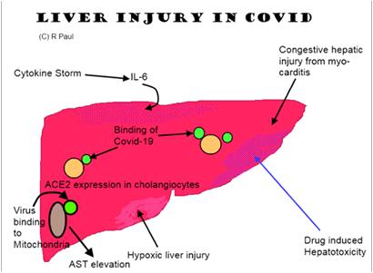
Visceral leishmaniasis (VL, also known as kala-azar) a neglected tropical and fatal parasitic disease caused by a parasite belonging to the Leishmania donovani complex and transmitted by infected female Phlebotomusargentipes sand flies. The main target of parasite is reticuloendothelial system, with infiltration of the spleen, liver, bone marrow and lymph nodes causing organomegaly and pancytopenia. Confirmation of diagnosis relies on invasive procedures like spleen or bone marrow aspirate, but most cases, having typical clinical fearures, can be detected using serological testing. Treatment of VL is very challenging because of few treatment options, long duration of treatment and drug toxicity.Treatment of choice is chemotherapy with single dose of Liposomal amphotericin B (LAmB) ormultidrug therapy (LAmB + miltefosine, LAmB + paromomycin (PM), or miltefosine + PM) for patients of VL in the Indian sub-continent.About 5-15% develop skin eruptions as sequelae of VL, known as Post Kala-azar Dermal Leishmaniasis, and ~5% have HIV-VL coinfection. Both these conditions do not have satisfactory treatment regimens.
[J Indian Med Assoc 2020; 118(6): 67-71]
Key words : Visceral leishmaniasis, L donovani and Leishmania infantum.
Visceral leishmaniasis (VL) is one among the various neglected tropical diseases. VL is caused by the Leishmania donovani complex, which includes L donovani and Leishmania infantum. L. donovaniisthe causative organism of VL in the India 1 .
It istransmitted in the Indian subcontinentby the bite of Phlebotomusargentipes (Sand fly). Persons of all ages can be affected by VL. The most common presentation of VL is an abruptonset moderate- to high -grade fever associated with rigor and chills which may continue for several weeks with decreasing intensity, and the patient may become afebrile for a short period before experiencing another bout of fever. The spleen may be palpable by the second week of illness and, may become hugely enlarged depending on the duration of illness. Hepatomegaly (usually moderate indegree) soon follows. In India, Lymphadenopathy is very rare. There is progressive anemia which may cause congestive heart failure , weight loss, hypoalbuminemia with edema, pancytopenia.Secondary infections such as measles, pneumonia, tuberculosis, bacillary or amebic dysentery and gastroenteritis arecommon. Herpes zoster, chickenpox, boils in the skin, and scabies mayalsooccur. It is fatal, if not treated.
1 MD, FRCP (London), FAMS, FNASc, FASc, FNA, FTWAS, FASTMH, Distinguished Professor of Medicine, Institute of Medical Sciences, Banaras Hindu University Varanasi 221005 and Corresponding Author 2 Department of Medicine, Institute of Medical Sciences, Banaras Hindu University, Varanasi 221005
Received on : 03/04/2020 Accepted on : 25/05/2020 Editor's Comment :
With the introduction of rK39 rapid diagnostics,
diagnosis has become simpler.
In the Indian subcontinent single dose of liposomal
amphotericin B (L-AmB) and multidrug therapy (L
AmB + miltefosine, LAmB + paromomycin [PM], or miltefosine + PM) are the preferred treatment of visceral leishmaniasis (VL).
PKDL and VL-HIV coinfection, have become
increasingly important because of their potential to trigger resurgence.
Vector control through IRS is one of the key
components of the current VL control strategy. Epidemiology :
Although the disease is endemic in over 67 countries, 90% of all reported cases occur in just six countries: Bangladesh, Brazil, Ethiopia, India, Sudan, and South Sudan. Of all the cases reported from India, the majority are from the state of Bihar 1 .
There was a dramatic decline in its incidence after extensive insecticide spraying in the 1950s by the National Malaria Eradication Programme but resurgence was noted from small area of North Bihar in the early 1970s and within next 10–15 years it spread to the entire state of Bihar, a few districts of Jharkhand and West Bengal, plus the eastern districts of Uttar Pradesh. In due course, Nepal and Bangladesh were also affected. Akala-azar elimination initiative was launched in 2005 jointly by the Governments of India, Bangladesh, and Nepal with target for elimination as annual VL incidence below 1/10,000 people at the upazilla level in Bangladesh, the subdistricts [block
67
JOURNAL OF THE INDIAN MEDICAL ASSOCIATION, VOL 118, NO 06, JUNE 2020
public health center (PHC)] level in India, and the district level in Nepal by the year 2015 – a deadline that was later reset to 2020 2 . Though there has been a dramatic decline in number of cases in India, and elimination target has been achieved in most of the endemic districts, barring few districts of Bihar and Jharkhand.
HIV-VL co-infection remains a major threat for the control of the disease, as the risk of developing active VL increases by >100 times. Initially, most of these cases were from southwestern Europe, but the number of cases is increasing in sub-Saharan Africa especially Ethiopia, Brazil and South Asia 3,4 . In India, 1.8–5.6% of VL patients were HIV-positive 3 (Fig 1).
DIAGNOSIS
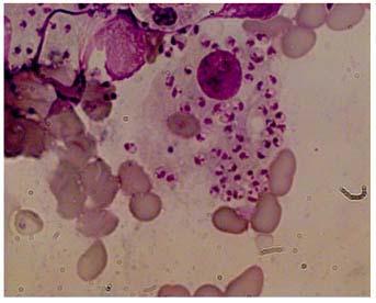
[i] Serological Tests: Through the development of the rK39 rapid diagnostic test (RDT) which has a sensitivity and specificity of 98% and 95%, respectively, serodiagnosis of kala-azar can be carried out in 20 minutes from a drop of blood. In elimination initiative, antirK39 RDT has been adopted for the diagnosis in combination with a clinical case definition 5 . However, there are two limitations, as anti K39 antibodies persist after cure for a long time (several years) thus it cannot be used in the diagnosis of relapses, further a significant proportion (~10%) of asymptomatic individuals, living in the endemic region, are also positive for serologic tests. [ii] Antigen Detection Tests: most studied test is the kala-azar latex agglutination test (KAtex), detecting a heat-stable leishmania antigen in the urine of VL patients. Its specificity was excellent but sensitivity was low (48%–87%). Attempts to improve the sensitivity and the format of the test are ongoing 6 . [iii] Molecular Diagnosis: Polymerase chain reaction (PCR)-based assays to detect parasite DNA are being increasingly used in high-income countries, particularly in Europe, but frequent demonstration of PCR-positive tests in asymptomatic infected individuals in disease-endemic regions hampers their sensitivity. Several modification of PCR like quantitative PCR or isothermal loop medicated PCR (LAMP) have been developed. This has been especially useful in the longterm monitoring of HIV-infected patients, as a way to reduce the need of invasive investigations for quantification of parasite burden 7,8 . [iv] Parasitologic Diagnosis: It is the gold standard for diagnosis, which is made by the visualization of the amastigote form of the parasites by microscopic examination of tissue aspirates (spleen, bone marrow, or lymph nodes) (Fig 2). Specificity of microscopy is high, but its sensitivity varies between spleen (93%–99%), bone marrow (52%–85%), and lymph node (52%–58%) aspirates 9,10 (Fig 2).
TREATMENT
Pentavalent antimonials (SbV) :
Sodium stibogluconate (Sbv) and meglumine antimoniate (MA) are two forms available, given in a
Fig 1 — Visceral leishmaniasis cases in India 68
JOURNAL OF THE INDIAN MEDICAL ASSOCIATION, VOL 118, NO 06, JUNE 2020
Fig 2 — Microphotograph showing intracellular and extracellular L. donovani bodies in splenic aspirate from a patient with visceral leishmaniasis
dose of 20mg/kg body weight for 28-30 days. But widespread resistance emerged in Bihar (India), and to some extent in adjoining Nepal, led to adoption of alternative treatment strategies for these regions. It's major limiting toxicities are cardiac arrhythmias, prolonged QT interval (QTc), ventricular premature beats, ventricular tachycardia, ventricular fibrillation and torsades de pointes. Other adverse effects include arthralgia, myalgia, increased pancreatic and liver enzymes 11 .
Though pentamidine was used for a brief period, however, its use was abandoned due to frequent serious toxicities like Insulin Dependent Diabetes Mellitus, unexplained hypotension and shock 12 .
AMPHOTERICIN B :
Amphotericin B deoxycholate (AB) was recommended as primary drug in patients hailing from SbV resistance region, later it was used for all patients. AB is recommended at doses of 0.75–1.0 mg/kg given for 15–20 intravenous infusions. Main toxicity are infusion reactions (in most patients), nephrotoxicity, hypokalemia, myocarditis and occasional death. Thus its infusion mandates close monitoring. Prolonged hospital stay limits the treatment to available beds and escalates the cost of therapy. To circumvent frequent and severe adverse events of AB, lipid formulations of amphotericin B have been developed with minimal side effects, and has made possible delivery of large daily doses of the drug 13 . Various trials on liposomal amphotericin B (LAmB) in India are shown in Table 1.There is considerable geographical variation in the total LAmB dose. In the Mediterranean region and South America, 18–21 mg/kg administered in various regimens, has been recommended , but in India single dose of 10 mg /kg has been shown to cure >95% VL cases 14 and it is the recommended drug in the elimination program in three countries of the Indian subcontinent.
For HIV-VL co-infection, LAmB is given at doses of 4 mg/kg for 10 doses (days 1–5, 10, 17, 24, 31 and 38) up to a total dose of 40 mg/ kg. Secondary prophylaxis is important and found to be effective in HIV-VL co-infected patients 15 . Better and shorter regimens for HIV-VL coinfection, are under clinical trials.
MILTEFOSINE :
It is an alkyl phospholipid (hexadecylphosphocholine) and the first oral antileishmanial agent registered for use following a Phase III trial in India from March 2002 at dose of 50 -100 mg/ day for 28 days, and resulted in a long-term cure rate of 94% 16 .
Study (year)
Thakur (2001) 24
Sundar (2004) 25
Sundar (2010) 14
Sundar (2014) 26 Table 1 — Liposomal Amphotericin B trials in VL in India
Trial arm and drug dosage Apparent cure Definitive 95% CI for definitive cure definitive cure (95% Confidence Interval)
L-AmB single dose 15 mg/kg AmB deoxycholate 1 mg/kg per day for 20 days 17/17 (100%) 17/17 (100%) 17/17–100 17/17 (100%) 77.1 (100%) 77.1–100
AmB deoxycholate 1 mg/kg per day for 15 days (every other day) 49/51 (96%) 49/51 (96%) 85.4–99.3 L-AmB 10 mg/kg (2 mg/kg × 5 days) 50/51 (98%) 49/51 (96%) 85.4–99.3 AmB lipid complex 10 mg/kg (2 mg/kg × 5 days) 51/51 (100%) 47/51 (92%) 80.2–97.4
L-AmB 10 mg/kg (single dose) 304/304 (100%) 291/304 (96%) 92.6- 97.6 AmB deoxycholate 1 mg/kg per day for 15 days (every other day) 106/108 (98%) 104/108 (96%) 90.2-98.8
AmB lipid emulsion 15 mg/kg (single dose) L-AmB 15 mg/kg (single dose) 354/376 (94%) 317/376 (84%) 80.1–87.8 122/124 (98%) 120/124 (97%) 91.4–99.0
69
JOURNAL OF THE INDIAN MEDICAL ASSOCIATION, VOL 118, NO 06, JUNE 2020
Because of its high cure rate and ease of administration, miltefosine was adopted by VL elimination programme in India, Nepal and Bangladesh. The recommended therapeutic regimens for patients on the Indian subcontinentare a daily dose of 50 mg for 28 days for patients weighing <25 kg, a twice-daily dose of 50 mg for 28 days for patients weighing >25 kg, and 2.5 mg/kg for 28 days for children 2-1 1 years of age.These regimens have resulted in a cure rate of 94% in India. After a decade of its use, the efficacy decreased and there was doubling of relapse rate 17 , further its efficacy from Nepal and Bangladesh was lower than observed in India. Main limitations of miltefosine are its relatively high cost, need for monitoring of gastrointestinal side effects and occasional hepatic toxicity and nephrotoxicity. It is teratogenic with a half life of >150 hours, so women of child-bearing potential have to observe contraception for the duration of treatment and for an additional 3 months. In 2014, use of miltefosine was stopped in the kala-azar elimination programme in favour of single dose LamB.
PARAMAMOMYCIN :
It is an aminoglycoside antibiotic, acts by interfering with protein synthesis in the ribosome of the target organism and also inhibit the respiration. In India, in a phase II study of VL patients, PM at a dose of 16 mg kg day-1 for 21 days led to cure in 93% 18 . In a Phase III trial PM at a dose of 15 mg kg-1 (11 mg base) for 21 days showed 95% cure rate [19]and was approved by the Indian government in August 2006 for the treatment of patients with VL This is the cheapest antileishmanial drug, and is produced in India. Its availability is an issue.
MULTIDRUG THERAPY :
Rationale of multidrug therapy is to shorten duration of therapy with reduced doses by adding synergistic drugs which lowers the adverse events, resistance, hospital stay and treatment cost. therapy has been studied in the India. In a randomized, non-comparative, group-sequential, triangulardesign study, 181 subjects were assigned to treatment with 5 mg/kg of L-AmB alone, 5 mg/kg of L-AmB followed by miltefosine for 10 days or 14 days or 3.75 mg/kg of L-AmB followed by miltefosine for 14 days. When efficacy of all regimens was apparent, 5 mg kg of L-AmB followed by miltefosine for 7 days were given 45 additional patients. All groups had similar cure rates (>95%) 20 .
Later a large Phase III study conducted in the India with three drug combinations: single injection of 5 mg/ kg LAmB and 7-day 50 mg oral miltefosine or 10-day 70 11 mg/kg intramuscular PM; or 10 days each of miltefosine and PM, were tested for the treatment of VL. Each of the combination showed an excellent CR (> 97%) 21 .
Current treatment guidelines for South Asia :
In 2010 WHO published the treatment recommendation based on the regional differences because the efficacy and required dosage of the antileishmanial agents vary in different areas 4 .
1. VISCERAL LEISHMANIASIS
Single (10 mg/kg) dose of L-AmBor multi-drug combination therapy are the preferred treatment options for the Indian subcontinent.
2. HIV-Leishmaniasis co-infection
Lipid formulations infused at a dose of 3 -- 5 mg/ kg/day or intermittently for 10 doses (days 1 -- 5, 10, 17, 24, 31 and 38) up to a total dose of 40 mg/kg are recommended. Antiretroviral therapy should be initiated and secondary prophylaxis should be given till the CD4 counts are > 200/µl (3 to 5 mg/kg every three week).
POST-KALA-AZAR DERMAL LEISHMANIASIS :
In Indian subcontinent and in Sudan and other East African countries, 2-50% of patients develop skin lesions concurrent with or after the cure of VL. Most common are hypopigmented macules, papules, and/ or nodules or diffuse infiltration of the skin and sometimes of the oral mucosa or mixed macular lesions with other types of manifestations.In PKDL, parasites are scanty in hypopigmented macules but may be seen and cultured more easily from nodular lesions. Cellular infiltrates are heavier in nodules than in macules.The diagnosis is based on history and clinical findings, but rK39 and other serological findings are positive in most cases 22 . Treatment is three to four courses of AmB spread over several months - but it is expensive and unacceptable for most patients. Oral miltefosine for 12 weeks, in the usual daily doses, cures most patients with Indian PKDL 23 .
Thus, VL is one of the major neglected and fatal infectious disease. There is emergence of drug resistance in disease endemic region, which is of concern and should be closely monitored. All suspect cases of kala-azar should be screened and diagnosed in an endemic area in an individual who has fever for more than 2 weeks, splenomegaly, and a positive serological rk39 test. There is increasing incidence of HIV-VL coinfection worldwide which further bring challenges to diagnosis and treatment. PKDL and HIVVL coinfections pose a threat to the elimination of VL
in this region, there is a need to develop simple and
JOURNAL OF THE INDIAN MEDICAL ASSOCIATION, VOL 118, NO 06, JUNE 2020
effective regimens for these conditions. Vector control through IRS is one of the key components of the current VL control strategy. There is need of scientists, funding agencies, implementers for universal approach to diagnosis and treatment and elimination of VL.
Funding : None
Conflict of Interest : None REFERENCES
1 Control of the Leishmaniases: Report of a Meeting of the
WHO Expert Committee on the Control of Leishmaniases.
Geneva: World Health Organization; 2010. Technical Report
Series Vol. 949. [2010 Mar 22-26]. 2 Accelerated Plan for Kala-azar Elimination-2017. [cited 2018
Sep 13]. Available from: http://nvbdcp.gov.in/Doc/Accelerated
Plan- Kala-azar1-Feb2017.pdf 3 Burza S, Mahajan R, Sanz MG, et al — HIV and visceral leishmaniasis coinfection in Bihar, India: an underrecognized and underdiagnosed threat against elimination. Clin Infect
Dis. 2014;59(4):552– 555. 4 de Sousa-Gomes ML, Romero GAS, Werneck GL — Visceral leishmaniasis and HIV/AIDS in Brazil: are we aware enough?
PLoSNegl Trop Dis. 2017;11(9):e0005772. 5 Sundar S, Reed SG, Singh VP, Kumar PC, Murray HW. Rapid accurate field diagnosis of Indian visceral leishmaniasis.
Lancet. 1998; 351: 563-5. 6 Sundar S, Agrawal S, Pai K, et al — Detection of leishmanial antigen in the urine of patients with visceral leishmaniasis by a latex agglutination test. Am J Trop Med Hyg 2005;73:269–71. 7 Mary C, Faraut F, DrogoulMP, et al — Reference values for
Leishmania infantum parasitemia in different clinical presentations: quantitative polymerase chain reaction for therapeutic monitoring and patient follow-up. AmJ Trop Med
Hyg 2006;75:858–63. 8 Riera C, Fisa R, Ribera E, et al — Value of culture and nested polymerase chain reaction of blood in the prediction of relapses in patients co-infected with leishmania and human immunodeficiency virus. Am J Trop Med Hyg 2005;73:1012–5. 9 Sundar S, Rai M — Laboratory diagnosis of visceral leishmaniasis. Clin Diagn Lab Immunol. 2002;9: 951-8. 10 Chappuis F, Sundar S, Hailu A — Visceral leishmaniasis: what are the needs for diagnosis, treatment and control? Nat Rev
Microbiol 2007;5:873–82. 11 Sundar S, More DK, Singh MK, Singh VP, Sharma S, Makharia
A, Kumar PC, Murray HW — Failure of pentavalent antimony in visceral leishmaniasis in India: report from the center of the
Indian epidemic. Clin Infect Dis. 2000;31: 1104-7. 12 Jha SN, Singh NK, Jha TK — Changing response to diamidine compounds in cases of kala-azar unresponsive to antimonial.
J Asso Phys India 1991;39:314–316.
13 Sundar S and Chakravarty J — An update on pharmacotherapy for leishmaniasis. Expert Opinion on
Pharmacotherapy 2015; 16(2): 237–252. 14 Sundar S, Chakravarty J, Agarwal D, Rai M, Murray HW —
Single-dose liposomal amphotericin B for visceral leishmaniasis in India. N Engl J Med 2010; 362: 504-12. 15 Lopez-Velez R, Videla S, Marquez M, Boix V, Jimenez-Mejias
ME, Gorgolas M, Arribas JR, Salas A, Laguna F, Sust M,
Canavate C, Alvar J — Amphotericin B lipid complex versus no treatment in the secondary prophylaxis of visceral leishmaniasis in HIV-infected patients. J Antimicrob
Chemother 2004;53: 540–543. 16 Sundar S, Jha TK, Thakur CP, et al — Oral miltefosine for
Indian visceral leishmaniasis. N Engl J Med 2002;347(22):1739- 46 17 Sundar S, Singh A, Rai M, Prajapati VK, Singh AK, Ostyn B,
Boelaert M, Dujardin JC, Chakravarty J — Efficacy of
Miltefosine in the Treatment of Visceral Leishmaniasis after a
Decade of use in India. Clin Infect Dis. 2012;55(4):543-50. 18 Jha TK, Olliaro P, Thakur CP, Kanyok TP, Singhania BL, Singh
IJ, Singh NK, Akhoury S, Jha S — Randomised controlled trial of aminosidine (paromomycin) v sodium stibogluconate for treating visceral leishmaniasis in North Bihar, India. BMJ 1998;316:1200–1205. 19 Sundar S, Jha TK, Thakur CP, Sinha PK, Bhattacharya SK.
Injectable paromomycin for Visceral leishmaniasis in India. N
Engl J Med. 2007;356:2571-81. 20 Sundar S, Rai M, Chakravarty J, Agarwal D, Agrawal N, Vaillant
M, Olliaro P and Murray HW — New treatment approach in
Indian visceral leishmaniasis: single-dose liposomal amphotericin B followed by short-course oral miltefosine.
Clin Infect Dis 2008;47:1000–1006. 21 Sundar S, Sinha PK, Rai M, et al — Comparison of shortcourse multidrug treatment with standard therapy for visceral leishmaniasis in India: an open-label, non-inferiority, randomised controlled trial. Lancet. 2011; 377:477-86. 22 Zijlstra EE, Musa AM, Khalil EAG, et al — Post-kala-azar dermal leishmaniasis. Lancet Infect Dis. 2003;3(2):87–98. 23 Sundar S, Sinha P, Jha TK, et al — Oral miltefosine for Indian post-kala-azar dermal leishmaniasis: a randomized trial. Trop
Med Intl Health 2013;18: 96–100. 24 Thakur CP.A single high dose treatment of kala-azar with
Ambisome (amphotericin B lipid complex): a pilot study. Int J
Antimicrob Agents. 2001; 17(1): 67-70. 25 Sundar S, Mehta H, Suresh AV, Singh SP , Rai M, Murray H W.
Amphotericin B treatment for Indian visceral leishmaniasis: conventional versus lipid formulations. Clin Infect Dis. 2004;38 : 377-83. 26 Sundar S, Pandey K, Thakur CP, Jha TK, Das VN, Verma N,
Lal CS, Verma D, Alam S, Das P — Efficacy and safety of amphotericin B emulsion versus liposomal formulation in Indian patients with visceral leishmaniasis: a randomized, openlabel study. PLoSNegl Trop Dis. 2014; 8(9): e3169.
71
JOURNAL OF THE INDIAN MEDICAL ASSOCIATION, VOL 118, NO 06, JUNE 2020
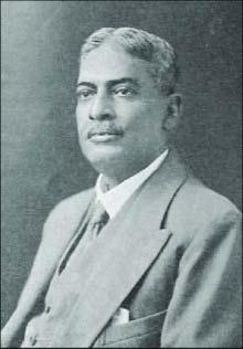
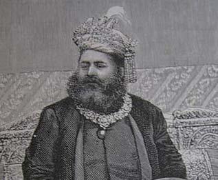
SIR UPENDRANATH BRAHMACHARI — THE FORGOTTEN MAESTRO
Sir Upendranath Brahmachari, an Indian scientist and leading medical researcher and practitioner was born on 19 December 1873 in Sardanga village near Purbasthali, district Burdwan of West Bengal. After completion of MD & PhD, he was appointed as a teacher at the Campbell Medical School, Calcutta (Now Nilratan Sircar Medical College and Hospital), where he carried out his monumental work on Kala-azar. He made the path breaking discovery of urea stibamine which drastically reduced the deaths caused due to kalaazar and which for many years was mankind’s only answer to the dreaded disease Kala-azar. Sodium stibogluconate, a newer form of this drug is still widely used globally for the treatment of kala-azar. In 1922, he discovered a new and deadly form of leishmaniasis, marked by the sudden appearance of eruptions on the face of the patient without fever or other complaints. It has been later termed as post-kala-azar dermal leishmaniasis.
Sir U.N Brahmachari was conferred with many awards including the prestigious Sir William Jones Medal of the Asiatic Society of Bengal, the Minto Medal from Calcutta School Of Tropical Medicine and the Griffith Memorial Prize from the University of Calcutta. He was awarded the title of Rai-Bahadur and was conferred the Kaisar-i-Hind Gold Medal. The British Government conferred him with the prestigious Knighthood in 1934.He was a nominee for the Noble Prize twice in 1929 and 1942 in the category of physiology and medicine. He spent many nights working in a room lit by a single kerosene lamp in the ill equipped room of Campbell Medical College with no research chemist as assistant, no water basin to wash hands and no modern equipments. Even bigger handicap was that no Indian till date had distinguished himself/herself in medical research which was the domain of British doctors, pharmacists and chemists. His only humble goal was to find the answer to a disease which had killed millions of his countrymen. 72