7 minute read
u

JOURNAL OF THE INDIAN MEDICAL ASSOCIATION, VOL 118, NO 06, JUNE 2020
Case Report An Atypical Manifestation of Post Streptococcal Glomerulonephritis
Advertisement
Uddalak Chakraborty 1 , Tarun Kumar Paria 2 , Purbasha Biswas 2 , Tanuka Mandal 3 , Atanu Chandra 4
Acute post streptococcal glomerulonephritis is a relatively common entity encountered by the internist in day to day practice. Aside the relatively common presentation of hematuria, oliguria and facial puffiness, one may present with convulsions and hypertensive encephalopathy. A 19 year old male patient was admitted with generalized tonic clonic seizure and elevated blood pressure, with some papular rash in different stages of healing in both lower limbs, MRI brain revealed bilateral symmetrical hyperintensities in frontal and parieto-occipital areas which resembled posterior reversible encephalopathy syndrome (PRES) like changes. The evaluation of hypertension in the young boy revealed microscopic hematuria and nephritic range proteinuria with decreased serum complements suggestive of acute glomerulonephritis. Symptomatic management ensured a complete recovery without any neurological deficit and resolution of MRI findings over a week. PRES is a rare clinico-radiological entity and may be a presenting symptom in patients with post streptococcal glomerulonephritis.
[J Indian Med Assoc 2020; 118(6): 58-9]
Key words : Post-streptococcal glomerulonephritis; PRES; hypertension; convulsions.
Acute post streptococcal glomerulonephritis (PSGN) is a nephritic syndrome which may present commonly with oliguria, cola colored urine, puffiness of face and eyelids, pedal edema, and hypertension. Apart from these classical symptoms, PSGN may present with atypical manifestations like myocardial dysfunction, acute renal failure, etc. Reversible encephalopathy due to dysfunction of cerebral autoregulation has been reported in children and juvenile patients with PSGN 1 . An adult patient presenting with reversible encephalopathy and posterior reversible encephalopathy syndrome like changes in MRI brain due to PSGN is relatively rare.
CASE REPORT
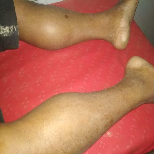
A 19 year old male patient presented to us in September, 2019 with his first episode of generalized tonic clonic seizure preceded by headache and nausea. He had a blood pressure of 160/100mmHg and complained of persisting headache and nausea, followed by which he again suffered another episode of generalized tonic clonic convulsion. Clinical examination revealed only a papular rash in both lower limbs in different stages of healing, which was accompanied by some pustular discharge from the rash 2
1 MBBS, Postgraduate trainee, Department of General Medicine, RG Kar Medical College and Hospital, Kolkata and Corresponding Author 2 MBBS, Post graduate trainee, Department of General Medicine, RG Kar Medical College and Hospital, Kolkata 3 MD, MACP, Senior Resident, Dept of Medicine, RG Kar Medical College Hospital, Kolkata 4 MBBS, Assistant Professor, Department of General Medicine, RG Kar Medical College and Hospital, Kolkata
Received on : 05/06/2020 Accepted on : 15/06/2020 Editor's Comment :
PSGN may present with hypertensive encephalopathy apart from the common symptoms of nephritic syndrome. PRES is a characteristic finding in brain imaging usually associated with hypertensive emergencies due to failure of cerebral autoregulation. PRES as a presentation of PSGN though seen commonly in children, may also be seen in young adults as in our case.
weeks prior to the current episode. Systemic examination did not reveal any abnormality.
A subsequent MRI brain revealed bilateral symmetrical hyperintensities in FLAIR (fluid attenuation inversion recovery) sequence in frontal and parieto-occipital areas (Figs 1,2), which predominantly involved the white matter. Further evaluation of hypertension in such a young male patient revealed microscopic hematuria with 8-9 RBC/ hpf with few dysmorphic RBC and pus cells 2-3/hpf with trace albuminuria in routine urinalysis, with negative cultures from urine. Urine output was around 700ml in 24 hours. An urinary albumin-creatinine ratio revealed a nephritic range proteinuria with 1685 mg/mcg (n=30-300mg/mcg). Serum complements revealed a diminished C3 level around 26mg/dl(n= 66-185mg/dl)with a C4 level in the normal range. Ultrasonographic imaging of kidneys did not reveal any abnormality. ASO titre was found to be raised around 600 units/ml (n<200 units/ml) and Anti DNAse B levels were raised as well around 300 units/ml (n<85 units/ml). Renal function tests were unaltered. All other routine investigations were non-contributory.
The patient was admitted with a hypertensive emergency and characteristic signal changes in brain imaging. On an
58
JOURNAL OF THE INDIAN MEDICAL ASSOCIATION, VOL 118, NO 06, JUNE 2020

PSGN is an immune-complexmediated disease with decline in the serum complement (C3) levels. Hypertension is found in majority of patients with PSGN and may also be associated with hypertensive encephalopathy in some cases. Toxic effects of streptococcus on the central nervous system may also lead to encephalopathy. Zaki et al. reported PRES as an unusual manifestation of PSGN in a 8 year old child 4 . Wirrell et al reported a series of 4 similar cases in children, however such
Fig 1 — A T2 Flair MRI showing bilateral Fig 2 — A T2 Flair MRI showing bilateral presentation in a young adult is frontal and occipital hyper intensities parietal and occipital hyper intensities relatively rare 5 . However, in this case, the attempt to work out the cause of hypertension in the young patient presented with convulsions and hypertension without individual, urinalysis revealed microscopic hematuria with any features of oedema, oliguria or frank hematuria. dysmorphic RBC and microalbuminuria with low urine Furthermore, the constellation of symptoms were not output. Decreased C3 levels led along with the supportive suggestive of glomerulonephritis and the lesions on the findings led us to the diagnosis of Acute glomerulonephritis skin with elevated Anti DNAse B levels, microscopic in the background of healing skin lesions, probably a post haematuria anddecreased levels of serum complement streptococcal sequale as evidenced by raised ASO and Anti clinched the diagnosis. Renal biopsy was not attempted in DNAse levels. our case as the symptoms resolved gradually and
The patient was diagnosed as a case of acute post complement levels normalized in due course of time. streptococcal glomerulonephritis with PRES as presenting T2 weighted MRI images showing diffuse, symmetrical manifestation. He started on enalapril and levetiracetam. reversible hyperintensities involving the white matter with He improved remarkably well with increased urine output relative sparing of the grey matter is suggestive of over the next 7 days. The proteinuria subsided and repeat PRES.Cerebral white matter is more susceptible to serum complement levels were in normal range by 2 weeks. vasogenic oedema. PRES with hypertensive A repeat MRI brain did not reveal any abnormality suggestive encephalopathy should be managed aggressively in order of transient abnormalities during hypertensive to avoid catastrophic complications and permanent residual encephalopathy. sequalae. Anti hypertensive therapy with anticonvulsants D ISCUSSION for symptomatic management is extremely beneficial.
PRES has been mainly attributed to vasogenic oedema, REFERENCES predominantly involving the white matter in the parieto1 Fux CA, Bianchetti MG, Jakob SM, Remonda L — Reversible occipital areas and has been classically described in encephalopathy complicating post-streptococcal ecclampsia and immunosuppressive therapy 2 . The pathophysiology of PRES is complex and highly debated. It is often attributed to autoregulatory failure of cerebral blood vessels leading to vasogenic oedema as seen in severe hypertension. The increased predilection of vasogenic oedema for the posterior cerebrum may be glomerulonephritis. Pediatr Infect Dis J 2006;25:85-7. [PubMed] [Google Scholar] 2 Hinchey J, Chaves C, Appignani B, Breen J — A posterior reversible leucoencephalopathy syndrome. N Engl J Med 1996; 334: 494-500. [PubMed] [Google Scholar] 3 Bartynski WS — Posterior Reversible Encephalopathy Syndrome, Part 2: controversies surrounding explained by the decreased density of sympathetic neurons pathophysiology of vasogenicedema. Am J Neuroradiol 2008; in the posterior cerebral artery territory 3 . At the onset of 29: 1043-49. [PubMed] [Google Scholar] symptoms, the blood pressure may be normal or minimally 4 Zaki, Syed Ahmed, and Preeti Shanbag — “Unusual elevated. The clinical manifestations of PRES are usually headache, vomiting, seizures, visual disturbances with altered sensorium among which seizures are the most consistent manifestation. PRES is most commonly seen in pregnancy induced hypertension, however it may be seen presentation of poststreptococcal glomerulonephritis as posterior reversible encephalopathy syndrome.” Journal of pediatric neurosciences vol. 9,1 (2014): 42-4. doi:10.4103/ 1817-1745.131484 5 Wirrell E, Hamiwka L, Hamiwka L, Grisaru S, Wei X — (2007). Acute Glomerulonephritis Presenting With PRES: A Report of in hypertensive encephalopathies as in our case but PRES 4 Cases. Canadian Journal of Neurological Sciences / Journal as a first presentation of PSGN is relatively rare. Canadien Des Sciences Neurologiques, 34(3): 316-21.
PSGN usually occurs after infection of throat or skin by doi:10.1017/S0317167100006740 nephritogenic strains of group-A beta hemolytic streptococci.
59