26 minute read
Vimal Raj, Jagalpathy Jagdish

JOURNAL OF THE INDIAN MEDICAL ASSOCIATION, VOL 118, NO 06, JUNE 2020
Imaging in Medicine Computed Tomography (CT) Chest imaging in Diagnosis and Management of patients with COVID-19
Advertisement
Vimal Raj 1 , Jagalpathy Jagdish 2
Over the last few weeks, there is an increase in the number of cases of Corona VIrus Disease- 2019 (COVID -19) reported in India. Every clinician is likely to manage one or other form of presentation of the disease in his or her practice. Therefore, every clinician needs to be aware of the imaging appearances of the COVID-19. Chest radiograph is not a good diagnostic tool and higher imaging modality such as Computed Tomography (CT) may be appropriate. The risk of spread of infection amongst patients and to the healthcare workers can be a challenge in performing CT studies for patients with COVID-19. In this review, we describe common CT appearances of COVID-19 infection and its complications. We also discuss the role of CT in assessing disease severity, prognosis and its utility in monitoring of disease progression. National and international guidelines relating to the use of CT imaging in COVID-19 are also highlighted.
[J Indian Med Assoc 2020; 118(6): 35-42]
Key words : COVID-19, CT, MDCT, Imaging, Chest CT, Novel Corona Virus
Novel coronavirus was identified, in January 2020, as a cause of pneumonia in many patients since December 2019 in China. The infection spread rapidly within China and in the rest of the world over the next few weeks. The World Health Organisation (WHO) termed the illness as Corona Virus Disease 2019 (COVID-19) and declared it as a global pandemic on 11 th of March 2020 1,2 .
By 12 th of June 2020, more than 74 lakh confirmed cases have been reported to WHO worldwide with a more than 4 lakh patients succumbing to the disease. diseased patients. India with more than 3 lakh cases was the fourth-worst effected country behind the United States of America, Brazil and Russia 3 . These numbers are likely to further increase in India as travel and lockdown restrictions are eased.
COVID-19 presents with symptoms of lower respiratory tract infection and many patients remain asymptomatic or with mild symptoms 4,5 .Isolation of virus in the respiratory samples using real-time reverse transcriptase-polymerase chain reaction (RT-PCR) is the testing of choice for detection of COVID-19. RTPCR has variable sensitivity ranging from 37% to 91% 6-11 . RT-PCR results can be falsely negative initially
1 FRCR,CCT (UK), EDM, PGDMLS, Department of Radiology, Narayana Hrudayalaya, Bangalore and Corresponding Author 2 Department of Radiology, Al Rahba Hospital, Seha, Abudhabi, UAE
Received on : 16/06/2020 Editor's Comment :
CT scans have variable sensitivity in detecting
patients with Covid-19 but is specially helpful for areas with limited availability of RT-PCR.
Ground glass opacities, consolidation, reticular
changes are most common finding.
Protection of healthcare workers of Radiology
Department is of prime importance.
and often require repeating of test. RT-PCR tests are also not easily available in different parts of the country and there can be significant delay in getting the results of these tests.
Chest imaging tests can be useful in the detection and management of patients with suspected or known COVID-19. Chest radiographs (CXR) are widely available and cost-effective but can also have variable sensitivity, with nearly 60% of patients having a normal CXR in one of the recent studies 10 12-14 . Computed Tomography (CT) imaging of the chest has a better resolution than CXR and may allow improved detection and assessment of patients with known or suspected COVID-19 (Figure 1). In this review, we describe common CT appearances of COVID-19 infection and its complications. We also discuss the role of CT in
Accepted on : 19/06/2020
assessing disease severity, prognosis and its utility in monitoring of disease progression. National and international guidelines relating to the use of CT imaging in COVID-19 are also highlighted.
35
JOURNAL OF THE INDIAN MEDICAL ASSOCIATION, VOL 118, NO 06, JUNE 2020
Figure 1: Chest radiograph (A) and coronal CT (B) images of a 42-year-old man with a one-week history of fever. The true extent of lung involvement is better appreciated on the CT compared to the CXR (red box) in this patient with COVID-19.
CT findings in COVID-19 :
CT chest appearances of COVID-19 can change with the severity of disease and the time course of presentation 15-17 . Commonly described CT chest findings in COVID-19 include ground-glass opacity, consolidation, reticular pattern and crazy paving. Various other CT findings are also reported in lesser frequency in patients with COVID-19.
Ground Glass Opacities (GGO) :
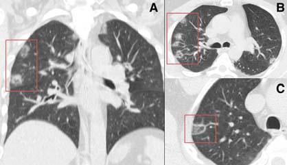
Ground Glass area on CT, appears as an area of hazy increased opacity of lung with preservation of bronchial and vascular margins 18 . This correlates with post mortem finding of pulmonary oedema and hyaline membrane formation in the lungs 19 . The finding of GGO has consistently been reported in numerous studies that have described imaging appearances of COVID19 6 15 20-22 .The frequency of GGO seen on imaging varied between 34 to 98% in various studies 16 23 . The GGO’s are often seen in the periphery of the lungs with lower zone predominance (Figure 2). Unfortunately, GGO is not a highly specific finding and can also be seen in other viral infective and non-infective conditions. GGO can be seen in isolation or in combination with other patterns inCOVID-19 6 22 .
Consolidation :
Consolidation is a marker of replacement of alveolar air with exudative tissue or other product of disease at histopathology. It is often considered synonymous with an infective pathology and is seen as an area of homogeneous increase in parenchymal density with obscuration of the vessel and airway walls 18 . In COVID19, there are multifocal areas of patchy or segmental consolidation distributed in subpleural areas or along the bronchovascular bundles (Figure 3). The frequency of involvement varies between 2-64% in the reported studies 15 16 20 23 . Bilateral involvement is more common, which is in contrast to other bacterial pneumonias which are often unilateral/unilobar in 36
distribution 24 . Many patients have a mixed pattern with areas of GGO intermixed with consolidation and other lung patterns (Figure 4).
Reticular Pattern:
Reticular pattern refers to the prominence of the interstitial structures with thickening of inter and intralobular septa. These are seen as increased ‘white lines’ in the chest CT and are a common feature seen in patients with interstitial lung disease. These are often associated with some dilatation/prominence of the adjoining bronchi (Figure 5). This has been reported in multiple studies as the third most common finding after GGO and consolidation 15 .
Crazy Paving Pattern:
This pattern refers to thickened inter and GGO 18 (Figure 6). This pattern has been reported less
Figure 2: Chest CT images in axial (A, B), Coronal (C) and Sagittal (D) planes in a 37-year-old male patient with COVID-19. He presented with a 7-day history of fever and cough and two days of shortness of breath. Bilateral peripheral ground-glass opacities can be seen (red boxed) with lower zone predominance.
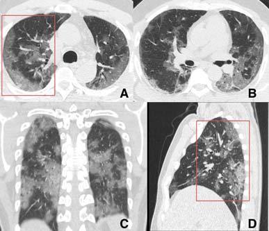

Figure 3: Chest CT axial images from three different patients of COVID-19, showing consolidation as the predominant pattern of involvement (red box). Notice the peripheral distribution of disease in A and peribronchial distribution in other cases. In the third patient (C, D) there are large confluent areas of consolidation in the central aspect of the lungs.
intralobular septa superimposed on a background of
JOURNAL OF THE INDIAN MEDICAL ASSOCIATION, VOL 118, NO 06, JUNE 2020
frequently in many studies with its presence in COVID19 varying between 10 to 36% 22,23 . Although, initially reported in patients with alveolar proteinosis this pattern can be seen in various other diffuse lung diseases and is not specific for COVID-19 25,26 .
Other patterns :
Various other CT patterns and findings have been described in patients with COVID-19. One such pattern is the ‘prominent vessel sign’ reported in few of the published studies 15,27 . In this pattern, there is a prominent/dilated vessel seen within the area of abnormality on lung CT (Figure 7). This may be secondary to damage and swelling of the capillary wall due to pro-inflammatory factors 15 . ‘Air bronchogram’ is a pattern wherein air-filled bronchi is present in the background of a consolidation lung or GGO. This also has been reported in some cases with COVID-19 15 28 (Figure 8). Lung nodules in isolation or associated with surrounding ground-glass change, the so-called ‘Halo’ sign have been reported in varying frequency in multiple studies (Figure 9). Lung nodules are often also seen in patients with other viral pneumonias and are not a very specific feature of COVID-19 6,15,21,29 . A ‘reverse halo sign’ has also been described in patients 37
Figure 6: CT images of a 53-year-old female with COVID-19. The patient presented with fever and the CT shows areas of crazy paving (A and zoomed image in B) in the periphery with linear opacities overlying the GGO.
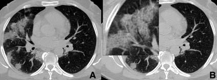
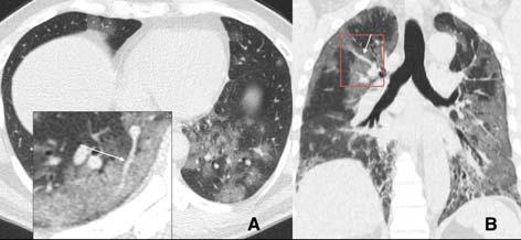
Figure 7: CT images of two different patients with COVID-19 with the ‘prominent vessel sign’ (white arrows in A and B) in the background of GGO.
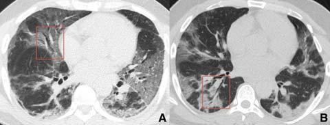
Figure 4: Chest CT axial images of a patient with COVID-19,with a mixture of GGO (red box in A) and consolidation (black box in A& B).
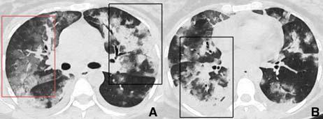
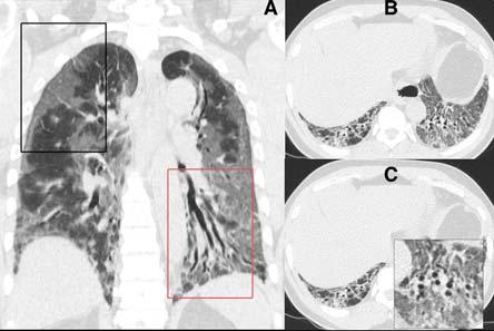
Figure 5: CT images of a 53-year-old male, who presented with a history of fever and shortness of breath for 1 week. He had high oxygen requirement at presentation with extensive peripheral ground glass changes (A- black box) and bibasal bronchial dilatation (B&C). The patient went on to be intubated and succumbed to the disease on day 28 of the presentation.
Figure 8: CT images of two different patients with COVID-19 with the ‘air bronchogram sign’ (red box in A and B) in the background of GGO and consolidation. with COVID-19. This may represent either progression of disease from GGO to consolidation or resolution of lesion with lower density in the center 15 (Figure 10, 11). An ‘air bubble sign’ may also be seen as a dark area containing air in the background of GGO. This could represent early changes of resolution of consolidation/GGO 15 (Figure 11). Pleural thickening, pleural effusion, lymphadenopathy and pericardial effusions are other patterns which have been described in patients with COVID-19. These are, however, not very frequent or a predominant feature in these patients. Abnormal coagulation has been increasingly reported in COVID-19 patients with some of them having a pulmonary embolism. The reported incidence of acute pulmonary embolism in COVID-19 patients is as high as 24% especially in patients with severe symptoms and those requiring intensive care therapy 30 31 . The Ddimer was found to be significantly elevated in these
patients.
JOURNAL OF THE INDIAN MEDICAL ASSOCIATION, VOL 118, NO 06, JUNE 2020
Learning Points • Ground glass opacification, consolidation and reticular changes are the most common findings on CT of patients with COVID-19. • Multifocal patchy areas of abnormalities involving both lungs are common. • Peripheral and peri-bronchovascular involvement is common. • Prominent vessel sign is a new sign described in patients with COVID-19. • Acute pulmonary embolism can be seen in as high as 24% of patients with severe symptoms of COVID-19.
Role of CT in COVID-19 :
CT in Diagnosis and Screening for COVID-19:
RT-PCR testing of the respiratory sample is considered a gold standard test in the diagnosis of COVID-19. As mentioned previously, there is significant variability in the sensitivity of the test, ranging from 37% to 91% 6-11 . Questions have been raised on the study design and error in the methodology of some of these studies 32 . However, with limited availability and long turn around time for RT-PCR reports, another
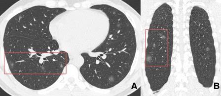
Figure 9: CT images (A-axial, B-coronal) of a 30-year-old male with COVID-19. Notice the well-defined ground glass nodules in both the lungs with some halo around some of the nodules (red box).
Figure 10: Images of a 48-year-old man who presented with cough and fever for 8 days. Multifocal peripheral areas of GGO are seen with central areas of clearing giving the classical
‘reverse halo’ sign (red box in A- coronal image and B/C- axial images). 38 modality for screening of patients is desirable. During the peak phase of disease in China, CT scans were used in the screening of patients suspected of being infected 33 . CT scans which were done as part of the ‘fever clinic’ had a significant role in controlling the epidemic in China 34 . A recently published metaanalysis looked at the sensitivity of CT in detecting patients with COVID-19. The sensitivity of CT was extremely high (96-99%) in studies conducted within Wuhan while it varied between 69 to 98% in other regions 33 . In a different meta-analysis, the pooled sensitivity of chest CT was 94% compared to 89% of RT-PCR 35 . The sensitivity of CT varied based on the number of asymptomatic patients and the severity of the illness/epidemic 33,35 . Contradictory results have been shown in some other studies wherein 56% of symptomatic patients had a normal scan in the early phase of the disease despite a positive RT-PCR 16 . Almost half (46%) of asymptomatic patients in the Diamond Princess cruise ship had a normal CT scan but were positive on RT-PCR testing 36 . CT findings of COVID-19 are not exclusive and can be seen in other viral pneumonias also. This can reduce the specificity of chest CT studies. However, sensitivity is more important than specificity in the context of emergency disease control 33 . The negative predictive value of chest ranges from 1.5% to 30% 35 .
CT is more than 95% while the positive predictive value
Learning Points • CT scans were routinely used for screening of patients with suspected COVID-19 in China due to the limited availability of RT-PCR testing kits. • CT scans have variable sensitivity in detecting patients with COVID-19. • Higher sensitivity is seen in patients with symptoms and areas with a higher prevalence of disease. • Poor sensitivity may be seen in young patients or those who are asymptomatic. • The use of CT scans in areas with limited availability of RT-PCR can help in faster diagnosis and better control of disease spread. • CT findings of COVID-19 can mimic other
viral pneumonias.
CT in assessing disease severity and progression
Multiple studies have looked at the CT appearances of COVID-19 based on the duration of symptoms 16,17,28 . Most of the patients initially presented with small peripheral GGO and nodules. These progressed into consolidation, reticular changes and crazy paving patterns as the time progressed and
JOURNAL OF THE INDIAN MEDICAL ASSOCIATION, VOL 118, NO 06, JUNE 2020
peaked after 10 days of initial symptom onset (Figure 12). With the progression of time, peripheral involvement with reticular changes and in some cases reverse halo sign was also seen more frequently (Figure 13 and 14).
Assessment of disease severity and its progression can also be objectively estimated with CT. Pan et al, used a simple scoring system to assess the severity of disease on CT 28 . In this system, each of the 5 lobes was visually scored from 0 to 5 as: 0- no involvement, 1: <5% involvement, 2: 5 to 25% involvement, 3: 26- 49% involvement, 4: 50-75% and 5: >75% involvement. A minimum score of 0 and a maximum score of 25 is possible. They found an increase in lung involvement and the severity score for the first two weeks after disease onset and before recovery. In a separate study, it was found that patients who had a higher CT severity score at presentation had a significantly lower rate of recovery and discharge 37 . Patients requiring intensive care unit (ICU) on admission had bilateral multilobular and sub-segmental consolidation in comparison to nonICU patients who presented with GGO and sub39
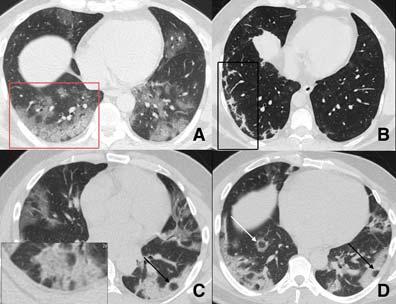
Figure 11: Composite axial CT images of different patients with COVID-19. Various patterns of involvement can be seen in these images. A- peripheral GGO (red box), B- peripheral consolidation/reticular changes (black box), C- Crazy paving (zoomed image) and air bubble sign (black arrow) and DReverse halo (white arrow) and air bubble sign (black arrow). segmental consolidation 38 (Figure 15). Other CT severity scores have also shown good correlation with clinical severity of disease 39,40 . The degree of lung inflation at the initial CT can also predict adverse outcomes in patients with COVID-19 41 .
Learning Points • Initial presentation on CT is with peripheral GGO. • Expansion of GGO and/or conversion into consolidation is a sign of disease progression. • Patients requiring ICU care on admission had bilateral multiple lobular and sub-segmental consolidation. • Patients with higher CT severity score at presentation had a significantly lower rate of recovery and discharge.
Guidelines on Use of CT in COVID-19 :
National and international guidelines have recommended the use of CT imaging only in patients with moderate to severe symptoms or in those with clinical deterioration 5,42-44 . Two of the more recent guidelines have also considered prevalent resource constraints relating to RT-PCR testing in their recommendations 8 45 . In India, there is a variable availability of RT-PCR testing and the time delay in getting the reports can also be significant. Contrary to most of the guidelines, few units have already started using CT for screening of patients on an ad hoc basis. on use of chest imaging are highlighted here 45 :
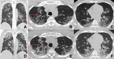
Figure 12: CT images of a 50-year-old man with COVID-19. Initial scan on day 6 after initiation of symptoms (A, B, C) showed peripheral GGO (red box) with the progression of the same on follow up scan (D, E, F) when patients symptoms worsened.

Figure 13: CT images of a 37-year-old patient presented with shortness of breath, fever and cough. His CT at presentation (A, B, C) shows mild peripheral GGO (red box). He had a repeat CT after 4 days due to the increasing requirement of oxygen. This showed progression of the GGO bilaterally into organized consolidation with reticular changes (D, E, F).
The main features of the WHO recent recommendations
JOURNAL OF THE INDIAN MEDICAL ASSOCIATION, VOL 118, NO 06, JUNE 2020
WHO recommendation on use of Chest Imaging • Chest imaging should not be used for diagnosis of COVID-19 in asymptomatic contacts. • For symptomatic patients, chest imaging should be used for COVID-19 diagnosis only when: RT-PCR test is either not available, test results are delayed or when the initial test is negative but there is high clinical suspicion. • Chest imaging should be used (along with clinical parameters) in non-hospitalized patients with mild symptoms to decide between hospital admission and home discharge. • Chest imaging should be used (along with clinical parameters) in non-hospitalized patients with moderate to severe symptoms to decide between ICU and ward admission. • Chest imaging should be used (along with clinical parameters) in patients with moderate to severe symptoms to inform therapeutic management. • Chest imaging is not recommended in hospitalized patients without symptoms to decide on decisions regarding discharge.
Machine Learning/Artificial Intelligence
Automation is becoming a new norm in all fields. There have been significant technological advances in CT imaging making it faster and improving the resolution of images acquired. CT images are very well suited for machine learning/artificial intelligence (ML/ AI) due to the availability of pixel-level density maps. There have been many initiatives around the world in making COVID-19 detection faster and accurate on imaging by use of ML/AI. These software work independent of the radiologist and can highlight

Figure 14: CT images of a 43-year-old female healthcare worker with COVID-19. CT study at presentation (A,B,C) and after 14 days (D,E,F) from the initial scan are depicted. She presented with 6 days history of cough and fever. Note temporal resolution of parenchymal changes from GGO at presentation to subpleural residual band like opacities. Her symptoms had also resolved (similar to the resolution of CT changes) and she was discharged following the second CT. 40 abnormal areas on a CT and predict the presence of COVID-19. CT severity scores can also be easily estimated by use of ML/AI algorithms and have shown to be useful in prognosis of patients 39 . Serial examination of patients and assessing disease progression is also easier by using ML/AI software. Some of these products are available commercially and even indigenously built in India (Figure 16).
Learning Points
Artificial Intelligence/Machine learning, based algorithms can quickly determine the presence/ absence of COVID-19 changes on CT and also help in quantifying the disease severity.
Infection Control
The novel coronavirus is highly contagious and transmission via droplets in radiology departments is a significant concern 46 . It is important to align local practices to adhere to the national and international guidelines on social distancing and the use of personal protective equipment (PPE) 45 . Appointment system should be followed to avoid unnecessary rush of patients in the department. The gap between the scans should be extended to allow for cleaning. Where ever possible all positive cases should be scanned in the evening hours after routine hours of working. Hand hygiene and continued education of staff on infection control should be reinforced. All patients coming to the department should be screened as per the local policies for fever, symptoms, travel history and exposure to known patient etc. Patients should wear a facemask and sanitize their hands-on entry into the department. The CT console room entry should be restricted and be treated as a clean zone. The CT gantry room should be treated as contaminated and staff should wear appropriate PPE when dealing with the patient. Appropriate decontamination steps should be taken as per the manufacturers advice after every positive patient 45 . The workflow of the department should be optimized so that known COVID-19 patients should not be made to wait in the department. Rotational duty of staffs is useful in restricted the risk
of exposure.
JOURNAL OF THE INDIAN MEDICAL ASSOCIATION, VOL 118, NO 06, JUNE 2020
Figure 15: CT images of two different patients with COVID-19. First patient (A) had minor symptoms at presentation and only had subtle peripheral GGO on CT (red box). He did not require hospital admission and was conservatively managed. Second patient (B) had extensive GGO (red box) and consolidation (black box) at the time of presentation and required prolonged ICU care.
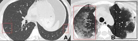
Figure 16: CT images of patients without (A/C) and with annotations (B/D) showing the utility of AI/ML software. The areas of GGO are marked in blue on the annotated images and consolidation as green. Quantification of lung involvement is also possible. Images courtesy of Predible Health.

Learning Points • Spread of infection from patient to healthcare workers and other patients is possible in the radiology department. • All radiology staff should adhere to social distancing norms, hand hygiene and use appropriate PPE. • Equipment should be regularly decontaminated as perthe unit policy.
Conclusion :
Commonly seen findings of COVID-19 on CT include GGO, consolidation, crazy paving and reticular pattern. In early stages and in those with milder symptoms GGO is more prominent. With disease progression areas of GGO increase or develop into consolidation/ crazy paving and reticular pattern. CT is a highly sensitive modality and it can be used in the diagnosis of COVID-19 when there is limited availability of RT41 PCR testing or delay in getting RT-PCR reports. CT is extremely useful in assessing the severity of disease and in prognosticating patients into those likely to require ICU therapy or not. Spread of infection amongst patients or to health care worker in the radiology department is a concern and appropriate steps should
R EFERENCES
1 Cennimo DJ, Bergman SJ — Coronavirus Disease 2019 (COVID-19). In: https://emedicine.medscape.com/article/ 2500114-overview - a1, ed. Medscape, 2020. 2 World Health Organisation. WHO Timeline- COVID -19 2020 [Available from: https://www.who.int/news-room/detail/27- 04-2020-who-timeline—covid-19 accessed 12 June 2020. 3 World Health Organisation. WHO Coronavirus Disease (COVID-19) Dashboard 2020 [Available from: https:// covid19.who.int/ accessed 12 June 2020. 4 Nasir MU, Roberts J, Muller NL, et al — The Role of Emergency
Radiology in COVID-19: From Preparedness to Diagnosis.
Can Assoc Radiol J 2020:846537120916419. doi: 10.1177/ 0846537120916419 5 Rodrigues JCL, Hare SS, Edey A, et al — An update on COVID19 for the radiologist - A British society of Thoracic Imaging statement. Clinical Radiology 2020;75(5):323-25. doi: 10.1016/j.crad.2020.03.003 6 Ai T, Yang Z, Hou H, et al — Correlation of Chest CT and RT
PCR Testing in Coronavirus Disease 2019 (COVID-19) in China:
A Report of 1014 Cases. Radiology 2020:200642. doi: 10.1148/radiol.2020200642 7 Li Y, Yao L, Li J, et al — Stability issues of RT-PCR testing of
SARS-CoV-2 for hospitalized patients clinically diagnosed with COVID-19. J Med Virol 2020 doi: 10.1002/jmv.25786 8 Rubin GD, Ryerson CJ, Haramati LB, et al — The Role of
Chest Imaging in Patient Management during the COVID-19
Pandemic: A Multinational Consensus Statement from the
Fleischner Society. Radiology;0(0):201365. doi: 10.1148/ radiol.2020201365 9 Long C, Xu H, Shen Q, et al — Diagnosis of the Coronavirus disease (COVID-19): rRT-PCR or CT? Eur J Radiol 2020;126:108961. doi: 10.1016/j.ejrad.2020.108961 10 Wong HYF, Lam HYS, Fong AH, et al — Frequency and
Distribution of Chest Radiographic Findings in COVID-19
Positive Patients. Radiology 2019:201160. doi: 10.1148/ radiol.2020201160 11 Liu R, Han H, Liu F, et al — Positive rate of RT-PCR detection of SARS-CoV-2 infection in 4880 cases from one hospital in
Wuhan, China, from Jan to Feb 2020. Clin Chim Acta 2020;505:172-75. doi: 10.1016/j.cca.2020.03.009 12 Chen N, Zhou M, Dong X, et al — Epidemiological and clinical characteristics of 99 cases of 2019 novel coronavirus pneumonia in Wuhan, China: a descriptive study. The Lancet 2020;395(10223):507-13. doi: 10.1016/s0140- 6736(20)30211-7 13 Ng M-Y, Lee EY, Yang J, et al — Imaging Profile of the COVID19 Infection: Radiologic Findings and Literature Review.
Radiology: Cardiothoracic Imaging 2020;2(1):e200034. doi: 10.1148/ryct.2020200034 14 Weinstock MB, Echenique A, Russell JW, et al — Chest X
Ray Fidnings in 636 Ambulatory Patients with COVID-19
be taken to mitigate this risk.
JOURNAL OF THE INDIAN MEDICAL ASSOCIATION, VOL 118, NO 06, JUNE 2020
Presenting to an Urgent Care Center: A Normal Chest X-Ray
Is no Guarantee. The Journal of Urgent Care Medicine 2020:13-18. [published Online First: 13 April 2020] 15 Ye Z, Zhang Y, Wang Y, et al — Chest CT manifestations of new coronavirus disease 2019 (COVID-19): a pictorial review. Eur Radiol 2020 doi: 10.1007/s00330-020-06801-0 16 Bernheim A, Mei X, Huang M, et al — Chest CT Findings in
Coronavirus Disease-19 (COVID-19): Relationship to Duration of Infection. Radiology 2020;295(3):200463. doi: 10.1148/ radiol.2020200463 17 Ding X, Xu J, Zhou J, et al — Chest CT findings of COVID-19 pneumonia by duration of symptoms. Eur J Radiol 2020;127:109009. doi: 10.1016/j.ejrad.2020.109009 18 Hansell DM, Bankier AA, MacMahon H, et al — Fleischner
Society: glossary of terms for thoracic imaging. Radiology 2008;246(3):697-722. doi: 10.1148/radiol.2462070712 19 Xu Z, Shi L, Wang Y, et al — Pathological findings of COVID19 associated with acute respiratory distress syndrome.
Lancet Respir Med 2020;8(4):420-22. doi: 10.1016/S2213- 2600(20)30076-X 20 Wu J, Wu X, Zeng W, et al — Chest CT Findings in Patients
With Coronavirus Disease 2019 and Its Relationship With
Clinical Features. Invest Radiol 2020;55(5):257-61. doi: 10.1097/RLI.0000000000000670 21 Pan Y, Guan H, Zhou S, et al — Initial CT findings and temporal changes in patients with the novel coronavirus pneumonia (2019-nCoV): a study of 63 patients in Wuhan, China. Eur
Radiol 2020;30(6):3306-09. doi: 10.1007/s00330-020-06731- x 22 Shi H, Han X, Jiang N, et al — Radiological findings from 81 patients with COVID-19 pneumonia in Wuhan, China: a descriptive study. Lancet Infect Dis 2020;20(4):425-34. doi: 10.1016/S1473-3099(20)30086-4 23 Li K, Wu J, Wu F, et al —. The Clinical and Chest CT Features
Associated With Severe and Critical COVID-19 Pneumonia.
Invest Radiol 2020;55(6):327-31. doi: 10.1097/
RLI.0000000000000672 24 Vilar J, Domingo ML, Soto C, et al — Radiology of bacterial pneumonia. Eur J Radiol 2004;51(2):102-13. doi: 10.1016/ j.ejrad.2004.03.010 25 Murch CR, Carr DH — Computed tomography appearances of pulmonary alveolar proteinosis. Clinical radiology 1989;40(3):240-3. doi: 10.1016/s0009-9260(89)80180-1 26 Rossi SE, Erasmus JJ, Volpacchio M, et al — “Crazy-paving” pattern at thin-section CT of the lungs: radiologic-pathologic overview. Radiographics 2003;23(6):1509-19. doi: 10.1148/ rg.236035101 27 Xie X, Zhong Z, Zhao W, et al — Chest CT for Typical 2019- nCoV Pneumonia: Relationship to Negative RT-PCR Testing.
Radiology 2020:200343. doi: 10.1148/radiol.2020200343 28 Pan F, Ye T, Sun P, et al — Time Course of Lung Changes On
Chest CT During Recovery From 2019 Novel Coronavirus (COVID-19) Pneumonia. Radiology 2020:200370. doi: 10.1148/ radiol.2020200370 29 Franquet T — Imaging of pulmonary viral pneumonia. Radiology 2011;260(1):18-39. doi: 10.1148/radiol.11092149 30 Grillet F, Behr J, Calame P, et al — Acute Pulmonary Embolism
Associated with COVID-19 Pneumonia Detected by Pulmonary
CT Angiography. Radiology 2020:201544. doi: 10.1148/ radiol.2020201544 31 Bompard F, Monnier H, Saab I, et al — Pulmonary embolism in patients with Covid-19 pneumonia. Eur Respir J 2020 doi: 10.1183/13993003.01365-2020 32 Waller JV, Kaur P, Tucker A, et al — Diagnostic Tools for
Coronavirus Disease (COVID-19): Comparing CT and RT-PCR
Viral Nucleic Acid Testing. AJR Am J Roentgenol 2020:1-5. doi: 10.2214/AJR.20.23418 33 Xu B, Xing Y, Peng J, et al — Chest CT for detecting COVID19: a systematic review and meta-analysis of diagnostic accuracy. Eur Radiol 2020 doi: 10.1007/s00330-020-06934- 2 34 Zu ZY, Jiang MD, Xu PP, et al — Coronavirus Disease 2019 (COVID-19): A Perspective from China. Radiology 2020:200490. doi: 10.1148/radiol.2020200490 35 Kim H, Hong H, Yoon SH— Diagnostic Performance of CT and
Reverse Transcriptase-Polymerase Chain Reaction for
Coronavirus Disease 2019: A Meta-Analysis. Radiology 2020:201343. doi: 10.1148/radiol.2020201343 36 Inui S, Fujikawa A, Jitsu M, et al — Erratum: Chest CT Findings in Cases from the Cruise Ship “Diamond Princess” with
Coronavirus Disease 2019 (COVID-19). Radiology:
Cardiothoracic Imaging 2020;2(2):e204002. doi: 10.1148/ ryct.2020204002 37 Liu Z, Jin C, Wu CC, et al — Association between Initial Chest
CT or Clinical Features and Clinical Course in Patients with
Coronavirus Disease 2019 Pneumonia. Korean J Radiol 2020;21(6):736-45. doi: 10.3348/kjr.2020.0171 38 Huang C, Wang Y, Li X, et al — Clinical features of patients infected with 2019 novel coronavirus in Wuhan, China. Lancet 2020;395(10223):497-506. doi: 10.1016/S0140- 6736(20)30183-5 39 Lyu P, Liu X, Zhang R, et al — The performance of chest CT in evaluating the clinical severity of COVID-19 pneumonia: identifying critical cases based on CT characteristics. Invest
Radiol 2020 doi: 10.1097/RLI.0000000000000689 40 Zhao W, Zhong Z, Xie X, et al — Relation Between Chest CT
Findings and Clinical Conditions of Coronavirus Disease (COVID-19) Pneumonia: A Multicenter Study. AJR Am J
Roentgenol 2020;214(5):1072-77. doi: 10.2214/AJR.20.22976 41 Colombi D, Bodini FC, Petrini M, et al — Well-aerated Lung on
Admitting Chest CT to Predict Adverse Outcome in COVID-19
Pneumonia. Radiology 2020:201433. doi: 10.1148/ radiol.2020201433 42 ACR Recommendations for the use of Chest Radiography and Computed Tomography (CT) for Suspected COVID-19
Infection, 2020. 43 ACEP COVID-19 Field Guide- CXR and CT Imaging, 2020. 44 Indian College of Radiology and Imaging. Imaging recommendations for COVID-19 patients. In: Chest Radiology
Subspecialty Group, ed., 2020. 45 World Health Organisation — Use of chest imaging in COVID19. A Rapid advice guide. 2020 46 Kooraki S, Hosseiny M, Myers L, et al — Coronavirus (COVID19) Outbreak: What the Department of Radiology Should
Know. Journal of the American College of Radiology 2020;17(4):447-51. doi: 10.1016/j.jacr.2020.02.008
42