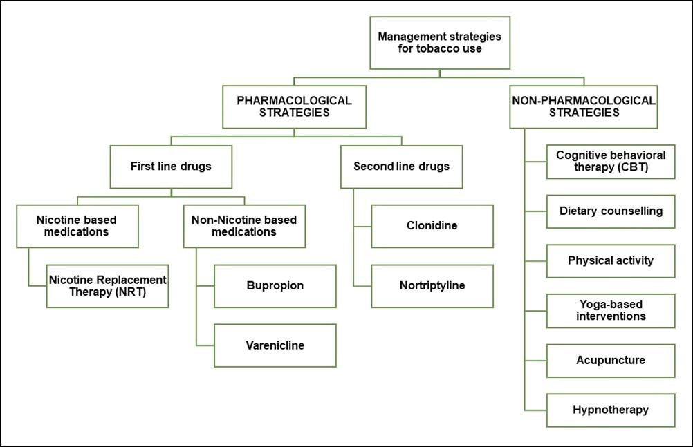
The Silent Epidemic : Chronic Hepatitis B and Its Global Impact
In the landscape of global health, certain diseases persist quietly, often overshadowed by more prominent health crises. Chronic Hepatitis B (CHB) is one such silent epidemic. Despite being preventable and treatable, it continues to claim over a million lives annually, primarily due to complications like cirrhosis and liver cancer. This editorial delves into the global impact of CHB, examining its prevalence, the challenges in diagnosis and treatment, and the urgent need for enhanced public health strategies.
Understanding Hepatitis B :
Hepatitis B is a viral infection that attacks the liver, leading to both acute and chronic diseases. The World Health Organization (WHO) estimates that approximately 254 million people worldwide were living with chronic hepatitis B infection in 2022, with 1.2 million new infections each year. The virus is most commonly transmitted from mother to child during birth and delivery, in early childhood, as well as through contact with blood or other body fluids during sex with an infected partner, unsafe injections, or exposures to sharp instruments.
Global Burden and Regional Disparities :
The global burden of Hepatitis B is disproportionately high in certain regions. In 2022, the WHO reported that 83% of the estimated 1.3 million deaths from viral hepatitis were attributed to Hepatitis B. The highest prevalence rates are found in the WHO Western Pacific Region and the WHO African Region, where 97 million and 65 million people are chronically infected, respectively. These regions also face significant challenges in healthcare infrastructure, leading to limited access to diagnosis and treatment.
In contrast, the WHO European Region and the Region of the Americas report significantly lower prevalence rates, highlighting the disparities in healthcare access and resources between regions.
Challenges in Diagnosis and Treatment :
One of the most pressing issues in combating chronic Hepatitis B is the low rates of diagnosis and treatment. According to the WHO, only 13% of people living with chronic Hepatitis B infection had been diagnosed by the end of 2022, and a mere 3% had received antiviral therapy. This underdiagnosis is compounded by the fact that many individuals with CHB remain asymptomatic for years, unknowingly
harboring the virus and unknowingly transmitting it to others.
The lack of widespread screening programs, especially in high-prevalence regions, exacerbates this issue. Additionally, the stigma associated with Hepatitis B, often linked to misconceptions about its transmission, further deters individuals from seeking testing and treatment.
Preventive Measures and Vaccination : Prevention remains the most effective strategy against Hepatitis B. The Hepatitis B vaccine is safe, effective, and widely available. Vaccination within 24 hours of birth prevents the spread of the virus from mother to child, significantly reducing the risk of chronic infection.
Despite the availability of the vaccine, global coverage remains uneven. In the WHO African Region, only 18% of newborns receive the Hepatitis B birth-dose vaccination, contributing to the high prevalence of the disease in that region. Increasing vaccination rates, particularly among newborns and high-risk populations, is crucial in the fight against hepatitis B.
Innovations in Treatment :
While a cure for Hepatitis B remains elusive, significant advancements have been made in treatment options. Antiviral therapies can effectively suppress the virus, reducing the risk of liver damage and transmission. However, these treatments require lifelong adherence and do not eliminate the virus entirely.
Researchers are exploring novel approaches, including gene-editing technologies like CRISPR, to develop potential cures. For instance, scientists in Melbourne are developing an mRNA-based treatment to prevent primary liver cancer by targeting Hepatitis B infections. Early trials are promising, and the treatment aims to be administered to people with serious liver inflammation due to Hepatitis B, identified through a simple blood test. Researchers hope this method will significantly reduce the risk of developing primary liver cancer, which is increasing worldwide and often diagnosed too late for curative treatments.
The Road Ahead : Global Strategies for Hepatitis B Elimination.
To combat the silent epidemic of chronic Hepatitis B, a multifaceted approach is necessary :
Enhanced Screening and Diagnosis :
Implementing widespread screening programs, especially in high-prevalence regions, can facilitate early detection and timely treatment.
Public Awareness Campaigns : Educating the public about Hepatitis B, its transmission and the importance of vaccination can reduce stigma and encourage individuals to seek testing and treatment.
Strengthening Healthcare Infrastructure : Investing in healthcare systems, particularly in low-resource settings, can improve access to diagnosis, treatment, and preventive services.
International Collaboration : Governments, international organizations, and non-governmental entities must collaborate to share resources, knowledge and strategies to combat hepatitis B globally.
Research and Development : Continued investment in research is essential to develop new treatments and, ultimately, a cure for Hepatitis B.
CONCLUSION
Chronic hepatitis B remains a significant global health challenge, silently affecting millions and claiming over a million lives annually. While progress has been made in prevention and treatment, much remains to be done. By enhancing screening, increasing vaccination coverage, and investing in research and healthcare infrastructure, the global community can work towards eliminating Hepatitis B and reducing its impact on future generations. The time to act is now, for inaction will only allow this silent epidemic to continue its devastating course.
FURTHER READING
1World Health Organization (2024). WHO sounds alarm on viral hepatitis infections claiming 3500 lives each day. Retrieved from WHO.
2World Health Organization (2024). Hepatitis B. Retrieved from WHO.
3Centers for Disease Control and Prevention. (2024). Fast Facts: Global Hepatitis B Vaccination. Retrieved from CDC
4Hepatitis B Foundation (ND). Hepatitis B Facts and Figures. Retrieved from Hepatitis B Foundation.
5Verywell Health (2022). Understanding If There’s a Cure for Hepatitis B. Retrieved from Verywell Health.
6Herald Sun (2022). Liver cancer could be stopped by ‘assassin’ treatment. Retrieved from Herald Sun.
Hony Editor, JIMA
Kakali Sen
Special Article
Clinical Application of Micronutrients in Recovery : A Practical Guidebook for Clinicians
Micronutrients play a vital role in enhancing recovery and improving overall health, particularly in individuals dealing with acute or chronic illnesses1,2. Adequate intake of vitamins and minerals supports immune function, tissue repair, metabolic regulation, and long-term health outcomes3,4,6. Despite substantial clinical evidence affirming their value, micronutrient supplementation remains limited in routine medical practice5,7,13. To address this gap, the Indian Medical Association (IMA) has developed comprehensive, evidence-based guidelines aimed at integrating micronutrient support into standard care for both adult and paediatric patients across medical and surgical settings8,10,11. These guidelines are designed to assist healthcare professionals in adopting targeted nutritional strategies, improving patient outcomes, reducing complications, and enhancing quality of life9,12. Authored by a multidisciplinary panel of experts—including General Practitioners, Paediatricians, Internal Medicine Specialists, and Surgeons—the guidelines are grounded in current research, clinical practices, and patient care outcomes10,12. Each chapter provides focused insight into specific micronutrients, outlining their physiological roles, implications of deficiencies, and evidence-based supplementation approaches tailored to various diseases1,3,5,14 In conclusion, the document emphasizes a holistic approach to recovery that incorporates both physical and psychological dimensions of patient well-being11,15. By prioritizing micronutrient adequacy in clinical care, these guidelines serve as a valuable resource for healthcare providers, reinforcing the essential role of nutrition in optimizing recovery and improving overall patient health across diverse care environments15,16
Key words : Micronutrients, Clinical Guidelines, Acute and Chronic Illness, Patient Recovery, Holistic Approach.
Recovery is the process of returning to normal routines and habits following illness or surgery, involving both physiological and psychological aspects. Physiological recovery involves tissue repair, restoration of organ function, and systemic balance through proper nutrition, hydration, and micronutrient support. Key recovery mechanisms include tissue
Editor's Comment :
! ! Micronutrient supplementation is crucial for enhancing recovery in both acute and chronic conditions.
! ! ! ! These IMA guidelines aim to bridge the implementation gap by offering evidence-based, disease-specific micronutrient strategies to improve patient outcomes.
1MBBS, Coordinator, Department of Family Medicine, IMA West, Mumbai, Maharashtra 400049 and Corresponding Author
2MD (Medicine), FCPS, FICP, FISE, FGSI, FDI, Professor, Department of Medicine, Health Harmony, Mumbai, Maharashtra 400064 3MD, Director, Department of Pulmonary, Brahma Kumari Global Hospital and Research Centre and Vora Clinic, Mumbai, Maharashtra 400058
4MS, President, IMA West, Department of Surgery, Benz Hospital, Mumbai, Maharashtra 400054 5MD (Medicine), FCPS, FICP, FISE, FGSI, FDI, Consultant Nephrologist and Renal Transplant Physician, Department of Nephrology, Manipal Hospital, Bangalore, Karnataka 560017
6MD (Medicine), FCPS, FICP, FISE, FGSI, FDI, Consultant, Centre for Child Health, Department of Pediatrics, Centre for Child Health Nanavati Max Super Speciality Hospital, Mumbai, Maharashtra 400056
7MD, Consultant, Critical Care Chief Intensivist and ICU Head, Department of Anaesthesia, KD Hospital, Ahmedabad, Gujarat 382421
8MD (Medicine), FCPS, FICP, FISE, FGSI, FDI, Director and Consultant Neonatologist, Department of Neonatology, Arpan Newborn Care Centre, Ahmedabad, Gujarat 380059
9MD (Medicine), FCPS, FICP, FISE, FGSI, FDI, Critical Care Specialist, Critical Care and Hospital Infection Control, Ahmedabad, Gujarat 10MD (Medicine), FCPS, FICP, FISE, FGSI, FDI, Senior Professor, Pulmonary and Critical Care Medicine, University of Health Sciences Rohtak, Haryana 124001
11MBBS, IMA West, Mumbai, Maharashtra 400049
Received on : 03/06/2025
Accepted on : 11/06/2025
How to cite this article : Clinical Application of Micronutrients in Recovery : A Practical Guidebook for Clinicians. Lele J, Mehta K, Vora A, Mehta R, Aggarwal G, Sanghvi J, Mehta J, Mehta A, Dileep P, Chaudhary D, Joshi S. J Indian Med Assoc 2025; 123(6): 16-9.
123, No 06, June 2025Journal
repair, immune function, energy metabolism, bone health, cognitive function, and antioxidant defence. Psychological recovery is influenced by mental health, as conditions like anxiety and depression can impair medication adherence, behaviour, and immunity. Micronutrients play a crucial role in metabolism, immune function, and the regulation of oxidative stress, thereby aiding overall recovery. A holistic approach integrating physical, mental, and emotional well- being, alongside pharmacological and nonpharmacological strategies, is essential for optimal recovery outcomes.
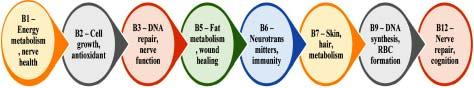
Key Micronutrient Roles (Fig 1) :
Micronutrients in Adults : The Vitamin B complex is essential for recovery across various conditions by supporting immune function, energy metabolism, nerve health, and tissue repair. Infections, burns, fractures, and post-surgical recovery benefit from B1, B2, B6, and B12, while B9 and B12 address anaemia linked to malabsorption. B3 aids DNA repair, B5 supports wound healing, and B7 promotes skin and hair health. In neurological conditions, B6 and B12 enhance nerve regeneration and cognition. Lysine complements these actions by boosting collagen synthesis and immunity, while zinc, selenium, iron, vitamin D, and calcium further support healing and bone strength.
Micronutrients in Disease Recovery :
In acute infections, Vitamin B complex (B1, B2, B6, B12) supports immune cell production, energy metabolism, and tissue repair, while lysine aids immune modulation. In burns, B9 and B12 help manage anemia and regeneration, with lysine promoting healing. In fever, Vitamin B complex, zinc, and electrolytes support gut integrity by reducing inflammation and restoring balance. Across all conditions, Vitamins C and D provide added support by boosting defense and reducing inflammation, with lysine aiding recovery.
Micronutrient Support in Chronic Conditions : Respiratory Diseases
•Tuberculosis : Vitamin B6 prevents neuropathy, B12 supports red blood cell formation, and B2 aids mucosal integrity. Vitamin C and Zinc assist in oxidative stress reduction and immune recovery.
Skin & Autoimmune Conditions
•Skin Infections : Vitamin B2 prevents eczema, B3 maintains the skin barrier, B6 aids immune defense, and B7 supports skin repair. Lysine enhances collagen synthesis, while Vitamin A and C aids in wound healing.
•Autoimmune Disorders : Vitamin B6 regulates immune response, B6 and B12 reduces inflammation, and B2 supports antioxidant defense. Lysine assists in tissue repair, while Vitamin D modulates immune activity.
Metabolic & Cardiovascular Health
•Diabetes : Vitamin B9 and B12 counteracts metformin-induced deficiencies, B3 improves lipid balance and B6 aids glucose metabolism. Vitamin C and D enhances insulin sensitivity, while Zinc supports metabolic function.
•Cardiovascular Disease : Vitamin B1 aids cardiac function. B6 regulates homocysteine levels and B12 supports red blood cell production. Magnesium and controlled sodium intake contribute to vascular health.
Kidney Disease
•Chronic Kidney Disease (CKD): Vitamin B complex (B1, B2, B6, B12) helps to counteract deficiencies due to dietary restrictions, while Vitamin D and Iron support bone and red blood cell health.
•End-Stage Renal Disease (ESRD): Vitamin B complex are essential to replenish losses during dialysis, with careful regulation of sodium, potassium, calcium, lysine, and iron for metabolic balance.
Fig 1
Lele J, et al. Clinical Application of Micronutrients in Recovery : A Practical Guidebook for Clinicians.
Liver Disease
•Alcohol-Associated Liver Disease (ALD): Vitamin B1 prevents Wernicke-Korsakoff syndrome, B9 addresses anemia, and B12 supports red blood cell formation. Lysine aids liver recovery alongside Vitamin A, D, Magnesium, and Zinc.
•Hepatitis: Vitamin B9 aids cellular regeneration and B12 enhances neurological function, and lysine supports liver repair, with additional support from Vitamins A, D, and Zinc.
•Metabolic-Associated Fatty Liver Disease (MAFLD) & Cholestatic Liver Disease: Vitamin B complex B1, B6, B12, and Folate optimizes energy metabolism, DNA synthesis, and detoxification, preventing liver dysfunction.
Malnourishment
•Malnutrition: Vitamin B complex B1, B3, and B9 supports metabolism and address deficiencies from chronic alcohol use, with additional needs for Calcium, Magnesium, Phosphorus, and Iron.
•Obesity: Vitamin B complex enhances energy metabolism, reduces fatigue, and supports nerve function in managing obesity-related complications.
Micronutrient Needs in Surgery :
The role of key micronutrients in recovery from certain surgeries, as covered in this chapter, includes:
•Bariatric surgery requires lifelong follow-up, including supplementation with Vitamin B9, B12, and iron to prevent anemia; Vitamin B complex, calcium, and Vitamin D3 for bone health; and zinc, Vitamin C, magnesium, lysine, and Vitamin K to support healing and collagen synthesis.
•Cardiac surgery benefits from Vitamin B6, B9, and B12 to regulate homocysteine levels; Vitamin B1 to support heart function; and Vitamin C, E, magnesium, zinc, selenium, and lysine to enhance vascular and immune health.
•Post-surgical recovery is supported by the Vitamin B complex for energy metabolism and immunity; iron, B9, Vitamin D, and calcium for blood and bone health; and lysine to accelerate overall healing.
Micronutrients for Paediatric Recovery :
Micronutrients are essential in managing paediatric conditions by supporting immunity, reducing oxidative stress, aiding tissue repair, and enhancing recovery.
This chapter highlights essential micronutrients and their significance in some of the acute and chronic paediatric conditions, such as:
Acute Conditions :
Hepatitis : Vitamin B complex supports energy metabolism and liver detoxification and Zinc and Selenium aids in liver regeneration.
Dengue Fever : Vitamin B complex supports nerve function, energy metabolism, and immunity, while Vitamin C improves vascular stability, and Vitamin D and Magnesium helps to reduce fatigue.
Chronic & Metabolic Disorders :
•Respiratory & Immune Support: Vitamin B6 and B12 helps to reduce inflammation, while B9 supports in cell regeneration. Lysine also aids in infection recovery.
•Metabolic & Endocrine Health: The Vitamin B complex (B1, B6, B12, and folate) is crucial for glucose metabolism, nerve function, and red blood cell synthesis, especially in diabetes. B9 and B12 help prevent anaemia, while lysine supports muscle maintenance. The Vitamin B complex also aids in thyroid function and reduces oxidative stress.
•Gastrointestinal & Nutrient Absorption: Vitamins B1 and B2 aid in energy production, while B5 supports fat metabolism. B9 and B12 are essential for cell repair and preventing deficiencies in conditions such as IBD or malabsorption syndromes. Lysine helps maintain protein balance and improves nutrient utilization.
•Blood & Kidney Function: Vitamins B6, B9, and B12 are vital for red blood cell formation, haemoglobin synthesis, and lowering homocysteine levels to reduce stroke risk. The Vitamin B complex supports kidney function by regulating energy metabolism and reducing nitrogen waste. Lysine aids in protein metabolism while minimizing kidney strain.
Surgical & ICU Recovery : Some of the key micronutrients that play a major role
Lele J, et al. Clinical Application of Micronutrients in Recovery : A Practical Guidebook for Clinicians.
Vol 123, No 06, June 2025Journal of the Indian Medical Association
in paediatric surgical and ICU recovery are listed below:
Postoperative Recovery: Vitamin B complex supports red blood cell production, nerve function, and tissue repair, preventing anemia and aiding metabolism. Lysine enhances collagen formation and accelerates healing. Additional support from Vitamin A, C, K, Zinc, and Magnesium.
Pediatric ICU: Vitamin B complex are critical for energy metabolism, neurological function, and red blood cell formation, helping sustain recovery in critically ill children. Vitamin D, Calcium, and Selenium provides additional metabolic support.
Drug interactions :
Fat-Soluble Vitamins – Avoid Vitamin A in pregnancy and liver disease; Vitamin D may reduce statin efficacy;
Vitamin E increases bleeding risk; Vitamin K needs caution in neonates and kidney disease.
Water-Soluble B Vitamins – B1, B2, B7, and B9 are generally safe; B3, B5, and B6 require caution in liver disease, pregnancy, and macrolide use.
Vitamin C – Caution in G6PD deficiency, thalassemia, and hemochromatosis due to oxidative stress or iron overload risk.
Minerals & Metabolism – Manganese toxicity risk increases with iron deficiency.
Iron & Zinc – Avoid excess iron in overload conditions; limit zinc to <40 mg/day in pregnancy.
Selenium – Requires caution in allergy-prone individuals.
CONCLUSION
Overall, micronutrients support immunity, repair, and recovery, with Vitamin B complex playing a key role
in metabolism, nerve function, and red blood cell production. Targeted supplementation in adults and paediatrics helps to prevent deficiencies and optimize health.
REFERENCES
1L Kathleen Mahan, Escott-Stump, S., & Raymond, J. (2011). Krause’s food & nutrition care process. (13th ed.). Saunders.
2Mudambi SR (2007) — Fundamentals of foods, nutrition and diet therapy. New Age International.
3Steinbrenner H, Al-Quraishy S, Dkhil MA, Wunderlich F, Sies H (2015) — Dietary selenium in adjuvant therapy of viral and bacterial infections. Advances in nutrition.
4Escott-Stump S (2008) — Nutrition and diagnosis-related care. Lippincott Williams & Wilkins.
5Shah B, & Shah S (2023) — Practical Recommendations on Micronutrient Deficiencies in Gastrointestinal Diseases. Journal of Nutrition Research.
6Chakrabarty K, Chakrabarty AS (2020) — Textbook of Nutrition in Health and Disease. Germany: Springer Nature Singapore.
7Joshi SA (2010) — Nutrition and Dietetics: With Indian Case Studies. Tata McGraw-Hill.
8Nutrient Requirements for Indians (2020) — Recommended Dietary Allowances, Estimated Average Requirements. ICMRNational Institute of Nutrition.
9Webster-Gandy J, Madden A, Holdsworth M (Eds)(2020) — Oxford handbook of nutrition and dietetics. Oxford University Press.
10American Academy of Pediatrics. (2014). Pediatric nutrition (pp. 123-139). RE Kleinman, & FR Greer (Eds.). Elk Grove Village, IL: American Academy of Pediatrics.
11Kliegman RM, Behrman RE, Jenson HB, Stanton BM (2007) — Nelson textbook of pediatrics e-book. Elsevier Health Sciences.
12Suskind D, Lenssen P (Eds) (2013) — Pediatric Nutrition handbook: an algorithmic approach. John Wiley & Sons.
13Harding KL, Aguayo VM, Webb P (2018) — Hidden hunger in South Asia: a review of recent trends and persistent challenges. Public health nutrition.
14L Kathleen Mahan, Escott-Stump S, Raymond J (2011) — Krause’s food & nutrition care process. (13th ed.). Saunders.
15dos Reis Santos M, Leite HP, Pereira AML, Cassão BDA, de Oliveira Iglesias SB (2016) — Factors associated with not meeting the recommendations for micronutrient intake in critically ill children. Nutrition,
16Ben XM (2008) — Nutritional management of newborn infants: practical guidelines. World journal of gastroenterology.
Original Article
Family Health Survey and e-Mamta
: Data Validation
Exercise in Districts of Western India
Mamtarani Verma1, S L Kantharia2
Abstract
Background : The Health and Family Welfare Department of Gujarat, has introduced a ‘Mother & Child’ name based tracking information management system called “e-Mamta” in collaboration with the National Rural Health Mission and National Informatics Centre.
Aims and Objectives : To do household survey of selected Anganwadis / Corporation and validate its records with family health survey register. To verify and validate the survey records with data entered in e-Mamta software.
Materials and Methods : Data validation of two districts namely Surat and Valsad was done by Community Medicine Department. For both districts, two PHCs “one best performing and the other worst performing” were decided to be taken on the basis of TT coverage. For data collection, all the houses of selected Anganwadi of Rural and Anganwadi of Corporation area were covered. Survey team members were faculty/resident Doctor and Health Worker. The data obtained in survey form was tallied with the entries in both Family health survey register and e-Mamta software. Missed entries and wrong entries in e-Mamta software were then identified.
Statistical Analysis : Data from survey forms was entered in excel software and frequencies/percentages were calculated.
Results : More than 80% of members’ entries have been made in the family health survey record register of Surat (87.82%) and Valsad (81.93%) districts. e-Mamta software records of Surat district was showing 80.63% entries while it was 78.14% in the Valsad district rest of the entries were missed.
Conclusion : The records of family health survey register which were entered in e-Mamta software showed a gap of 14.5-24.5%.
Key words : e-Mamta, Data validation, Family health survey, Records.
The Mother and Child Tracking System (MCTS) is a centralized web-based application launched by the Ministry of Health and Family Welfare in India to provide reliable data for effective decision-making through name-based tracking of each client1. More than 4.06 crore pregnant women and 3.3 crore children have been registered in the system since its inception2. Few experiences of MCTS implementation have been documented in the states of Gujarat (eMamta)3, Tamil Nadu (Pregnancy and Infant Cohort Monitoring and Evaluation System — PICME) 4 , Rajasthan5 and Chhattisgarh.
The Health and Family Welfare Department of Gujarat, has introduced a ‘Mother & Child’ name based tracking information management system
Department of Community Medicine, Government Medical College, Surat, Gujarat 395001 1MD (PSM), Assistant Professor and Corresponding Author 2MD, Ex-Professor and Head
Received on : 01/05/2024
Accepted on : 27/07/2024
Editor's Comment : There is need to update the records timely so that services among vulnerable population should not be delayed. Identification of the wrong entries in the e-Mamta software should be done on time and removed from the software after confirmation at different levels so as to avoid hurdles in smooth functioning of system.
called “e-Mamta” in collaboration with the National Rural Health Mission (NRHM) and National Informatics Centre (NIC). e-Mamta technology seeks to improve the lives of poor people by providing them timely services by involving Health Care Workers and people themselves6
"e-Mamta” was introduced by Government of Gujarat for the first time in India. This system generates facility-wise reports and provides real-time information3
The system aims at registering individual pregnant women, individual children in the age group 0-6 and adolescents along with their full details to ensure
How to cite this article : Family Health Survey and e-Mamta : Data Validation Exercise in Districts of Western India. Verma M, Kantharia SL. J Indian Med Assoc 2025; 123(6): 20-4.
123, No 06, June 2025Journal
complete service delivery of Antenatal Care (ANC), Child birth, Post Natal Care (PNC), Immunization, Nutrition and Adolescents services and to track the left outs7
In Gujarat, thousands of Rural Health Workers, trained by the National Rural Health Mission (NRHM), go door-to-door collecting information on pregnant women, infants and sends it using their mobile phone to State Rural Health Mission (SRHM), the Government body, which collates the data into a centralised repository. This data is then used to alert Rural Health Workers through SMS to make sure they reach out to the pregnant women and mothers regarding immunisation dates or medicines to be taken8
Our country is having the problem of poor record making. With the help of this technology (e-Mamta) we can keep record of each beneficiary; both women and child that will help in improving delivery and quality of services to beneficiaries. Microplanning can be done efficiently and all health workers can be given specific time bound targets each month which will ultimately help in correct and fast decision making. Looking to our aim of reducing the maternal mortality it is now to possible to track each pregnant women. Similarly the objective of reducing the infant and child mortality can be achieved with this ambitious online technology.
With this background study was planned to see how this recording system of family surveys and online eMamta is functioning.
AIMS AND OBJECTIVES
(1) To do household survey of selected Anganwadis / Corporation and validate its records with family health survey register.
(2) To verify and validate the survey records with data entered in E-Mamta software.
(3) To identify missed and wrong entries in e-Mamta software.
MATERIALS AND METHODS
Data validation work was assigned by Hon'ble Minister of Health & Family Welfare, Government of Gujarat to Community Medicine Departments of all Government Medical Colleges of Gujarat. Preliminary meeting for planning the research was held at
Ahmedabad which was attended by Professor & Head and one Assistant Professor from Community Medicine Department of six Government Medical Colleges of Gujarat.
Responsibility for data collection in areas geographically closet to each Medical College was given. Each college has to cover two regions namely one where the medical college is located and the 2nd which is most remote and distant in that region.
It was decided in the meeting to identify PHCs on the basis of performance of Tetanus Toxoid (TT) coverage in Antenatal Women taking it as alternate indicator of Reproductive and Child Health (RCH) services.
Data validation of two districts namely Surat and Valsad was done by Community Medicine Department. For both districts, two PHCs “one best performing and the other worst performing” were decided to be taken on the basis of TT coverage. List of Anganwadis under the identified PHCs was obtained and arranged alphabetically. Random numbers were generated with the help of excel software and one Anganwadi was selected. Similary Anganwadis were selected for all rest of three PHCs. Randomly selected PHCs were Motaponda PHC (Kaprada-Valsad), Timbhi road PHC (Sanjan-Valsad), Shekhpur PHC (Kathor-Surat) and Vadiya PHC (Naladhara-Surat).
e-Mamta software were 75.5%, 75.7%, 83.1% and 85.5% for Timbi road, PHC-Sanjan, Valsad, Motapanda-PHC, Kaprada, Valsad, Vadiya, PHCNaladhara, Surat and Shekhpur, PHC-Kathor, Surat respectively. Shekhpur, PHC-Kathor 52 (5.3%), Surat showed the highest proportion of wrong entries out of four PHCs.
To get an idea about situation in Municipal Corporation, one additional cluster in corporation area where Medical College is located was surveyed. Ambanagar Anganwadi (very old Anganwadi) of Surat Municipal Corporation was covered.
Data validation survey and cross verification was done.
For doing the data collection, all the houses of selected Anganwadi of Rural and Anganwadi of Corporation area were covered. Survey team members were faculty/resident Doctor of Community Medicine Department and Health Worker (Anganwadi worker/ASHA/Helper/ANM/MPHW). Predesigned questionnaire for collecting the information like names
Verma
123, No 06, June 2025Journal
of all family members in each household/gender/age/ whether beneficiary of RCH 2 (Y/N), summary of type of beneficiary in each household (15-45 years women, lactating women, pregnant women, infant, adolescent (10-19 years), children (1-5 years) /any other remarks was used. Preliminary information regarding selected Anganwadi like Anganwadi number, Name of village, sub-centre, PHC as well as name of Anganwadi worker with her full address including mobile/landline number was also recorded.
The data obtained in survey form was tallied with the entries in both Family health survey register and eMamta software. Accompanying Health Worker helped in comparing the survey data with the data records of family health survey register. For comparing the data with online e-Mamta records passwords were obtained from the concerned PHCs.
Missed entries and wrong entries in e-Mamta software were then identified. Data from survey forms was entered in excel software and frequencies/ percentages were calculated.
For the local Anganwadi (Municipal Corporation) it was possible to search each entry on net at UHC (Althan in our case) with more number of visits but for Rural areas visit at remote location it was not feasible so online UID search was even done after field work.
Overall it was not the one day/one cluster working as doing survey of approximately 200 houses & cross checking each & every record in family health survey register on the same day was a lengthy job for Rural remote areas like Valsad district.
Definitions used —
Registered in family health record — Write down the total number of family members in a household, who were registered in family health record (Register no 2) of MPHW and were cross-checked from the same.
Registered in e-Mamta — Write down the total number of family members, who were entered in the e-Mamta software.
Wrong entry — Write down the total number of family
members, who were neither present in the same family nor in the village.
Missing entry — Write down the total number of the family members, who were not entered in the software, but found out during our survey.
RESULTS
More than 80% of members’ entries have been made in the family health survey record register of Surat (87.82%) and Valsad (81.93%) districts. e-Mamta software records of Surat district was showing 80.63% entries while it was 78.14 % in the Valsad district rest of the entries were missed ie, 19.37% and 21.86% in the Surat and Valsad district respectively. Number of households surveyed and total family members in both Surat and Valsad are also shown in (Table 1).
In Table 2 shows PHCs wise number of household surveyed and total family members. Out of total four PHCs covered one of the PHC showed less than eighty percent registration of survey records in family health survey records. Family health survey records registered in e-Mamta software were 75.5%, 75.7%, 83.1% and 85.5% for Timbi road, PHC-Sanjan, Valsad, Motapanda-PHC, Kaprada, Valsad, Vadiya, PHC-Naladhara, Surat and Shekhpur, PHC-Kathor, Surat respectively. Shekhpur, PHC-Kathor 52 (5.3%), Surat showed the highest proportion of wrong entries out of four PHCs.
Entries of family health survey records missed in eMamta software were higher for covered PHCs of Valsad district as compared to PHCs of Surat District (Table 2).
Survey results of Anganwadi of Municipal Corporation showed quite different results. Number of records entered in family health survey records were very less 387 (45.53 %). Further entries of e-Mamta software 322 (37.88 %) were not consistent with the records of family health survey records. Missing entries were 528 (62.1%). One positive thing was that there was no wrong entry in the e-Mamta software records for this Anganwadi (Table 3).
Table 1 — Summarization of the family health survey records and E-Mamta software records for Surat and Valsad Districts
Table 2 — PHC wise records in family health survey records and e-Mamta software records for Surat and Valsad Districts
VillageNo ofTotalRegistered in familyRegistered inWrong entryMissed entry NamehouseholdsFamily health survey recorde-Mamta software surveyed members
In our study data validation showed lots of missing and wrong entries in family health survey register as well as online e-Mamta tracking system in both Rural as well as Urban areas.
In 87.82% and 81.93% population of Surat and Valsad districts data was registered in family health survey register while it was 45.53% for Anganwadi of Municipal Corporation.
In 80.63% and 78.14% entries of Surat and Valsad districts respectively were registered in e-Mamta Software while in Anaganwadi of Municipal Corporation it was 37.88%. For Urban area data entries in family health survey register as well as eMamta online system both was poor. There were approximately 30.75% wrong entries and 51% missed entries in the surveyed villages of Surat and Valsad districts. There was PHC wise variation in both Surat and Valsad district for family health survey records but PHC wise records were almost similar for eMamta online records for both districts.
In a study done by Divya Barot (2015), et al family health survey data validation in Sabarkantha district, 98.25% of the population was registered in family health survey register in Rural areas9. 85.56% family survey data was registered in e-Mamta Software. There were approximately 30.75% wrong entries and 51% missed entries in the surveyed villages of Sabarkantha district. 30% data entry gap and in Urban areas poor data collection was found in a review case study of UNICEF by Syed S Kazi10
In a study done by Nagarajan P (2016) gap leading to underutilization of Maternal and Child Tracking
System (MCTS) portal is the unavailability of standard recording registers with the frontline health functionaries. The data columns in Maternal and Child Health (MCH) registers-the basic tool available with Health Care Workers to maintain the records of clients for MCH services - do not match with the information required to be filled in the MCTS portal. Such instances may lead missing and wrong entries and non-clarity in online entering of data. There may be lots of confusion among Health Workers while collecting, recording and entering data11. They should be trained well, guided enough with continuous monitoring of their work.
Investigators team in our study faced lots of problems at the time of data validation due to no internet access/ poor connectivity in Rural as well as Corporation Anganwadi. Even staff working there stated lack of internet/poor connectivity as a hindrance to their work. Study done by Nagarajan P (2016) documented that interrupted supply of electricity and slow server speed as two major challenges in remote Rural areas. As per author a dedicated computer assistant with highspeed Internet connectivity is the basic requirement for regular entry and update of the database. Mother and Child Tracking System (MCTS) portal is an absolute online version and it cannot operate if there is no or poor Internet connectivity11
With the technology comes the problems of accessing the site, site getting hanged, poor accessibility of internet in remote areas even currently. Some of the problems faced during data validation in our study were, ID & Password we got from the PHC sometimes not working, If ID & password entered wrong more than 3 times, site gets blocked & automatically new
Verma
password is generated & we have to inquire for the new password telephonically, no internet access at some PHC’s at the time of visit. These problem needs to tackled and staff/workers need to be trained in such aspects.
Feedback was obtained from staff regarding improving recording system. Some of the suggestions were routine use of e-Mamta software in planning of Mamta Day as well as recording of the services and good internet connectivity. Suggestions for improvement in recording system has also been mentioned by Divya Barot (2015) study, emphasizes that family health survey register should be updated at least quarterly, identification of wrong entries of eMamta software and subsequent removal after confirmation9
We conclude that all family members were not covered in family health survey records, there were deficiencies in records of e-Mamta software. Records entered in e-Mamta were also not 100% accurate there are wrong entries. More deficiencies were found in records of Urban area.
Gap of around 6.4-21.6 % was found in data collected through survey and data registered in family health survey records of Rural districts (Surat and Valsad). The records of family health survey register which were entered in e-Mamta software showed a gap of 14.5-24.5 %.
Records of Anganwadi of Corporation area showed 387 (45.53 %) entries in family health survey records in comparison to survey records. 322 (37.88%) records were entered in e-Mamta software and 528 (62.1%) were missing entries. Good point was there was no wrong entry in Corporation area.
Reasons for missing and wrong entries were due to records not updated timely and accurately and migratory population. There is need to update the records timely so that services among vulnerable population should not be delayed. Identification of the wrong entries in e-Mamta software should be done on time and removed from the software after confirmation at different levels so as avoid hurdles in smooth functioning of system.
Acknowledgement
We are thankful to State Government for funding this study. We are thankful to Medical officers, AWWs,
ASHA, Helper, MPHW, ANM and FHW for helping out at various stages of data collection. We are thankful to family members of surveyed households. We are thankful to all team members and Resident Doctors of concerned Department for field work and data collection.
Source of support : Financial support from Government of Gujarat.
Permissions : Task has been assigned by Government of Gujarat.
Conflicting Interest : Nil
REFERENCES
1Ministry of Health and Family Welfare-GOI. Draft Operational Manual for Mother and Child Tracking. 2011; Available from: http://nrhm-mis.nic.in/Downloads.aspx. [Last accessed on 2012 Aug 08].
2Mother and Child Tracking System. Press Information Bureau, Government of India Ministry of Health and Family Welfare: New Delhi; 2013. Available from: http://pib.nic.in/ newsite /Print Release. aspx? relid=95999. [Last accessed on 2015 Jan 11].
3Mamta E — Name Based Mother and Child Tracking Application. An e-Governance Bulletin from Gujarat Informatics Ltd. 2010; 7(11). Available from: http://www. gujaratinformatics.com/ pdf/E-Mamta.pdf. [Last accessed on 2015 Jan 11].
4Pregnancy and Infant Cohort Monitoring Evaluation. Directorate of Public Health and Preventive Medicine, Tamil Nadu. Available from: http://picme.tn.nic.in/. [Last accessed on 2015 Jan 15].
5Department of Health and Family Welfare — Govt of Rajasthan. Pregnancy, child tracking and health services management system - challenges in rolling out. 2011; Available from: http://nrhm-is.nic.in/UI/MEActivities/MCHTracking/ PCTS- Rajasthan.ppt. [Last accessed on 2012 Aug 08].
6https://www.india.gov.in/e-mamta-mother-and-child-trackingsystem-gujarat-govt. [Last accessed on 2012 Aug 22].
7https://www.india.gov.in/e-mamta-mother-and-child-trackingsystem-gujarat-govt. [Last accessed on 2015 Jan 11].
8Economictimes.indiatimes.com/articleshow/12360459.cms? from=mdr&utm_source=contentofinterest&utm_medium= text&utm_campaign=cppst.[Accessed on 01/09/2019].
9Divya Barot, Anjali Mall, Khushbu Makadia, Geeta Kedia. Family Health Survey Data Validation in Sabarkantha District. International Journal of Multidisciplinary Research and Development 2015; 2(8): 368-71.
10Syed Kazi S, Ritu Srivastava — Mobile phones: A tool for social and behavioural change. New Delhi. Unicef, 2013, 3132
11Nagarajan P, Tripathy JP, Goel S — Is mother and child tracking system (MCTS) on the right track? An experience from a northern state of India. Indian J Public Health 2016; 60: 34-9.
Verma M et al. Family Health Survey and e-Mamta : Data Validation Exercise in Districts of Western India.
Original Article
Evaluation of the Effectiveness of CT / MRI Brain in Detecting Central Nervous System Tuberculosis
Afra Anzar1, Ravichandra Gopala Krishna2
Abstract
Background : Central Nervous System Tuberculosis (CNS TB) is complex, atypical and associated with mortality as well as sequelae. Newer imaging techniques aid in improving the diagnosis of various forms of CNS TB.
Aims and Objectives : In this research, we aim to evaluate the role of neuro-imaging in detecting Central Nervous System Tuberculosis and describe the different forms of CNS Tuberculosis including Meningitis, Cerebritis, Cerebral Abscesses, Tuberculomas, Vasculitis and Spinal or Calvarial involvement.
Materials and Methods : A retrospective review of medical records of 101 patients with CSF positive TB were analysed for their findings in CT and or MRI scans. Prior administrative approval obtained.
Results : Majority were men and most common age group was 20 to 29 years. Fever, altered sensorium and seizures were the common presenting symptoms, while 25% were retrovirus positive. Baseline CT of 18.2% of patients had Hydrocephalus, 18.2% had Cerebral edema and 13.6% had Tuberculoma. More than half had evidence of Tuberculoma or signs of Meningitis in baseline MRI.
Conclusion : MRI appeared sensitive in detecting Tuberculomas, Edema and Meningeal enhancements.
Key words : Vasogenic Edema, Tuberculosis, Central Nervous System Tuberculosis, Tuberculoma, Tuberculous Meningitis.
Tuberculosis (TB) is a major public health problem even after almost three decades of its declaration as a global public health1. Rightly named as the “captain of all men of death”2, TB has been a scourge of the humankind from time immemorial. Historically, even though several other diseases like Smallpox and Plague have killed millions of people, their reign has been relatively short-lived; TB has been ever present.It tops the list of infectious diseases killing people globally. About a quarter of these deaths occur in India3
Neurological Tuberculosis (TB) comprises 5-10% of the cases of extra-pulmonary TB4. The pandemic of Acquired Immunodeficiency Syndrome (AIDS) has resulted in an increased incidence of CNS TB worldwide with CNS involvement occurring in 2-5% patients with TB and in 10% of those with AIDS-related TB5. Involvement of the Central Nervous System (CNS) is one of the most serious forms of this infection and is responsible for a high mortality and morbidity.
Department of Radiology and Imaging, Yenepoya Medical College Hopsital, Mangalore, Karnataka 575018
1MBBS, Resident and Corresponding Author
2MBBS, DMRD, DNB, Professor
Received on : 12/02/2024
Accepted on : 23/02/2024
Editor's Comment :
MRI offers enhanced sensitivity for detecting subtle CNS tuberculosis changes compared to CT, which remains useful for identifying calcifications and acute complications. Both imaging techniques are most effective when combined with clinical and laboratory evaluations. Early and accurate neuroimaging is critical for guiding timely treatment and improving patient outcomes.
The spectrum of CNS-TB is wide. In the Brain, it can present as Tuberculous Meningitis (TBM), Tubercular abscess or Tuberculomas.
Granulomatous inflammatory reaction in CNS caused by Mycobacterium Tuberculosis may involve the Meninges, Brain, Spinal Cord, Vertebrae and Skull vault and may manifest clinically depending on the specific location of the disease process. The most common pathway for CNS entry of TB is by hematogenous spread of Mycobacterium Tuberculosis from a disease focus elsewhere in the body, such as Lung or Gastrointestinal Tract. Rarely, it can also spread directly from an intra- or extracranial focus.
Early and definitive diagnosis of TBM is difficult due to the subacute presentation with nonspecific clinical manifestations. The diagnosis of TBM cannot be
How to cite this article : Evaluation of the Effectiveness of CT / MRI Brain in Detecting Central Nervous System Tuberculosis. Anzar A, Krishna RG. J Indian Med Assoc 2025; 123(6): 25-30.
123, No 06, June 2025Journal
confirmed or excluded on the basis of clinical findings. Microbiology is time consuming and has a low sensitivity in CNS TB whether it is acid-fast bacillus smear or culture6
Imaging is the cornerstone of CNS TB diagnosis and its associated complications. Widespread availability and utilization of Computed Tomography (CT) and Magnetic Resonance Imaging (MRI), have facilitated an early diagnosis of complications4. It is also used in monitoring response to treatment. Contrastenhanced MRI is generally considered the modality of choice. The CT or MRI of the brain may reveal thickening and enhancement of basal meninges, hydrocephalus, infarction, oedema (often periventricular) and mass lesions due to associated Tuberculoma or TB abscess4
Bhargava, et al demonstrated the presence of Hydrocephalus (83%), Cerebral Infarction (28%) and Tuberculoma (10%) on CT in patients with TB meningitis7. Rajashekar, et al concluded in their research that based on clinical findings (evidence of raised intracranial tension and a progressive neurological deficit) and CT appearance (size, shape and association with a midline shift) it is possible to distinguish Tuberculomas and granulomas in a majority of patients presenting with seizures and single small enhancing lesions8.
Serial CT imaging is very helpful in assessing the course of Tuberculomas and Hydrocephalus. Gadolinium enhanced MRI is superior to the CT in detection of basal meningeal enhancement and small Tuberculomas4. Contrast enhanced MRI has been found to be superior to contrast enhanced CT in the detection of diffuse and focal meningeal granulomatous lesions. The MRI is also superior to CT in delineating focal infarcts of the basal ganglia and diencephalon. Furthermore, MRI is superior to CT in defining the presence, location and extent of associated brainstem lesions4
The clinical and radiologic manifestations of CNS TB may mimic other infectious and non-infectious neurological conditions, such as Brain Tumors and non-TB related granuloma such as Sarcoidosis. A better understanding and familiarity with CTand MRI features of CNS TB will aid prompt and accurate diagnosis of CNS Tuberculosis. If undiagnosed and untreated, severe complications can occur like obliterative Vasculitis, Cerebral Infarction, Cerebral Edema, Obstructive Hydrocephalus and Multiple
Cranial Nerve Palsies, resulting in increased morbidity and mortality. Therefore, familiarity with the imaging presentations of CNS Tuberculosis is essential for prompt and accurate diagnosis of this entity. The objective of this research was to describe the neuroimaging findings of patients with Central Nervous System Tuberculosis diagnosed based on CBNAAT analysis of their CSF.
MATERIALS AND METHODS
A cross sectional study was conducted in a Tertiary Care Hospital in Mangalore, Western Karnataka in India on patients diagnosed as positive for CNS Tuberculosis based on CSF analysis by CBNAAT. Among them, those who underwent radiological imaging such as CT and or MRI brain were included for the study. Those patients who were suspected for CNS TB but had a negative CSF analysis on CBNAAT were excluded.
Sample size was estimated to be 101 based on previous research 9 on radio-imaging in CNS Tuberculosis, taking p as 82%, with an absolute precision of 10% at 95% confidence levels and 10% non-response rate. A random sample of patients fitting the eligibility criteria was selected from the hospital information management system till the desired sample size was achieved.
Study variables included for the analysis were age, gender, time since onset of illness, status of retrovirus positivity, presence of Pulmonary Tuberculosis, baseline and follow up radiological findings. Imaging parameters included in the study were Computerised Tomogram (contrast enhanced) and Magnetic Resonance Imaging. The sequences of MRI performed include T1-weighted, T2-weighted, diffusion, perfusion, MRS and post gadolinium (0.2 mg/kg). The radiological findings were obtained from PACS software for evaluation. CT and MRI scans were reviewed by two Radiologists. The scans were assessed using a predesigned questionnaire with defined categories. The number of lesions, their localizations, dimensions, signal characteristics and contrast enhancement patterns were recorded.
Permission from Medical Superintendent of the hospital was obtained to access MRD files and CT/ MRI Brain findings of Tuberculosis patients prior to start of the study. Permission from the Ethics Committee was requested for waiver of informed patient consent.
The collected data was entered in Microsoft excel software and analysed using SPSS version 22.0. Categorical data was summarised in frequencies and percentages, while continuous data was summarised in mean and Standard Deviation.
RESULTS
Totally 101 patients with CNS Tuberculosis were included in the research during our study period. Of them, 64 (63.4%) were Men. The mean (±SD) age of the patients was 36.8 (±14.1) years. The most common presenting symptom was Fever (94.1%) followed by Altered sensorium (74.3%), Seizures (72.3%) and headache (21.8%). Neck stiffness was observed in two patients (2.0%) while one patient had Bell’s palsy (0.9%). About 25 patients (24.8%) were retrovirus positive as well. Of the 101 patients, 15 (14.9%) also had Pulmonary Tuberculosis as evidenced by CBNAAT (Table 1).
A baseline CT was done for 22 (21.8%) patients. Among them, four (18.2%) had hydrocephalus, four (18.2%) had Cerebral Edema, three (13.6%) had tuberculoma. About 14 (13.9%) patients had CT done in the follow up period, of which four (28.6%) were normal. Follow-up period from three months to 15 months. The follow-up CT had evidence of hydrocephalus in seven (50.0%) patients, while one each had Cerebral Edema (7.1%) and Tuberculoma (7.1%) respectively (Table 2).
A baseline MRI was available for 89 (88.1%) patients. Of them, 77 (86.5%) had meningeal enhancing
Table 1 — Clinical profile of study subjects (n=101)
SymptomFrequencyPercentage Fever9594.1
Altered sensorium7574.3
Seizures7372.3
Headache2221.8
Mono / hemiplegia33.0
Neck pain22.0 Others33.0
Table 2 — CECT Brain findings of study subjects
CT findingBaseline (n=22)Follow-up (n=14)
FrequencyPercentageFrequencyPercentage
Hydrocephalus418.2750.0
Cerebral edema418.217.1 Tuberculoma313.617.1
Infarct313.6 214.3
Periventricular CSF seepage00.0321.4
lesions, 48 (53.9%) had evidence of Tuberculoma, 47 (52.8%) showed signs of Meningitis and 31 (34.8%) had Cerebral Edema. About 21 (20.8%) patients had MRI done in the follow up period of which only one (4.8%) was normal. Follow-up period ranged from 1-14 months. Meningeal enhancing lesion (71.4%) was the commonest finding in the follow up MRI, followed by Cerebral Edema (42.9%) and Meningitis (33.3%) and Tuberculoma (28.6%) (Table 3).
The baseline CT as well as MRI was normal for only two (1.9%) patients. Eleven (10.9%) patients had completed both baseline CT and MRI investigation. MRI was more sensitive in picking up a larger number of Tuberculoma lesions, infarcts, vasogenic edema and meningeal enhancement as compared to CT (Fig 1).
Core hypo-intensity on T2-weighted and FLAIR images was related to extensive necrosis and the large number of cells in the lesion. The rims of Tuberculomas were composed of fibrous tissue and gliosis. The signal characteristics of the rims had no reliable correlation with fibrosis or gliosis. The pattern of enhancement was the same as in CT scans, showing marked variability, including homogenous, ringlike, and lobular patterns.
DISCUSSION
TB Meningitis is one of the common causes of chronic Meningitis in the developing countries and is a major public health problem due to its permanent neurological sequelae as well as mortality 10. The diagnosis of CNS TB is elusive and high index of suspicion is necessary for early diagnosis. It involves demonstrating M Tuberculosis on smear as acid fast bacilli or culture of the CSF. CSF acid fast bacillus has a low sensitivity of 20-40%11 and CSF culture is a time-consuming procedure.Moreover, CSF culture can be negative in 15-75% of cases12. A delay in
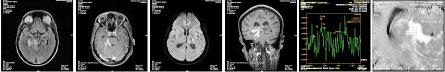
Fig 1 — MRI - a, b, c, d, e, f: Axial FLAIR, Axial T1 Contrast enhanced, Axial DWI, Coronal T1 Contrast enhanced Wt and MR Spectroscopy images. Multiple ring enhancing altered signal intensity lesions in bilateral cerebral and cerebellar hemispheres with mild perilesional edema, displaying hypo intense signal intensity on T1 and hyper intense signal intensity on FLAIR sequences. Enhancing lesion with conglomeration in the ambient and quadrigeminal cisterns with thick enhancing exudates. Diffuse restriction focus in left thalamus - TUBERCULOMAS WITH TUBERCULOUS MENINGITIS AND VASCULITIS.
treatment is often associated with high mortality, thus ensuring early recognition is of paramount importance as the clinical outcome depends upon the stage at which therapy is initiated13,14. Current antitubercular drugs are highly effective when the chemotherapy is provided prior to the onset of complications.
Cranial imaging is useful in diagnosing CNS TB, predicting its complications and also has a prognostic value15. Typical neuroradiological findings of CNS TB can aid in the diagnosis of this illness16. Despite of this, the diagnostic value of neuroimaging in CNS TB has not been fully validated in studies. Moreover, data on the utility of neuroradiology in predicting the outcome is even more limited. Researchers elsewhere have done studies comparing CT to MRI and have documented MRI as a superior diagnostic imaging modality for neuro-Tuberculosis17-19
Present study was conducted with 101 CSF positive CNS TB patients in Radiology Department of a Tertiary Care Centre, wherein their CT and or MRI findings were assessed for the various presenting features of CNS Tuberculosis.
Most common age group observed was between 20 to 29 years in the present study. Dinesh M, et al observed that 60% of their subjects were aged between 20 to 40 years5. The mean age of study subjects in present study was similar to that of Nabi S, et al6 and Aher, et al20. A male preponderance was observed in our research, similar to the study done in Nagpur20, though it was in contrast to that done on similar CNS TB patients in Chengalpattu5. It is even evident from the data of the national TB program that Tuberculosis had a male preponderance21
The triad of presenting symptoms in our research were Fever, Altered sensorium and Seizures. Etlik O, et al observed that all subjects in their study had presented with Fever and Lethargy, while four had cough22. Neurologic presentations included raised
intracranial pressure, vomiting, seizures, paresis, third cranial nerves palsy, nuchal rigidity and disturbance of consciousness. Meanwhile, Idris, et al documented that headache, convulsions and hemiparesis with or without hemisensory symptoms were commonest neurological symptoms in their study17. Increased intracranial pressure leading to papilledema was encountered in more than half of their patients.
The clinical presentation of TB Meningitis appears vague sometimes with nonspecific symptoms that are tough to distinguish it from other causes of bacterial meningitis. The typical sign of meningeal stiffness was present in very minimal proportion in our study. A longer duration of illness (more than a week) has previously been shown to be a clinical variable highly predictive of TB meningitis 9,10 , we have similar findings.
George, et al noted that age above years, a GCS score <8, absence of Headache, CSF protein below 60 mg% and Medical Research Council (MRC) Stage III at presentation were significant predictors of inhospital mortality in both HIV sero-positive as well as sero-negative patients 23 . Also, transtentorial herniation was observed in neuro imaging of a patient, who eventually expired.
The prevalence of HIV seropositivity in our study was 24.8%. Out of the 25 PLHIV, 21 had an abnormal baseline MRI. Among the PLHIV, 20 had enhancing lesions, 12 had features of Meningitis and nine had Tuberculoma in MRI. Research by Aher, et al in Nagpur20 on 50 CNS TB patients observed a higher prevalence of HIV (36.0%). Mortality was more in HIV positive patients (25%) with stage III disease. Of the 18 HIV patients in their study, 16 had Meningitis and two patients had Tuberculomas. Two patients had CD4 count below 50, eight patients had count between 51 to 100, six had counts between 101 to 150 while two patients had counts between 151 to 20020. It is
123, No 06, June 2025Journal
reported that HIV-infected patients have fewer Tuberculomas compared to non-HIV-infected patients24. These findings add to the hypothesis that Tuberculomas are formed because of a robust immunological response to Tuberculous infection. About 15% of the patients in our study also had TB infection coexisting in their Lung parenchyma, evident in sputum analysis. Etlik, et al observed a similar prevalence of PTB in their research (12.5%), although it was by Chest radiography 22. But studies done elsewhere observed a higher PTB prevalence of 44%20 to 46%25. The reason for low prevalence in present study could be attributed to absence of sputum analysis for some of our patients. This reiterates the importance of Chest X-rays and microbiological analyses on respiratory specimens in the diagnostic process of extra pulmonary Tuberculosis.
Hydrocephalus was the commonest CT imaging finding in the present study. In three out of four patients who had Hydrocephalus at baseline, it had resolved during the follow up period. A study by Botha et al in Cape Town on evaluating the sensitivity, specificity and reliability of CT imaging in diagnosing TB Meningitis observed that CT scan criteria for basal meningeal enhancement had good intra-rater agreement (k range 0.35-0.78) and fair to moderate inter-rater agreement (k range 0.20-0.52). The criteria for basal meningeal enhancement had better specificity (61.5% - 100%) but poorer sensitivity (5.9% - 29.4%)26.
Apart from enhancing lesions, more than half of our patients had MRI evidence of Tuberculoma and Meningitis, while a third of our patients had cerebral edema. Similar finding was observed by other authors as well20,22,25. Nabi, et al in their study on 100 TB Meningitis patients documented that Hydrocephalus (61%), Tuberculomas (54%), Leptomeningeal involvement (46%) and infarcts (13%) were the most frequent radiological signs on MRI of their patients6. Presence of infarct was significantly associated with mortality. Possible reason could be that majority of patients in that study had presented in MRC Stage II. MR scans especially DWI sequences were superior in detecting infarcts.
Christensen, et al observed that MRI scans proved more sensitive for identifying meningeal enhancement than CT scans (86% versus 0%) and Cranial CT scans seem to be just as sensitive as MRI scans in
identifying Hydrocephalus, Infarcts and Tuberculomas25
Our study is not without any limitations. It was not possible to have both the imaging scans for all patients, because the decision to perform the imaging in each patient was not according to predefined criteria, but at the discretion of the Physician. Also, follow up scans could not be performed in many patients due to attrition, which is unavoidable. Hence, status of neuroimaging after initiation of treatment could not be ascertained in them.
The treating Physician should interpret the CT imaging findings with caution and rely on other parameters as well for diagnosing CNS TB, with the understanding that a normal CT Brain imaging is not uncommon in initial phases of the disease particularly in adult patients.
CONCLUSION
CNS TB remains a serious disease of concern irrespective of the incidence of TB in our setting. The disease has a high mortality rate as well as sequelae among the survivors. Given the fact that diagnosis of this disease is difficult due to lack of specific tools, the clinician should remain vigilant to treat empirically if there is suspicion of CNS Tuberculosis.
ACKNOWLEDGEMENT
The authors would like to acknowledge the support of the management as well as the study subjects for the effective conduct of the study.
Funding : None
Conflict of Interest : None
REFERENCES
1World Health Organization. TB: A Global Emergency. WHO report on TB epidemic. Geneva. 1994. Available from URL: http://www.who.int/tb/publications/1994/en/ [Accessed on 21/ 02/22]
2Bhalwar RV — Textbook of Public Health and Community Medicine.1st ed. Pune: Department of Community Medicine, AFMC, Pune in collaboration with WHO India office New Delhi: 2009.
3Global tuberculosis report 2022. Geneva: World Health Organization; 2022. Available from URL: https://www.who.int/teams/ global-tuberculosis-programme/tb-reports/global-tuberculosisreport-2022[Accessed on 21/02/23]
Anzar A, et al. Evaluation of the Effectiveness of CT / MRI Brain in Detecting Central Nervous System Tuberculosis.
Vol 123, No 06, June 2025Journal of the Indian Medical Association
4Mathuranath PS, Radhakrishnan K — Neurological tuberculosis. In: Sharma SK, editor. Tuberculosis, 2nd ed. New Delhi: Jaypee Publishers; 2009. 304-29.
5Dinesh M, Alfred AJ, Murthy GS — Clinical and radiological manifestations of Central Nervous System (CNS) tuberculosis. European Journal of Molecular & Clinical Medicine 2022; 9(6): 2268-77.
6Nabi S, Badshah M, Ahmed S, Nomani AZ, Khattak I — Neuroradiology in tuberculous meningitisdiagnostic significance and prognostic value. Pakistan Journal of Neurological Sciences (PJNS) 2016; 11(2): 5-12.
8Rajashekhar V, Haran RP, Prakash GS, Chandy MJ — Differentiating solitary small cysticercus granulomas and tuberculomas in patients with epilepsy. Clinical and computerized tomographic criteria. J Neurosurg 1993; 78: 402-7.
9Ingole S, Morey C, Pote P, Ingole S, Chandak P — Radiological manifestations of central nervous system tuberculosis. MedPulse – International Journal of Radiology 2019; 11(2): 91-6.
10Marx GE, Chan ED — Tuberculous meningitis: diagnosis and treatment overview. Tuberc Res Treat 2011: 798764
11Iseman MD — A Clinician’s Guide to Tuberculosis, Lippincott Williams & Wilkins, Baltimore, Md, USA, 1999.
12Bernaerts A, Vanhoenacker FM, Parizel PM, Van Goethem JW, Van Altena R, Laridon A, et al — Tuberculosis of the central nervous system: overview of neuroradiological findings. European Radiology 2003; 13: 1876-90.
13George EL, Iype T, Cherian A, Chandy S, Kumar A, Balakrishnan A, et al — Predictors of mortality in patients with meningeal tuberculosis. Neurol India 2012; 60: 18-22.
14Qureshi HU, Merwat SN, Nawaz SA, Rana AA, Malik A, Mahmud MK, et al — Predictors of inpatient mortality in 190 adult patients with tuberculous meningitis. J Pak Med Assoc 2002; 52: 159-63.
15Wasay M, Farooq S, Khowaja ZA, Bawa ZA, Ali SM, Awan S, et al — Cerebral infarction and tuberculoma in central nervous system tuberculosis: frequency and prognostic implications. Journal of Neurology, Neurosurgery & Psychiatry 2014; 85(11): 1260-4.
16Thwaites GE, Hien TT — Tuberculous meningitis: many questions, too few answers. The Lancet Neurology 2005; 4(3): 160-70.
17Idris MN, Sokrab TE, Arbab MA, Ahmed AE, El Rasoul H, Ali S, et al — Tuberculoma of the brain: aseries of 16 cases treated with anti-tuberculosis drugs. Int J Tuberc Lung Dis 2007; 11: 91-5.
18Kalita J, Misra UK, Nair PP — Predictors of stroke and its significance in the outcome of tuberculousmeningitis. J Stroke Cerebrovasc Dis 2009; 18: 251-8.
19Haris M, Gupta RK, Husain M, Srivastava C, Singh A, Singh Rathore RK, et al — Assessment oftherapeutic response in brain tuberculomas using serial dynamic contrast-enhanced MRI. Clin Radiol 2008; 63: 562-74.
20Aher A, Paithankar M, Bhurke B — Study of Central Nervous System Tuberculosis. The Journal of the Association of Physicians of India 2018; 66(1): 41-4.
21Govt of India. TB India 2023. RNTCP Status Report. Central TB Division. Directorate General of Health Services, New Delhi: Ministry of Health and Family Welfare; 2023. Available from URL: https://tbcindia.gov.in/index1.php?lang=1&level= 1&sublinkid=4160&lid=2807[Accessed on 21/02/22]
22Etlik Ö, Evirgen Ö, Bay A, Yilmaz N, Temizöz O, Irmak H, et al — Radiologic and clinical findings in tuberculous meningitis. Eur J Gen Med 2004; 1(2): 19-24.
23George EL, Iype T, Cherian A, Chandy S, Kumar A, Balakrishnan A, et al — Predictors of mortality in patients with meningeal tuberculosis. Neurol India 2012; 60: 18-22.
24Wasay M, Kheleani BA, Moolani MK, Zaheer J, Pui M, Hasan S, et al — Brain CT and MRI findings in 100 consecutive patients with intracranial tuberculoma. Journal of Neuroimaging 2003; 13(3): 240-7.
25Christensen AS, Andersen ÅB, Thomsen VØ, Andersen PH, Johansen IS — Tuberculous meningitis in Denmark: a review of 50 cases. BMC Infectious Diseases 2011; 11(1): 1-6.
26Botha H, Ackerman C, Candy S, Carr JA, Griffith-Richards S, Bateman KJ — Reliability and diagnostic performance of CT imaging criteria in the diagnosis of tuberculous meningitis. PloS one 2012; 7(6): e38982.
Original Article
Comorbidities in COVID-19 Patients : Are these associated with Vitamin D Deficiency and SARS-CoV-2 Infection Grade ?
Background : Vitamin D, known for its immune benefits, is vital for bolstering defences. Evidence links COVID-19 comorbidities to low Vitamin D. Our goal is to assess COVID-19 patients, examining demographics, lab data, and postinfection comorbidities, focusing on vitamin D deficiency impact.
Materials and Methods : This was a cross-sectional study. Estimation of serum 25(OH)D was done in conjunction with other blood tests, including D-dimer and Complete Blood Count. All COVID-19 positive patients were checked for other health issues and medical emergencies. Data was analyzed using the Statistical Package for the Social Sciences (SPSS) Version 23 for Windows. The demographic variables, COVID-19 severity, Vitamin D level, and comorbidity were calculated in numbers and percentages. The ANOVA test was used to find significant differences in Vitamin D, D Dimer to COVID severity.
Results : Fifty patients who were clinically diagnosed with positive COVID-19 by RT-PCR were included in this study. 74% (n=37) of patients were Vitamin D deficient. Eight per cent of patients (n=4) were diagnosed with insufficient Vitamin D levels, and 18% of patients had adequate Vitamin D levels. It was noted that after acquiring SARS-CoV-2 infection, 62% (n=31) were Diabetic, 36% (n=18) were Obese and 42% (n=24) patients were suffering from Hypertension. Other medical conditions, such as NS (20%), TH (6%), TB (4%), CKD (2%), and COPD (2%) were observed.
Discussion : Correlation was observed in the severity grade of COVID-19 infection and comorbidities. Moreover, the positive correlation between the laboratory and demographic markers was also observed.
Conclusion : SARS-CoV-2 infection had an impact on individuals' medical health. Health comorbidities were associated with the COVID-19 severity. Plus, our study demonstrated that lower Vitamin D levels also had a significant impact on demographic markers indicative of low Vitamin D levels that may be associated and responsible for infection severity and comorbidities.
Key words : Vitamin D, Comorbidities, COVID-19, Correlation of Infection Severity, Association with Vitamin D.
The COVID-19 pandemic has presented an unprecedented Global health challenge, prompting intensive research to unravel the multifaceted factors influencing disease severity and outcomes. Among these factors, the role of Vitamin D deficiency has emerged as a critical and intriguing avenue of investigation. Vitamin D, renowned for its immunomodulatory properties, is implicated in the intricate interaction between the immune system and
Department of Anatomy, Dr D Y Patil Medical College, Hospital and Research Centre, Pune, Maharashtra 411018
1PhD Student and Corresponding Author
2MS, Professor and Head
3MSc (Medical Biochemistry), Associate Professor, Department of Biochemistry, B J Government Medical College, Pune, Maharashtra 411001
4MSc (Statistics), Assistant Professor, Department of Community Medicine, B J Government Medical College, Pune, Maharashtra 411001
Received on : 19/12/2023
Accepted on : 09/01/2024
Editor's Comment :
The COVID-19 pandemic has exposed various vulnerabilities in human health, focusing on the influence of vitamin deficits on illness penalties.
Vitamin D has garnered significant interest due to its crucial role in immune regulation and its potential impact on the severity of COVID-19.
Addressing their deficiencies through dietary interventions, supplementation, and public health measures is essential for improving health outcomes during the pandemic and beyond.
various comorbidities that heighten the risk of severe COVID-19 manifestations1
Comorbidities such as Diabetes Mellitus (DM), Obesity, Nephrotic Syndrome (NS), Thyroid Disorders (TH), Tuberculosis (TB), Chronic Kidney Disease (CKD) and Chronic Obstructive Pulmonary Disease (COPD) have been identified as significant contributors to the vulnerability of individuals to severe COVID-19 complications. This paper reviews the
How to cite this article : Comorbidities in COVID-19 Patients : Are these associated with Vitamin D Deficiency and SARS-CoV-2 Infection Grade ? Girish S, Sonje P, Jagtap A, Borle P. J Indian Med Assoc 2025; 123(6): 31-5.
123, No 06, June 2025Journal
existing literature to delineate the intricate connections between Vitamin D deficiency and these diverse comorbidities, shedding light on the potential mechanisms that underpin their collective impact on the course of COVID-192-5.
By exploring the nexus between Vitamin D, comorbidities, and COVID-19 infection grade, this research aims to provide a comprehensive understanding of the immunological dynamics at play, laying the groundwork for targeted interventions and therapeutic strategies to mitigate the severity of COVID-19 in individuals with underlying health conditions.
In the present study, we aim to understand how comorbidities post-COVID-19 infection and deficiency of Vitamin D are associated with infection grades. This study might open avenues for designing therapeutic strategies or lines of treatments for COVID-19 sufferers.
MATERIALS AND METHODS
The present study adopted a cross-sectional design spanning a duration of six months, with approval obtained from the Institutional Ethical Committee. Informed consent was diligently obtained from all participating patients.
In this investigation, we assembled a cohort consisting of 880 individuals who tested positive for COVID-19 and presented with concomitant Vitamin D deficiency. From this cohort, a targeted subset of 50 participants was purposively selected for an in-depth methylation study, with a particular focus on exploring the interplay between comorbidities and disease severity in relation to Vitamin D deficiency which required good quality of extracted DNA to perfume the experiment. The selection criteria for this subgroup encompassed a comprehensive assessment of comorbid conditions and disease severity levels, ensuring a nuanced representation of the diverse clinical spectrum observed in COVID-19 cases with Vitamin D deficiency. The investigation methodology adhered to established ethical guidelines and the selection process was designed to provide a robust foundation for examining the intricate relationships between Vitamin D status, comorbidities, disease severity and epigenetic modifications. The systematic approach employed in participant identification and subsequent methylation analysis aimed to unravel potential associations and shed light on the underlying
molecular mechanisms governing the intricate relationship between Vitamin D deficiency, comorbidities and COVID-19 severity. The statistical analysis was conducted using SPSS (Statistical Package for the Social Sciences) Version 23 for Windows. Demographic variables, COVID-19 severity, Vitamin D levels, and comorbidities were computed in both absolute numbers and percentages. The ANOVA test was employed to identify significant differences in Vitamin D, D Dimer and their association with the severity of COVID-19.
The study exclusively focused on clinically diagnosed COVID-19-positive individuals confirmed through RTPCR testing. Following the World Health Organisation (WHO) standards, patients were categorised based on the severity of symptoms, distinguishing between mild, moderate, and severe cases6
To ensure the specificity of the study, individuals with a history of severe illnesses such as Cancer, Respiratory disease, Gastrointestinal disease and Kidney disease were excluded. Pregnant women, patients on Vitamin D supplementation and those with missing data were also omitted from the analysis.
Sample collection and testing were conducted at the Biochemistry Centre Clinical Laboratory situated at BJ Government Medical College and Sassoon General Hospital in Pune. Plasma 25-hydroxyvitamin D (25(OH)D) levels were measured using ELISA (kit name). In addition to Vitamin D levels, various laboratory parameters, including D dimer, Complete Blood Count, Liver Function Tests and Kidney Function Tests, were observed.
According to established guidelines, individuals with serum 25(OH)D levels below 20 ng/ml were classified as vitamin D deficient. The data analysis was carried out using SPSS software version 23. To discern significant differences among the study groups, the Analysis of Variance (ANOVA) test was employed. This rigorous methodology ensures the reliability and validity of our findings, contributing valuable insights into the association between Vitamin D deficiency and the severity of COVID-19 symptoms.
RESULTS
This study comprised 50 individuals diagnosed with COVID-19 through RT-PCR. The majority of participants were male, with a male-to-female sex ratio of 2.1:1. The average age was 46.78 years, with
a Standard Deviation of ±22.10. Additionally, three newborns aged 1-2 weeks were included in the study population. SARS-CoV-2 severity-wise classification revealed that 32% of the patients fell within the mild category, while 38% were classified as moderate to severe cases. The remaining 32% presented with severe illness. Vitamin D deficiency, defined as serum levels below 20 ng/ml, was prevalent in a substantial majority, accounting for 74% of the patients.
Notably, 60% of the patients were identified as Obese. Diabetes Mellitus emerged as the predominant comorbidity among the study population, with 62% (n=31). Moreover, Neurological disorder (n=10) in 20%, Cancer in 18% (n=9), TH in 6% (n=3), TB in 2% (n=4) were dominantly found. Additionally, CKD, COPD, SLE, CCF and HIV were observed in 2% of patients, respectively (Table 1).
The severity of COVID-19 was observed in individuals with Diabetes Mellitus and Hypertension; the association did not achieve statistical significance. However, a statistically significant difference in age concerning COVID-19 severity was noted (P<0.05), indicating age as a potentially influential factor in the progression of the disease (Table 2). Furthermore, a positive correlation between Vitamin D levels and BMI was identified, although this correlation did not reach statistical significance.
Contrastingly, no statistically significant differences were found in Vitamin D levels or D dimer concerning COVID-19 severity (P>0.05). However, the relationship between vitamin D and other parameters can be potentially explored with a large population size (Table 3). These findings shed light on the demographic and clinical characteristics of the COVID-19 patients under investigation, providing
Table 1 — Comorbidities according to the Vitamin D levels : This table shows the association between the Vitamin D levels and comorbidities. Patients with deficiency with Vitamin D seem to have more in number after acquiring the COVID-19 infection were associated with high risk factor comorbidities
ParametersVitamin D (ng/ml) (%)P Value >3020-3010-20<10 (n=9)(n=4)(n=26)(n=11)
Sex (M/F)6/31/318/89/2
Cancer2 (22.2)1 (25)3 (11.5)3 (27.3)0.66
Table 2 — Comparison of comorbidity according to COVID severity
ComorbidityCOVID severity (%)Chi-square*P Value Grade IGrade IIGrade III (n=19)(n=15)(n=16)
DM10 (52.63)9 (60)12 (75)1.780.18
HTN7 (36.84)8 (53.33)9 (56.25)1.330.25
Obesity III8 (53.33)139 (56.25)7.620.11 202
*Linear by linear
Table 3(A) — Correlations of Vitamin D levels with Laboratory markers, Age and BMI
Correlation between Vitamin D andr s Value*P Value D Dimer-0.080.57 WBC0.3060.031
*Spearman rank correlation
Table 3(B) — Correlations of COVID-19 severity grade with Laboratory and Demographic parameters
Parameters Grade I versus Grade I versus Grade II versus Grade IIGrade IIIGrade III VIT D (ng/mL)0.6011 D Dimer (mg/L)111 WBC111
Lymphocyte (%)111 Age (Years)0.0120.141 BMI0.2410.89
By post hoc Bonferroni test: There is no significant difference of laboratory markers, age, BMI in Group I versus Group III, Group II versus Group III and Group I versus Group II except age as P>0.05. Group II had significantly more age than Group I as P<0.05.
valuable insights into the prevalence of comorbidities and vitamin D deficiency within this cohort (Table 4).
DISCUSSION
The observed increased trend in COVID-19 severity among individuals with Diabetes Mellitus 7,8 and Hypertension 9,10 while not achieving statistical significance, implies a potential association that warrants further investigation. These comorbidities have been previously identified as risk factors for severe outcomes in COVID-19, aligning with broader epidemiological patterns. The lack of statistical significance in this study may be attributed to the relatively small sample size, emphasising the need for larger cohorts to elucidate these relationships more definitively.
The significant difference in age concerning COVID-
Girish
Table
WBC9.556.11311.087.54510.096.2180.230.80
Lymphocyte (%)16.6812.62518.4513.07520.0414.2520.280.76 Age (Years)35.7618.70857.2221.52750.0621.6814.870.012 BMI29.993.97232.102.44530.813.4381.610.21
19 severity highlights the pivotal role of age as a determinant of disease progression. Advanced age has consistently been acknowledged as a critical factor influencing the severity of respiratory infections, including COVID-1911,12 . This finding aligns with existing literature and underscores the importance of age-stratified analyses in understanding disease dynamics.
Contrary to expectations, no statistically significant differences were found in Vitamin D levels and D dimer with respect to COVID-19 severity. While Vitamin D deficiency has been implicated in immune modulation, the absence of a significant association in this study suggests that the role of Vitamin D in COVID-19 severity may be more complex and multifactorial, warranting further exploration.
The positive correlation between Vitamin D levels and BMI, although not statistically significant, introduces an intriguing avenue for exploration. The relationship between Obesity, Vitamin D and COVID-19 outcomes is complex, with potential implications for immune function. The lack of statistical significance may be attributed to the need for more refined methodologies or larger sample sizes. The present literature has evidence for an association between Vitamin D, as a potent immunomodulator and SARS-CoV-2 infection severity, which underscores the potential role of Vitamin D in influencing immune responses. This connection provides a basis for exploring targeted interventions to optimise Vitamin D levels, potentially impacting the course of COVID-19. However, the association between Vitamin D and COVID-19 severity is complex and requires investigation at the genetic and molecular levels13-16. Our study also finds Cancer cases post-COVID-19 infection in Vitamin Ddeficient patients. The detection of Vitamin D deficiency in Cancer cases raises intriguing questions about its potential role in cancer development or progression. Further research is warranted to elucidate the complex relationship between Vitamin
D status and Cancer, offering insights that may inform preventive strategies and complementary therapeutic approaches17,18
The relationship between Vitamin D deficiency and COVID-19 is complex and multifactorial. Several factors contribute to Vitamin D deficiency in COVID19 patients, such as limited sun exposure, underlying health conditions, an inflammatory response, reduced outdoor activities and malabsorption. Understanding these interconnections is crucial for developing strategies to address Vitamin D deficiency in COVID19 patients. Supplementation, dietary adjustments, and recommendations for safe sun exposure are avenues that may be explored to optimise Vitamin D levels and potentially mitigate the severity of COVID19 outcomes. Biesalski, et al in 2020, concluded that Vitamin D levels are associated with Vitamin D levels, which was matched with our results presented in subsequent tabular forms. Therefore, it’s important to note that all comorbidities from the past COVID19 infection might have a relationship with depleted levels of Vitamin D19
In conclusion, Vitamin D levels are associated with comorbidities and COVID-19 infection severity. Our study results offer valuable insights into the interaction of comorbidities, age and biomarkers in the context of COVID-19 severity. The first limitation of the study was relatively small sample size due to expertise performing the actual experimentation and higher cost of methylation study. Second, Vitamin D may be more important in combination with other nutrients also known to interact with the epigenome. Moreover, present manuscript only investigates the relation between COVID-19 infection severity with Vitamin D levels and comorbidities. Percent methylation, epigenetic modification or gene expression will be the future aspects of the study and are under examination. Finally, this was a cross sectional study; thus, no causal inference can be established. Future longitudinal studies including placebo-controlled
Girish S, et al.
Vol 123, No 06, June 2025Journal of
supplementation studies are needed to establish causality.
CONCLUSION
The SARS-CoV-2 infection had an impact on individuals’ medical health. Health comorbidities were associated with the severity of COVID-19. Moreover, our study demonstrated that lower Vitamin D levels also had a significant impact on demographic markers. This study might provide a path and open research avenues in terms of Vitamin D association with disease severity. While the trends observed are consistent with existing knowledge, the study underscores the importance of continued research with larger cohorts and refined methodologies to unravel the intricacies of these relationships and inform more targeted clinical interventions.
Funding : None
Conflict of Interest : None
REFERENCES
1Pereira M, Dantas Damascena A, Galvão Azevedo LM, de Almeida Oliveira T, da Mota Santana J — Vitamin D deficiency aggravates COVID-19: systematic review and meta-analysis. Crit Rev Food Sci Nutr 2022; 62(5): 1308-16.
2Sousa LG de, Mcgrail DJ, Li K — Spontaneous tumor regression following COVID-19 vaccination. J Immunother Cancer 2022; 10(3). doi:10.1136/jitc-2021-004371
3Ejaz H, Alsrhani A, Zafar A — COVID-19 and comorbidities: Deleterious impact on infected patients. J Infect Public Health 2020; 13(12): 1833-9. doi:10.1016/j.jiph.2020.07.014
4Lima-Martínez MM, Carrera Boada C, Madera-Silva MD, Marín W, Contreras M — COVID-19 and diabetes: A bidirectional relationship. Clinica e Investigacion en Arteriosclerosis 2021; 33(3): 151-7. doi:10.1016/j.arteri.2020.10.001
5Walsh JS, Bowles S, Evans AL — Vitamin D in obesity. Curr Opin Endocrinol Diabetes Obes 2017; 24(6): 389-94. doi:10.1097/MED.0000000000000371
6 Clinical Spectrum of SARS-CoV-2 Infection . https:// www.covid19treatmentguidelines.nih.gov/
7Lim S, Bae JH, Kwon HS, Nauck MA — COVID-19 and diabetes mellitus: from pathophysiology to clinical management.
8Abumayyaleh M, Núñez Gil IJ, Viana-LLamas MC — PostCOVID-19 syndrome and diabetes mellitus: a propensitymatched analysis of the International HOPE-II COVID-19 Registry. Front Endocrinol (Lausanne) 2023; 14. doi:10.3389/ fendo.2023.1167087
9Matsumoto C, Shibata S, Kishi T — Long COVID and hypertension-related disorders: a report from the Japanese Society of Hypertension Project Team on COVID-19. Hypertension Research 2023; 46(3): 601-19. doi:10.1038/s41440-02201145-2
10Akpek M — Does COVID-19 Cause Hypertension? Angiology 2022; 73(7): 682-7. doi:10.1177/00033197211053903
11Kushwaha S, Khanna P, Rajagopal V, Kiran T — Biological attributes of age and gender variations in Indian COVID-19 cases: A retrospective data analysis. Clin Epidemiol Glob Health 2021; 11. doi:10.1016/j.cegh.2021.100788
12Statsenko Y, Al Zahmi F, Habuza T — Impact of Age and Sex on COVID-19 Severity Assessed From Radiologic and Clinical Findings. Front Cell Infect Microbiol 2022; 11. doi:10.3389/ fcimb.2021.777070
13Gibbons JB, Norton EC, McCullough JS — Association between vitamin D supplementation and COVID-19 infection and mortality. Sci Rep 2022; 12(1). doi:10.1038/s41598-02224053-4
14Martineau AR, Cantorna MT — Vitamin D for COVID-19: where are we now? Nat Rev Immunol 2022; 22(9): 529-30. doi:10.1038/s41577-022-00765-6
15Tomaszewska A, Rustecka A, Lipiñska-Opa³ka A — The Role of Vitamin D in COVID-19 and the Impact of Pandemic Restrictions on Vitamin D Blood Content. Front Pharmacol 2022; 13. doi:10.3389/fphar.2022.836738
16Karonova TL, Kudryavtsev I V., Golovatyuk KA — Vitamin D Status and Immune Response in Hospitalized Patients with Moderate and Severe COVID-19. Pharmaceuticals 2022; 15(3). doi:10.3390/ph15030305
17Easty DJ, Farr CJ, Hennessy BT — New Roles for Vitamin D Superagonists: From COVID to Cancer. Front Endocrinol (Lausanne) 2021; 12. doi:10.3389/fendo.2021.644298
18Jahankhani K, Ahangari F, Adcock IM, Mortaz E — Possible cancer-causing capacity of COVID-19: Is SARS-CoV-2 an oncogenic agent? Biochimie 2023; 213: 130-8. doi:10.1016/ j.biochi.2023.05.014
19Biesalski HK — Vitamin D deficiency and co-morbidities in COVID-19 patients – A fatal relationship? NFS Journal 2020; 20: 10-21. doi:10.1016/j.nfs.2020.06.001
Girish S, et al. Comorbidities in COVID-19 Patients : Are these associated with Vitamin D Deficiency and SARS-CoV-2 Infection Grade?
Original Article
Pulmonary Complications in Systemic Lupus Erythematosus Patients : A Cross Sectional Observational Study in a Tertiary Care Set-up
Background : Studies suggest that pulmonary complication is a well prevalent manifestation of Systemic Lupus Erythematosus (SLE), with most common complications being pleuritis - with or without effusion followed by Interstitial Lung Disease (ILD) and infections. Many patients continue to harbour pulmonary complications, with no clinical features. The present study aims to explore the pulmonary complications in SLE patients.
Materials and Methods : A cross-sectional, observational study included adult admitted cases of SLE, who were interviewed for basic demographic information and detailed medical history. Physical examination, haematological, biochemical, microbiological and serological tests were conducted. Patients were made to undergo Spirometry and Chest X-ray in all cases; and pleural fluid study and High-resolution CT (HRCT) Scan of Thorax was done selectively as required. Pulmonary complications were assessed and type of involvement was noted.
Results : Out of the 50 patients, 32% had pulmonary complication, with most common being pleural effusion noted in 18% patients, followed by ILD in 10%. Pulmonary manifestation on clinical presentation were however positive in 20% patients. On Chest X-ray, abnormality was noted in 22% patients whereas, PFT and HRCT showed pulmonary manifestations in 30% and 32% patients. Radiological imaging – HRCT and PFT detected pulmonary involvement in a significant number of patients who were asymptomatic. PFT was found to have restrictive lung pattern in 15 patients, while HRCT Chest was abnormal in 16 cases.
Conclusion : As almost one-third of SLE patients have pulmonary complications, so all patients of SLE should routinely undergo screening so as to exclude pulmonary complication if any.
Key words : Systemic Lupus Erythematosus, Pulmonary Complications, High Resolution CT scan, Spirometry, Chest X-ray.
SEditor's Comment :
Pulmonary complication is a well prevalent manifestation of Systemic Lupus Erythematosus (SLE), with most common complications being pleuritis - with or without effusion followed by Interstitial Lung Disease (ILD) and infections. Many patients continue to harbour pulmonary complications, with no clinical features.
All patients of SLE should routinely undergo screening so as to exclude pulmonary complication if any.
Department of Tropical Medicine, Calcutta School of Tropical Medicine, Kolkata, West Bengal 700073
1MD, Senior Resident
2MD, Associate Professor
3M Pharm, PhD Scholar, Department of Clinical & Experimental Pharmacology and Corresponding Author
4MD, Former Professor
5MD, Professor, Department of Dermatology, Venereology and Leprology
6MD, Professor, Department of Chest and Respiratory Medicine, Medical College and Hospital, Kolkata, West Bengal 700073
Received on : 09/02/2023
Accepted on : 08/05/2023
ystemic Lupus Erythematosus (SLE) is an autoimmune disease in which organs and cells undergo damage mediated by tissue-binding antibodies and immune complexes. The greatest prevalence of SLE is among women of child-bearing age. Among the ethnic groups, the greatest prevalence is noted in that of African-American and Afro-Caribbean people1. The pathogenesis of SLE includes genetic factors, epigenetic factors and environmental factors which cause abnormal immune response and development of autoantibody and immune complexes resulting in inflammation ultimately leading to tissue damage 1 . SLE can have a wide range of manifestations, involving virtually every organ or apparatus and its severity can vary from very mild disease without major organ involvement, to severe life-threatening conditions. Clinical manifestations may include cytopenia, fever, malar and other skin rashes, oral ulcers, polyarthralgia/non erosive arthritis, vasculitis, renal, neurological, cardiac and pleuropulmonary involvement2
How to cite this article : Pulmonary Complications in Systemic Lupus Erythematosus Patients : A Cross Sectional Observational Study in a Tertiary Care Set-up. Chatterjee R, Gonjhu D, Mukherjee S, Pramanik N, Haldar S, Das SK. J Indian Med Assoc 2025; 123(6): 36-41.
123, No 06, June 2025Journal
According to study by Hannah, et al 3 in 2019 on pulmonary complications of SLE, pulmonary involvement was reported to be well prevalent and seen in 50 to 70% of SLE patients. In 4-5% of patients, it has been the presenting feature of SLE. Around 12% of patients was proposed to have evidence of permanent lung damage by 10 years postdiagnosis. Pulmonary complications are broad and include pleural disease, Interstitial Lung Disease (ILD), vasculitis, pulmonary hypertension, large airway disease and infection. This manifestation, when mild, may respond to treatment with Non-steroidal Antiinflammatory Drugs (NSAIDs); when more severe, patients require a brief course of glucocorticoid therapy. Pulmonary infiltrates also occur as a manifestation of active SLE and are difficult to distinguish from infection on imaging studies. Lifethreatening pulmonary manifestations include interstitial inflammation leading to fibrosis, shrinking lung syndrome, and intra-alveolar haemorrhage; all of these probably require early aggressive immunosuppressive therapy as well as supportive care1 Some of the immuno-suppressive agents have also been linked to drug-induced lung injury3
Literature search suggests that pulmonary complication is a well prevalent manifestation of SLE, with most common complications being pleuritis- with or without effusion followed by ILD and infections. It is stated that HRCT Chest is more sensitive in diagnosing the complications than PFT and Chest Xray. Nevertheless, many patients continue to harbour pulmonary complications, with no clinical features –ie, subclinical. Thus, the present study aims to assess the prevalence of pulmonary complications in SLE patients and further determine the proportion/type of each pulmonary manifestations occurring in the SLE patients.
MATERIALS AND METHODS
A cross-sectional, observational study was conducted in the Inpatient and Outpatient Department of Tropical Medicine and Dermatology at School of Tropical Medicine, Kolkata over a period of one year. Permission for the conduct of the study was obtained from the Institutional Ethical Committee of the institute prior commencement (vide Approval No CREC-STM/ 573). Written informed consent was obtained from each individual prior participation. The study included patients over 18 years of age of both sexes fulfilling
the SLICC 2012 classification criteria4 of Systemic Lupus Erythematosus and admitted in Indoor and attending the designated Outdoor Departments. Patients with HIV, HBV or HCV co-infection; those with current or past malignancy within the past 5 years; those suffering from known pulmonary disorder that is not due to SLE; pregnant or lactating females and those unwilling to participate were excluded. A convenient sample size of 50 was selected for the study based on inclusion and exclusion criteria.
For each patient, basic demographic information and detailed medical history was noted. Physical examination in terms of general survey and systemic examination, haematological, biochemical, microbiological and serological tests were conducted. The preliminary assessment tried to determine whether patient has pulmonary manifestation of SLE. After clinical evaluation, patients were made to undergo Spirometry and Chest X-ray in all cases; and pleural fluid study and High-resolution CT (HRCT) Scan of thorax was done selectively as required. Pulmonary complications were assessed and type of involvement was noted.
Data were statistically analysed. Categorical data were presented as frequency and percentages, while continuous data were presented as mean ± Standard Deviation (SD). All statistical analysis for various measures were performed using various statistical software packages like Statistical Package for the Social Sciences (Windows version 21.0; SPSS Inc, Chicago, IL, USA) and Microsoft Excel.
RESULT
A total of 50 diagnosed patients of SLE were included in this study for analysis. The patients were diagnosed as SLE on fulfilling the SLICC criteria. The age distribution of the study population revealed that maximum number of patients belonged to the age group of 25 years to 35 years with 34 patients (68%) falling under this category. The mean age was 28.42 ± 5.6 years. There is a female preponderance with 96 % of the study population ie, 48 patients being female. Most of the patients (n=20) had a disease duration of 37 to 60 months followed by 14 patients with a disease duration of 13 to 36 months and 10 patients with disease duration of 61 to 84 months. The mean disease duration is 46 ± 24.37 months. (Table 1) Among the 50 patients included in our study, Chatterjee R et
Table 1 — Patient Characteristics
No of patients (n=50)
Gender
Female48 (96%)
Male2 (4%)
Age (in years)
<2511 (22%)
25-3534 (68%)
>355 (10%)
SLE Disease Duration (in Months)
<124 (8%)
13-3614 (28%)
37-6020 (40%)
61-8410 (20%)
>842 (4%)
Presence of Nephritis40 (80%)
Presence of musculoskeletal manifestations35 (70%)
Presence of Skin, hair and mucous membrane manifestations36 (72%)
Alopecia16 (32%)
Oral ulcer15 (30%)
Presence of Serositis (ascites/ pleural effusion/ pericardial effusion)11 (22%)
Presence of Neuropsychiatric manifestation2 (4%)
Presence of Haematological abnormality
Anaemia35 (70%)
Leukopenia16 (32%)
Thrombocytopenia12 (24%)
12 were newly diagnosed cases. Remaining 38 patients were already diagnosed receiving treatment. All patients of the study were ANA positive. Anti-ds DNA were positive in 39 patients of our study.
Pulmonary Complications in SLE Patients :
Out of the 50 patients included in our study, total of 10 patients, ie, 20% had symptoms pertaining to pulmonary involvement. Of them, 8(16%) had Chest pain, 8 (16%) had Shortness of breath or dyspnoea, 6 (12%) had Cough, 5 (10%) had Fever and 1 patient had Haemoptysis as manifestation (Table 2).
Clinical examination of respiratory system revealed abnormality in 10 patients (20%). Radiological examination included Chest X-ray and HRCT thorax. Chest X-ray were abnormal in 11 subjects with most common abnormality being pleural effusion. HRCT thorax revealed abnormality in 16 cases (32%)(Table 3).
Pulmonary Function Test (PFT) done in all subjects of the study population showed abnormality in 15 patients (30%). PFT was of restrictive pattern in all the cases, with FEV1/FVC >0.7. Pleural fluid analysis was done in subjects with pleural effusion. Analysis
Table 2 — Presenting symptom related to respiratory system in the study population
No ofNo ofPresenting Pulmonary SymptomsNo of Clinicalpatientspatients
Symptoms
11Only Chest Pain1
22Chest Pain + Dyspnoea2
35Fever+ Cough+ Dyspnoea1
>32Chest Pain+ Fever+ Cough+ Dyspnoea2
Chest Pain + Fever + Dyspnoea1
Chest Pain + Cough + Dyspnoea2
Fever + Cough + Haemoptysis1
Table 3 — Clinical and Radiological Findings in SLE Patients
Chest X Ray11 (22%)Pleural Effusion8(2-Unilateral, 6-Bilateral)
Opacification/ Infiltration4 Lymphadenopathy1
HRCT Thorax16 (32%)Pleural effusion9
Pleural thickening1 LRTI4 ILD5
of pleural fluid showed to be exudative in nature in all cases as per Light’s criteria.
Sputum analysis in patients with Cough were done. Sputum CBNAAT was positive in one patient. In the four patients of Pneumonia, sputum Gram stain and culture revealed isolate in 2 cases (Streptococcus pneumonia – 1; Klebsiella pneumonia – 1).
Only 1 patient of our study had pulmonary arterial hypertension evidenced by loud P2 on clinical examination and verified by 2D Echocardiography.
So, a total of 16 patients (32%) of the study population had presence of pulmonary complications. Out of the 16 patients of our study population detected finally to have presence of pulmonary complication by clinical, microbiological and radiological assessment, 9 patients (18%) were found to have pleural effusion. Out of the 9 cases of pleural effusion, 2 had unilateral pleural effusion and rest 7 had bilateral fluid accumulation. Among the 9 patients of pleural effusion, 7 had only pleural effusion. Rest 2 patients had effusion co-existing with Pneumonia. In all the cases, the fluid was exudative in nature. Overall, the most common manifestation was pleural effusion
detected in 9 patients (18%) followed by ILD in 5 cases (10%) and Pneumonia in 4 patients (8%). Pulmonary tuberculosis, PAH and pleural thickening was detected in 1 patient each respectively. Out of the 5 ILD diagnosed cases, 2 had only ILD; 1 had ILD with PAH. In 1 patient with ILD, Pneumonia was also present and 1 had pulmonary TB with ILD. So, of the 16 patients diagnosed to have pulmonary involvement, 10 had only 1 pathology; rest 6 patients had more than one manifestation (Fig 1).
Out of the 16 patients finally diagnosed with pulmonary involvement, 10 were clinically symptomatic. Chest X-ray revealed abnormality in 11 cases. PFT was found to have restrictive lung pattern in 15 patients. HRCT Chest was abnormal in 16 cases.
DISCUSSION
The present cross-sectional, observational study revealed that 32 % of the included SLE patients had pulmonary complication, with most common complication being pleural effusion noted in 18% patients, followed by ILD in 10%. Infections was the next common manifestation- with Pneumonia seen in 4 cases and Pulmonary TB in 1 case. PAH was noted in only 1 patient. Pleural effusion was mostly bilateral and exudative in nature. From our study, we conclude that pulmonary manifestation on clinical presentation were positive in 20% patients. On Chest X-ray, abnormality was noted in 22% patients whereas, PFT and HRCT showed pulmonary manifestations in 30% and 32% patients. Thus, radiological imaging – HRCT and PFT detected pulmonary involvement in a significant number of patients who were asymptomatic. Out of the 16 patients finally diagnosed with pulmonary involvement, 10 were clinically symptomatic. Chest X-ray revealed abnormality in 11 cases. PFT was found to have restrictive lung pattern in 15 patients. HRCT Chest was abnormal in 16 cases.
Our study revealed 34 patients (68%) in the age group of 25 to 35 years with a mean age of 28.42 ± 5.59 years and a female preponderance of 48 patients (96%). This is in concurrence with similar studies as conducted by Teh, et al in 20185 for 4 years with 125 patients, which showed a mean age of 33.4 ± 14.2 years and a female predominance of 89.6 %. Another study by Skare, et al 6 in 2016 of 144 patients conducted and followed up for 5 years showed a mean
age of 39.15 ± 11.65 years with a female predominance of 93.8 %. Most studies on SLE have documented female preponderance and involvement of mainly female of reproductive age group7. While the gender predisposition was comparable in all the 3 studies, the higher mean age in both the reference studies can be attributed to a larger study population and longer study period. The reason for a significant female predominance is because of the genetic buildup of the present population which favours development of this autoimmune disease. Our study showed 20 patients (40%) who were admitted had lupus duration of 37 to 60 months. The mean disease duration was 46 ± 24.375 months.
The patients had various kinds of manifestations attributable to SLE. Nephritis was present in 40 patients (80%) of the study population with evidence of proteinuria on urine examination. In the study by Teh, et al5, among 125 patients, Nephritis was present in 81 patients (64.8%). A case-control study by Jung, et al8 in 2018 showed in a total population of 360 patients, 244 patients (67.8%) had Nephritis. In the same study 241 patients (66.9%) had Arthritis, which was quite in similarity to our findings (70%). The present study noted cutaneous manifestations in 72% study population, with the most common rash being erythematous, maculopapular and butterfly shaped rash on the face. While 16 patients (32%) had diffuse alopecia, 15 patients (30%) had chronic oral ulcer. This finding was in line with the study by Teh, et al5 , which showed the presence of mucocutaneous manifestations in 60 % of their population. The study by Skare, et al6 showed that photosensitivity was present in 70.7%, malar erythema was present in 48.9% and oral ulcers were present in 45.6% of their study population of 144.
Ten patients presented with symptoms pertaining to
Fig 1 — Pulmonary involvement in SLE Patients
Chatterjee R et al. Pulmonary Complications in Systemic Lupus Erythematosus Patients.
123, No 06, June 2025Journal
respiratory system in our study which constituted 20% of the study population. A study by Kakati, et al9 found 23.68% of the study subjects to have sign/symptom indicating pulmonary involvement. Out of the 10 patients having symptoms of respiratory manifestations, 9 had Chest pain, 8 had Shortness of breath/dyspnoea, cough was present in 6 cases and Fever in 5 cases. Only 1 patient gave history of haemoptysis. So, the most common symptom observed was Chest pain followed by Dyspnoea and Cough. Similar findings were reported by Samuel, et al10, Al Abbad, et al 11 and Omer, et al7 in their study. Samuel, et al 10 observed exertional dyspnoea, productive Cough and Chest pain as commonest presenting symptom. Delgado, et al12 also observed similar symptoms in their study. A study conducted by Ghosh, et al13 found the commonest respiratory symptom to be dyspnoea.
Clinical examination of the patients with detailed respiratory system examination found the commonest clinical manifestation to be pleural effusion. By clinical examination alone, 6 patients were diagnosed to have pleural effusion. 4 patients were clinically diagnosed to have LRTI /Pneumonia. Features of ILD was found in 2 patients on clinical examination. A single centre cross-sectional observational study by Ghosh, et al13 in a Tertiary Care Hospital of same study region also reported the commonest respiratory manifestation to be pleural effusion. Chest X-ray revealed abnormality in 11 patients (22% of the study population), PFT revealed abnormality in 15 patients (30%) and HRCT Chest revealed abnormality in 16 patients (32%) of the study population. So, HRCT was found to be more sensitive than Chest X-ray in diagnosing the pulmonary complications. Studies conducted by Ghosh, et al13, Kakati, et al9 , Sant, et al14 also noted similar findings. In the study by Ghosh, et al13, among the finally diagnosed ILD cases diagnosed by HRCT Chest, 50% had normal Chest X-rays. Kakati, et al9 found HRCT abnormalities in 55% cases while PFT and Chest X-rays were abnormal only in 29% and 18% cases respectively.
In our study, a total of 16 patients, ie, 32% -about 1/ 3rd of the study population was finally diagnosed to have pulmonary complication. Two large studies also showed similar prevalence of pulmonary involvement. A 10-year study conducted retrospectively in Arab involving 180 patients showed 33% patients to have pulmonary involvement 7 Data from Spanish
Rheumatology Society which included data of 3215 SLE patients noted 31% patients to have at least 1 pleuro-pulmonary manifestation most common of them being pleural disease15. Higher prevalence rated of up to 50% was also noted in some studies. According to Hannah, et al3, pulmonary complications may be seen in 50-70% of SLE cases and around 12% of patients will have evidence of permanent lung damage by 10 years postdiagnosis3. In 16 patients diagnosed to have pulmonary involvement, 10 had only 1 pathology; rest 6 patients had more than one manifestation. The commonest pulmonary complication found in our study was pleural effusion seen in 9 cases (18%), followed by ILD in 5 patients (10%). Pleura-pulmonary infections was the next common manifestation. Pneumonia was diagnosed in 4 cases; Pulmonary TB was diagnosed in 1 patient. 1 patient with ILD was also found to have PAH. Studies by Ghosh, et al13 , Mittoo, et al16 also found the commonest manifestation to be pleural effusion. Among the 16 patients diagnosed with pulmonary complication, all were females. This fact may be explained by the fact that females largely outnumber male SLE cases.
Analysis of pleural fluid revealed it to be exudative in nature in all cases. Of the 9 pleural effusion cases, 7 cases had bilateral involvement. Ghosh, et al13 also noted pleural effusion to be bilateral in 80% of the cases. The second most common manifestation was ILD seen in 5 patients, which means 10% of the study population. Ghosh, et al 13 also noted same prevalence – 10% of study subjects had ILD. All the patients who were diagnosed to have ILD had disease duration for more than 60 months. A large-scale study conducted in China found a significant association between ILD and duration of disease17 The present study noted that HRCT and PFT were superior to routine Chest X-ray in diagnosis of pulmonary complications.
Our study had a few limitations which need a bit of attention. Firstly, the sample size was small to extrapolate and relate the findings of the study to a much larger population. Secondly, time duration of the study was small to consider any other major factors which may have influenced the results of the study. Thirdly, the observational design of the study did not allow for comparison of variables. Fourthly, whether injection cyclophosphamide had any role on development of ILD in the patients who were already
Vol 123, No 06, June 2025Journal of
treated with the same as per protocol could not be clearly ascertained. Further prospective studies with larger sample size and longer duration among the Indian population might have a better understanding of the subject. This study thus sparks the need for conduct of future studies with improved study design to determine risk factors for development of pulmonary complications in SLE for an improved Quality of Life in these patients.
CONCLUSION
As almost one-third of SLE patients have pulmonary complications, so all patients of SLE should routinely undergo screening so as to exclude pulmonary complication if any.
Source of Funding : Nil
Conflict of Interest : None Declared
REFERENCES
1Jameson JL, Kasper DL, Longo DL, Fauci AS, Hauser SL, Loscalzo J — Harrison’s Principles of Internal Medicine. 20th edition. The United States of America: Cenveo Publisher Services; 2018. Chapter 349, Systemic Lupus Erythematosus; 2515-25.
2Zucchi D, Elefante E, Calabresi E, Signorini V, Bortoluzzi A, Tani C — One year in review 2019: systemic lupus erythematosus. Clin Exp Rheumatol 2019; 37(5): 715-22.
3Hannah JR, D’Cruz DP — Pulmonary Complications of Systemic Lupus Erythematosus. Semin Respir Crit Care Med 2019; 40(2): 227-34.
4Petri M, Orbai AM, Alarcón GS, Gordon C, Merrill JT, Fortin PR, et al — Derivation and validation of the Systemic Lupus International Collaborating Clinics classification criteria for systemic lupus erythematosus. Arthritis Rheum 2012; 64(8): 267786.
5Teh CL, Wan SA, Ling GR — Severe infections in systemic lupus erythematosus: disease pattern and predictors of infection-related mortality. Clin Rheumatol 2018; 37(8): 2081-6.
6Skare TL, Dagostini JS, Zanardi PI, Nisihara RM — Infections and systemic lupus erythematosus. Einstein (Sao Paulo) 2016; 14(1): 47-51.
7Alamoudi OS, Attar SM — Pulmonary manifestations in systemic lupus erythematosus: association with disease activity. Respirology 2015; 20(3): 474-80.
8Jung JY, Yoon D, Choi Y, Kim HA, Suh CH — Associated clinical factors for serious infections in patients with systemic lupus erythematosus. Sci Rep 2019; 9(1): 9704.
9Kakati S, Doley B, Pal S, Deka UJ — Pulmonary manifestations in Systemic Lupus Erythematosus (SLE) with special reference to HR CT. J Assoc Physicians India 2007; 55: 83941.
10Samuel S, Mohsen M, Gamal El-Din RM, Hosny H — Pulmonary function Tests in patients with SLE. Alexandria Journal of Pediatrics 2005; 19(2): 277-81.
11Al-Abbad AJ, Cabral DA, Sanatani S, Sandor GG, Seear M, Petty RE, et al — Echocardiography and pulmonary function testing in childhood onset systemic lupus erythematosus. Lupus 2001; 10(1): 32-7.
12Delgado EA, Malleson PN, Pirie GE, Petty RE — The pulmonary manifestations of childhood onset systemic lupus erythematosus. Semin Arthritis Rheum 1990; 19(5): 285-93.
13Ghosh A, Das T, Ghosh A, Karmakar P, Pal J — Evaluation of respiratory manifestations in systemic lupus erythematosus with special reference to pulmonary interstitial involvement. J Indian Med Assoc 2012; 110(2): 109-11.
14Sant SM, Doran M, Fenelon HM, Breatnach ES — Pleuropulmonary abnormalities in patients with systemic lupus erythematosus: assessment with high resolution computed tomography, chest radiography and pulmonary function tests. Clin Exp Rheumatol 1997; 15(5): 507-13.
15Narváez J, Borrell H, Sánchez-Alonso F, Rúa-Figueroa I, López-Longo FJ, Galindo-Izquierdo M, et al — RELESSER Study Group. Primary respiratory disease in patients with systemic lupus erythematosus: data from the Spanish rheumatology society lupus registry (RELESSER) cohort. Arthritis Res Ther 2018; 20(1): 280.
16Mittoo S, Fell CD — Pulmonary manifestations of Systemic Lupus Erythematosus. Semin Respir Crit Care Med 2014; 35(2): 249-54.
17Chen Y, Wang Y, Chen X, Liang H, Yang X — Association of Interstitial Lung Disease with Clinical Characteristics of Chinese Patients with Systemic Lupus Erythematosus. Arch Rheumatol 2020; 35(2): 239-46.
Chatterjee
Original Article
Opportunistic Screening, Assessment of Awareness and Proportion of Hypertension among Patients in a Tertiary Care Hospital in Vadodara
Deya Ghosh Chatterji1, Sangita V Patel2, Vipulkumar Taviyad3
Abstract
Background : Hypertension is one of the major risk factors and the cause of mortality and morbidity.
Aims and Objectives : To find the proportion of newly diagnosed cases and known cases of Hypertension among the patients and to assess their awareness regarding complications and control of Hypertension.
Materials and Methods : A cross-sectional study was conducted in the CPGP OPD of a Tertiary Care Hospital. 400 participants more than 18 years of age were included over a period of 5 months. Interviewer-administered pre tested semi-structured questionnaire was used.
Results : Proportion of Hypertensives and pre-hypertensives was 18% and 19.7% respectively. 53% were male of which 19.25% were Hypertensive and 23% were Prehypertensive. Among females, 16.5% were hypertensive and 16% were in the pre-hypertensive range. In 9.58% were found to be hypertensive on that visit. In 5.64%, 22.45% and 48.26% were Hypertensive in the age groups of 18-39 years, 40-59 years and above the age of 60 years respectively. 21% had never had a check-up for their Blood Pressure levels before this visit. Among the previously diagnosed Hypertensives, 60% were diagnosed at a previous hospital visit and 21.43% had stopped medication after few months of starting the medication. 31% patients had no knowledge regarding complications of uncontrolled Hypertension. Only 8.5% participants knew that Hypertension can be managed with a combination of medication and life style modifications.
Conclusion : Proportion of Hypertension was high in the studied population. Screening helped to diagnose large proportion of new cases more than 40 years of age. Awareness of Hypertension and Knowledge regarding management of Hypertension with medication and lifestyle modification was poor. Key words : Opportunistic Screening, Hypertension, Knowledge, Awareness.
Hypertension, commonly known as High Blood Pressure (BP), is a significant public health issue affecting millions of people Worldwide. It is a leading risk factor for Cardiovascular disease, Stroke, Eye disease and Kidney failure, and is responsible for a significant burden of morbidity and mortality. Despite its high prevalence, Hypertension often goes undiagnosed and untreated.
According to American Heart Association (AHA) High Blood Pressure is when the force of blood flowing through blood vessels is consistently too high1. WHO has given definition of Hypertension as Systolic BP greater than 140 mm of Hg and diastolic BP greater than 90 mm of Hg. Worldwide, an estimated 1.28 billion adults aged 30-79 years Worldwide have
Department of Community Medicine, Medical College Baroda, Vadodara, Gujarat 390001
1MBBS, MD, Senior Resident and Corresponding Author 2MD, Associate Professor 3Junior Resident
Received on : 17/08/2023
Accepted on : 19/02/2024
Editor's Comment :
The proportion of hypertension in the study population was notably high. Screening efforts were effective in identifying a significant number of previously undiagnosed cases, particularly in individuals over 40. However, awareness and understanding of hypertension, including its management through medication and lifestyle changes, remain insufficient.
hypertension2. In India, 21% of women and 24% of men of over the age of 15 years have Hypertension3. In Gujarat, in Western India, 20.45% adults are Hypertensive4. Opportunistic screening can serve as a model for other low- and middle-income countries facing similar challenges in the diagnosis and management of Hypertension.
However, there is limited information about the diagnosis of Hypertension and the perceptions and knowledge of patients about the condition in Gujarat. By understanding the perceptions about Hypertension, healthcare providers can enhance patient education leading to better self-care and improved health outcomes.
How to cite this article : Opportunistic Screening, Assessment of Awareness and Proportion of Hypertension among Patients in a Tertiary Care Hospital in Vadodara. Chatterji DG, Patel SV, Taviyad V. J Indian Med Assoc 2025; 123(6): 42-8.
123, No 06, June 2025Journal
This study aimed to find the prevalence of hypertension by opportunistic screening among the patients visiting Curative and Preventive General Practice Outpatient Department (CPGP-OPD) and to explore their perceptions and knowledge of patients about Hypertension in a Tertiary Care Hospital in Central Gujarat.
MATERIALS AND METHODS
A Cross sectional study was conducted in the Curative and Preventive General Practice OPD (CPGP). As per hospital protocol, all patients after registering at the hospital are managed first in this OPD for primary care and screening prior to referral to speciality clinics. In 400 patients assuming that proportion of Hypertension based on pilot study. (Sample Size calculated by 4pq/L2 )(p=20, q=80, L=5)(400)(20%) allowable error based on 20% prevalence rate) visiting CPGP were selected purposively during both morning and afternoon shifts for five months. Patients interviewed for medical history and perceptions related to hypertension as a non-communicable disease using a prevalidated semi structured questionnaire. Patients unwilling to participate, give consent or get Blood Pressure checked during their visit were excluded.
Verbal informed consent was taken from each patient prior to filling the questionnaire. All questions were asked in vernacular language in either Hindi or Gujarati. Basic demographic data such as Age, Gender, Socio-economic Condition, Occupation, Addictions, Unknown/ known case of Hypertension or not, name of Hypertensive medication if patient was already taking medication, family history of Hypertension was noted. After that patient was made sit in upright position and Blood Pressure was measured using both palpatory over the radial artery and auscultatory method on the left arm cubital fossa over the brachial artery. Calibrated Diamond Mercury Sphygmomanometer kept at the patient’s heart level on a table was used to take measurements. Single reading was taken in patients found in Normotensive range. Patients who were found to be in Hypertensive range or were known Hypertensive patients, a second reading on same arm after asking the patient to sit for 10 minutes prior to reapplying Blood Pressure cuff was taken. Average of the 2 readings was taken as the final reading. Data was collected in hardcopy and kept in lock and key to maintain privacy of the patients. Statistical analysis : Data entered and analysed
using MS excel and Epi Info software version 7.2.5.05. Confidence interval was calculated at 95%. Chi square was applied to find the association between risk factors and outcome.
Patients were counselled simultaneous regarding management and control of Hypertension and the need of frequent monitoring of Blood Pressure. Study was conducted after getting due permissions from the Institutional Ethical Committee and OPD incharge.
Definitions: As per Joint National Committee (JNC) 7 classification6.
Total of 400 participants were included in the study out of which 213(53%) (CI=48.36-58.14) were Male and 187(47%) (CI=41.86-51.64) were Female participants. Overall, 195(48.75%) (CI=43.85-53.65) participants were between 18-39 years, 147(36.75%) (CI=32.03-41.47) were between 40-59 years and 58(14.50%) (CI=11.05-17.95) participants were 60 years and above.75(18.75%) (CI=14.93-22.57) participant from the sample were Illiterate and the rest were Educated upto Primary level and above. 87(21.75%) (CI=17.71-25.79) of them were previously diagnosed with Hypertension. 210(52.5%) (CI=47.6157.39) participants understood that there is correlation of increasing age with Hypertension while the others 190 (47.5%) (CI=42.61-52.39) answered they don’t think there is association between the two.
Age wise and Gender wise distribution of participants based on Blood Pressure measurements taken during their visit to the OPD (n=400). Maximum number of Prehypertensive (12.25%) and Hypertensive (8.25%) patients were found in the age group of 40-59 years. Thus regular screening and long term management related counselling must be targeted in this age group to reduce the burden of Hypertension (Table 1).
Association between Gender and Hypertension based on Blood Pressure measurements during screening (n=400). Applying chi-square test, no association between the Gender and the Hypertension and gender was found (Table 2).
DG et al. Opportunistic Screening, Assessment of Awareness and Proportion of Hypertension among Patients.
Chatterji
Table 1 — Age Group and Gender wise distribution of participants based on blood pressure measurements (n=400)
Age group
NormotensivePre-hypertensiveHypertensiveTotal 18-39164(41%)20(5%)(CI=0.030-0.076)11(2.75%)(CI=0.01-0.05)195(48.75%)(CI=0.437-0.537) 40-5965(16.25%)49(12.25%)(CI=0.092-0.158)33(8.25%)(CI=0.057-0.11)147(36.75%)(CI=0.32-0.416) 60 and above20(5%)10(2.5%)(CI=0.092-0.158)28(7%)(CI=0.047=0.099)58(14.50%)(CI=0.112-0.183)
Male41(10.25%)172(43%)213at degree of Female31(7.75%) 156(39%)187freedom=1
Total72 (18%)328 (82%)400
Proportion of Old and New cases and Blood Pressure findings during screening (n=400). In 4(13.33%) (CI=1.17-25.50) of the new cases were diagnosed from the 30-39 years of age group while 14(46.67%) (CI=28.81-64.52) and 12(40%) (CI=22.47-57.53) new cases were diagnosed from the 40-59 years of age group and >60 years of age group respectively (Table 3).
Association between family history of Hypertension and diagnosed case of Hypertension. On applying Chi-square test, no association was found the association between family history and hypertensive status (Table 4).
122(30.50%) (CI=25.99-35.01) participants said that they understood the terms Systolic and Diastolic Blood Pressure. However, only 39(9.75%) (CI=6.84-
Table 3 — Proportion of Old and New cases and Blood Pressure findings during screening (n=400)
Total New CaseTotal Old cases = 313 (78.3%)87 (21.7%)
Table 4 — Association between family history of Hypertension and diagnosed case of Hypertension
Any knownHypertensiveNon- Total χ2=0.594 case ofhypertensivep=0.4407
Hypertensionat degree of in familyfreedom=1
Yes30 (7.5%)118 (29.5%)148 No42 (10.5%)210 (52.5%)252 Grand Total72328400
12.66) participants knew that Systolic BP of more than 140mmHg and 54(13.5%) (CI=10.15-16.85) participants knew that Diastolic BP more than 90 means Hypertension. Upon asking whether BP remains constant or variable throughout the day, 71.25%(CI=66.81-75.69) knew that it is variable.
Time (n=400) and place of previous Blood Pressure for known Hypertensive patients (n=313) check-up by the participants. 60%(CI=55.20-64.80) were diagnosed at a hospital visit while 19%(CI=15.1622.84) were diagnose coincidentally at their work place or homes while 21%(CI=17.01-24.99) had never checked their BP previously.
Knowledge of participants regarding complications of uncontrolled Hypertension (n=400). On questions regarding complications of uncontrolled Hypertension, 51.25% (CI=46.35-56.15) answered Dizziness, 29.5% (CI=25.03-33.97) answered Heart attck while 30.75%(CI=26.23-35.27) had no idea, other answers were Stroke (23.25%)(CI=19.11-27.39), Kidney failure (5.75%)(CI=3.47-8.03), Memory loss (6.75%) (CI=4.29-9.21), Rupture of arterties (9.75%)(CI=6.8412.66), Eye damage (8.5%) (CI=5.77-11.23), all of the given options (4.75%)(CI=2.67-6.83) and None of the given options (3.25%)(CI=1.51-4.99).
In 55(13.75%) (CI=10.38-17.12) need more salt than the rest of the family and add salt on top of their food while eating.
Among the 42(10.5%) (CI=7.50-13.50) known cases of Hypertension, 30(75%) (CI=57.87-85.09) patients were taking daily medication as prescribed, 4(9.52%) (CI=0.65-18.40) said they took medicine whenever they remembered to take it and 9(21.43%) (CI=9.0233.84) had stopped medication after few months of starting the medication. 248(62%) (CI=57.24-66,76) participants think that there are side effects to antihypertension medication.
Regarding duration of antihypertensive medication,
Chatterji DG et al. Opportunistic Screening, Assessment of Awareness and Proportion of Hypertension among Patients.
No 06, June 2025Journal
126(31.5%) (CI=26.96-36.05) said the medication should be taken lifelong, 129(32.25%) (CI=27.6736.83) had no idea about duration, 59(14.75%) (CI=11.27-18.23) thought it should continue as long as symptoms persists, 45(11.25%) (CI=8.15-14.35) said as much as mentioned in the prescription and there is no need to followup with the doctor, 35(8.75%) (CI=5.98-11.52) said no symptoms means no medication, while 5(1.25%) (CI=0.16-2.34) and 1(0.25%) (CI=0.00-0.74) said one month and two months respectively.
Knowledge of participants regarding alternate methods/ ways to control Hypertension (n=400). 9.75% mentioned that Hypertension does not need any sort of treatment. Only 8.5% participants that Hypertension can be managed with a combination of diet changes, exercise, stress control and yoga/ meditation along with medication.
DISCUSSION
The World Health Organization (WHO) has estimated that 46% of the adults Worldwide are unaware that they have Hypertension and only 42% (less than half) adults are diagnosed and treated. Despite treatment only 21% of the patients of Hypertension have it under control2. This projects the magnitude of Global burden of disease which is a silent killer.
Opportunistic screening in our study found 18% patients to be Hypertensive out of which 10.25% were Men and 7.75% were Women. 12.25% Men and 4.29% Women were found to be pre-hypertensive. National Family Health Survey-5 (NFHS-5) conducted between 2019-2021 revealed that 21% of women and 24% of Men over 15 years of age in India have Hypertension while 49% Men and 39% Women were found to be prehypertensive3
19.24% of the male participants and 16.57% of the female participants from our study were found Hypertensive. However, association between Gender and the Hypertension status between the two was not found significant in our study. A study conducted in rural South Gujarat also concluded that there was no significant relation of Gender with Hypertension7 A review article published in 2018 to explore emerging trends of Hypertension in India used two of the surveys NFHS-4 and DLHS-4 (District Level Household Survey). In which, DLHS-4 had found 27.4% prevalence of Hypertension in Men and 20.0% in Women. NFHS-4 had showed 13.8% prevalence
in Men and 8.8% prevalence in Women8. One such study conducted at OPD at Tertiary Care Centre in Jodhpur city of India resulted in prevalence of Hypertension 6.38% and 8.8% in Men and Women respectively 9 . In Gujarat, one such study was conducted in Rajkot to assess prevalence of Hypertension in Bank employees, which found 27.63% of Men to be Hypertensive and 2.75% of the women to be hypertensive10. A systematic review and meta-analysis conducted in Bangladesh published in 2020 showed that weighed pooled prevalence among males was 17% and among the Females was 21%11
A pooled analysis of 1201 Worldwide studies found that in 2019, age-standardized prevalence of Hypertension in the age group of 30-79 years of age was 34% in Men and 32% in Women12. Prevalence was almost similar in the Males while Females have lower prevalence in our study population.
One of the major findings of our study was that 48.26% of the elderly participant (60 years and older) were found Hypertensive, while 22.45% of the 40-59 years of age group were found Hypertensive and it decreased even further in the age group 18-39 years (5.64%). Data from National Health and Nutrition Examination Survey conducted in US (United States) found that prevalence of Hypertension was 22.4% in the age group of 18-39 years, 54.5% in 40-59 years of age group and 74.5% in 60 years and above age group13. Comparing the data from the two studies we can understand that prevalence of Hypertension was higher in each age group in the US. NFHS-5 found that 49.25% of the elderly (60 years and above) have Hypertension, 33.67% of the 40-59 years of age group were Hypertensive and 10.68% of the 20-39 years of age group were Hypertensive3. The data from NFHS5 resonated with the findings of our study except for the younger age group and middle age group where the prevalence was found slightly higher in the NFHS5, prevalence in the elderly population was almost identical in our study and the NFHS-5 data.
Our study was OPD based study, in which 21.75% of participants were previously diagnosed with Hypertension. Data from the Longitudinal Ageing Study in India (LASI) found that age-sex adjusted selfreported Hypertension was 25.8% with significant variation within states14. Another study conducted at a referral hospital in North-West Ethiopia had observed 21% of participants previously diagnosed with Hypertension 15. It implies that proportion of previously diagnosed/self-reposted Hypertensive
Chatterji DG et al. Opportunistic
123, No 06, June 2025Journal
patients are almost equal in the studies reviewed Globally.
On assessment of Knowledge regarding management and compliance to treatment, we found 75% and 13.5% participants knew about upper limits of Systolic BP and Diastolic BP respectively. A study conducted at community Psychiatry Clinics in Haryana and Punjab found that 81.1% of individuals had knowledge that BP above 140/90 mm of Hg is considered higher 16. A Knowledge, Attitude and Practice (KAP) study among community in Shanghai, China concluded that 55.5% of the participants exactly knew about the diagnostic criteria for Hypertension17 which indicates less knowledge about the diagnostic criteria of Hypertension in our study population.
Our study also found that 53.25% of participants were of the opinion that Hypertension is correlated with age. One such study conducted in Rural community Psychiatry Clinic in Haryana and Punjab found that 28.33% participants correctly answered to the question about the correlation of Hypertension with age and the need of taking medication with regard to it16 . A study conducted in Iran to evaluate health literacy and awareness about Hypertension showed that 62.9% of the participants linked increasing age with Hypertension18 which is similar to our study findings.
One study conducted in Chennai, India to find adherence to the anti-hypertensive medication found that 72% of the diagnosed individuals were taking their medication daily19. A study conducted in Sri Lanka showed 74% of all diagnosed patients were adherent to their Anti-Hypertensive medicine20. Our study found that 75% of the previously diagnosed patients were taking medicine as per prescription which is almost equal compared to the studies in Chennai and in Sri Lanka.
41.67% of the participants diagnosed with Hypertension had family history of Hypertension suggestive of association of Hypertension with positive family history, but no significant association was found between the two in our study. In a study conducted in Varanasi district of India, 30.7% of the hypertensive participants had positive family history and with no significant association was found21. A case control study done in Pakistan found 49.5% of the Hypertensive patients had positive family history22 Another study of Jordan found 44% Hypertensive participants having positive family history if
Hypertension23. So, the percentage of Hypertensive patients having positive family history of the same is relatively equal in the articles reviewed with no significant association between the two.
Of the newly diagnosed cases, 40% of them were from the 60 years and above age group while 46.67% were from the age group of 40-59 years.
On questioning on knowledge about anti-hypertensive medication, 14.75% participants answered that as long as symptoms persists, they have to take medication, 11.25% said that medication should be taken till the time mentioned in the prescription and no longer further, 31.5% answered that it should be taken lifelong, 32.75% had no idea about the time duration of medication intake, while very few of them answered one month, two months and if no symptom is there no medication should be there. So, it can be seen easily that there is very little awareness about anti-hypertensive medication among the participants. A study from Chennai, India found that 23% participants thought that medication was the best effective way to control Hypertension, 43% answered that medication should be stopped from time to time and 52% answered that medication was one of the three best ways to control Hypertension19. In the study conducted in Iran where they used Hypertension Knowledge Level Scale (HK-LS) prepared by Erkoc, et al24, 69.4% correctly answered to the question that Hypertensive individuals should only take medication when they fell ill. 81.8% correctly answered to the question that medication to the Hypertensive individuals should be continued throughout their life and 62.9% individuals correctly answered to the question that Hypertension is related to ageing so that taking medication is unnecessary18. So, it can be understood that the knowledge regarding antihypertensive medication is poor among our participants and in the article we reviewed, it can be seen that awareness is better compared to our participants.
In our study, upon questioning on when the participants last measured their Blood Pressure their responses were, few years ago (13%), never (21%), one week ago (4%), within this month (27%), within this week (9%) and within the same year (26%). And further questioning of the last place where they get their BP measured, the answers were, at home (3.5%), at work/educational institute (15.75%) and during last hospital visit (60%). We can see here that participants were irregular regarding their BP check-
Chatterji DG et al.
up and mainly they get their BP checked during hospital visits. To overcome this limitation, in this digital era, we have wearable devices available to us which can be worn on wrist or arm and thus Blood Pressure can be measured at any place or time. There are certain advantages and disadvantages related to the use of this devices. Advantages include correct diagnosis of Masked Hypertension and White-coat Hypertension, large number of collections of BP measurements, well acceptance by participants and usefulness in long term monitoring. Disadvantages includes its price which could not be affordable by many in the country like India25,26. There is a need of study in the country like India to identify the need of these new devices in the population of India and its effectiveness in the Indian population. Not much studies are available on self Blood Pressure monitoring devices available in the Indian market. On assessing the knowledge of the participants regarding complications of uncontrolled Hypertension, the answers were, No idea (30.75%), Stroke (23.25%), Sexual dysfunction (0.25%), Kidney failure (5.75%), Memory loss (6.75%), Heart attack (29.5%), Rupture of arteries (9.75%), Eye damage (8.5%), Dizziness (51.25%), all of the above (4.75%) and none of the above (3.25%). We can see that the most known complications were dizziness, heart attack and stroke, while a large number (30.75%) of participants had no idea about the complications. A study conducted in a Health Centre in Wroclaw, Poland used HK-LS questionnaire for knowledge assessment, which found that regarding comorbidities following Hypertension, 40.8 % and 36.1% participants had low and high knowledge respectively about Diabetes Mellitus, 34.0% and 33.7% had low and high knowledge respectively regarding Chronic Obstructive Pulmonary Disease (COPD), 31.3% and 15% had low and high knowledge respectively about Ischemic Heart Disease and 23,1% and 30.2% had low and high knowledge respectively about Kidney Failure27. This implicates that there is little awareness about complications/comorbidities of Hypertension in our participants.
In our study, when asking question on knowledge on other ways to control Hypertension apart from using medication, participants answered that diet like cutting down on salty and oily food intake (10.25%), Exercise (12.5%), Stress control (8.5%), yoga/meditation (5.25%), none of the given option (9.75%) and all of the given option (8.5%). A study conducted in 3
districts of 3 states (Gujarat, Rajasthan and Bihar) concluded that Ayurveda medication along with lifestyle management and Yoga effectively controls Systolic and Diastolic BP and helps in reducing or discontinuing antihypertensive medication in the patients of essential Hypertension28. In one of the study conducted in Ghana, on asking how can the participant prevent Hypertension, the answers were, by Exercise (4.3%), by checking Diet (72.3%), by checking BP regularly (13.8%), by taking enough rest (2.7%) and by reducing Stress (2.7%)29. In both the studies we can see that awareness regarding BP control is not proper in both the populations.
All the data was collected by the same interviewer, by using the same calibrated instrument after standardization thus eliminating bias.
Since the diagnosis has been made in our study using WHO guidelines, 24-hour ambulatory BP method which is gold standard for the diagnosis of Hypertension was not used, so the White Coat Hypertension and masked Hypertension cannot be ruled out. Generalizability of the data is difficult.
CONCLUSION
Proportion of Hypertension was high in the studied population. Screening helped to diagnose large proportion of new cases more than 40 years of age. Awareness of Hypertension and knowledge regarding management of Hypertension with medication and lifestyle modification was poor.
Funding : None
Conflict of Interest : None
REFERENCES
1High Blood Pressure | American Heart Association [Internet]. [cited 2023 Apr 26]. Available from: https://www.heart.org/en/ health-topics/high-blood-pressure
2Hypertension [Internet]. [cited 2023 Apr 26]. Available from: https://www.who.int/news-room/fact-sheets/detail/hypertension
3International Institute for Population Sciences. National Family Health Survey(NFHS-5) India 2019-21. Minist Heal Fam Welf Natl 2022; 1-714.
4Patel S, Patel P — Trend of hypertension in Gujarat–Understanding the NFHS-4 and NFHS-5 Data. Natl J Community Med [Internet]. 2021 Jan 31 [cited 2023 Apr 12]; 12(01): 8-10. Available from: https://njcmindia.com/index.php/file/article/ view/280
Chatterji DG et al. Opportunistic Screening, Assessment of Awareness and Proportion of Hypertension among Patients.
Vol 123, No 06, June 2025Journal of
6Chobanian AV, Bakris GL, Black HR, Cushman WC, Green LA, Izzo JL, et al — The Seventh Report of the Joint National Committee on Prevention, Detection, Evaluation, and Treatment of High Blood Pressure: The JNC 7 Report. JAMA [Internet] 2003 May 21 [cited 2023 Apr 26];289(19):2560–71. Available from: https://jamanetwork.com/journals/jama/ fullarticle/196589
7Damor A — Prevalence of hypertension in rural south gujarat and risk factors associated with it. Natl J Community Med 2021; 12(2): 28-32.
8Gupta R, Gaur K, S. Ram CV — Emerging trends in hypertension epidemiology in India. J Hum Hypertens [Internet] 2019; 33(8): 575-87. Available from: http://dx.doi.org/10.1038/ s41371-018-0117-3
9Ramawat Y, Kumar V, Kumar N, Pareek S — Jodhpur 1,2 , Senior Nursing Officer, AIIMS, Jodhpur 3, Nursing Superintendent. Railw Heal Serv [Internet]. [cited 2023 Apr 17];4. Available from: http://dx.doi.org/10.24327/ijrsr.2020.1102.5112
10Savani NM, Chauhan RB, Chudasama RK — A Study to Assess the Prevalence and Risk Factors of Hypertension among the Bank Employees of Rajkot City, Gujarat, India. Natl J Community Med [Internet] 2020 Mar 31 [cited 2023 Apr 12]; 11(03): 118-21. Available from: https://njcmindia.com/index.php/file/ article/view/228
11Chowdhury MZI, Rahman M, Akter T, Akhter T, Ahmed A, Shovon MA, et al — Hypertension prevalence and its trend in Bangladesh: Evidence from a systematic review and metaanalysis. Clin Hypertens [Internet]. 2020 Jun 1 [cited 2023 Apr 17]; 26(1): 1-19. Available from: https://link.springer.com/ articles/10.1186/s40885-020-00143-1
12Zhou B, Carrillo-Larco RM, Danaei G, Riley LM, Paciorek CJ, Stevens GA, et al — Worldwide trends in hypertension prevalence and progress in treatment and control from 1990 to 2019: a pooled analysis of 1201 population-representative studies with 104 million participants. Lancet [Internet] 2021 Sep 11 [cited 2023 Apr 19]; 398(10304): 957-80. Available from: http:/ /www.thelancet.com/article/S0140673621013301/fulltext
13Ostchega Y, Fryar CD, Nwankwo T, Nguyen DT — Hypertension Prevalence Among Adults Aged 18 and Over: United States, 2017-2018. NCHS Data Brief 2020; (364): 1-8.
14Lee J, Wilkens J, Meijer E, Sekher TV, Bloom DE, Hu P — Hypertension awareness, treatment, and control and their association with healthcare access in the middle-aged and older Indian population: A nationwide cohort study. PLOS Med [Internet] 2022 Jan 1 [cited 2023 Apr 18]; 19(1): e1003855. Available from: https://journals.plos.org/plosmedicine/ article?id=10.1371/journal.pmed.1003855
15Belachew A, Tewabe T, Miskir Y, Melese E, Wubet E, Alemu S, et al — Prevalence and associated factors of hypertension among adult patients in Felege-Hiwot Comprehensive Referral Hospitals, northwest, Ethiopia: A cross-sectional study. BMC Res Notes [Internet] 2018 Dec 10 [cited 2023 Apr 17]; 11(1): 1-6. Available from: https://link.springer.com/articles/10.1186/ s13104-018-3986-1
16Malik Y, Singh SM, Naskar C, Shukla S — Assessment of Knowledge of Hypertension among Patients with Mental Illness Approaching a Community Psychiatry Service. J Postgrad Med Educ Res 2020; 54(1): 8-11.
17Gong D, Yuan H, Zhang Y, Li H, Zhang D, Liu X, et al — Hypertension-Related Knowledge, Attitudes, and Behaviors among Community-Dwellers at Risk for High Blood Pressure in Shanghai, China. Int J Environ Res Public Heal [Internet] 2020 [cited 2023 Apr 26]; 17: 3683. Available from: www.mdpi.com/journal/ijerph
18Chajaee F, Pirzadeh A, Hasanzadeh A, Mostafavi F — Relationship between health literacy and knowledge among patients with hypertension in Isfahan province, Iran. Electron Physician [Internet] 2018 Mar 25 [cited 2023 Apr 17]; 10(3): 6470. Available from: /pmc/articles/PMC5942567/
19Sudharsanan N, Ali MK, McConnell M — Hypertension knowledge and treatment initiation, adherence, and discontinuation among adults in Chennai, India: a cross-sectional study. BMJ Open [Internet] 2021 Jan 1 [cited 2023 Apr 18]; 11(1): e040252. Available from: https://bmjopen.bmj.com/content/11/ 1/e040252
20Ralapanawa U, Bopeththa K, Wickramasurendra N, Tennakoon S — Hypertension knowledge, attitude, and practice in adult hypertensive patients at a tertiary care hospital in Sri Lanka. Int J Hypertens 2020; 2020.
21Vijna, Mishra CP — Prevalence and predictors of hypertension: Evidence from a study of rural India. J Fam Med Prim Care [Internet] 2022 [cited 2023 Apr 18]; 11(3): 1047. Available from: /pmc/articles/PMC9051678/
22Mubarak R, Gilani M — Hypertension associated risk factors in Pakistan: A multifactorial case-control study. Artic J Pakistan Med Assoc [Internet] 2019 [cited 2023 Apr 17]; Available from: https://www.researchgate.net/publication/334680035
23Khader Y, Batieha A, Jaddou H, Rawashdeh SI, El-Khateeb M, Hyassat D, et al — Hypertension in Jordan: Prevalence, Awareness, Control, and Its Associated Factors. Int J Hypertens 2019; 2019.
24Erkoc SB, Isikli B, Metintas S, Kalyoncu C — Hypertension Knowledge-Level Scale (HK-LS): a study on development, validity and reliability. Int J Environ Res Public Health [Internet] 2012 [cited 2023 Apr 18]; 9(3): 1018–29. Available from: https:/ /pubmed.ncbi.nlm.nih.gov/22690180/
25Khoong EC, Commodore-Mensah Y, Lyles CR, Fontil V — Use of Self-Measured Blood Pressure Monitoring to Improve Hypertension Equity. Curr Hypertens Rep [Internet] 2022 Nov 1 [cited 2023 Apr 18]; 24(11): 599-613. Available from: https:/ /link.springer.com/article/10.1007/s11906-022-01218-0
26Railey AF, Dillard DA, Fyfe-Johnson A, Todd M, Schaefer K, Rosenman R — Choice of home blood pressure monitoring device: the role of device characteristics among Alaska Native and American Indian peoples. BMC Cardiovasc Disord [Internet] 2022 Dec 1 [cited 2023 Apr 18]; 22(1): 1-8. Available from: https://bmccardiovascdisord.biomedcentral.com/articles/10.1186/s12872-021-02449-w
27Jankowska-Polañska B, Uchmanowicz I, Dudek K, Mazur G — Relationship between patients’ knowledge and medication adherence among patients with hypertension. Patient Prefer Adherence [Internet] 2016 Dec 7 [cited 2023 Apr 13]; 10: 2437–47. Available from: https://www.tandfonline.com/action/ journalInformation?journalCode=dppa20
28Sharma R, Goyal A, Singh R, Khanduri S, Ota S, Goel S, et al Effect of Ayurveda intervention in the integrated management of essential hypertension- a retrospective observational study. J Ayurveda Integr Med [Internet] 2021 Jul 1 [cited 2023 Apr 20]; 12(3): 521-8. Available from: https:// pubmed.ncbi.nlm.nih.gov/34362604/
29Agyei-Baffour P, Tetteh G, Quansah DY, Boateng D — Prevalence and knowledge of hypertension among people living in rural communities in Ghana: a mixed method study. Afr Health Sci [Internet] 2018 Nov 29 [cited 2023 Apr 18]; 18(4): 931-41. Available from: https://www.ajol.info/index.php/ahs/article/view/ 180227
Chatterji DG et al. Opportunistic Screening, Assessment of Awareness and Proportion of Hypertension among Patients.
Original Article
A
Novel Teaching Learning Methodology in Medical Education : Personification (Role Play) in Biochemistry – A Pilot Study
Deepika Gulati1, Kusum Singla2, Bishambar D Toora3
Abstract
Background : The perception of Biochemistry among medical Undergraduates is that it is dry and uninspiring, with retention of the subject being the least as a medical student progresses to a medical Graduate. To change this perception and make the subject more interesting, it was decided to experiment with role-play and develop a new TeachingLearning Methodology - Personification.
Aims and Objectives : To introduce the concept of personification in biochemistry and assess the response of the undergraduates.
Materials and Methods : The topic chosen was Heme Synthesis and study of Porphyrias. The day before class, each student was given a role of products, enzymes, cofactors, and all other factors involved in Heme Synthesis. Each student was asked to be prepared to talk two lines about their assigned role on the topic, for eg, Uroporphyrinogen decarboxylase and also had a placard informing others of their role. The students spoke as per the order of involvement in the cycle and deficiencies of factors and enzymes were also discussed. Ultimately, feedback was collected from the undergraduates and analyzed.
Results : In 43.2% found the concept of role-play somewhat useful in understanding Heme Synthesis. In 41.1% understood porphyrias very clearly with this method. 42.1% expressed the desire to have future classes similarly.
Conclusion : Even though the Undergraduates were introduced to personification for the first time, it aroused their curiosity and ensured their involvement in the process of learning. More metabolic cycles need to be taught using personification for an in-depth analysis of this method. Moreover, feedback from faculty should also be included in the future.
Key words : Role Play, Personification, Medical Education, Biochemistry.
The perception of Biochemistry among medical Undergraduates is that it is dry and uninspiring with retention of the subject being the least as a medical student progresses to becoming a medical Graduate even though it was felt that the knowledge of Biochemistry is important as a medical professional1,2. In a bid to change this perception and to make the subject interesting, it has become imperative to develop new innovative methods for teaching Biochemistry. Role play is one such teaching learning methodology which has traditionally been utilized by clinical subjects for displaying doctor patient interactions and even various forms of communication with positive outcome3,4. The aim of the project is to make Biochemistry more relatable and real as here
Department of Biochemistry, Army College of Medical Sciences, New Delhi 110010 1MD, Associate Professor 2MD, Associate Professor and Corresponding Author 3MD, Professor
Received on : 22/05/2024
Accepted on : 24/06/2024
Editor's Comment : Personification (Role Play) can become an important tool for teaching Biochemistry as it involves the learner and makes learning easy.
each student will impersonate a metabolite/ enzyme. This would remove the abstract feeling that a medical student perceives while learning metabolic cycles. It will create an interactive learning process with enhanced retention and understanding. Hence, it is a felt need to try to experiment with this method as few medical educators have utilized this teaching methodology in the field of Biochemistry with positive outcomes5. To develop and implement this novel method of Teaching Learning MethodologyPersonification, it was imperative to conduct a pilot study to determine the acceptability among Undergraduates.
MATERIALS AND METHODS
The topic of Biochemistry chosen as per CBME was
How to cite this article : A Novel Teaching Learning Methodology in Medical Education : Personification (Role Play) in Biochemistry –A Pilot Study. Gulati D, Singla K, Toora BD. J Indian Med Assoc 2025; 123(6): 49-51.
123, No 06, June 2025Journal
‘Describe the functions of Heme in the body and describe the processes involved in its metabolism and describe Porphyrin metabolism’ in which Heme Synthesis and study of Porphyrias was selected. The day before class, each student was given a role of products, enzymes, cofactors and all other factors involved in Heme Synthesis. Each student was asked to be prepared to talk two lines about their assigned role in relation to the topic, for eg Uroporphyrinogen decarboxylase and also had a placard informing others of their role. The students spoke as per the order of involvement in the cycle and deficiencies of factors and enzymes were also discussed. Simultaneously the topic of Jaundice was selected from the CBME ‘Describe different types of Anaemias & Jaundice’ to be taught as Case-based Learning and Haemoglobinopathy was selected from the CBME ‘Describe the major types of Haemoglobin and its derivatives found in the body and their physiological/ pathological relevance’ to be taught as Seminar wherein students prepared and presented the same with guidance of faculty. At the end, feedback was taken from the Undergraduates and analyzed. The feedback was taken after the lecture in the form of a google survey form with the responses on a Likert Scale.
RESULTS
In 70.5% found the class good/excellent as against 29.5% who found the class fair to poor (Fig 1). 36.8% found the topic of Jaundice taught by case based learning the best as compared to 32.6% who found all three topics taught by different methodologies acceptable. However, on direct questioning 54.7% found case based learning the best as against 21.1% who found all three methods acceptable. 43.2% found the concept of role-play somewhat useful in understanding Heme synthesis as against 23.1% who
did not find it useful (Fig 2). 41.1% understood Porphyrias very clearly with this method as against 15.8% who did not understand the topic by this method (Fig 3). In 42.1% expressed the desire to have future classes similarly.
DISCUSSION
Biochemistry is a basic science subject which is the part of first MBBS and is taught for one year in India. It forms the foundation on which the future knowledge acquired during the next three and a half years is built. It is one of the key subjects on which a clinical diagnosis is based 6 . Traditionally, the different teaching methods utilized for learning Biochemistry are traditional lectures, PowerPoint presentations, whiteboards, tutorials and practicals. These traditional teaching methods allow for a monologue from the teachers end with no input from the learner’s side, causing the learner to lose the learning initiative and creativity7. With the advent of Competency-based Medical Education, the different teaching-learning methods advocated are Lecture cum Demonstration, Laboratory experiments, Case study, Mastery Learning, Creative projects, Small group discussions, Tutorials Simulation, Self-directed Learning and Roleplay8. Most of the methods mentioned above are already being implemented in Departments of Biochemistry across the country as mandated by NMC in its curriculum. Role play is one of the methods which has rarely been used 5 for teaching Biochemistry. It has routinely been utilised by clinical specialities to demonstrate and develop patient doctor interactions and promotes and improves communication skills 3,4. Role play is a teaching strategy which encompasses active learning and promotes critical thinking. It is an activity which engages learners cognitively and effectively and allows them to work together to resolve issues. The
Fig 1 — Overall, how would you rate the class? (95 responses)
Fig 3 — How clearly were you able to understand porphyrias? (95 responses)
Fig 2 — How useful was the role play in making cycle of heme synthesis interesting? (95 responses)
Gulati D, et al. A Novel
123, No 06, June 2025Journal
learners role play will interact with different persons and share information collaboratively which will remove the ambiguities and inaccuracy of concepts9 In the present pilot study, an attempt was made to introduce the concept of Personification and simultaneously other methodologies were also used to determine the acceptability of this novel method. This study was primarily a sensitisation of the Undergraduate students to the concept of Role Play and Personification. In this maiden attempt at implementation of the method of personification, 41.1% of Undergraduate students (Fig 3) expressed increased understanding. This was positive feedback which motivates the faculty to further streamline the method and to implement it in other metabolic cycles. 43.2% found the concept of role-play somewhat useful in understanding Heme Synthesis implying the acceptability of this novel method among Undergraduates (Fig 2). 42.1% of Undergraduates expressed the desire to have future classes similarly. A drawback of this study was that feedback of the faculty was not included. It would be important to determine the usefulness of this novel method of personification from the faculty’s point of view.
CONCLUSION
Even though the Undergraduates were introduced to Personification for the first time, it aroused their curiosity and ensured their involvement in the process of learning. More metabolic cycles need to be taught using personification for an in-depth analysis of this method. Moreover, feedback from faculty also should be included in the future.
Funding : None
Conflict of Interest : None
REFERENCES
1Gupta S, Gupta A, Verma M, Kaur H, Kaur A, Singh K — The attitudes and perceptions of medical students towards basic science subjects during their clinical years: A cross-sectional survey. International Journal of Applied and Basic Medical Research 2014; 4: 16.
2D’Souza JM, Raghavendra U, D’Souza DH, D’Souza ND — Teaching Learning in Biochemistry: Medical College Students’ Perceptions and Opinions. Educ Med J 2013; 5(2): 45-53.
3Manzoor I, Mukhtar F, Hashmi NR — Medical Students ’ Perspective About Role-Plays As A Teaching Strategy in Community Medicine 2012; 22(4): 222-5.
4Rønning SB, Bjørkly S — The use of clinical role-play and reflection in learning therapeutic communication skills in mental health education: An integrative review. Advances in Medical Education and Practice 2019; 10: 415-25.
5Narad SG, Chari S, Gupta M — Designing & implementing role play as a method of teaching biochemistry for first MBBS students. 2019; 6(December): 66-71.
6Kanani D, Mishra A, Patel V, Patel A, Patel N — Learning medical biochemistry by combination of traditional & modern teaching methods: students perceptions. Int J Clin Biochem Res 2020; 7(1): 25-9.
7Khosrowbaki A — Students’ Preffered Teaching Method For Biochemistry 2010; 7: 2255-8.
8Aruna V — Teaching - Learning methods in medical education merits and demerits. Int J Res Rev 2019; 6(8): 215–21.
9Rashid S, Qaisar S — Role Play/ : A Productive Teaching Strategy to Promote Critical Thinking 2017; 39(2): 197-213.
Gulati
Original Article
Knowledge and Awareness of Medical Students Regarding Snakebite and Its First Aid Management in a Tertiary Care Hospital of Western Maharashtra
Background : Snakebite is a severe, time-sensitive medical emergency. It is a risk to public health that can be avoided. For the condition to have as little of an impact as possible, effective management is a must. In order to provide appropriate and effective care in the future, medical students and interns working in the medical field need to be fully aware of the latest guidelines.
Aims and Objectives : The aim of this study is to determine the knowledge and perception of Indian Medical Students about the features and first aid management of Snakebite.
Materials and Methods : A cross-sectional study was done on the medical students and interns of a Medical College of Pune, India to assess their knowledge and attitude about Snakebite first aid management. The sample size was calculated to be 157, but data was collected from about 171 students. The study was carried out using a semi structured questionnaire which was circulated among the students via google forms and the data was analyzed via Epi-infoTM 7.2.3.0 software.
Results : The mean age of the participants was 21.32 years. A majority of the participants that is 95.91% knew that all snakes are not venomous. Whereas 27.49% thought that the snake will catch the image of the victim in its eyes and take revenge later. Almost 74.56% had the idea to wash the bite site with soap and water. While 80.98% knew that there should be no incisions made at the bite site, 46.43% felt that anti venom therapy should not be given to the patient even if the patient is allergic to it
Conclusion : The current undergraduate medical education in India, about the management of Snakebite is leading to gaps in knowledge in several key areas. It is important to highlight a clinically oriented approach appropriate for the Indian context.
Key words : First Aid, Medical Students, Public Health, Snakebite.
When Alexander the Great conquered India in 326 BC, he was astonished by the knowledge of Indian physicians, especially in the treatment of Snakebites. Since that time, India has continued to
Department of Community Medicine
1MBBS, MD, Senior Resident, Kasturba Medical College, Mangaluru, Karnataka 575001
2MBBS, MD, Senior Resident, Dr D Y Patil Medical College, Hospital & Research Centre, Dr D Y Patil Vidyapeeth, Pune, Maharashtra 411018 and Corresponding Author
3MBBS, MD, Senior Resident, Amrita Institute of Medical Sciences, Kochi, Kerala 682041
4MBBS, MD, Senior Resident. Dr D Y Patil Medical College, Hospital & Research Centre, Dr D Y Patil Vidyapeeth, Pune, Maharashtra 411018
5MBBS, MD, Professor, Dr D Y Patil Medical College, Hospital & Research Centre, Dr D Y Patil Vidyapeeth, Pune, Maharashtra 411018
6MBBS, MD, Professor and Head, Dr D Y Patil Medical College, Hospital & Research Centre, Dr D Y Patil Vidyapeeth, Pune, Maharashtra 411018
Received on : 23/09/2023
Accepted on : 04/01/2024
Editor's Comment :
There are significant knowledge gaps among Indian medical students regarding snakebite first aid and management, including persistent myths and inadequate understanding of appropriate interventions.
Strengthening practical, evidence-based education in undergraduate curriculum is crucial to improving early snakebite care and reducing preventable morbidity and mortality.
be known as a country with deadly and poisonous snakes and the consequences of being bitten by one. More than 60 different venomous snake species may be found in India, some of which are common and can cause significant envenoming1 Snakes are very important for preserving the ecological harmony of the ecosystem. Because they are timid animals, snakes frequently bite when frightened or provoked. Snakes are frequently encountered in rural areas, where the majority of Indians live. In tropical and subtropical regions,
How to cite this article : Knowledge and Awareness of Medical Students Regarding Snakebite and Its First Aid Management in a Tertiary Care Hospital of Western Maharashtra. Akhil R, Ray S, Nair GR, Nagar A, Shrivastava K, Rathod H. J Indian Med Assoc 2025; 123(6): 52-6.
123, No 06, June 2025Journal
Snakebites are a frequent and undertreated public health issue that mostly affects persons from lower socio-economic groups. It is one of the occupational injuries because it primarily affects farmers and those who work in the fields. Globally, Snakebite public health issues are often ignored2. leading to its addition to the World Health Organization’s list of neglected tropical illnesses in June, 20173
A Snakebite is a serious medical emergency that can cause anything from local tissue damage to the involvement of nearly all of the body’s vital organs, which can threaten one’s ability to breathe. It can also cause serious bleeding issues that can result in fatal hemorrhages, permanent renal failure, extensive local tissue destruction and even permanent disability or limb loss. Before going to the hospital, Snakebite sufferers frequently seek home medicines and receive inadequate first assistance. The lack of knowledge on how to treat victims properly is to blame for the death rate from Snakebites. One of the most efficient strategies to reduce death in snakebite patients is to administer proper first assistance. A timely first aid response is crucial in the management of life-threatening Snakebite cases. Despite the existence of established national guidelines, it was deemed necessary to evaluate the clinical staff’s knowledge with the national Snakebite management procedure since it might have an impact on the victims’ clinical results3
The general public is less knowledgeable of the environment, behavior and potentially lethal outcomes of a Snakebite, which include the victim’s death. Sometimes, spectators or the family members of snake bite victims squander time visiting traditional healers and administering ineffective first aid. Even while a sizable percentage of Snakebite victims seek medical attention, it is also clear that both the general public and medical students – the future doctors –have a very limited understanding of Snakebite, its prevention and how to handle first aid in such cases.
The significance of this study lies in its focus on a subject that offers a significant chance to enhance medical schools’ quality of instruction in India by filling critical knowledge gaps. It aims to determine the knowledge and perception of Indian Medical Students about the features and first aid management of Snakebite.
AIMS AND OBJECTIVES
To assess medical students’ knowledge about the
diagnosis and the first aid management of Snakebite injuries and their perception about the Snakebite.
MATERIALS AND METHODS
A cross-sectional study was conducted via semi structured questionnaire. The study population were the medical students of 1st year, 2nd year, 3rd year, Final year and Interns of a Tertiary Care Hospital and Medical College of Pune. The study was conducted over a period of 1 month that is July 2022.
Since the majority of gifted students in our nation are enrolled in medical schools who are the future doctors, who will serve the nation, we chose medical undergraduates (MBBS) students since their answers to questions about Snakebite knowledge would, if other streams were taken into consideration, show the highest percentage of students who correctly answered. In the third year of the MBBS, the students are taught Forensic medicine and Toxicology which acts as the formal education concerning Snakebite treatment as well as information on the various species of snakes and the myths and realities surrounding them. They have a platform through their internship where they were exposed to Snakebite management scenarios where they work in the health care sector.
Approval of Institutional Ethical Committee was obtained before the start of the study. Consent of the participants was taken before the start of the study. Google form included questions about sociodemographic data, general information about snakes, knowledge about Snakebite, its first aid management. Considering the proportion of good knowledge among medical students “An assessment of medical students’ proficiency in the diagnosis and management of Snakebites: a cross-sectional study from Palestine”4 as 11.5 %, with a confidence interval of 95% CI and accepted difference of 5%, sample size calculated was 157. Software used was WinPepi version 11.38. The authors got in touch with the students after their regular classes ended to explain the study’s goals and how to fill out the questionnaire. Students were advised to answer the form truthfully and as completely as possible. The students were also informed that the answers would be kept anonymous and that no identifiers would be used on the forms when filling out the questions in order to encourage them to respond confidently. Consecutive sampling was done and around 30 students from each
Revikumar A, et al. Knowledge and Awareness of Medical Students Regarding Snakebite and Its First Aid Management.
123, No 06, June 2025Journal
batch were selected. Data was collected using Google forms after obtaining consent. Consecutive sampling was done.
Statistical Analysis :
Data was entered in Microsoft-excel and analyzed using Epi-info TM 7.2.3.0 software.
RESULTS
About 220 medical students and Interns of the Tertiary Care Hospital and Medical College, Pune was approached with the google form. Out of which, only 171 gave the consent and filled out the forms completely. The mean age of the respondents was 21.32 (1.72). Out of 171 participants, 67.25% were females. 21.63% respondents were of 2nd year and final year respectively, followed by 21.05% from 3rd year, with 18.12% from internship batch and 17.54% from 1st year.
In this survey we found out that 88 (51.46%) respondents had their knowledge from Medical Education, 26(15.20%) from internet, 22 (12.87%) from Television, 18 (10.53%) from books and magazines and 8 (4.68%) from family and friends (Table 1).
Among the study population, survey showed that 155 (90.64%) students were not able to identify the venomous snakes correctly. Further, majority of students 121(70.76%) were aware that, “The snake will catch the image of the victim in its eyes and take revenge later” is a misbelief (Table 2).
Survey revealed that regarding the knowledge about Snakebite and first aid 132 (77.19%) students had the knowledge that you should not put local incisions or pricks/punctures be made over the wound site. Whereas majority of students, 138 (80.70%) lacked the knowledge that you should not put tourniquets or
Identify all the venomous161550171 snakes correctly (9.35%) (90.64%)(100%)
The snake will catch the471213171 image of the victim in its (27.49%) (70.76%) (1.75%) (100%) eyes and take revenge later
other tight bands be applied to the limb closest. In 125 (73.10%) students had the knowledge that a healthy volunteer should not suck out the blood from Snakebite site. For the question of whether a victim should run towards first aid, 48 (28.07%) student had the right knowledge. Further assessing the knowledge regarding whether the wound should be washed with soap and water majority of students, 126(73.68%) students gave the right answer. 100 (58.48%) students expressed that alcohol application at the bite site is not beneficial. For assessing the right knowledge regarding use of topical instillations, application of herbs and usefulness of electrocautery at the bite site, 91 (53.22%) and 126 (73.68%) students had correct knowledge regarding such practices (Table 3).
Survey revealed that regarding the knowledge about first test to recommend in a case of Snakebite case, 80 (46.78%) students responded that it should be 20minute whole blood clotting test, whereas 39(22.81%),
Table 3 — Knowledge about Snakebite and its First Aid
Should local incisions or pricks/punctures be made over the wound site?31 (18.83%)132 (77.19%)8 (4.68%)171 (100%)
Should tourniquets or other tight bands be applied to the limb closest138 (80.70%)31 (18.13%)2 (1.17%)171 (100%) to the bite site?
Should healthy volunteer suck blood out?44 (25.73%)125 (73.10%)2 (1.17%)171 (100%)
Should victim run towards first aid?122 (71.35%)48 (28.07%)1 (0.58%)171 (100%)
Should you wash with soap and water?126 (73.68%)43 (25.15%)2 (1.17%)171 (100%)
Is the application of alcohol at the site of bite beneficial?64 (37.43%)100 (58.48%)7 (4.09%)171 (100%)
Is topical instillation or application of herbs beneficial?72 (42.11%)91 (53.22%)8 (4.68%)171 (100%)
Is electric cautery at the site of bite useful?37 (21.64%)126 (73.68%)8 (4.68%)171 (100%)
Revikumar A, et al. Knowledge and Awareness of Medical Students Regarding Snakebite and Its First Aid Management.
24 (14.04%), 16(9.36%) of students responded that first test to recommend is full blood count, urinalysis for myoglobinuria and blood group and cross matching respectively.
Further to assess the knowledge of study population regarding the management of Snakebite, question on use of anti-venom to treat envenomation and whether anti-venom can be given for a person who is allergic to anti-venom was asked. 137 (80.12%) students correctly answered regarding use of anti-venom against envenomation and 35 (20.47%) students had right knowledge regarding use of anti-venom in a patient who is allergic to anti-venom (Table 4).
DISCUSSION
The current cross-sectional, questionnaire-based study was carried out among students at a medical college and Tertiary Care Hospital in Pune with the intention of evaluating their familiarity with snakes and Snakebite, their ability to treat Snakebite victims, and their attitude toward snakes in general. The main goals of administering first aid for Snakebite victims are to preserve life, stop further damage and hasten recovery.
Avadhesh Singh Malik found from his study done on medical practitioners practicing in India that 36.4% knew to identify the venomous snakes whereas on the current study, only 9.35% could identify correctly. There is a dire need of proper knowledge which needs to be imparted to the upcoming doctors. The medical school is a medium to provide education that is integral in content, deep in its concern and sensitive to the issue5.
A study was undertaken by Saad S Alqahtani, Senior Students (third year and above), Jazan University Medical, Pharmacy, Nursing or Emergency Medicine Colleges. However, this study also identified a knowledge gap in the students’ understanding of firstaid measures for Snakebites, as evidenced by their answers to questions about recommending tourniquets, sucking venom from the wound, applying topical herbs, and whether pricks were to be made at the bite site. This knowledge gap could help determine
the level of training that should be given in the future to the students. In comparison to the present study where there was a higher level of knowledge which was seen in these parameters6
Nuwadatta Subedi conducted a study in Gandaki Medical College, Nepal. 54.98% of the respondents obtained their knowledge from medical textbooks, whereas in the present study, around 51.46 % had their knowledge from medical textbooks which was almost of the same. But only 7.04 % had the belief that the victim who teases the snake will be captured in its vision, and it will avenge them, whereas in the present study 27.49% thought that. This is a grave misconception which needed to be clarified and corrected among the medical students7
In a study done by Sunil Sapkota, where all the healthcare professionals working in the emergency room and IPD (In-Patient) wards of a BHU, Dzongkhags, general hospitals, and referral hospitals in Bhutan, where they were interviewed by him who had the following findings that among the health professionals, 92% learned the fundamentals of snake identification and snakebite management from textbooks throughout their professional training, 20% of respondents claimed that discussions and folktales in their families and villages assisted them with snake identification and snakebite management, whereas 62% of respondents said they learned information through the internet, watching television, and listening to radio programs, where as in the present study, participants 15.20% from Internet, 12.87% from Television, 10.53% from Books and Magazines, 4.68% from Family and Friends8
CONCLUSION
Envenomation from Snakebites is a frequent cause of morbidity and mortality in India. According to World Health Organization, the best method of lowering Snakebite morbidity and mortality is to increase community awareness about Snakebite prevention. By offering region-specific guidelines and case management training regimens, adequate supply of anti-venoms, the existing burden of Snakebite morbidity can also be lessened. Support resources
Revikumar A, et al. Knowledge and Awareness of Medical Students Regarding Snakebite and Its First Aid Management.
Table 4 — Knowledge about Snakebite Management
Vol 123, No 06, June 2025Journal of
for medical professionals at all levels of the healthcare system could aid in the dissemination of accurate scientific information regarding the treatment of Snakebites9,10
This study suggests that, despite the failure or delay in getting the victims to medical facilities, the knowledge and awareness of medical practitioners acting as the first point of care is limited to concepts learned in undergraduate curricula, which are restricted to largely theoretical aspects with little or no practical exposure to clinical cases. Undergraduate medical education should cover the treatment of Snakebites using the most recent epidemiological and clinical recommendations that are uniquely justifiable to the Indian setting.
Limitations :
One of the study’s drawbacks is that we only evaluated the participants’ theoretical understanding of treating Snakebites; we neglected seeking information about their actual abilities, particularly with regard to first aid.
Financial Support and Sponsorship : Nil.
Conflict of Interest : None.
REFERENCES
1Mohapatra B, Warrell DA, Suraweera W, Bhatia P, Dhingra N, Jotkar RM, et al — Snakebite mortality in India: a nationally representative mortality survey. PLoS Negl Trop Dis [Internet] 2011; 5(4): e1018. Available from: http://dx.doi.org/10.1371/ journal.pntd.0001018
2Williams D, Gutiérrez JM, Harrison R, Warrell DA, White J, Winkel KD, et al — The Global Snake Bite Initiative: an antidote for snake bite. Lancet [Internet] 2010 [cited 2023 Feb 22]; 375(9708): 89-91. Available from: https:// pubmedncbi.nlm.nih.gov/20109867/
4Kharusha IK, Sulaiman SS, Samara AM, Al-Jabi SW, Zyoud SH — Assessment of knowledge about first aid methods, diagnosis, and management of snakebite among nursing students: A cross-sectional study from Palestine. Emerg Med Int [Internet] 2020 [cited 2023 Feb 22];2020:8815632. Available from: https://www.hindawi.com/journals/emi/2020/8815632/
5Chatterjee K, Malik A — Awareness of Indian medical practitioners about snakebite and its management – Is there a need to re-evaluate medical training? Med J Dr DY Patil Vidyapeeth
[Internet] 2020 [cited 2023 Feb 22];13(5):519. Available from: https://www .mjdrdypv.org/article.asp?issn=25898302;year=2020;volume=13;issue=5;spage= 519;epage=524;aulast=Malik
6Alqahtani SS, Banji D, Banji OJF, Syed MH, Syed NK, Meraya AM, et al — Knowledge and attitude of first-aid treatments for snakebites, and the perception of snakes among the students of health sciences at Jazan University, Saudi Arabia. Healthcare (Basel) [Internet]. 2022 [cited 2023 Feb 22];10(11):2226. Available from: https://www.mdpi.com/22279032/10/11/2226
7Subedi N, Paudel IS, Khadka A, Shrestha U, Mallik VB, Ankur KC — Knowledge of first aid methods and attitude about snake bite among medical students: a cross sectional observational study. J Occup Med Toxicol [Internet] 2018; 13(1): 26. Available from: http://dx.doi.org/10.1186/s12995-018-0210-0
8Sapkota S, Pandey DP, Dhakal GP, Gurung DB — Knowledge of health workers on snakes and snakebite management and treatment seeking behavior of snakebite victims in Bhutan. PLoS Negl Trop Dis [Internet]. 2020;14(11):e0008793. Available from: http://dx.doi.org/10.1371/journal.pntd.0008793
If you want to send your queries and receive the response on any subject from JIMA, please use the E-mail or Mobile facility. Know Your JIMA Website:https://onlinejima.com www.ejima.in
For Reception:Mobile : +919477493033
For Editorial :jima1930@rediffmail.com
Mobile : +919477493027
For Circulation :jimacir@gmail.com
Mobile : +919477493037
For Marketing :jimamkt@gmail.com
Mobile : +919477493036
For Accounts:journalaccts@gmail.com
Mobile : +919432211112
For Guideline:https://onlinejima.com
Review Article
Moderating Effect of Tobacco Dependence on Pharmacological Management of Tuberculosis : A Narrative Review
Tobacco dependence is one of the reasons for exacerbations of Pulmonary Tuberculosis (PTB), which is still a global health challenge. Tobacco dependence is itself highly prevalent, which makes one’s body susceptible to Tuberculosis Infection (TBI). This review aims to study the moderating effect of Tobacco dependence on the pharmacological management of PTB. Tobacco smoke contains several harmful compounds that can interfere with the metabolism of anti-tuberculosis (TB) drugs, potentially alleviating their action and efficacy. Moreover, Tobacco use is a significant contributor to the development of PTB infections associated with poorer treatment outcomes, including higher rates of treatment failure and drug resistance. The immunosuppressive effects of Tobacco tend to worsen PTB symptoms and delay the healing of lung tissues. Addressing Tobacco dependence and the inclusion of Tobacco cessation should be crucial components of PTB management strategies to improve treatment outcomes and reduce the PTB burden globally.
Key words : Tobacco Dependence, Tuberculosis, Tobacco Cessation.
Global and Indian Scenarios of Tobacco Use :
Tobacco use is the most prevalent risk factor for premature mortality and morbidity that is readily preventable. Globally, Tobacco use is associated with 5,400,000 deaths every year; by 2030, this number is likely to increase to 8,300,0001. The Global Adult Tobacco Survey (GATS 2-2016-17) reported that the prevalence of smoking is 10.38% and SLT use is 21.38% in India2. More intriguingly, Tobacco use is associated with increased risks of infectious diseases, an increase in respiratory symptoms, and several adverse effects (Fig 1).
Global and Indian Scenarios of Pulmonary Tuberculosis (PTB) :
According to the WHO report, an estimated global total of 10,600,000 people fell ill with PTB in 2022,
King George’s Medical University, Lucknow, Uttar Pradesh 226003
1PhD, Senior Research Fellow, Department of Physiology
2MD, Professor and Head, Department of Respiratory Medicine and Corresponding Author
3MD, Professor, Department of Respiratory Medicine
4MD, Additional Professor, Department of Pharmacology, Dr Ram Manohar Lohia Institute of Medical Sciences, Lucknow, Uttar Pradesh 226010
5MD, Professor, Department of Psychiatry
6MD, Dean, Hind Institute of Medical Sciences, Sitapur, Uttar Pradesh 261303
7MBBS, Junior Resident, Department of Psychiatry, Andhra Medical College, Visakhapatnam, Andhra Pradesh 530002
Received on : 03/08/2024
Accepted on : 08/09/2024
Editor's Comment :
Addressing tobacco dependence should be an integral part of PTB management to improve treatment outcomes and reduce the burden of PTB globally.
equivalent to 133 incident cases per 100,000 population. In 2022, eight countries accounted for more than two-thirds of global PTB cases, India being one of them with 27% of the global number of incident PTB cases in 20193. In 2022, reported death totals of notified cases for PTB were 3,31,0004.
How to cite this article : Moderating Effect of Tobacco Dependence on Pharmacological Management of Tuberculosis : A Narrative Review. Singh A, Kant S, Verma AK, Singh A, Tripathi A, Verma N, Tripathi P. J Indian Med Assoc 2025; 123(6): 57-61.
123, No 06, June 2025Journal
Comorbidity of Tobacco Dependence and PTB :
A study among Pulmonary Tuberculosis (PTB) patients in urban Primary Health Centres (PHCs) in Pondicherry reported a prevalence of smoking and SLT of 35.3% and 9.8%, respectively5. In line with these findings, a prospective study conducted in the Belgaum district of Karnataka documented the prevalence of smoking in newly diagnosed PTB patients to be 32.21% 6 . Apart from the studies conducted in the southern part of India, a study from the northern state of Haryana conducted at the Tuberculosis unit of Ballabgarh among newly diagnosed PTB patients showed that one-fifth of the patients were current Tobacco users with high Nicotine dependence levels7.
Tobacco Dependence and PTB : A Double Whammy :
Tobacco use not only contributes to adverse health effects but is also an important risk factor for PTB. Recent epidemiological evidence indicating a link between Tobacco and PTB has now been established.
Tobacco smoke contains numerous harmful chemicals that weaken the body’s immune response to combat the Tuberculosis (TB) bacteria ( Mycobacterium tuberculosis ). Nicotine, a major component of Tobacco, has immunosuppressive effects that impair the ability of the body’s immune cells to defend against TB bacteria. Thus, a weakened immune system exacerbates the susceptibility of an individual to TBI upon exposure to TB bacteria 8 Mechanistically, Tobacco smoke, upon entering the respiratory tract, causes chronic inflammation in the lungs and airways, causing structural changes and impairing the function of cilia (tiny, hair-like structures that help to clear pathogens and debris from the Lungs). This damage is conducive for TB bacteria to establish an infection in the respiratory tract, which leads to disease progression9
Moreover, inhaling smoke as a passive smoker causes serious harm to the health of second-hand smokers and poses a risk factor for TBI caused by TB bacteria and developing PTB disease10. Studies indicate a high prevalence of Tobacco use among PTB patients, negatively influencing their response to TB treatment.
Thus, the current narrative review aims to study the effect of Tobacco dependence on the symptoms, course, outcome and management of tuberculosis.
Moderating Effect of Tobacco Dependence :
Tobacco dependence can significantly impact the use of pharmaceuticals in the treatment of PTB in several ways discussed below:
(1) Drug Metabolism Interference
Tobacco smoke contains numerous chemicals, such as Polycyclic Aromatic Hydrocarbons (PAHs) and benzopyrene, that can induce the activity of certain Liver enzymes responsible for drug metabolism, particularly the enzyme cytochrome P450 (CYP). These enzymes play a crucial role in metabolizing many drugs, including those used in PTB treatment (rifampicin and isoniazid)11. When Tobacco smoke induces these enzymes, it accelerates the TB drug metabolism, leading to decreased drug concentrations in the bloodstream and potentially reducing the effectiveness of TB medications to kill TB bacteria. This results in treatment failure or relapse12
(2)
Reduced Treatment Efficacy
Tobacco dependence can alter the pharmacokinetics of TB drugs in the body, affecting their absorption, distribution, metabolism and excretion13. Tobacco smoke contains nicotine, which can affect the permeability of cell membranes and alter the absorption of drugs in the Gastrointestinal tract. Additionally, Nicotine can influence blood flow and tissue distribution, potentially impacting the distribution of TB drugs to target sites of infection. These changes in pharmacokinetics can lead to variability in subtherapeutic drug levels in the body and efficacy, particularly in patients who are highly dependent on Tobacco14
(3)
Increased Drug Resistance
Tobacco use is known to weaken the immune system. A compromised immune response in PTB patients who smoke may lead to slower recovery and clearance of TB bacteria, providing a longer exposure time for the bacteria to mutate and develop resistance to the drugs being used8. Subtherapeutic drug levels may fail to effectively suppress the growth of TB bacteria, potentially giving rise to drug-resistant strains. Other factors, such as the specific drugs used in treatment, the presence of multidrug-resistant TB strains and individual variations in drug metabolism, also play significant roles15.
(4)
Worsened Respiratory Symptoms
When individuals with PTB smoke Tobacco, the exposure to these irritants exacerbates existing airway inflammation, airflow obstruction, lung capacity, a reduced ability of the cilia to remove mucus and trapped particles out of the lungs and impaired gas exchange, which worsens respiratory symptoms such as persistent coughing, sputum production, and shortness of breath, which are already common in PTB patients16. The existing damage to the respiratory epithelium makes PTB patients more susceptible to respiratory infections such as Pneumonia, Bronchitis, Influenza, etc, complicating the ongoing PTB treatment17 . The combination of TB-induced lung inflammation and Tobacco smoke-induced airway irritation can result in a synergistic effect, making respiratory symptoms more severe. Smoking-related respiratory problems might make people uncomfortable or reluctant to take their medicine, which can further lower their quality of life and treatment compliance for Tuberculosis16
(5) Delayed Healing of Lung Tissues
Smoking slows down the healing process and prolongs the recovery time of lung tissues, which is crucial for recovery from TBI8. Additionally, delayed healing of TB-related lung lesions may result in chronic lung damage, impaired lung function and increased susceptibility to secondary respiratory infections such as lung abscesses and bronchiectasis, leading to long-term respiratory complications18
(6) Incomplete Treatment and Poor Adherence
Tobacco dependence is often associated with behavioural factors that can impact treatment adherence. Tobacco dependence can also indirectly interfere with TB drug metabolism by affecting treatment adherence. Smokers may be more likely to miss doses of PTB medications or have irregular treatment schedules due to the addictive nature of Tobacco and the associated cravings as well as withdrawal symptoms. Discontinued treatment or poor treatment adherence can result in subtherapeutic drug levels in the body, allowing TB bacteria to survive and potentially develop drug resistance19
Management Strategies for Tobacco Use in Patients with PTB :
There are several strategies for smoking or Tobacco cessation (Fig 2).
Pharmacological Strategies :
Pharmacological strategies for smoking or tobacco cessation are preferably used because of their higher efficacy. These include both first-line and second-line drugs. Among the first-line treatments, Nicotine Replacement Therapy (NRT) is commonly used to help individuals reduce their dependence on Nicotine. NRT works by delivering controlled doses of nicotine through various NRT products like gums, patches, lozenges, nasal sprays, inhalers, tablets or injections, gradually decreasing nicotine receptor activity and reducing psychological cravings20. Besides nicotinebased medications, non-nicotine-based alternatives like bupropion are also effective. Originally developed as an antidepressant, bupropion helps manage Nicotine addiction by inhibiting the neuronal reuptake of norepinephrine and dopamine neurotransmitters in the brain, which improves neurotransmission and reduces nicotine cravings as well as withdrawal symptoms. Its action as an antagonist at nicotinic acetylcholine receptors (nAChRs) further supports tobacco cessation 21 . Another first-line option is varenicline, a partial agonist of α 4 β 2 nAChRs. It partially stimulates these nAChRs to a lesser extent than Nicotine, competing with Nicotine for the same binding spots, which causes a moderate dopamine release while blocking nicotine’s binding to nAChRs, thereby diminishing the reward response associated with Nicotine intake and easing withdrawal symptoms 22 . Second-line medications include clonidine, which acts on the Central Nervous System by stimulating alpha-2 adrenergic receptors in the
Fig 2 — Management strategies for tobacco use in patients with PTB
Vol 123, No 06, June 2025Journal
brain, thereby lowering the release of norepinephrine and reducing the severity of Tobacco withdrawal symptoms experienced during Tobacco cessation, making Nicotine intake less rewarding 23. Lastly, nortriptyline, a tricyclic antidepressant, is used offlabel to assist in tobacco cessation. It increases norepinephrine and serotonin levels, which potentially improves mood and reduces depressive symptoms, thereby attenuating Nicotine urges, making it easier for Tobacco users to abstain from Tobacco use23
Furthermore, non-pharmacological strategies for Tobacco cessation can be highly effective and can complement pharmacological treatments or be used alone.
Non-pharmacological Strategies :
In the primary care setting, a brief strategy, following the “rule of 5A’s (Ask, Advise, Assess, Assist, and Arrange),” is recommended for Tobacco users who are willing to quit, and the “rule of 5 R’s (Relevance, Risks, Rewards, Roadblocks, and Repetition)” helps encourage the motivation to quit Tobacco use. Among the Non-pharmacological strategies, one widely used method is Cognitive Behavioural Therapy, which helps Tobacco users identify their Tobacco triggers–such as specific situations, thoughts, or activities –that prompt Tobacco use and teaches them coping techniques to manage their Tobacco urges and prevent relapse20. Dietary counselling also plays a crucial role by promoting a balanced diet that includes complex carbohydrates, lean proteins, healthy fats, and a variety of fruits and vegetables, preventing weight gain and helping manage withdrawal symptoms. Healthy snacking and increased omega-3 fatty acid consumption have been associated with reduced Tobacco dependence, while adequate hydration supports detoxification and abstaining from the consumption of dehydrating drinks such as alcohol and caffeinated drinks20,24,25. Physical activity is another powerful tool that acts as a natural stress reliever and mood enhancer. Exercise not only distracts from cravings but also stimulates the production of neurotransmitters such as serotonin and dopamine, alleviating Nicotine withdrawal symptoms and thus decreasing the likelihood of Tobacco use. Yoga-based interventions, which combine deep breathing, meditation, physical movement and relaxation exercises, enhance mindfulness and stress reduction, facilitating individuals’ recognition and management of Tobacco use triggers. The calming
effects of yoga practice support emotional well-being, the body’s natural detoxification, and overcoming addictive behaviours, making the cessation process more manageable. Acupuncture, through stimulation of specific acupoints, modulates the release of neurotransmitters (such as dopamine, serotonin, and endorphins) to reduce Stress, Anxiety and Cravings, easing the Tobacco withdrawal process. Lastly, hypnotherapy works by accessing the subconscious mind to induce a change in the perception towards tobacco use among Tobacco users, instilling motivation, confidence and self-efficacy to quit tobacco use while addressing the root causes of tobacco urges and highlighting the negative consequences of Tobacco use20. Together, these holistic strategies significantly support long-term Tobacco cessation.
CONCLUSION
Healthcare providers should be aware of the moderating effects of Tobacco use on the conventional treatment of PTB patients as well as consider the same while prescribing TB medications. In addition to TB medications, professional counselling explaining the negative impact of Tobacco on the current health status of the patient must be ensured by the physician directing the patient to quit tobacco. Subsequently, the concept of the 5D’s, ie, Delay, Distract, Deep breathing, Drink water and Discuss; could be elucidated to the patients to ride out the craving when encountering Tobacco urges. Moreover, if a PTB patient needs assistance with Tobacco cessation, pharmacological or nonpharmacological strategies could be given as per the need.
Funding Acknowledgement : None
Conflict of interest : None
REFERENCES
1Mathers CD, Loncar D — Projections of Global Mortality and Burden of Disease from 2002 to 2030. Samet J, editor. PLoS Med 2006; 3(11): e442.
2Tata Institute of Social Sciences (TISS), Mumbai and Ministry of Health and Family Welfare, Government of India. GlobalAdult-Tobacco-Survey-Second-Round-India-2016-2017. Available from: https://ntcp.mohfw.gov.in/assets/document/surveys-reports-publications/Global-Adult-Tobacco-Survey-Second-Round-India-2016-2017.pdf
Singh A, et al. Moderating Effect of Tobacco Dependence on Pharmacological Management of Tuberculosis.
Vol 123, No 06, June 2025Journal of the Indian Medical Association
3World Health Organization. Global Tuberculosis Report 2023. Available from: https://www.who.int/teams/global-tuberculosisprogramme/tb-reports/global-tuberculosis-report-2023/tb-disease-burden/1-1-tb-incidence
4Ministry of Health and Family Welfare. 2023. Available from: https://www.pib.gov.in/PressReleaseIframePage.aspx? PRID=1975724
5Mariappan VA, Sahu SK, Sarkar S, Kattimani S, Soundappan G — Smoked and smokeless tobacco use among pulmonary tuberculosis patients under RNTCP in urban Puducherry, India. Indian Journal of Tuberculosis 2016; 63(3): 158-66.
6Mahishale V, Patil B, Lolly M, Eti A, Khan S — Prevalence of Smoking and Its Impact on Treatment Outcomes in Newly Diagnosed Pulmonary Tuberculosis Patients: A Hospital-Based Prospective Study. Chonnam Med J 2015; 51(2): 86.
7Kumar R, Kant S, Chandra A, Krishnan A — Tobacco use and nicotine dependence among newly diagnosed pulmonary tuberculosis patients in Ballabgarh tuberculosis unit, Haryana. J Family Med Prim Care 2020; 9(6): 2860.
8Bai X, Stitzel JA, Bai A, Zambrano CA, Phillips M, Marrack P, et al — Nicotine Impairs Macrophage Control of Mycobacterium tuberculosis Am J Respir Cell Mol Biol 2017; 57(3): 32433.
9Leopold PL, O’Mahony MJ, Lian XJ, Tilley AE, Harvey BG, Crystal RG — Smoking is associated with shortened airway cilia. PLoS One 2009; 4(12): e8157.
10Öberg M, Jaakkola MS, Woodward A, Peruga A, Prüss-Ustün A — Worldwide burden of disease from exposure to secondhand smoke: a retrospective analysis of data from 192 countries. The Lancet 2011; 377(9760): 139-46.
11Luo K, Luo X, Cao W, Hochalter JB, Paiano V, Sipe CJ, et al— Cigarette smoking enhances the metabolic activation of the polycyclic aromatic hydrocarbon phenanthrene in humans. Carcinogenesis 2021; 42(4): 570-7.
12Hukkanen J, Jacob Iii P, Peng M, Dempsey D, Benowitz NL — Effect of nicotine on cytochrome P450 1A2 activity. Brit J Clinical Pharma 2011; 72(5): 836-8.
13Miller LG — Cigarettes and drug therapy: pharmacokinetic and pharmacodynamic considerations. Clin Pharm 1990; 9(2): 125-35.
14Wang EY, Arrazola RA, Mathema B, Ahluwalia IB, Mase SR — The impact of smoking on tuberculosis treatment outcomes: a meta-analysis. Int J Tuberc Lung Dis 2020; 24(2): 170-5.
15Khan A, Fox G, Menzies D — Drug-Resistant Tuberculosis. In: Gotte M, Berghuis A, Matlashewski G, Wainberg M, Sheppard D (eds). In: Handbook of Antimicrobial Resistance. Springer, New York, NY; 2014. Available from: 10.1007/9781-4939-0667-3_13-1
16Perriot J, Underner M, Peiffer G — Tuberculosis and tobacco smoking. Journal of Tuberculosis 2018; 1(01): 17-22.
17Dasaraju PV, Liu C — Infections of the respiratory system. Medical Microbiology 4th edition. 1996;
18Arcavi L, Benowitz NL — Cigarette smoking and infection. Archives of Internal Medicine 2004; 164(20): 2206-16.
19Burusie A, Enquesilassie F, Addissie A, Dessalegn B, Lamaro T — Effect of smoking on tuberculosis treatment outcomes: A systematic review and meta-analysis. Glantz SA, editor. PLoS ONE 2020; 15(9): e0239333.
20Kant S, Singh A, Verma N, Kumar Verma A, Tripathi A — Non-pharmacological Modalities for Tobacco Cessation: An Overview. IJAR 2022; 10(05): 176-86.
21Mooney ME, Sofuoglu M — Bupropion for the treatment of nicotine withdrawal and craving. Expert Review of Neurotherapeutics 2006; 6(7): 965-81.
22De Moura FB, McMahon LR — The contribution of á4â2 and non-á4â2 nicotinic acetylcholine receptors to the discriminative stimulus effects of nicotine and varenicline in mice. Psychopharmacology 2017; 234(5): 781-92.
23Pajai DD, Paul P, Reche A — Pharmacotherapy in Tobacco Cessation: A Narrative Review. Cureus 2023; 15(2): e35086.
24Singh A, Verma N, Kant S, Verma AK, Tripathi A, Bhardwaj K — Impact of Flaxseed Oil Supplementation on Tobacco Dependence, Craving, and Haematological Parameters in Tobacco-Dependent Subjects. Cureus 2024; 16(3): e57101.
25Singh A, Verma N, Kant S, Verma AK, Tripathi A, Bhardwaj K — The Effects of Alpha Linolenic Acid (Omega-3 Fatty Acid) on Tobacco Craving Intensity, Withdrawal Symptoms and Biochemical Markers in Tobacco Users. Ind J Clin Biochem 2024; 30.
Case Report
Measles Pneumonia in Adults : A Case Report and Review of Literature
Pneumonia, in the context of measles, can lead to severe morbidity and mortality among adults. Although childhood vaccination efforts have substantially decreased global measles incidences, adults might remain susceptible due to incomplete vaccination histories or absence of natural immunity. This complexity challenges physicians when considering differential diagnoses in adults. Here we present a case of atypical viral Pneumonia in a 45-year-old male who presented with persistent fever and exanthematous rash for ten days. Subsequent workup of fever revealed measles as the cause of his viral Pneumonia.
Key words : Measles, Adult Measles, Pneumonia, Atypical Pneumonia.
Measles remains a major public health concern with an estimated 1,36,000 deaths reported Worldwide in 2022. It is caused by a virus and is characterised by Fever, Malaise, Cough, Coryza and Conjunctivitis, followed by a maculopapular rash 1 . Measles can result in complications such as Pneumonia, Encephalitis and Death.
Children aged between 6 months to 5 years are most susceptible to measles virus infection2. However, there has been a recent increase in the percentage of measles cases in adults since 2010 probably due to rampant vaccination. Pneumonia due to measles in adults is uncommon3
We report a case of atypical measles characterized by Fever, Pneumonia along with polymorphic rash in a 45year-old male.
CASE REPORT
A 45-year-old male with no prior medical illness, presented to the Emergency Department with a six-day history of Fever with Chills and Rigors, Dry cough, Headache, Body ache and Conjunctival suffusion. He had no history of insect bites or recent travel.
On examination, the patient was afebrile with tachycardia of 102 beats/minute. His Blood Pressure was 130/80 mmHg and Oxygen saturation was 98% at room air. Local examination revealed exanthematous rashes on his face, chest, trunk, and limbs. Thrombocytopenia (146x103 µL), Lymphopenia (12%), and C reactive protein elevated at
Department of Medicine, Healthway Hospitals Pvt Ltd, Old Goa, Goa 403402
1MD (Medicine), Senior Consultant and Corresponding Author
2MSc (Biomedical Genetics), Research Officer, Department of Research
3MBBS, Junior Resident
Received on : 14/10/2024
Accepted on : 20/03/2025
Editor's Comment :
Measles should be considered in adults presenting with febrile rash illnesses and respiratory symptoms, especially when common viral and bacterial causes are excluded. Adult measles can manifest atypically, leading to delayed diagnosis and unnecessary antimicrobial use. Awareness of vaccination history gaps and early serologic testing are key to timely diagnosis and management.
76.9 mg/L were noted. Pharyngeal swabs tested for influenza and COVID-19 were negative. Sputum and blood cultures were found to be sterile. Arterial blood gas demonstrated hypoxia. Dengue, Malaria, Leptospira and Weil Felix tests were negative. In view of persistent Fever, CT scan of Thorax was done which revealed subpleural subsegmental atelectasis involving inferior lingula, and small consolidation involving the medial basal segment of the right lower lobe.
How to cite this article : Measles Pneumonia in Adults : A Case Report and Review of Literature. Nayak C, Pereira MI, Prakash PK, Noushad M. J Indian Med Assoc 2025; 123(6): 62-3.
Fig 1 — Exanthematous rash on chest
Fig 2 — Exanthematous rash on foot
123, No 06, June 2025Journal
Fever persisted for one week despite treatment with Oseltamivir and antibiotics which were started in view of suspicion of viral Pneumonia. In view of persistent Fever and Rash, the patient was tested for IgM measles on day ten of his illness and the results were positive. The patient showed resolution of Fever and rash on days 11 and 12. Pneumonia resolved radiologically after two weeks. The patient did not have any neurological symptoms Postresolution of Pneumonia and Post-recovery. His vaccination status against measles in childhood was not known.
DISCUSSION
While measles cases and mortality have decreased since the 1960s when vaccines became widely available, adult measles is currently on the rise and presents unique challenges and considerations distinct from its pediatric counterpart. Despite widespread vaccination efforts, outbreaks among adults continue to occur, underscoring the importance of understanding and addressing this public health concern3
The epidemiology of adult measles reveals a shifting landscape, with incidence rates influenced by factors such as waning immunity and pockets of unvaccinated individuals. While childhood immunization programs have significantly reduced measles cases globally, adults may remain vulnerable due to incomplete vaccination status or lack of natural immunity. Additionally, travel-related exposures contribute to sporadic cases and outbreaks in adult populations3
Clinical presentation of measles in adults often differs from that in children, with symptoms potentially being less severe or atypical. Fever, Cough and Malaise may precede the characteristic maculopapular rash, leading to diagnostic challenges, especially in settings where measles is not routinely encountered. Complications, including Pneumonia and Encephalitis, can occur more frequently and may result in severe morbidity or mortality in adults. Central Nervous System complications of measles include Acute Disseminated Encephalomyelitis (ADEM), Measles Inclusion Body Encephalitis (MIBE) and Subacute Sclerosing Panencephalitis (SSPE)4
In measles cases, Pneumonia has been reported in up to 57% of instances. However, most of these cases involve secondary bacterial infections, whereas primary measles Pneumonia, which occurs in 3-4% of cases, is particularly
associated with individuals who have compromised immune systems. When individuals who received a measles vaccination with a measles virus-killed vaccine are exposed to natural measles, it can result in Atypical Measles Pneumonia. On the other hand, there have been documented occasions where patients were not vaccinated against measles. A high Fever, Headache, Cough, Myalgia and abdominal pain typically appear two to three days after the onset of atypical measles Pneumonia. In contrast to standard measles, patients also exhibit a rash. Centripetally, it spreads from the palms and soles. The densest in the lower extremities are pruritic, vesicular and petechial. Limb edema may also occur. Chest radiographs may show lobular or segmental infiltration along with hilar lymphadenopathy. Pleural effusion might also be visible on these radiographs. These abnormalities can persist for several months, even after the patient has recovered5
CONCLUSION
Measles Pneumonia in the current era should be considered as one of the differential diagnosis along with other viral etiologies like COVID-19 and Influenza in adults. Hence, understanding the clinical features of adult measles is crucial for developing effective prevention, diagnosis and treatment strategies at population level.
REFERENCES
1Zhao L, Wang Y, Chen X, Yang L, Cai M, Zhang Z, et al — Clinical characteristics of adult inpatients with measles in Beijing from 2010 to 2021: A retrospective analysis. BMC Infect Dis 2023; 23(1): 312. doi: 10.1186/s12879-023-08256-2.
2Premaratna R, Luke N, Perera H, Gunathilake M, Amarasena P, Chandrasena TG — Sporadic cases of adult measles: A research article. BMC Research Notes 2017; 10(1) doi:10.1186/s13104-017-2374-6
3Karakeçili F, Akin H, Çikman A, Özçiçek F, Kalkan A — Measles outbreak in the adult age group: Evaluation of 28 cases. Mikrobiyoloji Bulteni 2016; 50(1): 112-21. doi:10.5578/ mb.10692
4Kondamudi NP, Waymack JR — Measles. [Updated 2023 Aug 12]. In: StatPearls [Internet]. Treasure Island (FL): StatPearls Publishing; 2024 Jan-. Available from: https:// www.ncbi.nlm.nih.gov/books/NBK448068/
5Albarello F, Cristofaro M, Busi Rizzi E, Giancola ML, Nicastri E, Schininà V — Pulmonary measles disease: old and new imaging tools. La Radiologia Medica 2018; 123: 935-43. Nayak C, et
Short Communication
Midlife Issues in Women — Crisis or Celebration
Surajit Bhattacharya1, Kaushik Bhattacharya2
Abstract
Background : Ageing is an inevitable natural process often linked with declining health conditions. Healthy ageing does not have a universally laid criterion but can broadly be regarded as maintaining robust physical, mental and social health, leading to overall well-being. Midlife represents a critical period of health transition requiring optimal health attention. This escalates among women due to the Socio-economic and cultural barriers and their physiological needs, which require intensive interventions to combat. Poorer Quality of Life in midlife can deteriorate work productivity and associated economic loss. The fact that it can at times curtail longevity goes without saying.
Key words : Quality of Life, Midlife Crisis, National Health Mission, Osteoporosis.
The changes of ageing are inherent everywhere and in all life forms 1 . The changes may be developmental or transitional. Midlife is one such transitional period, which brings about menopause in women and demands significant changes in the Quality of Life of ladies everywhere in the world. Menopause is a transitional time in a woman’s life leading to both physical and emotional challenges which affects the Quality of Life. It is accompanied by biological and psychological changes that affect women’s health and sense of wellbeing. Indian women, particularly from rural backgrounds, are often ignorant about the changes taking place in their reproductive system. Religion and culture of our society also inhibits to express these changes. Healthcare professionals have a great role in addressing these issues to prepare women to face the challenges of reproductive health. There is lack of awareness of the causes, effects, and management pertaining to it. Awareness programs need to be conducted to overcome these issues.
The demographic and epidemiological transition in low-and-middle-income countries like India has led to the rise in co-existing two or more long-term conditions known as multimorbidity. The burden of multimorbidity often increases with a rise in life expectancy. India witnessed a dramatic increase in life expectancy from 42.27 years in 1960 to
2MS, DNB, MNAMS, FAIS, FACS, FRCS (Glasgow), FRCS (Edin), Associate Professor, Department of Surgery, MGM Medical College and LSK Hospital, Kishanganj 855107 and Corresponding Author
Received on : 01/09/2023
Accepted on : 28/11/2023
Editor's Comment :
Midlife in women should be seen not as a crisis, but as a critical transition that offers opportunities for empowerment, self-awareness and health optimization. While biological and psychological changes – especially menopause – pose challenges to Quality of Life, informed support and timely intervention can turn this period into a phase of renewal.
In India, Socio-cultural stigma, lack of awareness, and inadequate healthcare policies often leave midlife women, especially in rural areas, underserved. The gap in care and understanding is not just urban-rural, but also developmental. Midlife is a time to shift focus from mere survival to holistic well-being.
Health systems must extend care beyond reproductive years, ensuring awareness, access and dignity. Women must be educated, supported, and celebrated—not silenced.
69.16 years in 2017 due to healthcare technology and quality advancements. But multimorbidity ensures that a mere increase in life expectancy does not guarantee a healthy life. Then again, as of today, inequality in life expectancy across gender in India makes women outlive men. Women seldom have a say in their decision-making process, including their health and when left alone after their husband’s demise, if they are not financially independent their issues of their own health may go neglected.
Broadly, the Government’s focus on women’s health remains confined to sexual and reproductive health, with almost no importance garnered to Postmenopausal or health at mid-life. Reproductive, Maternal, Neonatal, Child and Adolescent Health (RMNCH + A) program under National Health Mission (NHM) is a strategy to promote interventions throughout lifecycle approach but does not cover health beyond reproductive age. Around midlife,
How to cite this article : Midlife Issues in Women — Crisis or Celebration. Bhattacharya S, Bhattacharya K. J Indian Med Assoc 2025; 123(6): 64-5.
123, No 06, June 2025Journal
women may develop several conditions such as Osteoporosis, Depression and Urinary incontinence due to menopausal transition. In India, chronic Noncommunicable Diseases (NCDs) - Cardiovascular Diseases, Cancers, Chronic Respiratory Diseases, and Diabetes, typically start a decade earlier (around 45 years and older) than in high income countries. Multimorbidity steeply rises in middle-aged adults with the accumulation of different chronic conditions, which plateaus among the elderly.
In the affluent countries though the NCDs are less frequent and better managed on mass scale, the Postmenopausal symptoms experienced by women are same. This is irrespective of their place of residence urban or rural. The reason could be the influence of mass media such as TV, Radio, Newspapers and Social Media which have deep rooted impact on the lifestyles of the people everywhere. In olden days the women had a close knitted interaction with the family members with whom they were sharing their reproductive problems and issues. Today they can do so with a wider virtual group. The rural ladies of the developing countries are perhaps not yet tech savvy enough and so a rural – urban divide in their midlife quality is understandable. The healthcare industry has a vital role in assessing, informing, and maintaining an improved Quality of Life. The family, society and the community must take a constructive role in supporting a woman as she passes through this crucial period.
Health personnel can educate women to have modification in the lifestyle practices such as having well balanced diet, regular exercises, decreased fat and salt intake, avoidance of self-medication, fruits and vegetable consumption, blood pressure control, and increased daily water consumption, practicing relaxation through Yoga, Pranayama and meditation. This helps them to identify and adapt to the various changes taking place in the body, so that the women will be better equipped to face the changes and minimize the risk of this potentially disruptive period. A wide gap in the knowledge has been documented on the women from developed and developing countries. And this gap is even wider in women among rural and urban communities.
The family support during this transition period is very significant as the women have changes occurring in physical, psychological, and social domains of life2. Women experience feelings of guilt and embarrassment in the menopause. These feelings
are guiding the behaviour and affecting the image of one in the eyes of others. Both feelings are enhanced by interpersonal and family relationships. A few alternative medical disciplines including homeopathy, naturopathy, acupuncture and traditional medicine, Yoga and Meditation have developed remedies for Postmenopausal problems and to enhance the Quality of Life3. We should be open to them and freely discuss them so that doctors, nurses, and social workers can convey them to the target audience.
Psychologically too midlife is a watershed for everyone. This is the age when men become boring and predictable or outrageously unpredictable and flashy. This is the aged women start to become “invisible” – their value, sexuality and power supposedly diminish by the vanishing of youth. If they feel at all more visible than ever before, it is because both men and women add a few inches to their equatorial girth.
Life in a middle-class family in India is a struggle, an enjoyable struggle if the couple struggles together. But would we like to turn the clock and relive those days when we know for certain that happier times lie ahead. Today, next to God we need to fear no person, no situations and no circumstances and the only approval we need is our own. This hard-won sense of self-acceptance is one of the joys of being a middleaged person. But it’s a narrative often drowned out by the same marketeers who try to peddle us their diet pills, miracle face creams and breathable Yoga pants – as if self-love is a purchasable commodity. Bereft of most family responsibilities midlife doesn’t feel like crisis. It’s feels like a celebration. The question is, are my sisters in rural India celebrating too?
Funding obtained : Nil
Conflict of Interest : Nil
REFERENCES
1Shek DT — Midlife crisis in Chinese men and women. J Psychol 1996; 130(1): 109-19. doi: 10.1080/ 00223980.1996.9914993.
2Wong LP, Awang H, Jani R — Midlife crisis perceptions, experiences, help-seeking, and needs among multi-ethnic malaysian women. Women Health 2012; 52(8): 804-19. doi: 10.1080/03630242.2012.729557.
3Mirmirani P — Hormonal changes in menopause: do they contribute to a ‘midlife hair crisis’ in women? Br J Dermatol 2011; 165 Suppl 3: 7-11. doi: 10.1111/j.13652133.2011.10629.x.
Bhattacharya S, et al. Midlife Issues in Women — Crisis or Celebration.
Letter to the Editor
[The Editor is not responsible for the views expressed by the correspondents]
Mentee Programme for Indian Medical Students : Opportunities and Challenges
SIR, — Medicine is by far one of the most respected and noble profession. But in this ever-changing society adaptability is extremely important. Keeping these points under consideration the National Medical Commission of India (NMC), the apex body regulating Medical Education in India is bringing in a revolutionary change in the medical curriculum to have a hand holding between the medical students and the senior doctors through the proposed Mentor-Mentee programme.
A closer look at a few facts about the stress faced by Indian medical students during training makes us understand the requirement of such a programme. An online survey conducted by the students of the Koppal Institute of Medical Sciences (KIMS), comprising 1,001 MBBS students from 70 medical colleges across India, revealed that 43% of them felt the urge to drop out from the course1. In the last 5 years, unfortunately there have been 122 cases of suicides by medical students and 1,270 dropouts according to the NMC2
The Anti-Ragging Committee of the NMC set up a National Task Force which launched an online survey regarding the mental health of medical students. They received more than 37,000 applications from medical students and faculty stating that they are suffering from mental health and psychological ailments3
These figures highlight the immense work pressure and stress the medical students deal in India. Prioritizing mental health is the only way to improve the current scenario. Therefore, the Mentor-Mentee program is the need of the hour. It aims at building an informal relationship between the student and the Medical teacher, where the student can openly discuss not only their academic issues but also their emotional, social, and psychological issues. There is also growing evidence of the mentoring program in professional and personal development of students.(4) Whenever the students are under any kind of stress, it is expected that they would always be able to discuss it freely with their mentors who can guide them through the crisis period.
In our opinion, a few changes can be made for this program to make it more student-friendly and effective.
Under the new Competency Based Medical Education (CMBE), a structured mandatory Mentorship Training
program for the faculty is required to sensitize them about the guidelines and procedures. Currently many mentors are not aware of their roles and responsibilities during this program. The mentees must also be made aware of their role and the expected outcome4. Success Indicators such as suicide avoidance rate and drop out avoidance rate can be calculated at a regular interval by the Medical College to understand the utility of this program. Initiation of “Best Mentor Award” by a Medical College can motivate mentors to contribute effectively.
The students should be allowed to share their experience and feedback about this program in a structured format. Their feedback is essential to improve the quality of mentoring in the long run.
Such steps would greatly contribute to successful implementation of the Mentor-Mentee program and contribute to Indian medical students in days to come.
REFERENCES
1Akhil Kadidal D — Nearly 50% of MBBS students want to opt out midway. Deccan Herald [Internet]. 2020 Apr 28 [cited 2024 May 24]; Available from: https://www.deccanherald.com/india/ karnataka/nearly-50-of-mbbs-students-want-to-opt-out-midway-831168.html
2Jaiswal A — 122 medical student suicides and 1,270 dropouts in past 5 years: NMC [Internet]. Times Of India. 2024 [cited 2024 May 24]. Available from: https:// timesofindia.indiatimes.com/city/delhi/122-medical-studentsuicides-and-1270-dropouts-in-past-5-years-nmc/articleshow/ 107964916.cms
3Sharma P — With 37,000 medical students giving details about their mental health illnesses, worried NMC [Internet]. mint. 2024 [cited 2024 May 24]. Available from: https:// www.livemint.com/news/india/with-37-000-medical-studentsgiving-details-of-their-mental-health-illnesses-worried-nmcplans-gate-keeper-programmes-11715494494888.html
4Guhan N, Krishnan P, Dharshini P, Abraham P, Thomas S — The effect of mentorship program in enhancing the academic performance of first MBBS students. J Adv Med Educ Prof 2020; 8(4): 196-9.
Department of Pharmacology, Souryadeep Sardar1 KPC Medical College & Hospital, Ankita Roy2 Kolkata, West Bengal 700032 Arpan Ganguly3 1MBBS, Student Subhrojyoti Bhowmick4 2MBBS, Postgraduate Trainee 3MD, Assistant Professor 4MD, Professor
Mentee Programme for Indian Medical Students : Opportunities and Challenges. Sardar S, Roy A, Ganguly A, Bhowmick S.