


A Spectacle of Faith, Culture and Challenges
India is home to some of the largest religious gatherings in the world, with Kumbh Mela and Gangasagar Mela standing as two internationally recognized fairs that attract millions of devotees from across the country and beyond. These sacred pilgrimages not only showcase India’s deep-rooted spiritual traditions but also present significant challenges in terms of public health, sanitation, crowd management, and environmental sustainability.
While Kumbh Mela, held every twelve years at Prayagraj, Haridwar, Nashik, and Ujjain, is the largest gathering on Earth, Gangasagar Mela, held annually in West Bengal’s Sagar Island, is the second-largest religious congregation in India. Every year, on Makar Sankranti, millions of devotees take a holy dip at the confluence of the Ganga and the Bay of Bengal, mirroring the sacred bathing ritual of Kumbh.
The 2025 Maha Kumbh in Prayagraj and the annual Gangasagar Mela have set new benchmarks in public health, safety, and sustainable event management, ensuring that faith and responsibility go hand in hand.
A Timeless Tradition of Devotion :
Both Kumbh Mela and Gangasagar Mela trace their origins to Hindu mythology and the belief that a holy dip in the sacred waters washes away sins and grants salvation. While Kumbh Mela is linked to the legendary Samudra Manthan (churning of the ocean) and the battle for the divine nectar, Gangasagar is associated with Bhagirath’s penance, which led to the descent of the Ganges from the heavens to purify the souls of his ancestors. Both fairs witness the participation of millions of pilgrims, sadhus (Hindu ascetics), religious scholars, and tourists, who come together in a unique confluence of faith, tradition, and cultural heritage.
Maha Kumbh 2025 & Gangasagar : A New Era of Public Health and Hygiene
The 2025 Maha Kumbh in Prayagraj introduced major advancements in public health, sanitation, and environmental sustainability, setting an example for religious gatherings worldwide. Similarly, Gangasagar Mela has adopted many of these measures on an annual basis, ensuring safe and hygienic conditions for pilgrims.
Key Health and Safety Initiatives at Maha Kumbh 2025 & Gangasagar Mela : Bio-Toilets & Sanitation Systems: Thousands of bio-toilets were installed at both fairs, ensuring clean, odor-free, and eco-friendly sanitation.
Regular Cleaning Systems: Ground-cleaning teams worked round the clock to maintain hygiene, sweeping and disinfecting areas multiple times a day.
Plastic-Free Zones: Strict enforcement of a plastic ban, with biodegradable alternatives provided for food packaging and other essentials.
Abundant Health Camps: Large-scale medical camps offered free check-ups, first aid, and emergency medical assistance.
Free Medicine Distribution: Pilgrims received essential medicines to prevent and treat dehydration, infections, and other health conditions.
Mobile ICU Vans & Ambulances: Well-equipped ICU vans and ambulances ensured quick response to medical emergencies.
Safe Water Supply: Clean drinking water stations were set up at both events to prevent waterborne diseases.
Gangasagar Mela’s Unique Governance Model :
While Kumbh Mela is known for its massive scale and periodic occurrence, Gangasagar Mela is unique because it happens every year and still manages to implement high standards of public health and governance.
Key Features of Gangasagar Mela’s Management:
Temporary Hospitals: Every year, a fully functional temporary hospital is set up at Gangasagar to handle medical emergencies and provide health services to pilgrims.
Temporary Judiciary System: A makeshift judicial court is established to resolve disputes and ensure law and order on-site, offering a swift justice mechanism.
Advanced Security Measures: Thousands of police personnel, drones, and CCTV cameras monitor the crowd to prevent stampedes and maintain order.


Eco-Friendly Infrastructure: Sustainable measures, including biodegradable toilets, solar-powered lighting, and plastic waste management, have been implemented. A Shared Vision: Faith with Responsibility
The 2025 Maha Kumbh and Gangasagar Mela exemplify how India’s largest religious gatherings can be managed with modern governance, strong public health policies, and sustainable practices. The successful implementation of medical facilities, sanitation systems, safe water supply, and eco-friendly solutions has transformed these fairs into models for future mass gatherings worldwide.
The Environmental Cost of Faith and Sustainable Solutions :
While faith brings millions together, it also creates a huge environmental footprint. However, both Kumbh and Gangasagar Mela are now leading the way in sustainable event management.
Key Environmental Measures at Maha Kumbh 2025 & Gangasagar Mela :
River Cleaning Initiatives: Continuous monitoring and large-scale cleaning of the Ganga and Bay of Bengal to prevent pollution.
Plastic-Free Zones: Strict enforcement of the plastic ban with alternatives like cloth bags, leaf plates, and biodegradable utensils.
Waste Management Systems: Advanced garbage disposal and recycling units to minimize pollution.
Eco-Friendly Toilets: Biodegradable toilets to prevent contamination of the sacred water bodies.
Renewable Energy Solutions: Increased use of solar lighting and eco-friendly transport options to reduce the carbon footprint.
A Global Symbol of Unity, Heritage and Responsible Governance :
Both Kumbh Mela and Gangasagar Mela have gained international recognition for their spiritual significance and large-scale management. In 2017, UNESCO recognized Kumbh Mela as an Intangible Cultural Heritage of Humanity, while Gangasagar Mela continues to attract millions of international visitors, scholars, and researchers interested in its unique governance model.
The Path Forward : Setting Global Standards for Mass Gatherings
As India moves forward, balancing faith with modernization, the success of Maha Kumbh 2025 and

Gangasagar Mela provides a blueprint for future largescale religious gatherings. The integration of smart technology, advanced healthcare infrastructure, improved crowd management, and eco-friendly practices should become standard procedures for managing such events.
Key Takeaways for Future Religious Gatherings:
A Well-Coordinated Public Health System: Temporary hospitals, ICU vans, and free medical care ensured a safe environment.
Advanced Sanitation Infrastructure: Bio-toilets, regular cleaning, and waste disposal improved hygiene and cleanliness.
Emergency Response Teams: Quick medical and security response prevented health crises and safety threats.
Sustainable Practices: Eco-friendly measures minimized environmental impact and preserved sacred rivers.
With these efforts, Kumbh Mela and Gangasagar Mela continue to be the greatest spiritual spectacles on Earth— prioritizing public health, sustainability, and the well-being of millions of devotees. By embracing modern governance while respecting ancient traditions, India is setting a global example for balancing faith with responsibility.
In our life time we could experience one most tragic incident Covid 2019, an most holy event of the Century Mahakumbha.
Lastly Kudos to our fraternity who proved in both the happenings how they render selfless services to the society in need.

Rohan Jobanputra1, Archana U Gandhi2, Aayushi Rajani3
Background : Thalassemia major is an autosomal recessive inherited blood disorder of defective synthesis of Beta Chain of Hemoglobin. With increasing medical facilities, life expectancy of Thalassemia major patients has increased. This study was carried out to study clinical and investigative profile of patients with Beta Thalassemia major patients attending Medicine OPD.
Materials and Methods : Patients of Beta Thalassemia major attending Medicine Outpatient Department were included in this retrospective and Cross-sectional Study. Detailed history and examination were done and patients’ previous medical data was checked. Patients were subjected to routine hematological, biochemical investigations and S Ferritin. Data was entered in Microsoft Excel. Presentation of the Categorical variables was done in the form of number and percentage and presentation of the continuous variables was done as mean ± SD and median values.
Results : Our study population had a mean age of 22.7 years with more males than females. 40% of the patients had short stature. 80% of the patients had tanner staging <2 suggestive of poor development of secondary sexual characteristics. Majority of the patients belonged to the Sindhi, Lohana and the Muslim communities. Symptoms of fatigue, generalized weakness and skin pigmentation were noted in many patients. The mean PCV requirement per month was 2.08. Mean hemoglobin was 9.7 ± 1.47 gm/dl. A large part of the study population had higher ferritin levels.
Conclusion : Patients of Thalassemia major suffer from multitude of symptoms due to chronic anemia and iron overload. Safe and adequate transfusion and proper iron chelation can prevent various complications of the disease.
[J Indian Med Assoc 2025; 123(2): 13-8]
Key words :Skin Hyperpigmentation, Short Stature, Tanner Staging, Blood Transfusion, Splenectomy, Iron Chelating Agents, Ferritin.
Beta Thalassemia is broadly classified in three categories, Thalassemia minor/Thalassemia trait, which is asymptomatic carrier state, Thalassemia intermediate, which is less severe form of Thalassemia and thalassemia major. Thalassemia major is an autosomal recessive inherited blood disorder of defective synthesis of Beta Chain of Hemoglobin.
India has approximately 1 lakh patients living with Beta Thalassemia major, amongst them 6000 patients are from Gujarat, where our study has been conducted 1 . It is estimated that there are approximately 10,000-15,000 babies are born with beta thalassemia major each year in India2
Manifestation starts at 6 months of life, when the switchover from Fetal Hemoglobin (HbF) to adult Hemoglobin (HbA) occurs physiologically in normal individuals3. In Thalassemia major due to the defect in beta globin gene, ineffective erythropoiesis occurs
Department of Medicine, Government Medical College, Vadodara
390001
1MD (Medicine), Senior Resident
2MD (Medicine), Associate Professor and Corresponding Author
3MBBS Student
Received on : 22/09/2023
Accepted on : 08/09/2024
Editor's Comment :
Beta thalassemia major is more common in Sindhi, Lohana and Muslim communities in Gujarat although it has now penetrated other communities also. Male patients of thalassemia major seek medical care more often than female patients.
Patients of thalassemia major suffer from symptoms due to chronic anaemia and iron deposition in various organs and endocrine glands.
Safe and adequate blood transfusion and adequate iron chelator therapy can reduce morbidity and mortality in patients of thalassemia major.
which leads to anemia, bone marrow expansion, skeletal deformities and increased GI iron absorption4
The diagnosis of Beta Thalassemia is based on Hb electrophoresis and genetic mutation analysis4. Treatment options include safe and adequate transfusion of packed red cells to maintain pretransfusion Hemoglobin >10 gm/dl and iron chelation to remove excess iron from the body which accumulates due to hemolysis and transfusion of packed cells4. Transfusion of packed red cells leads to accumulation of iron in Liver, Heart, Pituitary, Thyroid, Gonads and other Organs of body, which can result in cardiomyopathy, liver dysfunction,
Vol 123, No 02, February 2025Journal
Hypothyroidism, Hypopituitarism, Growth Retardation, Diabetes and delayed puberty or failure of development of secondary sexual characteristics. Iron chelators have their own side effects like Agranulocytosis, Bone Marrow Suppression, Raised Liver Enzymes, Joint Pain, Decreased Hearing, Ophthalmic Complication, Injection Site Reaction, etc. Thus, patients of thalassemia suffer from multitude of signs and symptoms ranging from that due to chronic anemia, due to iron overload of various organs and side effects of iron chelators5
With increasing medical facilities life expectancy of patients with Thalassemic major has reached up to the 4th-5th decade of life6. Therefore increasing number of Thalassemia major patients are visiting physicians. This study was undertaken to study clinical and investigative profile of Thalassemia major patients attending medicine Outpatient Department (OPD) or admitted in Medicine wards.
MATERIALS AND METHOD
Study Design :
Descriptive, Retrospective and Cross-sectional Observational Study.
Study Population and Sample Size :
Patients of Beta Thalassemia major patients visiting Medicine Outpatient Department of Sir Sayajirao General Hospital (SSGH), Vadodara were enrolled in the study. A total of 50 patients of Beta Thalassemia major patients who fulfilled inclusion and exclusion criteria and gave written and informed consent were included in the study which was done from January, 2020 to December, 2020.
Inclusion Criteria :
(1)Beta Thalassemia major patients.
(2)Patients of more than 12 years of age and attending Medicine OPD.
Exclusion Criteria :
Thalassemia intermediate and minor patient.
Data Collection :
Permission of Institutional Ethics Committee for Human Research was taken for carrying out the study. Study was retrospective and cross sectional. After explaining the purpose and the method of the study, written and informed consent of patient about enrolment in the study was taken. After maintaining adequate privacy and confidentiality, all the patients were subjected to detailed history taking and examination by predesigned and pretested proforma. Demographic details like Name, Age, Gender, Caste,
Residence etc. were noted. Patients were inquired for presence of any complaints. Detailed treatment history regarding age at first transfusion, transfusion requirement, splenectomy, use of iron chelation medicines, vaccination and previously known illness was also noted.
Family history was taken to find out whether any other family member of the patient is also suffering from thalassemia. Anthropometric examination (Height, Weight and BMI) of parents was carried out to find mid-parental height which was used to determine whether growth retardation is present in the patient.
Detailed general examination was done and presence of Pallor, Icterus, Clubbing, Lymphadenopathy, Pedal Edema, Nail Changes, Facial Appearance (presence of chipmunk facies/ frontal bossing/depressed nasal septum), bony deformities (presence of outgrowth of skull/ paravertebral masses osteoporosis/pathological fractures/spine deformities/nerve compression), pigmentation was looked for.
All patients were subjected to anthropometric examination like height, weight and BMI. Short stature was assessed by matching patient’s height with target height obtained from mid-parental height. Midparental height for boys = (Mother’s height + Father’s height)/2 + 6.5cm ± 8cm and mid-parental height for girls = (mother’s height + father’s height)/2 – 6.5cm ± 8cm were obtained using above formula. Patient’s height falling below mid-parental height was classified as short stature. Secondary sexual characteristics were assessed using tanner staging which includes parameters of external genitalia development (Males), breast development (Females) and growth of pubic hair (both Males and Females). Systemic examination of Respiratory system, Cardio-vascular system, Nervous system and per abdomen examination to look for organomegaly was done.
Patients’ previous medical data was checked and available investigations were noted. Patients were subjected to Hemogram, Fasting and Postprandial Blood Glucose, Urine Examination, Renal Function Test, Liver Function Test, Serum Ferritin Level. Statistical Analysis :
The presentation of the Categorical variables was done in the form of number and percentage (%). On the other hand, the presentation of the continuous variables was done as mean ± SD and median values. The association of the variables which were qualitative in nature was analyzed using Chi-Square test/Fisher’s Exact test. The association of the variables which
123, No 02, February 2025Journal of
were quantitative in nature was analyzed using independent t test for two groups and ANOVA for more than two groups.
The data entry was done in the Microsoft EXCEL spreadsheet and the final analysis was done with the use of Statistical Package for Social Sciences (SPSS) software version 21.0. For statistical significance, p value of less than 0.05 was considered as significant.
As per Table 1, patients were divided in three age groups, <20 years (34%), 21-25 years (28%) and 2630 years (38%). Mean age was 22.76 years. In 74% subjects were male and 26% subjects were female. (Fig 1)
Table 2 shows the caste wise distribution of subjects with more number of patients from Sindhi (24%), Lohana (16%) and Muslim (12%) community but it is seen in many other castes also.
We can see the distribution of clinical features faced by thalassemic patients in Table 3. Generalized Weakness (88%) and Skin Pigmentation (74%) are found to be two of the major complaints faced by patients.
Table 4 analyses the treatment history of the study subjects. In 16% of the patients were splenectomised in our study and 84% patients didn’t go for Splenectomy. In 52% of study subjects were taking combination therapy comprising of more than one medicine out of available three medicines tablet
Table 1 — Distribution of Age (years) of study subjects

Table 2 — Distribution of Caste of study subjects CasteFrequencyPercentage Brahmin24.00% Goswami12.00% Jain48.00% Kurmi12.00% Lohana816.00% Marwadi12.00% Mistry12.00% Mochi12.00% Muslim612.00% Patel12.00% Prajapati48.00% Punjabi24.00% Rajput24.00%
Table 3 — Distribution of Clinical Features of study subjects
Mean ± SD5.95 ± 1.48
Median (25th-75th percentile)6(5-6.75) Range4-12
Blood transfusion requirement (per month) :
Mean ± SD2.08 ± 0.54
Median (25th-75th percentile)2(2-2) Range1-3
deferiprone, tablet Deferasirox and injection Desferioxamine for iron chelation. The mean age at first transfusion was 5.95 months and mean PCV requirement per month was 2.08 for our study subjects. Stature of patients was assessed by matching patient’s height with target height obtained from midparental height. Patient’s height falling below midparental height was classified as short stature. Analysis of stature of study subjects is shown in Fig 2. In 60% of patients had normal stature and 40% of patients

had short stature.
Here, tanner staging was used as a marker of appearance of secondary sexual characteristics. 80% of patients had tanner staging of <2 suggestive of poor development of secondary sexual characteristics and underlying hormonal abnormalities (Fig 3).
Table 5 shows the descriptive statistical analysis of hematological and biochemical parameters of the study subjects. Mean hemoglobin was 9.7±1.47 gm/dl. Mean level of fetal hemoglobin in HPLC at the time of diagnosis of study population was 91.06% of total HB, suggesting that most of the patients belonged to severe variety of Beta Thalassemia major. The mean blood sugar in our study population was 90.92 mg/dl. Mean total bilirubin and indirect bilirubin were 1.66±0.59 and 1.29±0.5 respectively. Kidney Function Tests were normal in the study population. In this study investigative hormonal profile was not known for many patients due to financial issues. So, hormonal level of patients could not be studied. S Ferritin level of more than 1000 was seen in 74% of

patients indicating that large part of study population was inadequately chelated.
In this study, patients of more than 12 years were taken as study subjects and the mean age was 22.7. Alireza Ansari-Moghaddam, et al studied 5,491 medical records of patients with thalassemia in Iran. (3936 Beta Thalassemia major, 999 Beta Thalassemia intermedia and 89 sickle Beta Thalassemia) 6. Alireza Ansari-Moghaddam, et al observed the average age of thalassemia patients in their study of 23.81±11.32 years6 Thalassemia major once thought to be disease of pediatrics has now become major hematological disorder of medicine and hematology owing to increased life expectancy of patients due to advances in medical field and better self-care of patients. Mean survival rate of 52.42 years and 41.97 years was reported in studies done in Iran and Tehran respectively6,7
Table 5 — Descriptive statistics of Biochemical Parameters of study subjects
Hemoglobin (g/dL)9.7 ± 1.479.7(8.45-10.8)7.4-13.9
Total Leucocyte Count (per cubic mm)8877.96 ± 4261.688120(5925-10200)3760-26000
Platelet Count (lakhs)3.28 ± 1.033.1(2.8-3.45)1.78-8.16
HPLC Fetal Hemoglobin (%)91.06 ± 3.5392(90-93.75)80-98
Random Blood Sugar/ Fasting Blood Sugar (mg/dL)90.92 ± 29.4684(80-93)68-232
Total Bilirubin (mg/dL) 1.66 ± 0.59 1.6(1.2-1.875)0.9-3.1
Direct Bilirubin (mg/dL)0.37 ± 0.22 0.3(0.2-0.4)0.2-1.3
Indirect Bilirubin (mg/dL)1.29 ± 0.51.2(1-1.5)0.3-2.6
SGPT (U/L)55.88 ± 28.11 48(33.25-77.5)14-134
SGOT (U/L)42.7 ± 22.74 38(25.25-57.5)10-90
ALP (U/L)127.16 ± 35.91120(102.5-138.75)36-234
Ferritin (µg/L)2725.6 ± 2160.681882(1015-4152.5)386-8688
This hospital-based study had a greater number of male patients than females. (74% males versus 26% females) Alireza Ansari-Moghaddam, et al showed that when larger Thalassemic population (n=5491) is taken into consideration incidence of Thalassemia among male and female are almost equal 7 . Gender discrimination for reaching for long lasting treatment of Thalassemia major by few families of remote villages may be
123, No 02, February 2025Journal
one of the reasons for a smaller number of female patients in this study.
Patel AG, et al screened 32,857 students and observed prevalence of Beta Thalassemia trait as 4.7% in Muslims, 4.4% in Hindus and 4% in Jain community 8 . Amongst Hindu communities they observed higher prevalence of Beta Thalassemia trait in Gamit, Chaudhary and Vasava tribal communities followed by Lohana and Sindhi communities8. This study had majority of patients from Sindhi, Lohana and Muslim communities. Although it has now penetrated to other castes also and that may be due to the rise in inter-caste marriages.
Renzo Galanello, et al noted that major symptoms of Thalassemia major patients were fatigue, poor musculature, growth retardation, abdominal distention and skeletal changes9. This study also showed that various complaints faced by Thalassemia patients were Generalized Weakness, Fatigue, Skin Pigmentation, Growth Retardation and Bony Deformity. Classically, individuals with severe Beta Thalassemia have been presented with variable but often very severe degrees of anemia, expansion of the Bone Marrow spaces secondary to erythroid hyperplasia, hepato-splenomegaly and extramedullary hematopoiesis10. Iron overload can lead to Cardiac Dysfunction, Endocrine Abnormalities, Particularly Hypogonadism, Low Growth Hormone, Hypothyroidism and Diabetes Mellitus10
Dhanya R, et al (N=1087) noted that median age at first transfusion in their study was 8 months and 10.24% patients were splenectomised in their study11 In this study we observed that 16% of patients were splenectomised, mean age of first transfusion was 5.95 months. Current guidelines in management of Thalassemia do not recommend usual practice of splenectomy unless there is absolute indication, due to increased risk of infection, thrombosis, portal and pulmonary hypertension and gall stone formation post splenectomy. Indications of splenectomy are splenomegaly (size >20cm below costal margin), hypersplenism leading to pancytopenia or neutropenia or thrombocytopenia and blood transfusion requirement >220ml/kg/year leading to severe symptomatic chronic anemia and growth failure12
Dey P, et al (N=50) noted that mean BMI of 20.21kg/m2 in their study population13. Mean BMI of our study population was 22.29 kg/m2, indicating that BMI of patients was not significantly affected by Thalassemia. Ehsahn Sabani, et al noted prevalence of short stature to be 52.3% amongst 2,446 Iranian
thalassemia major patients14. In this study 40% of patients had short stature. Anemic status and pituitary iron deposition lead to growth retardation in Thalassemia major patients. Adequate blood transfusion and iron chelator therapy can help these patients to have normal stature in their adult life.
Romana Chowdhury, et al observed prevalence of Hypogonadism as 35.11% by tanner staging in their study population of 96 patients of transfusion dependent Beta Thalassemia patients 15 . Tanner staging less than or equal to 2 was observed in 80% of patients in this study. Iron overload in pituitary gland leads to suppression of FSH, LH and GH which in turn causes reduced production of sex hormones and delayed/absence of puberty in form of Hypogonadism and non-appearance of secondary sexual characteristics.
Ayyash H, et al (N=65) noted severe anemia with mean Hemoglobin level of 7.4 ± 0.8 g/dL and 7.36 ± 1.57 g/dL in males and females patients with Beta Thalassemia major respectively16. In our study pre transfusion Hb<8gm/dl was taken as cut-off for anemia, according to that 54% of patients were found to be anemic, suggesting that most of patients were not adequately transfused.
In this study, patients had unconjugated Hyperbilirubinemia due to hemolysis consistent with Beta Thalassemia major. Ayyash H, et al (N=65) also noted Thalassemia major patients had significantly worsened Liver Function Tests16
Ayyash H, et al (N=65) observed that serum ferritin levels in their study subjects were 7162.4 ± 3297.3 and 7068.7 ± 3826.0 ng/ml in the males and females Beta Thalassemia major patients, respectively16. In this study also a large part of study population was inadequately chelated and had higher Ferritin levels. Ineffective erythropoiesis and frequent blood transfusion in patients of Beta Thalassemia major patients lead to iron accumulation in body. Serum Ferritin is a marker which can effectively predict iron accumulation in the body quantitatively. Adequate iron chelation therapy is needed to control iron overload in these patients. But owing to inadequate knowledge of patients and their relatives about the need for iron chelation therapy as well as cumbersome lifelong therapy may lead to noncompliance and nonadherence to iron chelation therapy and its subsequent consequences.
Endocrinological investigations like FSH, LH, Testosterone, Estrogen and Progesterone were not available with patients and were not analyzed due to cost constraints in this study.
123, No 02, February 2025Journal
Thalassemia major is more common in certain communities like Lohana, Sindhi, Muslim communities in Gujarat although it has penetrated other communities also. Patients of Thalassemia suffer from multiple symptoms due to chronic anemic state and iron deposition in various organs and endocrine glands.
Safe and adequate transfusion to maintain pretransfusion Hemoglobin >10gm/dl and regular iron chelation to maintain serum Ferritin <500ug/L are main cornerstone of the treatment of Thalassemia which can prevent various complications of the disease.
Apart from Bone Marrow Transplantation, which is costly and has a low success rate with high chances of graft rejection and mortality, there is no definite cure of Beta Thalassemia major at present, so prevention of the disease by awareness and policy making is the key to reduce this disease burden in the society.
1Colah, Roshan & Italia, Khushnooma & Gorakshakar, Ajit — Burden of thalassemia in India: The road map for control. Pediatric Hematology Oncology Journal 2017; 2.10.1016/ j.phoj.2017.10.002.
2National Health Mission guidelines on Hemoglobinopathies in India. Prevention and control of Hemoglobinopathies in India-Thalassemias, Sickle cell disease and other variant hemoglobins. Ministry of Health and Family welfare, Government of India (2016).https://nhm.gov.in/images/pdf/in-focus/ NHM_Guidelines_on_Hemoglobinopathies_in_India.pdf
3Sankaran VG, Orkin SH — The Switch from Fetal to Adult Hemoglobin. Cold Spring Harb Perspect Med [Internet] 2013; 3(1). Available from: /pmc/articles/PMC3530042/
4Cappellini MD, Cohen A, Eleftheriou A — Guidelines for the Clinical Management of Thalassaemia [Internet]. 2nd Revised edition. Nicosia (CY): Thalassaemia International Federation; 2008. Foreword. Available from: https://www.ncbi.nlm.nih.gov/ books/NBK173960/
5Al-Khabori M, Bhandari S, Al-Huneini M, Al-Farsi K, Panjwani V, Daar S — Side effects of Deferasirox Iron Chelation in Patients with Beta Thalassemia Major or Intermedia. Oman Med J [Internet] 2013; 28(2): 121. Available from: /pmc/articles/ PMC3628199/
6Ansari-Moghaddam A, Adineh HA, Zareban I, Mohammadi M, Maghsoodlu M — The survival rate of patients with betathalassemia major and intermedia and its trends in recent years in Iran. Epidemiol Health [Internet] 2018; 40: e2018048. Available from: /pmc/articles/PMC6335498/
7Khoei RA, Bakhshi E, Azarkeivan A, Biglarian A — Survival analysis of the thalassemia major patients using parametric and semiparametric survival models. Journal of Health Administration (JHA) 2015; 18(59)
8Patel AG, Shah AP, Sorathiya SM, Gupte SC — Hemoglobinopathies in South Gujarat population and incidence of anemia in them. Indian J Hum Genet 2012; 18(3): 294-8. doi: 10.4103/0971-6866.107979. PMID: 23716936; PMCID: PMC3656517.
9Galanello R, Origa R — Beta-thalassemia. Orphanet J Rare Dis [Internet] 2010; 5(1): 1-15. Available from: https:// ojrd.biomedcentral.com/articles/10.1186/1750-1172-5-11
10Nienhuis AW, Nathan DG — Pathophysiology and Clinical Manifestations of the β-Thalassemias. Cold Spring Harb Perspect Med [Internet] 2012; 2(12). Available from: https:// pubmed.ncbi.nlm.nih.gov/23209183/
11Dhanya R, Sedai A, Ankita K, Parmar L, Agarwal RK, Hegde S, Ramaswami G, et al — Life expectancy and risk factors for early death in patients with severe thalassemia syndromes in South India. Blood Adv 2020; 4(7): 1448-57. doi: 10.1182/ bloodadvances.2019000760. PMID: 32282881; PMCID: PMC7160270.
12Taher A, Vichinsky E, Musallam K, Cappellini MD, Viprakasit V — Guidelines for the Management of Non Transfusion Dependent Thalassaemia (NTDT) [Internet]. Weatherall D, editor. Nicosia (Cyprus): Thalassaemia International Federation; 2013. PMID: 24672826.
13Dey P, Konwar G, Sarkar B — Body Mass Index in Thalassemia Children. Original Research Article J Evolution Med Dent Sci 2019; 8.
14Badfar, Gholamreza — Prevalence of Short Stature, Underweight and Delayed Puberty in Iranian Patients with Thalassemia Major: A Systematic Review and Meta-Analysis. Iranian Journal of Pediatric Hematology and Oncology 2017; 7: 24559.
15Chowdhury R, Iktidar MA, Ahmed MN, Hasan MM, Hoque MM, Tapan, et al — Prevalence of hypogonadism in transfusion-dependent β-Thalassemia patients of Bangladesh: investigating the role of serum ferritin level as a diagnostic tool, Hematology. Transfusion and Cell Therapy 2022; ISSN 25311379, https://doi.org/10.1016/j.htct.2022.06.010.
16Ayyash H, Sirdah M — Hematological and biochemical evaluation of β-Thalassemia Major (βTM) patients in Gaza Strip: A cross-sectional study. Int J Health Sci (Qassim) 2018; 12(6): 18-24. PMID: 30534039; PMCID: PMC6257880.
Dinesh Mehta1, Ajit Yadav2, Sameer Singhal3, Rakesh K Chawla4, Chavi Mehta5, Yashika Bansal6, Niki Gianniou7
Background : Persistent Bronchopleural Fistula (BPF) is a challenging complication of various pulmonary diseases and surgery. Traditional surgical approaches have been the mainstay of treatment, but bronchoscopic interventions have gained popularity as less invasive alternatives. This study aims to evaluate the efficacy of bronchoscopic sealing of persistent BPF using absolute alcohol or silver nitrate in a cohort of 120 cases over a 13-year period.
Aims and Objectives : The primary objective of this study was to assess the success rate of bronchoscopic sealing in closing persistent BPFs using absolute alcohol or silver nitrate. Secondary objectives included evaluating the safety and feasibility of the procedure and identifying factors associated with treatment success.
Materials and Methods : A retrospective analysis was conducted on 120 consecutive cases of persistent BPF treated bronchoscopically using absolute alcohol and silver nitrate. Patient demographics, underlying pulmonary conditions, fistula characteristics, procedural details and outcomes were reviewed. The bronchoscopic sealing technique involved direct instillation of absolute alcohol or silver nitrate application to the fistula site.
Results : Out of the 120 cases, successful bronchoscopic sealing of persistent BPF was achieved in 114 cases, resulting in a remarkable success rate of 95%. The mean age of the patients was 45 years, with a male predominance. Underlying pulmonary conditions included postoperative BPF (n=4), necrotizing pneumonia (n=25), empyema (n=20), and traumatic injury (n=15), secondary pneumothorax (n=50). Complications were minimal, including mild bronchospasm in two cases and transient fever in three cases.
Conclusion : Bronchoscopic sealing with absolute alcohol or silver nitrate is a highly effective and safe technique for treating persistent BPF. This study demonstrates a remarkable success rate of 95% in closing BPFs using this approach. Bronchoscopic intervention should be considered as a first-line treatment option in selected cases, providing a less invasive alternative to surgery. Further prospective studies are warranted to validate these findings and refine the bronchoscopic sealing technique for optimal outcomes.
[J Indian Med Assoc 2025; 123(2): 19-22]
Key words :Bronchopleural Fistula, Bronchoscopy, Absolute Alcohol, Silver Nitrate.
BPFs are communications between the pleural space and the bronchial tree. Persistent Bronchopleural Fistula (BPF) is a complex and challenging complication that can arise from various pulmonary conditions, including postoperative
1MBBS, MD, FERS, Professor and Head, Department of Respiratory Medicine, Adesh Medical College and Hospital, Kurukshetra, Haryana 136135
2MBBS, MD, Assistant Professor, Department of Respiratory Medicine, Maharishi Markandeshwar Institute of Medical Sciences & Research (MMIMSR), Ambala, Haryana 133203
3MBBS, MD, Professor and Head, Department of Respiratory Medicine, Maharishi Markandeshwar Institute of Medical Sciences & Research (MMIMSR), Ambala, Haryana 133203
4MBBS, MD, Senior Consultant, Jaipur Golden Hospital, Rohini, Delhi 110085
5 MBBS, DLO, Senior Consultant, Chavi ENT Centre, Yamunanagar, Haryana 135001
6MBBS, MD 3rd year Resident, Department of Respiratory Medicine, Adesh Medical College and Hospital, Kurukshetra, Haryana 136135
7MD, Department of Critical Care and Pulmonary Services, Evangelismos Hospital, Athens, Greece 10676
Received on : 11/09/2023
Accepted on : 23/09/2023
Editor's Comment :
Bronchoscopic sealing of BPF with absolute alcohol and silver nitrate offers a minimally invasive, safe and highly effective approach for managing Persistent Bronchopleural Fistulas, with a lower complication rate compared to traditional surgical methods, particularly for small and less complex fistulas.
This technique should be considered the first-line treatment, not restricted to patients who are poor surgical candidates. However, it does require a learning curve, and surgery remains the gold standard for larger or more complex fistulas.
complications, Necrotizing Pneumonia, Empyema, secondary to various lung diseases most notably Tuberculosis and Traumatic Injuries. It is characterized by an abnormal communication between the bronchial tree and the pleural space, leading to the continuous leakage of air or fluid. The failure to heal BPF may be from improper initial closure or inadequate blood supply. Impaired respiratory mechanics and contralateral lung contamination further contribute to its poor outcome. BPF poses significant clinical problems, such as
123, No 02, February 2025Journal
prolonged hospital stays, increased morbidity and mortality and impaired Quality of Life for affected individuals 1 Traditionally, surgical interventions, including thoracotomy and muscle flap reconstruction, have been the standard approach for treating persistent BPF. However, these procedures are associated with considerable invasiveness, morbidity and prolonged recovery periods. Patients have failure rates as high as 35-40% and and overall mortality of up to 20% 2-4 . In recent years, bronchoscopic techniques have emerged as less invasive alternatives for managing persistent BPF. These interventions aim to seal the fistula from within the airways, offering the potential for faster recovery, reduced complications and improved patient outcomes. One such bronchoscopic approach involves the use of absolute alcohol or silver nitrate for sealing persistent BPFs. Absolute alcohol is a sclerosing agent The mechanism of action of silver nitrate on bronchial mucosa to treat large BPF was studied on dogs and it was seen that immediate application led to bronchial mucosa swelling and ulceration with microscopic examination showing necrosis of the bronchial epithelium with some degenerative changes in the musculature that causes tissue inflammation and Fibrosis5. This therapy has shown promise in closing persistent BPFs through bronchoscopic intervention.
The present study aimed to evaluate the success rate, safety and feasibility of sealing persistent BPFs bronchoscopically with absolute Alcohol or Silver Nitrate. The study reviewed a total of 120 cases treated, mostly referred patients with intercostal drain in situ with free air leak positive over a 13-year period. The outcomes of interest included the closure rate of BPFs, procedural complications and factors associated with treatment success.
Understanding the effectiveness of bronchoscopic sealing with absolute Alcohol and Silver Nitrate in treating persistent BPFs could have significant clinical implications. If proven to be a reliable and successful technique at various centres, it could offer a less invasive option for patients, potentially reducing the need for surgical interventions and their associated complications. Author have experienced and used various other sealants of which anecdotal reports of success have been published earlier and found comparatively these two agents highly successful with minimal failure rate and immediate results in bpf closure compared to most other agents. Therefore, this study aims to contribute valuable insights into the management of persistent BPFs, potentially guiding
clinical decision-making and improving patient care.
MATERIALS AND METHODS
Study Design :
This study is a retrospective analysis of 120 consecutive cases of persistent Bronchopleural Fistula (BPF) treated bronchoscopically with absolute Alcohol and Silver Nitrate. The study was conducted at a Tertiary Care Center over a 13-year period.
Data Collection :
Patient records, medical charts and radiological reports were reviewed to collect relevant data. The following information was extracted for each case: patient demographics (age, gender), underlying pulmonary conditions leading to BPF (postoperative BPF, Necrotizing Pneumonia, Empyema, Traumatic injury, Secondary Spontaneous Pneumothorax (SAP), fistula characteristics (location, size), procedural details and treatment outcomes.
Bronchoscopic Sealing Technique :
The bronchoscopic sealing technique involved the following steps: in cases of Pneumothorax in which already intercostal drainage has been done without expansion of lung and a persistent Bronchopleural Fistula suggested by free air leak in icd bag; after lignocaine spray (no other anaesthesia given because patients intact cough reflex is paramount to success of the procedure), a flexible bronchoscope was inserted through the airway to identify the site of the persistent BPF. The Fistula characteristics, such as size and location, were assessed. The slowest step of the procedure was to identify the Fistula or sub sub segment leading to Fistula. This step is never hurried as unlike Postsurgical cases the site is not known in spontaneous pneumothorax and hard to be found. Diluted methylene blue dye was injected through the intercostal drainage tube after clamping and a total of 50-100 ml was needed in various cases. The dye can be identified bronchoscopically (Fig 1) and the site can be localised. Wedging of various segment and subsegment using ballon was another technique applied. In rare cases Fistula was directly visible.Absolute alcohol was directly instilled into the Fistula (Fig 2) using a catheter or injection needle after withdrawing the scope a little using 1ml aliquots 3-4 in number. After a short period, Silver Nitrate was applied to the Fistula site using a dedicated applicator or spray catheter using 0.3% 1ml aliquots 2 in number. There was immediate blanching of the mucosa followed by inflammation and swelling (Fig 3) and the free air leak checked by patient coughing stopped or decreased drastically on the table itself. By the next day the free air leak stopped in nearly all cases and 20
123, No 02, February 2025Journal
on inspecting the patient bronchoscopically visible swelling of mucosa was seen. Check procedure performed after two weeks showed significant fibrosis obliterating the lumen with resolution of inflammation (Fig 4) There was no Postprocedural collapse of lung in any case.
Outcome Measures :
The primary outcome measure was the success rate of bronchoscopic sealing in closing persistent BPFs. Success was defined as the absence of air or fluid leakage through the Fistula, confirmed by bronchoscopy and/or Chest imaging. The secondary outcome measures included procedural complications such as bronchospasm, bleeding, or infection.
A total of 120 cases of persistent Bronchopleural Fistula (BPF) treated bronchoscopically with absolute Alcohol and Silver Nitrate were included in this retrospective analysis. The mean age of the patients was 45 years, with a predominance of males. Out of the 120 cases, successful bronchoscopic sealing of persistent BPF was achieved in 114 cases, resulting in a remarkable success rate of 95%. Underlying pulmonary conditions included postoperative BPF (n=4), Necrotizing Pneumonia (n=25), Empyema (n=20), and Traumatic injury (n=15), Secondary Pneumothorax (n=50). Closure of the Fistula was achieved through the instillation of absolute alcohol directly into the Fistula, followed by the application of Silver Nitrate to the Fistula site.
Overall, procedural complications were minimal. Two cases experienced mild bronchospasm following the bronchoscopic sealing procedure, which resolved with appropriate management. Additionally, three cases had transient fever following the procedure, but without any signs of infection or systemic complications.
The findings of this study highlight the high success rate of bronchoscopic sealing with absolute alcohol and silver nitrate in closing persistent BPFs. This less invasive technique offers a promising alternative to

traditional surgical approaches, providing faster recovery, reduced morbidity and improved patient outcomes. These results support the consideration of bronchoscopic intervention as a first-line treatment option in selected cases of persistent BPF.
It is important to note that this study is limited by its retrospective design. Further prospective studies are needed to validate these findings and refine the bronchoscopic sealing technique.
Persistent Bronchopleural Fistula (BPF) is a challenging complication that can result from various pulmonary conditions, leading to significant morbidity and mortality. According to the consensus statement of the American College of Chest Physicians, patients should be operated on at the second occurrence or in case of persistent air leaks of >4 days (BPF)6 Traditional surgical interventions have been the mainstay of treatment, but bronchoscopic techniques have emerged as less invasive alternatives.Initially it was thought to be proposed for patients with poor general conditions and high operative risk however during the present study, authors are of the opinion that it should be the first line treatment of all persistent BPF. Various materials have been used in sealing bpf like polidocanol-1, Silver Nitrate7, Fibrin and Acrylic Glue 8-10 , Methylene Blue 11 In this retrospective analysis of 120 cases, we evaluated the efficacy of bronchoscopic sealing with absolute Alcohol and Silver Nitrate in closing persistent BPFs and found a remarkable success rate of 95%.
The high success rate observed in our study underscores the effectiveness of bronchoscopic sealing with absolute Alcohol and Silver Nitrate along with diluted methylene blue through icd tube for localising which incidentally also has been used in a study for sealing BPF through ICD tube in the management of persistent BPFs. The mechanism of action involves the use of absolute Alcohol and Silver Nitrate as a sclerosing agent, causing tissue inflammation and Fibrosis and also provides local


antimicrobial effects. This combination therapy effectively sealed the Fistula, preventing the continuous leakage of air or fluid into the pleural space. The success rate achieved in our study is comparable to or even higher than those reported in previous studies utilizing different bronchoscopic techniques for BPF closure. For instance, studies evaluating the use of fibrin sealants or endobronchial valves have reported success rates ranging from 40% to 85%12. Our findings suggest that the use of absolute Alcohol and Silver Nitrate may offer a superior sealing capability, potentially making it a preferred and first option for bronchoscopic closure of persistent BPFs.
The benefits of bronchoscopic sealing with absolute Alcohol and Silver Nitrate extend beyond the high success rate. The technique is less invasive than traditional surgical approaches, mini 13,14 mising patient trauma, reducing hospital stay duration and potentially enabling faster recovery. Furthermore, the low incidence of procedural complications in our study supports the safety and feasibility of this approach. The few cases of mild bronchospasm and transient fever observed were manageable and did not lead to significant adverse outcomes.
The selection of patients suitable for bronchoscopic sealing with absolute Alcohol and Silver Nitrate is an important consideration. In our study, we included cases with persistent BPFs of various etiologies, such as Postoperative BPF, Necrotizing Pneumonia, Empyema, Secondary Spontaneous Pneumothorax Post TB and Traumatic Injury, however, very large BPF are not amenable to closure by the bronchoscopic means.
During the course of study the authors also tried a number of other sealants including but not limited to n -butyl cyanoacrylate, fibrin, amplatz, watnabe etc, but found them to be inferior to absolute Alcohol and Silver Nitrate in terms of results both immediate and follow up combined which later led to complete cessation of using of these products by the authors.. Authors also had the advantage of initially having a number of patients with icd In situ for more than a month who were showing at various institutes nearby which enhanced the confidence in the procedure.
Despite the promising results of this study, several limitations should be acknowledged. Firstly, the retrospective nature of the analysis introduces inherent biases, including selection bias and incomplete documentation of certain variables. Prospective studies with standardized protocols and longer follow-up periods are warranted to confirm our findings. Secondly, multi-center studies involving diverse patient populations would provide a more comprehensive assessment of the technique’s efficacy and safety.
In conclusion, bronchoscopic sealing with absolute Alcohol and Silver Nitrate demonstrates an impressive success rate of 95% in closing persistent BPFs in our case series. This technique offers a less invasive alternative to traditional surgical approaches with potential benefits including faster recovery, reduced morbidity and improved patient outcomes. These findings support the consideration of bronchoscopic intervention as a first-line treatment option for selected cases of persistent BPF. Further prospective studies are needed to validate these results, assess longterm outcomes, and refine the bronchoscopic sealing technique for optimal patient care.
1Sarkar P, Chandak T, Shah R — Diagnosis and management bronchopleural fistula. Indian J Chest Dis Allied Sci 2010; 52: 97-104
2Malave G, Foster ED, Wilson JA, Munro DD — Bronchopleural fistula - present day study of an old problem. Ann Thoracic Surg 1971; 11: 1-10.
3Hankins J, Miller JE, Attar S, Satterfield JR, McLaughlin JS — Bronchopleural fistula, thirteen year experience with 77 cases. J Thorac Cardiovasc Surg 1978; 76: 755-60.
4Menard JW, Prejean CA, Tucker WY — Endoscopic closure of Bronchopleural fistulas using a tissue adhesive. Am J Surg 1988; 155: 415-6.
5Hanif J, Tasca RA, Frosh A — Silver nitrate: histological effects of cautery on epithelial surfaces with varying contact times. Clin Otolaryngol Allied Sci 2003; 28: 368-70.
6Baumann MH, Strange C, Heffner JE, et al; AACP Pneumothorax Consensus Group. Management of spontaneous pneumothorax: an American College of Chest Physicians Delphi consensus statement. Chest 2001; 119: 590-602.
7Roksvaag H, Skalleberg L, Nordberg C, Solheim K, Hoivik B — Endoscopic closure of bronchial fistula. Thorax 1983; 38: 696-7.
8Torre M, Chiesa G, Rasini M, Vercelloni M, Belloni PA — Endoscopic gluing of bronchopleural fistula [Published erratum appears in Ann Thorac Surg 1987; 43: 691]. Ann Thorac Surg 1987; 43: 295-7.
9Glover W, Chavis TV, Daniel TM, Kron IL, Spotnitz WD — Fibrin glue application through the flexible fiberoptic bronchoscope:closure of bronchopleural fistulas. J Thorac Cardiovasc Surg 1987; 93: 470-2.
10Menard JW, Prejean CA, Tucker WY — Endoscopic closure of bronchopleuralfistulas using a tissue adhesive. Am J Surg 1988; 155: 415-6.
11Hsu JT, Bennett GM, Wolff E — Radiologic assessment of bronchopleural fistula with empyema. Radiology 1972; 103: 41.
12Chandra D, Stamm JA, Taylor B, Ramos J, Mehta AC, Colt H — Fibrin sealant application in bronchopleural fistula: a systematic review. Chest 2018; 153(5): 1148-58. doi:10.1016/ j.chest.2017.12.036
13Varoli F, Roviaro G, Grignani F, Vergani C, Maciocco M, Rebuffat C — Endoscopic treatment of bronchopleural fistulas. Ann Thorac Surg 1998; 65(3): 807-9. PMID: 9527218 DOI: 10.1016/s0003-4975(97)01427-6
14Madsen KH, Schulse S, Pedersen VM, Halkier E — Managementof bronchopleural fistula following pneumonectomy. Scand J Thorac Cardiovasc Surg 1984; 18: 263-6.
Arupratan Ghosh1, Pankaj Kumar Mandal2
Background : Axial Spondyloarthritis leads to progressive loss of spinal mobility. A study was done to assess the improvement in spinal mobility among the patients with Axial Spondylarthritis undergone Supervised Rehabilitation Programme (SRP).
Materials & Methods : A parallel concurrent randomized controlled trial was conducted after relevant approval from the Institutional Ethics Committee. 63 participants with Axial Spondyloarthritis within 18-45 years, were randomly allocated in two groups. Intervention group participants were undergone SRP for 3 months, control group participants were on multimodal home exercises. Spinal mobility was measured by Bath Ankylosing Spondylitis Metrology Index (BASMI) & Chest expansion, at baseline & 3 months. After exclusion of drop outs, each group consisted 30 participants (Male 25, Female 5). Master chart done in Microsoft office excel 7 and analyzed by SPSS version 20.
Analysis and Results : Variables were tested for normal distribution by Shapiro-wilk test. Then the appropriate test of significance used. Overall BASMI significantly improved in intervention group (p=0.001) compared to control group. Significant improvements were seen with intervention group in respect to Cervical Rotation (CR) score (p=0.006), Intermalleolar Distance (IMD) score (p=0.004) & Tragus to Wall (TW) score (p<0.001). Significant improvements in Chest expansion (p=0.001), Modified Schober test (MST) score (p=0.013) & Lateral Flexion (LF) score (p<0.001) were seen among intervention group, but on intergroup analysis, no significant change in Chest expansion (p=0.126), MST score (p=0.100) or LF score (p=0.086) recorded over control group.
Conclusion : Spinal mobility measured by BASMI in patients with Axial Spondyloarthritis, had a significant improvement after Supervised Rehabilitation Programme (SRP).
[J Indian Med Assoc 2025; 123(2): 23-7]
Key words :Axial Spondyloarthritis (Ax-SpA), Supervised Rehabilitation Programme (SRP), Bath Ankylosing Spondylitis Metrology Index (BASMI), Spinal Mobility.
AEditor's Comment :
Spinal stiffness is a major challenge for young patients with Axial Spondyloarthritis which hinders their Socio-economic productivity & Quality of Life, Pharmacotherapy alone can not address the Spinal mobility component which has their own adverse effects on prolong use. Supervised Rehabilitation Programme (SRP) is a cheap, affordable, accessible, acceptable comprehensive programme consisting of Institutional Supervised Multimodal exercises in groups, occupational therapy, Cognitive Behavioral therapy, lifestyle modification counseling, posture care advises etc. This study shows that Spinal mobility is significantly improved with participants undergoing 3 months Supervised Rehabilitation Programme over controls. Thus, SRP could be a very important management tool which drives the persons with Axial Spondyloarthritis from disability to productive life.
1MBBS, DPH, MD, Assistant Professor, Department of Physical Medicine and Rehabilitation, Medical College & Hospital, Kolkata 700073 and Corresponding Author
2MBBS, MD, Professor, Department of Physical Medicine and Rehabilitation, Murshidabad Medical College and Hospital, Baharampore, West Bengal 742101
Received on : 17/04/2023
Accepted on : 28/11/2023
xial Spondyloarthritis is a group of inflammatory rheumatological disorder which includes Ankylosing Spondylitis, Enteropathic Spodyloarthritis, Juvenile Spondyloarthritis, Reactive Arthritis, Psoriatic Arthritis etc. The inflammatory low back pain with involvement of axial skeleton especially sacroiliac joints, vertebra, costochondral joints, leads to progressive loss of spinal mobility. Disease onset is mostly in between second to fourth decade of life, most important period for initiation of Socio-economic productivity in young people. Thus, disability limitation & deformity prevention need to be addressed as early as possible. Comprehensive Rehabilitation Programme could be an accessible, affordable & effective tool for improvement in spinal mobility. A systemic review by Cochrane musculoskeletal group1 suggested that an Individual home based or supervised exercise programme is better than no intervention and supervised group exercise is better than home exercise. The American College of Rheumatology/Spondylitis Association of America/ Spondyloarthritis Research and Treatment Network
123, No 02, February 2025Journal
Recommendations2 also go in favor of land based supervised active exercises. Most of the studies in this context were of different duration of intervention, & lack standardized multidisciplinary rehabilitation approach. So, a study was planned with multidisciplinary Supervised Rehabilitation Programme to assess the improvement in Spinal mobility among the patients with Axial Spondyloarthritis.
A parallel concurrent randomized controlled trial was conducted after relevant approval from the Institutional Ethics Committee at Department of Physical Medicine & Rehabilitation. Total 63 participants, (53 male, 10 female) diagnosed as Axial Spondyloarthritis by ASAS classification criteria 3 , within the age group of 18-45years were included in this study. Exclusion criteria were, active noninflammatory spinal disease, Hip & Knee deformities, postsurgical history on Axial skeleton or peripheral Joints, Hypertension, Diabetes, Psychiatric illness, Heart diseases, Equilibrium disturbances & Pregnancy. The participants were randomly allocated in two groups by using serially numbered opaque concealed envelope technique. Total 32 participants
allocated in intervention group & 31 participants in control group. The participants of intervention group were undergone multidisciplinary Supervised Rehabilitation Programme (SRP) for 3 months at the Department of Physical Medicine & Rehabilitation. They also continue this rehab advices including exercises at home during rest of the days in week. Participants of control group were demonstrated conventional multimodal home exercises for a period of initial 3months (The interventions summarized in Table 1A & 1B). After that period, participants of control group were invited in Supervised Rehabilitation Programme so that they should not be deprived. All the participants did not have any history of Biologic therapy before & during study period. Short course of Non-Steroidal Anti-inflammatory drugs used not more than two times during the 3 months period of rehabilitation (as and when required).
The multimodal exercise programme was consisted of total 50 minutes aerobic, stretching & pulmonary exercises, thrice weekly for 3 months in presence of experienced physical therapists. The participants of intervention group were also advised to continue the same exercises at their home in rest of the days. Spinal mobility was measured by Bath
Table 1A — Interventions done
Supervised Rehabilitation ProgrammeHome Exercise Programme
Supervised Multimodal Exercises thrice weekly, Aggressive Lifestyle Modification with regular supervision thrice weekly, Group therapy classes thrice weekly, Counselling weekly,
Supervised Joint protection Technique weekly, Supervised Energy Conservation Technique weekly, Active Environmental modification weekly, Cognitive Behavioural Therapy for pain management weekly follow up session, Physical modalities for Pain management as required.
Multimodal Exercise Programme (demonstrated monthly, Lifestyle modification advises monthly, counselling monthly, Environmental modification advises monthly, Joint protection advises monthly, Energy conservation advises monthly. Physical Modalities for Pain management in Outpatient basis as required.
Table 1B — Descriptions of the multimodal exercise program for Axial Spondyloarthritis4,5
Warm-up: 10 minutes of step exercises (each motion repeated 10 times) + 5 minutes of stretching exercises. Main period: 20 minutes of step exercises (each motion repeated 10 times).
Cool-down: 10 minutes of pulmonary exercises + 5 minutes of stretching exercises. Step Aerobic exercisesStretching exercises
1.March
2.Tap up-tap down
3.V step
4.Step touch
5.Turn step
6.Grapevine
7.Grapevine with knee up
8.Grapevine with leg curl
Pulmonary exercises
1.Deep breathing, Diaphragmatic breathing, Fast breathing Exercises.
2. Resistance exercises for the inspiratory pulmonary muscles.
1.Forward and backward head stretch.
2.Sideways head stretch.
3.Chest and shoulders stretch.
4.Deltoid muscle stretch.
5.Triceps muscle stretch.
6.Overhead stretch.
7.Lateral trunk muscle stretch.
8.Arched back stretch.
9.Leg extensor and pelvic flexor stretch.
10.Spinal twist stretch.
11.Para vertebral muscle stretch.
12.Loosen-up stretch.
13.Upper back prayer.
14.Double knee-to-chest stretch.
123, No 02, February 2025Journal
Ankylosing Spondylitis Metrology Index (BASMI) & Chest expansion (in centimeters) independently. The components of BASMI consist of 5 measurements with their respective scores, Lateral Flexion (LF), Tragus to Wall (TW), Modified Schober Test (MST), Intermalleolar Distance (IMD) & Cervical Rotation (CR), corresponding scores are calculated from BASMI index by Standardized Bath tool6
The baseline data collected from drop outs (2 from intervention group & 1 from control group) were not included in analysis. Each group after exclusion of drop outs, had 30 participants (Male 25, Female 5).
Adherence to the Supervised Rehabilitation Programme by participants of intervention group was 66% or more. Data were collected at the time of entering the study (baseline) & end of the study (ie, after 3 months) from both groups. Master chart done in Microsoft office excel 7 and analyzed by SPSS version 20.
Variables were tested for normal distribution by Shapiro-wilk test. Then the appropriate test of significance used. Discrete variables were analyzed by Chi Square tests. Continuous variables were analyzed by appropriate paired/unpaired T-test (for normally distributed variables) and non-parametric Mann-Whitney U test (for skewed distributed variables). Where p value <0.05 is taken as statistically significant change.
Baseline characteristics in both groups were similar. There was no significant difference in baseline distribution of BASMI & Chest expansion measurements between participants of both groups (vide Table 2).
After 3 months rehabilitation, data from study variables in both groups analyzed. The result of the
postintervention outcome analysis is shown in Table 3.
Improvement in spinal mobility was interpreted as decrease in BASMI score & increase in chest expansion. Overall BASMI significantly improved in intervention group (p=0.001) compared to control group at the end of the study. Significant improvements were seen with intervention group over control group in respect to CR score (p=0.006), IMD score (p=0.004) & TW score (p<0.001).
There was significant improvement seen in MST score (p=0.013) & LF score (p<0.001) among intervention group at end of the study, but on intergroup analysis, no significant change in MST score (p=0.100) or LF score (p=0.086) with intervention group over control group.
After 3 months of Supervised Rehabilitation, significant improvement (p=0.001) in Chest expansion was seen among intervention group from baseline measurement. Upon intergroup analysis, there was no significant improvement (p=0.126) in chest expansion established with intervention group compared over control group.
All the spinal mobility components tested in this study showed that participants of intervention group had significant improvement at the end of the study from baseline. Whereas, no significant improvement in any component of Spinal mobility from baseline was noted with control group.
Intergroup analysis showed that Spinal mobility measured by BASMI improved significantly (p=0.001) with Supervised Rehabilitation Programme, where CR score, IMD score & TW score were the main contributor to improvement in spinal mobility, but no significant improvement was noticed at MST score & Lateral Flexion score.
Table 2 — Comparison in Baseline characteristics between two groups
Completed years of Education (years) 10.17±4.519.5±4.110.50†
Body Mass Index (BMI) (Kg/m2)23.51±3.3123.35±3.520.929†
Duration of low back pain (months)39.33±30.3239.13±35.760.761†
(cm)3.96±1.414.34±1.600.44
*Two tailed unpaired t-test. †Mann- Whitney U tests. BASMI=Bath Ankylosing Spondylitis Metrology Index, LF=Lateral Flexion, TW=Tragus to Wall, MST=Modified Schober Test, IMD=Intermalleolar Distance, CR=Cervical Rotation, SD=Standard Deviation.
Spinal mobility was measured by Bath Ankylosing Spondylitis Metrology Index (BASMI) & Chest expansion (in centimeters) independently. After, end of the 3 month Supervised Rehabilitation, intergroup analysis showed that intervention group had significant improvement in BASMI compared to control group, whereas no significant improvement in BASMI in control group. When analyzed separately for each component of BASMI, intervention group had significant improvement
Table 3 — Effects of Supervised Rehabilitation Programme in mobility parameters
VariablesIntervention Group (n=30)Control Group (n=30)Differences between outcome in two groups
Baseline3months p-value
Baseline3months p-valuep-value (Mean±SD)(Mean±SD)(Mean±SD)(Mean±SD)
BASMI3.45±1.361.87±1.00<0.0013.07±1.332.68±1.120.1270.001
LF Score4.67±2.622.1±1.90<0.0013.90±2.092.9±1.790.0700.086
TW Score2.2±0.961.43±0.68<0.001 2.1±0.892±0.700.711<0.001
MST Score5.07±2.433.43±2.030.0134.57±2.164.33±2.040.7040.100
IMD Score2.33±1.610.88±1.05<0.0012.03±1.471.80±1.400.4340.004
CR Score2.97±1.631.53±1.31<0.0012.73±2.022.37±1.520.6990.006
Chest Expansion (cm)3.96±1.415.33±1.390.0014.34±1.604.73±1.370.2750.126
As data were not distributed normally, they were analyzed by non-parametric Mann- Whitney U test. SD=Standard deviation. BASMI=Bath Ankylosing Spondylitis Metrology Index, LF=Lateral Flexion, TW=Tragus to Wall, MST=Modified Schober Test, IMD=Intermalleolar Distance, CR=Cervical Rotation.
over control group in Tragus to Wall Distance (TWD) Score, Intermalleolar Distance (IMD) Score & Cervical Rotation (CR) Score. Significant improvement was seen in Lateral Flexion (LF) Score & Modified Schober Test (MST) Score with intervention group by intragroup analysis, but intervention group had no significant improvement in intergroup analysis over control group.
There was significant improvement in Chest expansion in intervention group by intragroup analysis, whereas intergroup analysis failed to show any significant improvement over control group. Similar duration studies show variable outcomes. Meryem Özbaþ Günay, et al7 reported significant improvement in spinal mobility measured by BASMI within the study group after a 3 months Rehabilitation Programme consisting of breathing & posture exercises, where no significant improvement registered with only posture exercise & conventional exercise group. Ince G, Sarpel T, et al5 reported a significant improvement in Schober test, Chest expansion. occiput to wall distance and chin to Chest distance. Study by Fernandez-de-Las-Penas C, et al8 reported that significant improvement in all the clinical measures of BASMI in global posture re-education group, where in conventional exercise group, improvement in Tragus to Wall Distance & Lumber Lateral Flexion were statistically significant, although the rest of the components also improved but failed to reach a significant level. Whereas, Silje Halvorsen Sveaas, et al9, showed no significant improvement in BASMI after 12 weeks rehabilitation with exercise group. Analay Y, et al10, reported significant difference in Schober test.
Few short-term studies also showed satisfactory improvements. Yndis A. Staalesen Strumse, et al11 reported significant improvement in spinal mobility components (they use modified Scober test, lateral
Flexion & Chest expansion) from baseline in both exercise groups, where chest expansion improved significantly in Mediterranean group compared to others. Significant improvement reported in both groups in respect to modified Schober test & Lateral Flexion, where improvement was significantly higher in Mediterranean group. Siv Grødal Eppeland, et al12 showed that there was a significant improvement in BASMI with their study population after a 2-week inpatient rehabilitation programme. whereas S Berea, C Ancuþa, et al13 showed that axial mobility improved in supervised Pilate group & conventional exercise groups in 10 days, but it had no statistically significant difference, as assessed by Modified Schober test & Index-ground distance & Chest expansion.
Different Supervised Rehabilitation Programmes showed improvement in Spinal mobility, though some studies not reported any significant improvement in Lateral Flexion, Modified Schober test & Chest expansion measurements.
Most of the studies where Supervised Rehabilitation Programme, continued for 12weeks or more, showed significant improvement in BASMI.
Supervised Rehabilitation Programme, is beneficial for improving spinal mobility due to certain factors like, strict maintenance of durations of multimodal exercises, supervised Step aerobic exercises, stretching & endurance exercises, posture care supervisions, peer group effects in group exercise, adequate & appropriate pain control measures including cognitive behavioral therapy, regular counselling & doubt clearing sessions as well as personalized optimization.
From the above references, it was evident that the outcomes in this study were comparable with previous similar studies & it showed a significant improvement in spinal mobility within the patients of Axial Spondylarthritis with this structured 3 month Supervised Rehabilitation Programme.
Vol 123, No 02, February 2025Journal of the Indian Medical Association
Spinal mobility measured by Bath Ankylosing Spondylitis Metrology Index (BASMI) had a significant improvement with Supervised Rehabilitation Programme in patients with Axial Spodyloarthritis, whereas Modified Schober test, Lateral Flexion & Chest expansion separately not significantly improved.
1Dagfinrud H, Hagen KB, Kvien TK — Physiotherapy interventions for ankylosing spondylitis. Cochrane Database Syst Rev 2009; 1: CD002822. Copyright © 2009 The Cochrane Collaboration. Published by JohnWiley & Sons, Ltd. Available online http://www.thecochranelibrary.com.
2Ward MM, Deodhar A, Gensler LS — Update of the American College of Rheumatology/Spondylitis Association of America/ Spondyloarthritis Research and Treatment Network Recommendations for the Treatment of Ankylosing Spondylitis and Nonradiographic Axial Spondyloarthritis. Arthritis Rheumatol 2019; 71:1599.
3Rudwaleit M, van der Heijde D, Landewe´ R, Listing J, Akkoc N, Brandt J, et al — The development of Assessment of SpondyloArthritis international Society classification criteria for axial spondyloarthritis (part II): validation and final selection. Ann Rheum Dis 2009; 68: 777-83.
4Barteck O — All Around Fitness. Neue Stalling, Oldenburg, Germany: Ko¨nemann Verlagsgesellschaft MbH; 1999: 130132, 136, 152.
5Ince G, Sarpel T, Durgun B, Erdogan S — Effects of a multimodal exercise program for people with ankylosing spondylitis. Physical Therapy 2006; 86(7): 924-35.
6Sieper J, Rudwaleit M, Baraliakos X, Brandt J, Braun J, BurgosVargas R, et al — The Assessment of SpondyloArthritis international Society (ASAS) handbook: a guide to assess

spondyloarthritis. Ann Rheum Dis 2009; 68: ii1-ii44. doi:10.1136/ard.2008.104018. http://ard.bmj.com/cgi/content/ full/68/Suppl_2/ii1
7Özbaþ Günay M, Bal S, Barýþ Bayram K, Harman E, Ebru Dalgýç E, Koçyiðit H, Gürgan A — The Effect of Breathing and Posture Exercise on the Clinical, Functional Status and Disease Related Quality of Life in Patients with Ankylosing Spondylitis. Medicine Science 2012; 1(2): 103-17. doi: 10.5455/medscience.2012.01.103-117.
8Fernandez-de-Las-Penas C, Alonso-Blanco C, MoralesCabezas M, Miangolarra-Page JC — Two exercise interventions for the management of patients with ankylosing spondylitis: a randomized controlled trial. Am J Phys Med Rehabil 2005; 84: 407-19.
9Sveaas SH, Berg iJ, Provan SA, Semb AG, Hagen KB, Vøllestad N, et al — Efficacy of High Intensity Exercise on Disease Activity and Cardiovascular Risk in Active Axial Spondyloarthritis: A Randomized Controlled Pilot Study. PLoS ONE 2014; 9(9): e108688. doi:10.1371/journal.pone.0108688.
10Analay Y, Ozcan E, Karan A, Diracoglu D, Aydin R — The effectiveness of intensive group exercise on patients with ankylosing spondylitis. Clinical Rehabilitation 2003; 17(6): 6316.
11Staalesen Strumse YA, Nordvåg BY, Stanghelle JK, Røisland M, Winther A, Pajunen PA, et al — Efficacy of rehabilitation for patients with ankylosing spondylitis : comparison of a fourweek rehabilitation programme in a Mediterranean and a Norwegian setting. J Rehabil Med 2011; 43: 534-42.
12Eppeland SG, Diamantopoulos AP, Soldal DM, Haugeberg G — Short term in-patient rehabilitation in axial spondyloarthritis - the results of a 2-week program performed in daily clinical practice. Eppeland et al. BMC Research Notes 2013; 6: 185. http://www.biomedcentral.com/1756-0500/6/185.
13Berea S, Ancuþa C, Miu S, Chirieac R — The Pilates Method In Ankylosing Spondylitis. Revista Românã De Reumatologie An 2012; xxi Nr2: 80-3.
Thingujam Sarjeet Singh1, Ranjith Kumar Saravanan2, Ettiyan Shanmugavalli3
Background : Events like laryngoscopy, tracheal intubation and extubation are critically involved in provoking transient, but significant sympathoadrenal response leading to hypertension and tachycardia. Any major surgeries would cause great tissue damage and have a high incidence of postoperative pain, complications and thus delay in recovery. There have been numerous ways to blunt the hemodynamic changes in response to these stressful conditions but not without unwanted side effects. Dexmedetomidine, a highly selective alpha-2 adrenergic agonist, is a sedative, analgesic and anxiolytic with unique sedation with no major respiratory depression. It provides conscious sedation and diminishes the intraoperative requirement of analgesics. It also provides a smooth transition from the time of reversal to the post-extubation period by suppressing the sympathetic activity of the central nervous system, leading to highquality extubation with minimum hemodynamic changes. The aim of the study is to determine the effectiveness of dexmedetomidine on analgesia and maintaining a stable hemodynamic profile in the perioperative period.
Materials and Methods : Eighty patients of ASA I-III scheduled for elective surgeries lasting for 2-3 hours were randomly allotted into Group A to receive a bolus infusion of dexmedetomidine 1 mcg/kg/h, followed by infusion at the rate of 0.6 mcg/kg/hour intra-operatively and Group B to receive a normal saline infusion. Anesthesia was maintained with nitrous oxide in oxygen, atracurium and isoflurane. Hemodynamic parameters were recorded. Sedation was assessed by Ramsay Sedation Score immediately after extubation. Time of first rescue analgesia was recorded. Collected datas were analysed for statistical significance.
Results : The demographic variables, baseline mean Heart Rate (HR) & Mean Arterial Pressure (MAP) were statistically similar in both groups. Mean HR & mean BP were significantly lower in Group A than Group B throughout the procedure (p<0.05). Sedation was more in Group A in comparison to Group B in immediate post extubation (p<0.05). Group A has a longer duration till the first rescue analgesia of 64.88±7.72 min compared to Group B of 17.00±7.41min.
Conclusion : Dexmedetomidine is effective in maintaining hemodynamic stability and blunting the hemodynamic stress response induced by intubation and extubation. It can be administered as a loading dose of 1 mcg/kg prior to induction and as a maintenance infusion of 0.6 mcg/kg/hour throughout the procedure. Additionally, it extended the time frame for the first round of postoperative rescue analgesia. Therefore, dexmedetomidine can be used as a supplement to General Anesthesia in a variety of surgical procedures with minimal risk of adverse effects like Respiratory Depression.
[J Indian Med Assoc 2025; 123(2): 28-32]
Key words :Dexmedetomidine, Hemodynamics, General Anesthesia, Postoperative Analgesia.
In 1940, Ried & Brace were the first to report the circulatory responses to laryngeal and tracheal stimulation in an anesthetized person1. Laryngoscopy and endotracheal intubation are usually necessary in individuals receiving General Anesthesia. These stimulate stimuli that cause sympathetic activation and catecholamine release, resulting in cardiovascular alterations, such as tachycardia, arterial hypertension, and arrhythmias2,3 These reflexes are mediated by the vagus and glossopharyngeal nerves, which convey afferent signals from the epiglottis and infraglotic areas
Department of Anesthesiology, Aarupadai Veedu Medical College & Hospital, Puducherry 607402
1MD, Postgraduate Trainee
2MD, Associate Professor and Corresponding Author
3MD, Associate Professor
Received on : 15/03/2023
Accepted on : 12/10/2023
Editor's Comment :
Dexmedetomidine infusion by decreasing Heart Rate produce stable intraoperative hemodynamics, smooth recovery during extubation and postoperative analgesia.
and activate the vasomotor centre 4 . The stress responses induced by surgical injuries also cause hyper-stimulation of the sympathetic part of the Central Nervous System and an increase in anxiety hormones such as catecholamines and pro-inflammatory cytokines. Therefore, intra-operative management to modify the stress response is important for improving postoperative outcomes 5 Although this reflex is transient, inconsistent and unpredictable, it can have negative effects, such as a hypertension crisis, myocardial ischemia, elevated Intracranial Pressure, and cerebrovascular accidents6,7
Dexmedetomidine is an imidazole derivative that
Vol 123, No 02, February 2025Journal
binds highly selectively to alpha 2 receptors. They prevent the sympathetic terminal from releasing norepinephrine, which causes hypertension and bradycardia and promotes analgesia8. It has 7-8 times more affinity for alpha-2 receptor than clonidine. It has a unique property as a sedative as it has limited respiratory depression9. In the recent few years, IV Dexmedetomidine has been extensively reckoned for its efficiency in enhancing hemodynamic composure before, during and after surgical procedures and postoperative pain relief in patients undergoing surgical procedures under general anesthesia10
Dexmedetomidine when given as pre-anesthetic medication prior to induction and infusion during surgery, helps in attenuation of pressure response to diverse noxious stimuli and maintains hemodynamic composure. But its tranquilizing property delays recovery after surgery11. Frequently mentioned dose in the literature is a bolus of 1 mcg/kg/hour over 10 min, come next by an infusion of 0.2 to 0.7 mcg/kg/ hour12. Apart from using as an adjunct, intra-operative dexmedetomidine lowers the Minimum Alveolar Concentration (MAC) of inhaled anesthetics leading to no overt evidence of intra-operative awareness13 This randomized controlled trial was devised to determine the clinical potency of Dexmedetomidine in maintaining stable hemodynamics when used in conjunction with general anesthesia during major elective surgery.
After getting the Institutional Ethical Committee approval, with IEC No. AV/IEC/2020/125, CTRI registration was done under registration no. CTRI/ 2021/01/030676. Informed written consent was acquired. The study was executed on randomly selected 80 adults who fall under the American Society of Anesthesiologists (ASA) physical status I – III and planned for major surgeries lasting for 2-3 hours like modified radical mastectomy, radical neck dissection, resection anastomosis, staging laparotomy, etc under General Anesthesia except for laparoscopic surgeries. Utilizing a computer-generated arbitrary number procedure, the patients were split up into two groups, A and B. Intravenous solutions containing either Dexmedetomidine or 0.9% saline were concocted by an Anesthesiologist (not a part of the researcher’s team). Dexmedetomidine, 2 ml ampoules containing 100 mcg/ml, was mixed with 48 ml of normal saline to get a concentration of 4 mcg/ml. A 50 ml amount of 0.9% saline solution was prepared for each subject in control group. As a part of the standard ASA monitoring, Non-invasive Blood Pressure, Electrocardiography,
peripheral oxygen saturation and end-tidal carbon dioxide were all attached to the patient. Patients in Group A were infused a loading dose of Dexmedetomidine (1 mcg/kg) within 10 min before the induction of anesthesia, whereas patients in Group B were administered the similar volume of 0.9% saline at the similar infusion rate in the same duration.
Following the loading dose, all of the patients in both groups were premedicated according to the institutional protocol with IV Ondansetron 0.08 mg/kg, IV Midazolam 0.02 mg/kg and IV Glycopyrrolate (0.004 mg/kg). In 2mcg/kg of IV fentanyl was administered as a preoperative analgesic. Induced with propofol at doses of 2-3 mg/kg. In 2 mg/kg of intravenous succinylcholine was used to aid intubation. Adequate muscle relaxation was maintained with repeated dosage of atracurium (0.5 mg/kg) by using capnograph. The patients were placed on mechanical ventilation with a tidal volume of 6-8 ml/kg, at the rate of 14 breaths/min and fresh gas flow of 3 L/min that contained 70% N2O in O2. Isoflurane was initiated at a rate of 0.6% in both groups. In 0.6 mcg/kg/hr injection Dexmedetomidine maintenance infusion was started in Group A while Group B was given Injection saline infusion at the same dose as Dexmedetomidine infusion. The following timelines were used to assess Heart Rate (HR) and Mean Arterial Pressure (MAP): baseline, after the loading dosage, immediately after intubation, every 10 min for the first hour, during the second and fourth postoperative hours, and immediately after extubation.
To alleviate intraoperative hypotensive episodes, crystalloids were administered intravenously and the concentration of isoflurane was decreased. Dexmedetomidine infusion was terminated along with the conclusion of skin closure and volatile agent was halted ten minutes before the termination of the procedure. Reversal achieved with 0.05 mg/kg of neostigmine and glycopyrrolate (0.01 mg/kg). All patients were extubated, transferred to the recovery area and kept under observation for up to 4 hours following surgery, while their hemodynamic parameters, postoperative analgesia, drowsiness, and time of first rescue analgesia were documented. Immediately after extubation, the Ramsay Sedation Scale (RSS) 14 was used to assess the level of sedation. RSS score >3 was termed Excess sedation.
A 10-point Visual Analog Scale (VAS), with 0 representing no pain and 10 representing the most agonizing pain imaginable, was used to measure the degree of postoperative pain. If the VAS score >3, tramadol (100 mg) was administered intravenously. Emesis was treated with IV ondansetron (4 mg).
123, No 02, February 2025Journal
Statistical Analysis :
Categorical variables were expressed as frequency and percentages. Continuous variables were synopsized as mean ± Standard Deviation. Normality of the data were assessed using Kolmogorov Smirnov’s test. Independent 't' test was used to compare the continuous variables such as HR, MAP between the comparing groups. The p value less than 0.05 was considered as statistically significant. SPSS software version 28 was used for data analysis.
In this double-blinded, randomized study, 80 adults in total were allowed into two groups. Group A: IV bolus of Injection Dexmedetomidine (1 mcg/kg), following with infusion of 0.6 mcg/kg/hour as a maintenance dose. Group B: IV bolus dose of 0.9% saline with a maintenance infusion later.
The trial was conducted on adults posted for conventional major surgeries done under General Anesthesia. The comparison was done using the bolus dose followed by an infusion of Dexmedetomidine (Group A) or 0.9% saline (Group B) for their efficacies in attenuating stress response to endotracheal intubation. The following were also evaluated: intraoperative hemodynamic stability, obtunding the stress response of extubation, post-operative recovery characteristics, and postoperative analgesia. Table 1 demonstrates zero significant discrepancies in either group’s age, sex, BMI, ASA grade or surgical time (p>0.05). Surgery took an average of 145.75 ± 24.19 min in Group A and 144.50 ± 22.38 min in Group B.
The baseline MAP in Group A was 96.40 ± 6.74 while in Group B, it was 94.25 ± 6.66 and it was depicted in Table 3 to be not statistically significant. The baseline mean HR in group A was 84 ± 10.09 in Group A and 81.78 ± 10.16 in Group B (Table 2). So there was no significant difference between the Groups.
In comparison to Group B, a substantial decrease in HR was observed in Group A (p<0.05) following the loading dosage, after intubation, right after extubation, 2nd hour in the PACU and 4th hour in the PACU, as shown in Table 2. The MAP greatly lessened in group
Table 1 — Comparison of Demographic Variables
Demographic variablesGroup AGroup BP values
Age43.00±12.1944.08±12.300.348
BMI21.97±2.921.69±3.250.345
Duration of Surgery(min)145.75±24.19144.50±22.380.406
Sex (Male/Female)14/2612/280.633
ASA (I / II / III)11/14/1511/11/180.729
RSS after extubation (1/2/3) 10/19/1217/17/50.902
BMI = Body Mass Index, SD = Standard Deviation, ASA = American Society of Anesthesiologists, RSS = Ramsay Sedation Scale
A compared to Group B after the loading dosage, during intubation, immediately following extubation, and during the second and fourth hours in the PACU. (Table 3) The first rescue analgesia was administered at 64.88 ± 7.72 min in Group A and 17.00 ± 7.41 min in Group B, which is statistically relevant (p<0.05), according to Table 4. In Group A, 12 patients had sedation scores that were on greater side, but none of them had excessive sedation. Therefore, the values had no significance level.
The present study showed that pre-operative bolus and intra-operative infusion of Dexmedetomidine are beneficial in lowering stress reactions to diverse unpleasant stimuli in patients undergoing major procedures conducted under general anesthesia. Since a substantial reduction in mean HR and MAP in comparison with placebo was observed throughout the procedure, Dexmedetomidine (0.6 mcg/kg/hour) infusion was beneficial in decreasing HR and BP brought on by the stressful events. (p<0.05). Its hemodynamic effects occur through peripheral vasoconstriction and central sympatholysis. 15 It decreases arterial pressure and Heart Rate and reduces serum norepinephrine concentrations in a dose-dependent manner. Without any negative effects, it alters sympathetic activity. It also reduces physiological responses to endotracheal intubation and extubation. It accomplishes this by increasing norepinephrine metabolism, stimulating vasomotor centre receptors, and inhibiting sympathetic outflow. Additional effects are produced by centrally stimulating parasympathetic discharge and suppressing sympathetic discharge from the locus coeruleus. Numerous studies have shown that dexmedetomidine reduces the hemodynamic reactions to endotracheal intubation during general anesthesia16 The alteration in metabolic and immune system functions brought by surgical trauma include Sympathetic Nervous System activation, increased pituitary hormone production, insulin resistance and the release of inflammatory cytokines17 An inimical postoperative outcome, such as postoperative hypertension, vascular graft blockage and the
Table 2 — Duration of First Rescue Analgesia and Sedation status
Variables
Mean ± SDP Values Group AGroup B
Duration of first rescue analgesia64.88±7.7217.00±7.41<0.001* Ramsay sedation at the time of extubation1.88±0.721.88±0.760.5
*P < 0.05 : Statistically Significant
123, No 02, February 2025Journal
emergence of morbid cardiac events, were all associated with intra-operative catecholamine concentrations18. Surgical results were enhanced by improved organ function caused by a reduction in the sympathetic hormonal response to acute unpleasant stimuli. Owing to the possible positive impacts on postoperative prognosis, there has been a great deal of interest in altering the intraoperative stress response 19 . Patients who were administered dexmedetomidine had lower levels of epinephrine and norepinephrine during and after surgery, maintained their blood pressure levels, and experienced less pain in the primary postoperative phase.
Dexmedetomidine, a extremely selective alpha-2 adrenergic receptor agonist, has negligible effects on breathing and has sedative, analgesic and opioidsparing effects. It activates presynaptic 2 receptors, which prevent sympathetic synaptic locations from releasing epinephrine and norepinephrine 20 . In patients undergoing surgery, Dexmedetomidine enhanced hemodynamic stability and decreased the amount of circulating plasma catecholamines in a dose-dependent manner. Dexmedetomidine has also been shown to reduce systemic inflammatory reactions in several circumstances, including cardiac bypass, severe sepsis and surgery21
According to Basantwani, et al22 intra-operative HR and MAP in the Group receiving a 0.5 mcg/kg/ hour Dexmedetomidine infusion were found to be lesser than baseline values and corresponding values compared to those in the group receiving normal saline. A higher percentage of patients in the group receiving 0.9% saline infusion had got to receive rescue propofol and fentanyl (36.6% and 30% versus 6.6% and 3.3%, respectively). The recovery profile was better in the Dexmedetomidine-infused category.
The observations of the current trial are again supported by the findings of the trial conducted by Bala, et al23. The intervention group was administered a bolus dose of Dexmedetomidine(1mcg/kg) over 10 min, followed by 0.5 mcg/kg/hour infusion. Normal
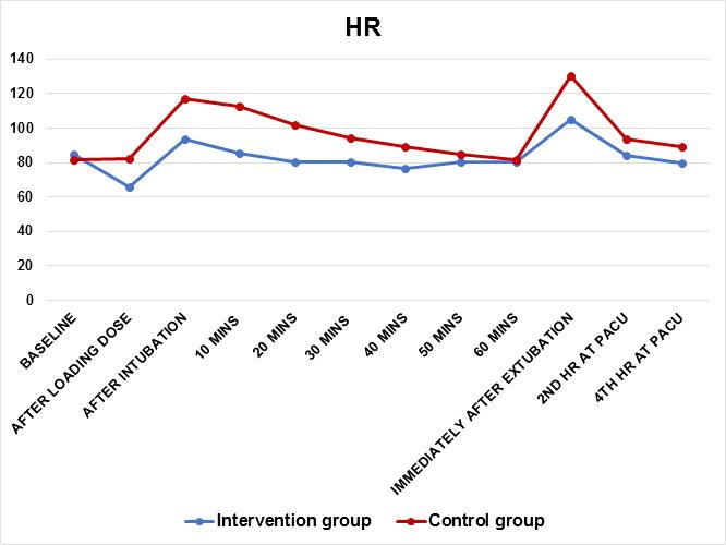
saline was administered to the control group in a similar fashion. They observed an increase in HR and BP following direct laryngoscopy, endotracheal intubation, insertion of a nasal speculum and extubation in both groups, but it was more profound in the control group. The need for analgesics, muscle relaxants and volatile agents during the intra-operative period was much less in the intervention group. The time of emergence from anesthesia and VAS score at the time of emergence were also negligible in the Dexmedetomidine group.
Dexmedetomidine and remifentanil’s effects on hemodynamic stability, sedation, and postoperative pain management were investigated by Jung, et al24 in 50 patients scheduled for total laparoscopic hysterectomy who were randomly assigned to receive either remifentanil (0.8 to 1.2 mg/kg) over 1 min followed by 0.05 to 0.1 mg/kg/min or Dexmedetomidine (1 mg/kg) over ten minutes followed by 0.2 to 0.7 mg/kg/hr infusion. In the postoperative room, the BP and HR were considerably curtailed in the Group receiving Dexmedetomidine compared to the Group receiving remifentanil. When comparing the postoperative hemodynamic stability between Dexmedetomidine and remifentanil, the doses utilized in this study showed a clear advantage for Dexmedetomidine.
The findings of this study are further supported by Panchgar, et al25 who investigated the efficiency of Dexmedetomidine in preserving hemodynamic stability throughout the peri-operative phase in randomized patients undergoing Laparoscopic Surgery. They came to the conclusion that the Dexmedetomidine group successfully obtunded the hemodynamic reactions to the stressful situations. The demand for postoperative analgesics was incredibly less in the intervention group. No adverse effects reported outside the two instances of bradycardia in this category.
There have been few limitations in our study. First, we conducted this study on different types of surgeries

Vol 123, No 02, February 2025Journal
ranging from modified radical mastectomy to staging laparotomy. So, it could have been done in more standardized manner. Second, we excluded patients with Cardiovascular diseases. So it’s not appropriate to comment on the effects of Dexmedetomidine on these types of patients. Third, the comparison was done between Dexmedetomidine and a placebo which resulted in the obvious outcome. As a result, we can contrast the potency of Dexmedetomidine with that of other medications in a future part of this study.
It is concluded that Dexmedetomidine is effective in maintaining hemodynamic stability and blunting the hemodynamic stress response induced by intubation and extubation. It can be administered as a loading dose of 1 mcg/kg prior to induction and as a maintenance infusion of 0.6 mcg/kg/hour throughout the procedure. Additionally, it extended the time frame for the first round of postoperative rescue analgesia. Therefore, Dexmedetomidine can be used as a supplement to General Anesthesia in a variety of surgical procedures with minimal risk of adverse effects like Respiratory Depression.
1Reid LC, Brace DE — Irritation of the respiratory tract and its reflex effect upon the heart. Surg Gynecol Obstet 1940; 70: 157-62.
2Mendonça FT, Silva SL, Nilton TM, Alves IR — Effects of lidocaine and esmolol on hemodynamic response to tracheal intubation: a randomized clinical trial. Brazilian Journal of Anesthesiology 2022; 72: 95-102.
3Seangrung R, Pasutharnchat K, Injampa S, Kumdang S, Komonhirun R — Comparison of the hemodynamic response of dexmedetomidine versus additional intravenous lidocaine with propofol during tracheal intubation: a randomized controlled study. BMC anesthesiology 2021; 21(1): 1-1.
4Lakhe G, Pradhan S, Dhakal S — Hemodynamic response to laryngoscopy and intubation using McCoy laryngoscope: a descriptive cross-sectional study. JNMA: Journal of the Nepal Medical Association 2021; 59(238): 554-7.
5Kalaskar VP, Ruparel DH, Wakode RP — Effects of dexmedetomidine infusion in low dose on dose reduction of propofol, intraoperative hemodynamics, and postoperative analgesia in patients undergoing laparoscopic cholecystectomy. Anesthesia, Essays, and Researches 2021; 15(4): 391394.
6Akheela MK, Chandra A — Comparative Evaluation of Nalbuphine and Fentanyl for Attenuation of Pressor Response to Laryngoscopy and Tracheal Intubation in Laparoscopic Cholecystectomy. Cureus 2021; 13(5): e15142.
7Misra S, Behera BK, Mitra JK, Sahoo AK, Jena SS, Srinivasan A — Effect of preoperative dexmedetomidine nebulization on the hemodynamic response to laryngoscopy and intubation: a randomized control trial. Korean Journal of Anesthesiology 2021; 74(2): 150-7.
8De Cassai A, Boscolo A, Geraldini F, Zarantonello F, Pettenuzzo T, Pasin L, et al — Effect of dexmedetomidine on hemodynamic responses to tracheal intubation: A meta-analysis with meta-regression and trial sequential analysis. Journal of Clinical Anesthesia 2021; 72: 110287.
9Paul GB — Clinical Anesthesia.8th ed. Philadelphia : Wolters Kluwer 2017; 1275-80.
10Chilkoti GT, Karthik G, Rautela R — Evaluation of postoperative analgesic efficacy and perioperative hemodynamic changes with low dose intravenous dexmedetomidine infusion in patients undergoing laparoscopic cholecystectomy–A randomised, double-blinded, placebo-controlled trial. Journal of Anaesthesiology, Clinical Pharmacology 2020; 36(1): 72-7.
11Patel CR, Engineer SR, Shah BJ, Madhu S — Effect of intravenous infusion of dexmedetomidine on perioperative haemodynamic changes and postoperative recovery: A study with entropy analysis. Indian Journal of Anaesthesia 2012; 56(6): 542-6.
12Kundra S, Taneja S, Choudhary AK, Katyal S, Garg I, Roy R — Effect of a low-dose dexmedetomidine infusion on intraoperative hemodynamics, anesthetic requirements and recovery profile in patients undergoing lumbar spine surgery. Journal of Anaesthesiology, Clinical Pharmacology 2019; 35(2): 248-53.
13Okello MO, Mung’ayi V, Adam R, Kabugi J — A comparison of risk of hypotension using standard doses of remifentanil versus dexmedetomidine infusions in adult patients undergoing surgery under general anaesthesia at the Aga Khan University Hospital, Nairobi. African Health Sciences 2018; 18(4): 1267-82.
14Liu LL,Gropper MA — Postoperative analgesia and sedation in the adult intensive care unit. Aguide to drug selection. Drugs 2003; 63(8): 755-67
15Talke P, Richardson CA, Scheinin M, Fisher DM — Postoperative pharmacokinetics and sympatholytic effects of dexmedetomidine. Anesthesia & Analgesia 1997; 85(5): 113642.
16Scheinin B, Lindgren L, Randell T, Scheinin H, Scheinin M — Dexmedetomidine attenuates sympathoadrenal responses to tracheal intubation and reduces the need for thiopentone and peroperative fentanyl. British Journal of Anaesthesia 1992; 68(2): 126-31.
17Desborough JP — The stress response to trauma and surgery. British Journal of Anaesthesia 2000; 85(1): 109-17.
18Parker SD, Breslow MJ, Frank SM, Rosenfeld BA, Norris EJ, Christopherson R, Rock P, et al — Catecholamine and cortisol responses to lower extremity revascularization: correlation with outcome variables. Critical Care Medicine 1995; 23(12): 1954-61.
19Kennedy BC, Hall GM — Neuroendocrine and inflammatory aspects of surgery: do they affect outcome? Acta Anaesthesiologica Belgica 1999; 50(4): 205-9.
20Coursin DB, Coursin DB, Maccioli GA — Dexmedetomidine. Current Opinion in Critical Care 2001; 7(4): 221-6.
21Uyar AS, Yagmurdur H, Fidan Y, Topkaya C, Basar H — Dexmedetomidine attenuates the hemodynamic and neuroendocrinal responses to skull-pin head-holder application during craniotomy. Journal of Neurosurgical Anesthesiology 2008; 20(3): 174-9.
22Basantwani S, Patil M, Govardhane B, Magar J, Tendolkar B — Effect of dexmedetomidine infusion on hemodynamic responses in microsurgery of larynx. Journal of Anaesthesiology, Clinical Pharmacology 2018; 34(1): 51-7.
23Bala R, Chaturvedi A, Pandia MP, Bithal PK — Intraoperative dexmedetomidine maintains hemodynamic stability and hastens postoperative recovery in patients undergoing transsphenoidal pituitary surgery. Journal of Neurosciences in Rural Practice 2019; 10(04): 599-605.
24Jung HS, Joo JD, Jeon YS, Lee JA, Kim DW, In JH, et al — Comparison of an intraoperative infusion of dexmedetomidine or remifentanil on perioperative haemodynamics, hypnosis and sedation, and postoperative pain control. Journal of International Medical Research 2011; 39(5): 1890-9.
25Panchgar V, Shetti AN, Sunitha HB, Dhulkhed VK, Nadkarni AV — The effectiveness of intravenous dexmedetomidine on perioperative hemodynamics, analgesic requirement, and side effects profile in patients undergoing laparoscopic surgery under general anesthesia. Anesthesia, Essays and Researches 2017; 11(1): 72-7.
Rumpa Banerjee1, Indranil Dutta2
Background : The shoulder joint has a wide range of motion in multiple planes - stability is compromised for mobility. It is an incongruous (large) ball and (small) socket joint without any fixed axis of rotation. Although a number of clinical tests are described for diagnosing painful shoulder, considered accurate in determining the location of periarticular lesions, shoulder pathology may be difficult to diagnose by physical examination alone.
Magnetic Resonance Imaging (MRI) and Ultrasonography have supplanted arthrography in the evaluation of the integrity of the rotator cuff.
Materials and Methods : The data for the study was collected from patients complaining of shoulder pain and restriction of movements, suspected for rotator cuff tears referred for Ultrasonography (USG) examination to the department of Radiodiagnosis, AIIMS, Mandya.
Results : In this study group which comprised of a total number of 50 patients, The most common symptom in patients who presented to the Orthopaedics Department with rotator cuff injuries was shoulder pain and restriction of movements comprising 72% and 28% of cases respectively. Among 50 patients who underwent ultrasound examination, 14 patients were diagnosed as having Full Thickness Tears (FTT) and 30 patients showed Partial Thickness Tears (PTT). In relation supraspinatus tendon, USG detected 34 truly positive supraspinatus tears (14 full & 30 PTT).
Conclusion : Based on our results, it can be concluded that Ultrasonography is an effective imaging modality that has a positive effect on the management of many patients presenting with shoulder pain and/or disability. MRI can be used as second line Diagnostic Tool.
[J Indian Med Assoc 2025; 123(2): 33-9]
Key words :Musculo Skeletal (MSK), Acromio Clavicular Joint (ACJ), Positive Predictive Value (PPV), Negative Predictive Value (NPV), Partial Thickness Tears (PTT), Full Thickness Tears (FTT).
The shoulder joint has a wide range of motion in multiple planes - stability is compromised for mobility. It is an incongruous (large) ball and (small) socket joint without any fixed axis of rotation. The muscles and their tendons are subjected to severe strain causing excessive “wear and tear” during sports and games activities. The spectrum of causative lesions that can give rise to shoulder pain ranges from acute trauma to degenerative disorders as well as impingement syndrome. Although a number of clinical tests are described for diagnosing painful shoulder, considered accurate in determining the location of peri-articular lesions, shoulder pathology may be difficult to diagnose by physical examination alone. Clinical diagnosis has low accuracy in comparison with arthroscopy1,2.
Rotator cuff injury has a distinct range of chronic disorders. These disorders are related to the osseo-
1MBBS, MD (Radiodiagnosis), Assistant Professor, Department of Radiodiagnosis, Gouri Devi Medical College Hospital & Research Institute, Durgapur, West Bengal 713212 and Corresponding Author
2MBBS, MS, PGDHHM, PGDMLS, Professor and Unit Head, Department of Obstetrics and Gynaecology, IQ City Medical College Hospital, Durgapur, West Bengal 713206
Received on : 11/06/2024
Accepted on : 30/06/2024
Editor's Comment :
Ultrasonography is an effective imaging modality that has a positive effect on the management of many patients presenting with shoulder pain and/or disability. There was a good agreement between USG and MRI in diagnosing rotator cuff pathologies
tendinous and musculotendinous part of the Rotator cuff and the anatomically narrow sub-acromial space. Due to this problem Ultrasound is often recommended as primary Diagnostic Tool. Ultrasound technology has taken leaps and advances in its course over the years and has contributed immensely to help us diagnose cases of rotator cuff pathology. Main advantage lies in the fact that ultrasound is non invasive, easy to perform, less expensive and has good sensitivity in diagnosing both rotator cuff and non rotator cuff disorders3. Another benefit also lies in the fact that it is also a non-ionizing modality.
Inspite of above advancements plain film radiography serves as basic and first line in most of cases particularly in rural areas where the above modality is easily available and prescribed for bony trauma and most athropathies. Magnetic Resonance Imaging and Ultrasonography have supplanted arthrography in the evaluation of the integrity of the
123, No 02, February 2025Journal
rotator cuff4. The overall joint structure assessment and detection of both subtle and obvious internal derangement has become easier with MRI being available and is considered as “Gold Standard” in diagnostic modality3. MRI have a advantage because of its multi-planar capability and exquisite soft tissue detail which helps us with detailed diagnosis.
In last 20 years or more Ultrasound technology has been the upfront in field of sports medicine and rheumatology and it has rightfully gained its place in literature along with MRI. The main advantage in USG is its real time capability in conducting dynamic studies in the shoulder as well as the patient’s ability to point out the discomfort caused by particular movement or position.
In this study approximate 50 patients who had shoulder joint pain and restriction of movements with a suspicion of rotator cuff injuries clinically were advised both USG and MRI of the shoulder joint. The main aim of this study was to show that ultrasound technology was as effective as MRI in the evaluation of shoulder pain, especially in cases of rotator cuff injuries.
MATERIALS AND METHODS
Source of Data : The data for the study was collected from patients complaining of shoulder pain and restriction of movements, suspected for rotator cuff tears referred for Ultrasonography (USG) examination to the Department of Radiodiagnosis, Sri Adichunchanagiri Hospital and Research Centre, B G Nagara, Nagamangala Taluk, Mandya District.
Study Design : Descriptive study.
Study area : Sri Adichunchanagiri Hospital and Research Centre, B G Nagara.
Study period : January, 2016 to August, 2017
Sample Size : 50
Statistical analysis : The agreement between the two methods was assessed using kappa coefficient.
Inclusion Criteria :
All patients in any age group.
All patients with shoulder pain, restriction of movements of shoulder and suspected rotator cuff injuries.
Exclusion criteria :
Other trauma/ open wounds.
Malignant and infectious conditions.
Patient having a history of claustrophobia. Patient having a history of metallic implant or cardiac pacemaker insertion or any metallic foreign body embedded in the tissues.
Equipment :
USG – GE Voluson S6 Pro with linear probe of frequency range 7-12 MHz.
MRI – Siemens 1.5 Tesla MRI superconducting magnet.
Scanning Technique for USG :
The patient is seated opposite to examiner in a high revolving chair; the probe which is used is a high frequency linear type, a broadband probe like L5-12 is ideal. The basic principles of MSK USG is that it is necessary to follow the curve of the bone, identify useful bony landmarks like the acromion process, biceps tendon, and the labrum4,5. Each structure has to be examined in 2 planes and maintaining perpendicularity to tendon fibers, should be followed. Following sequence when examining the shoulder joint to followed —
Biceps tendon
Subscapularis
Supraspinatus (including test for impingement)
Infraspinatus
Teres minor
Posterior glenoid labrum and glenohumeral space
Coraco-acromial ligament
The Acromioclavicular joint.
Criteria for Diagnosis of Rotator Cuff Tear :
Various criteria’s have been defined over the years for rotator cuff tears. The criteria devised and suggested by Middleton are easy to follow and report6,7. One should always examine the integrity, thickness and echogenicity of the cuff and look out for calcification. The size of the tear, its orientation, and its location relative to the biceps tendon has to be reported.
Major Criteria :
(1)Non-visualization of the Cuff (Fig 1) : If the cuff is not visualized totally then it may suggest a complete tear. But the problem lies in the fact that a novice person can easily miss this finding since the entire supraspinatus tendon is missing and the deltoid with the subdeltoid bursa then rests on the head of the humerus. The best way to confirm this is by comparing with the other side. An important indirect clue in this situation is the loss of the normal convexity of the deltoid and bursa with dipping of the same (with concavity) in the empty space. This is the most important indirect sign. Bursal thickness and fluid in the subdeltoid bursa and along the biceps are very commonly associated. The hyaline cartilage on the humeral head stands out. This often referred to as the naked tuberosity or double arc sign.

(2)Focal non-visualization : Focal nonvisualization of the tendon is relatively easy to diagnose, as the remaining tendon is seen adjacent to it. Within this gap, the subdeltoid bursa can also dip. Confirmation in two planes is important. Most rotator cuff pathologies occur in the terminal 1 cm of the tendon, which is the critical zone.
(3)Discontinuity : Discontinuity of fibers may be seen, the gap filled with fluid or reactive tissue. A stress test, by positioning the elbow with the arm internally rotated, is the best way to check for discontinuity of the fibers. It is also apparent by the compressibility of the tendon compared to a normal tendon, which is non compressible. (The subscapularis tendon is best examined while performing passive external rotation and internal rotation. In external rotation the tendon becomes more prominent, as it is medial to the biceps tendon. It must be examined in two planes. Injuries of the subscapularis are rare, except in the event of direct trauma)
(4)Focal abnormal Echogenicity : Full or Partial Thickness Tears (PTT) is usually associated with this. The area of increased echogenicity is thought to result from granulation tissue, hypertrophied synovium, and hemorrhage. The radiologist should be able to confirm this is real and not an artifact or rotator cuff calcification.
:
(1)Reliable sign of rotator cuff injury can be elicited by presence of fluid along the biceps tendon sheath and in subdeltiod bursa and the joint. Fluid along the biceps tendon sheath and in the subdeltoid bursa and the joint is a very reliable sign of rotator cuff injury. Cuff tear can be suspected by presence of fluid along the biceps tendon and in the joint7,8
(2)Concave subdeltoid bursal contour.
(3)Irregularity of the greater tuberosity9
(4)Compressibility.
Partial-thickness Tears :
For the evidence of PTT, Middleton’s group uses either a distinct hypoechoic or mixed hyper and hypoechoic defect visualized in two planes at the deep articular side of the cuff, or minimal flattening of the bursal side of the cuff. When tears involve bursal surface, intra substance or articular surface tears then they fall under definition of partial tears. Around 80% of accuracy has been noted in the diagnosis of PTT8.
Some of the reports suggests that USG is a significantly better modality for the evaluation of partial-thickness tears than MRI.
Imaging Technique for MRI :
Imaging Parameters
SequenceFOV(cm)SliceIntervalTRTE Thickness
Coronal PDFS164mm0.5mm185032
Axial T2164mm0.5mm536797
Coronal T2164mm0.5mm270090
Axial PDFS164mm0.5mm196726
Sagittal T1164mm0.5mm11012
Sagittal PDFS164mm0.5mm240035
Age Distribution of Patients in the Study :
In this study group which comprised of a total number of 50 patients, the age at presentation ranged from 25 to 65 years. Most patients belonged to the 36-45 years and 46-55years age groups with 16 and 19 patients respectively. There were 51 Males and 9 Females in the patients included in the study group. Males comprised 86% and Females comprised 14% of the group.
Symptoms :
Most of the patients who had presented to Orthopaedics Department with rotator cuff injuries had shoulder pain and restriction of movements comprising 72 % and 28% of cases respectively. Etiology and Predisposing Factor :
History of trauma was observed in the 38 patients. Among 50 patients Diabetes Mellitus was seen in 20 patients presenting to the Department with shoulder
123, No 02, February 2025Journal
pain or restriction of movements.
Rotator Cuff Tears in Relation to Dominant Hand :
Out of 32 patients who were right handed, 28 patients had rotator cuff tear involving the right shoulder, 2 had rotator cuff tear involving the left shoulder and 2 patients had no tear. In 12 patients had rotator cuff tear involving the left shoulder out of 18 patients who were left handed. Four had rotator cuff tear involving the right shoulder and 2 patients had no tear. It suggests that dominant arm is more susceptible to wearing effects and thus leads to rotator cuff tear.
Supraspinatus Tear :
Fourteen patients were diagnosed as having full thickness tears and 30 patients showed Partial Thickness Tears (PTT) among 50 patients who underwent ultrasound examination. 14 patients had articular surface PTT and 16 patients were found to have bursal surface PTT. On MRI 14 patients showed full thickness tears and 32 patients showed PTT. Fifteen patients were diagnosed with articular surface PTT and 17 patients with bursal surface PTT on MRI. Seven patients had tendnosis and impingement which was detected both on USG and MRI. Both USG and MRI had good diagnostic capability for rotator cuff tears (Table 1).
Table 1 — Agreement between USG and MRI for the diagnosis of Supraspinatus Tears MRI
FTTPTTTendi-Impinge- No tearTotal nosisment
Subscapularis Tear :
Among 50 patients who underwent ultrasound examination, 3 patients were diagnosed as having Full Thickness Tears (FTT) and 6 patients showed PTT. On MRI 3 patients showed full thickness tears and 7 patients showed partial thickness tear. 4 patients had tendnosis which was detected both on USG and MRI. USG findings showed good correlation with MRI findings (Table 2).
Infraspinatus Tears :
Among 50 patients who underwent Ultrasound examination, 2 patients were diagnosed as having PTT on USG. On MRI 3 patients showed PTT. FTT of the tears of the Infraspinatus were not detected both on USG and MRI. USG findings showed good correlation with MRI findings (Table 3).
Teres Minor Tears :
Two patients had partial thickness tear of teres minor on USG. However on MRI, 3 patients showed partial thickness tears of the teres minor. Full thickness tears of the tears of the Infraspinatus were not detected both on USG and MRI. USG findings showed good correlation with MRI findings (Table 4). Sensitivity and Specificity Analysis of USG in Relation to Rotator Cuff Tears with respect to findings in MRI :
In relation supraspinatus tendon, USG detected 34 truly positive supraspinatus tears (14 full & 30 PTT)
Table 3 — Agreement between USG and MRI for the diagnosis of Infraspinatus Tears MRI FTTPTTNo tearTotal
Kappa= 0.954, SE of kappa = 0.032, 95% confidence interval: From 0.891 to 1.000
Table 2 — Agreement between USG and MRI for the diagnosis of subscapularis tears MRI
FTTPTTTendinosisNo tearTotal FTT31004
Tendinosis00404
No tear00044
Total374418
Kappa=
Kappa= 0.741, SE of kappa = 0.198, 95% confidence interval: From 0.352 to 1.000
Table 4 — Agreement between USG and MRI for the diagnosis of teres minor tears
MRI
FTTPTTNo tearTotal
Kappa= 0.741, SE of kappa = 0.198, 95% confidence interval: From 0.352 to 1.000
2 falsely negative as normal while 4 patients were diagnosed as normal (True negative). No false positive cases were noted. The sensitivity, specificity and accuracy of USG against the MRI for Full thickness finding are 100%, 100% and 100% respectively. The sensitivity, specificity and accuracy of USG against the MRI for Partial thickness finding are 93.75%, 100% and 94.4% respectively. In relation subscapularis tendon, USG detected 8 truly positive supraspinatus tears (3 full & 6 PTT) 1 falsely negative as normal while 4 patients were diagnosed as normal (True negative). No false positive cases were noted. The sensitivity, specificity and accuracy of USG against the MRI for Full thickness finding are 100%, 100% and 100% respectively. The sensitivity, specificity and accuracy of USG against the MRI for Partial thickness finding are 85.7%, 100% and 90.9% respectively.
In relation infraspinatus and teres minor tendons, USG detected 1 truly positive infraspinatus and teres minor PTT, 1 falsely negative as normal for both muscles, while 4 patients were diagnosed as normal (True negative). No false positive cases were noted. The sensitivity, specificity and accuracy of USG against the MRI for PTT of infraspinatus and teres minor tendon are 50%, 100% and 83.3% respectively. Sensitivity, specificity of USG could not be assessed as FTT of infraspinatus muscle was not found even on MRI.
The overall sensitivity, specificity, PPV and NPV of Ultrasound compared to MRI in detection of full thickness rotator cuff tear was 100% in each category respectively. The overall sensitivity, specificity, PPV and NPV of ultrasound compared to MRI, for detection of rotator cuff tear was 84.4%, 100%, 100% and 36% respectively. The accuracy of ultrasound compared to MRI in diagnosis of FTT and PTT was 100% and 85.7% respectively.
Acromio-clavicular (AC) Joint Arthrosis and Impingement :
Among 46 patients with rotator cuff tear, AC joint arthrosis was seen in 25 on USG examination. On MRI 29 patients were diagnosed with AC joint arthrosis. On dynamic USG examination impingement of supraspinatus tendon was seen in 7 patients. However in MRI impingement was observed in only 4 patients.
Patients who present with rotator cuff injuries usually present to OPD with shoulder pain and restriction of movements and account for 30 to 70
percent of these cases10,11. The integrity of rotator cuff has to be attained along with extent of tear while assessing these patients. Thus it will help the surgeons to plan up the cases. Arthrography has been used for the diagnosis of rotator cuff tears12,13.
Ultrasonography of the shoulder was first reported by Seltzer, et al14. Since then several authors have been discussing and refining this method15,16. There are several advantages of USG over MRI. Because Ultrasonography has been more dynamic form of imaging compared to MRI, it has been preferred widely for the diagnosis. Main advantage lies in USG is its portability, quick, and a more cost-effective method. Thus it enables USG to be well accepted by patients and also enables patient to point out the main area of trauma or pain to have a better visualization and thus helping in the diagnosis. Portable USG Scanners though difficult to acquire in our country, the advent of portable USG scanners has made it easier for Orthopaedicians to help acquire the knowledge and skill and also perform USG in the clinic at the first point of contact. This actually helps reduce the burden from the Radiology Department and also saves enormous amount of energy, time and money.
Al-shawi, et al17 have reported a sensitivity and specificity of 96.2% and 95.4%, respectively in 143 consecutive patients with shoulder problems, who underwent shoulder USG by an Orthopedic Surgeon, and Joseph, et al18 reported the use of office-based ultrasonography by an Orthopedic Surgeon with 88% accuracy for full-thickness and 70% for PTT.
The ages of all the cases ranged from 25 to 65 years. Out of these 50 patients; 6(12%) patients were in 25-35 years age group, 16(32%) were in 36-45 years age group, 19(38%) in the were in the 46-55 years age group while 9(18%) patients were in 56-65 years age group. Hence, outcome was an increase in the prevalence of shoulder pain and rotator cuff tears with advancing age. Another study by Jerosch, et al19 suggested similar findings in their study.
Yamamoto A20, et al had reported in their study that rotator cuff tears were commonly associated with male polulations20. Our M:F Ratio was 4.5:1 (Out of 50 patients, 41 were males (82%) and 9 (18%) were females) which was comparable to above study by Yamamoto A20, et al
Out of 32 patients who were right handed, 28 patients had RCT involving the right shoulder and 2 had RCT involving the left shoulder. Out of 18 patients who were left handed, 12 patients had RCT involving the left shoulder and 4 had RCT involving the right
123, No 02, February 2025Journal
shoulder. It suggests that dominant arm is more susceptible to wearing effects and thus leads to RCT. The findings of above suggested that right shoulder was more involved than left shoulder. As suggested by Bouaziz21 , et al who suggested right shoulder involvement (68%) more than left shoulder involvement (32%)21 and is hence comparable to above.
Diabetes was seen in approx 45% of patients and was noted as one of significant predisposing factors pushing rotator tendons to tears. Trauma was identified as the major etiological factor, present in 86.6% of RCT. Contralateral shoulder was also checked via USG for comparision. It was Rutten, et al 22 who recommended comparison with the contralateral shoulder as an additional support for avoiding misinterpretation of normal anatomic differences as tears22
Among 50 patients, tears involving the supraspinatus tendon were more common seen in 92% of patients. Subscapularis was next most commonly involved tendon seen in 20% of the patients. Infraspinatus and teres minor was seen 4% of the patients respectively. Similar findings were seen in the study conducted by Bashir, et al23
On USG out of 44 patients , 30 patients had PTT, 14 had FTT. When MRI was conducted in these patients, it showed 14 FTT and 32 PTT, which means that 2 patients were falsely diagnosed as normal on USG that turned out to be PTT on MRI. Both methods of MRI and USG were assessed and compared using Kappa Co-efficient. There was a very good agreement (k=0.954) between USG and MRI. Out of 50 patients, 9 patients were diagnosed with subscapularis tears of whom 3 patients had full-thickness tears and 6 patients had PTT on USG.
One patient was falsely diagnosed as normal on USG which was found to be PTT on MRI. MRI was conducted in these patients, it showed 3 full-thickness tears and 7 PTT. Similar results were obtained by Alasaarela, et al24 and Rutten22. Evaluation of 31 painful shoulders of 30 patients and reporting a good agreement between US and MRI for diagnosis of FTT and intra-substance abnormalities of supraspinatus tendon (the kappa co-efficient =0.73) by Alasaarela, et al24. In 68 patients who underwent MRI and surgery following USG examination and reported that agreement between US and MRI was high (the kappa coefficient was calculated to be 0.78) in the study conducted by Rutten, et al22
Our study showed sensitivity, specificity, PPV and NPV of 100% respectively for high resolution USG
when compared to MRI for full thickness. The accuracy of USG in diagnosing a FTT is 100% and 85.7% for partial thickness tears. The current study also showed similarity to studies conducted by Kenn, et al28 and Lach, et al29 and proved that ultrasound is accurate in diagnosing rotator cuff pathologies. Ultrasound was as accurate as MRI for assessment of tears of the rotator cuff, both full- or partial-thickness tears according to Saraya, et al25. In our study USG showed ACJ arthrosis in 25 patients (86.2%) in comparison to 29 patients (93.5%) on MRI, proving that though USG is easily available, it’s still less accurate compared to MRI for ACJ evaluation.
MRI detected supraspinatus impingement in only 4 patients. USG was more accurate than MRI in detecting supraspinatus impingement. The findings in our study were comparable with the study conducted by Nathalie J, et al26. The advantage lies in the fact that MR imaging can quantify the degree of muscle atrophy by assessing the occupancy ratio of the supraspinatus muscle in the supraspinatus fossa27. Oblique sagittal T1W view in MRI best depicts chronicity of a rotator cuff injury
So the above wide comparative study noted results suggesting that USG is an effective imaging modality that has a positive effect on the management of many patients presenting with shoulder pain or disability. Ultrasound of shoulder has a good accuracy in diagnosing tears of the rotator cuff and in differentiating partial from full-thickness tears. Also important to note that there was good agreement (kappa value=0.79) between USG and MRI in diagnosing rotator cuff pathologies.
From the study there were notable advantages noted in USG over MRI such as USG is available on a larger scale, portable, quick and a much more costeffective imaging method. Most importantly is well tolerated by patient because they can point out the main area which is affected to the Radiologist thus helping in accurate diagnosis.
Ultrasound allows instant comparison to contralateral side and it helps in dynamic evaluation of the same. However, sonography of the shoulder joint is operator dependent. Hence small transducer related errors and angulation can easily obscure small abnormalities within and around the cuff and give rise to false positive and false negative results. The learning curve is also difficult for shoulder Sonography. If the observer has good knowledge about the anatomy of shoulder then above problem of diagnosing with USG can be easier. Sonography
123, No 02, February 2025Journal
is an effective, reliable and noninvasive means of detecting rotator cuff tears.
Hence, both USG and MRI are considered as comparable modalities for diagnosis, USG can be used for first line investigation whereas MRI can be used as second line tool, either following an equivocal shoulder USG or for delineation of anatomy in cases where surgical correction is needed.
1Murrell GA, Walton JR — Diagnosis of rotator cuff tears. Lancet 2001; 357: 769-70.
2Resnick D — Shoulder arthrography. Radiol Clin North Am 1981; 19: 243-52.
3Middleton WD — Ultrasonography of the shoulder. Radiol Clin North Am 1992; 30: 927-40.
4Mack LA, Nyberg DA, Matsen FA 3rd. Sonographic evaluation of the rotator cuff. Radiol Clin North Am 1988; 26: 161-77.
5Winter TC, Richardson MI, Matsen FA — Rotator cuff ultrasound. Radilogic Soc Am Electronic Jr 1996.
6Teefey SA, Hasan SA, Middleton WD, Patel M, Wright RW, Yamaguchi K — Ultrasonography of the rotator cuff. A comparison of ultrasonographic and arthroscopic findings in one hundred consecutive cases. J Bone Joint Surg Am 2000; 82(4): 498-504.
7Middleton WD, Reinus WR, Tott y WF, Nelson GL, Murphy WA — Ultrasonographic evaluation of the rotator cuff and biceps tendon. Bone Joint Surg Am 1986; 68: 440-50.
8Hollister MS, Mack LM, Patt ern RM, Winter TC 3rd, Matsen FA 3rd, Veith RR — The association of sonographically detected subacromial/subdeltoid bursal eff usion and intraarticular fluid with rotator cuff tear. Am J Roentgenol 1995; 165: 605-8.
9Wohlwend JR, van Holsbeeck M, Craig J, Shirazi K, Habra G, Jacobsen G, et al — The association between irregular greater tuberosities and rotator cuff tears: A sonographic study. Am J Roentgenol 1998; 171: 229-33.
10Mitchell C, Adebajo A, Hay E, Carr A — Shoulder pain: Diagnosis and management in primary care. BMJ 2005; 331: 11248.
11Macfarlane GJ, Hunt IM, Silman AJ — Predictors of chronic shoulder pain: A population based prospective study. J Rheumatol 1998; 25: 1612-5.
12Mink JH, Harris E, Rappaport M — Rotator cuff tears: Evaluation using double- contrast shoulder arthrography. Radiology 1985; 157: 621-3.
13Goldman AB, Ghelman B. The double-contrast shoulder arthrogram: A review of 158 studies. Radiology 1978; 127: 65563.
14Seltzer SE, Finberg HJ, Weissman BN — Arthrosonography: Technique, sonographic anatomy and pathology. Invest Radiol 1980; 15: 19-28.
15Crass JR, Craig EV, Bretzke C, Feinberg SB — Ultrasonography of the rotator cuff. Radiographics 1985; 5: 941-53.
16Middleton WD, Reinus WR, Totty WG, Melson CL, Murphy WA — Ultrasonographic evaluation of the rotator cuff and biceps tendon. Bone Joint Surg Am 1986; 68: 440-50.
17Al-Shawi A, Badge R, Bunker T — The detection of full thickness rotator cuff tear using ultrasound. J Bone Joint Surg Br 2008; 90: 889-92.
18Iannotti JP, Ciccone J, Buss DD, Visotsky JL, Mascha E, Cotman K, et al — Accuracy of office-based ultrasonography of the shoulder for the diagnosis of rotator cuff tears. J Bone Joint Surg Am 2005; 87: 1305-11.
19Jerosch J, Müller T, Castro WH — The incidence of rotator cuff rupture. An anatomic study. Acta Orthop Belg 1991; 57: 124-9.
20Yamamoto A, Takagishi K, Osawa T — Prevalence and risk factors of a rotator cuff tear in the general population. J Shoulder Elbow Surg 2010; 19: 116-20.
21Bouaziz MC, Jabnoun F, Chaabane S, Ladeb MF — Diagnostic accuracy of high resolution ultrasound in communicating rotator cuff tears. Iran J Radiol 2010; 7: 153-60.
22Matthieu JCM Rutten — General introduction Study aims outline of thesis. In: Matthieu J.C.M. Rutten, editor. Ultrasound of the shoulder; efficacy studies. 1st ed. The Netherlands: Ipskamp Drukkers; 2010; 15-9.
23Bashir S, Firdose SR, Kamal Y — Correlation between high resolution ultrasonography and MRI in rotator cuff tear diagnosis. Int J Health Sci Res 2014; 4(8): 103-112.
24Alasaarela E, Takalo R, Tervonen O, Hakala M,Suramo I — Sonography and MRI in the evaluation of painful arthritic shoulder. Br J Rheumatol 1997; 36(9): 996-1000.
25Saraya S, Bakry RE. Ultrasound: Can it replace MRI in the evaluation of the rotator cuff tears. Egyptian Journal of Radiology and Nuclear Medicine 2016; 47: 193-201.
26Nathalie J — Dynamic Sonography Evaluation of Shoulder Impingement Syndrome. American Journal of Roentgenology 2006; 187: 216-20.
27Thomazeau H, Rolland Y, Lucas C — Atrophy of the supraspinatus belly: Assessment by MRI in 55 patients with rotator cuff pathology. Acta Orthop Scand 1996; 67: 264-68.
28Kenn W, Hufnagel P, Muller T, Gohlke F, Bohm D — Arthrography, ultrasound and MRI in rotator cuff lesions: a comparison of methods in partial lesions and small complete ruptures. Rofo 2000; 172(3): 260-6.
29Lach W, Dudarenko G,Brzosko M, Cyrylowski L, Flicinski J — Usefulness of ultrasonographic and MR imaging in diagnosis of shoulder impingement. Chir Narzadow Ruchu Ortop Pol 2003; 68(5): 307-11.
Kanakeswar Bhuyan1, Aritra Bhattacharjee2, Punadhar Deori3, Pulak Kumar Das4
Background : Procalcitonin (PCT) and C-reactive Protein (CRP) are the most frequently used biomarkers for critically ill patients. Changes in these biochemical markers may be useful in predicting therapeutic response and prognosis in septic patients. The aim of this study was to assess utility of CRP and PCT as predictors of outcome in critically ill patients with sepsis treated in ICU.
Materials and Methods : The study included 100 patients treated in ICU with sepsis. Data on Serum CRP and PCT level on day one and day seven were collected and compared with the outcome. Student 't' test, AUC and Pearson’s correlation coefficient were applied to study its significance.
Results : The male female ratio was 61:39 with age group of 18 to 80 years. The mean CRP on day 1 was 54.5±60.3 and on day 7 was 22.9±34.7 (p:0.0001). The mean PCT on day 1 was 7.2±3.6 and on day 7 was 1.7±1.4 (p:0.0021). It was observed that both PCT (mean) and CRP(mean) decreased significantly on day 7 compared to day 1 amongst the survivors. The co-relation of outcome with CRP level was found to be more significant. The Pearson’s correlation coefficient showed significant positive correlation with poor outcome of 28 patients (28%) in this study.
Conclusion : Changes in PCT and CRP concentrations were associated with outcome of critically ill patients with sepsis. The serum CRP was found to be a reliable biomarker for prediction of outcome in sepsis.
[J Indian Med Assoc 2025; 123(2): 40-2]
Key words :Sepsis, Procalcitonin (PCT), C-reactive Protein (CRP), Morbidity, Mortality.
Sepsis is the systemic inflammatory response to microbial infection1. An exaggerated immune response with overproduction of inflammatory mediators influence that results of diffuse injury of healthy tissues, major organs dysfunction and associated mortality2. Procalcitonin (PCT) and Creactive Protein (CRP) are the most frequently used biomarkers for critically ill patients with sepsis. If sepsis is well controlled, PCT and CRP may show decreasing patterns. A dynamic approach of assessing these biomarkers may provide more information on treatment outcome or modification of treatment in patients with sepsis. The aim of this study was to assess predictive values based on changes in PCT and CRP concentrations in patients with sepsis and to identify a single and cost-effective biomarker.
MATERIALS AND METHODS
This study was conducted on data collected from 100 consecutive patients treated in surgical ICU for sepsis of varied reasons in Gauhati Medical College & Hospital covering a period of six months. The
Department of Surgery, Gauhati Medical College and Hospital, Guwahati 781032
1MS, Professor and Corresponding Author
2MBBS, Postgraduate Trainee
3MS, Associate Professor
4MS, Assistant Professor
Received on : 01/10/2022
Accepted on : 08/06/2023
Editor's Comment :
PCT and CRP are two important biomarkers to prognosticate patients in sepsis. It can predict ICU requirements for these groups of patients. and modulation of antibiotics during the course of treatment.
primary endpoint of this study was to determine the outcome of treatment on the basis of serum CRP and PCT level on day 1 and after 7 days from day of ICU admission. The laboratory data of serum PCT and CRP on day 1,2 and day 5,7. Serum PCT concentrations were measured using enzyme-linked fluorescent assays (Mini VIDAS method) with the lower reference limit was 0.05 ng/mL. Serum CRP concentrations were measured using latex agglutination principle and the lower reference limit was 10mg/L. The initial baseline PCT and CRP levels defined as peak levels from day1 to 2 and subsequent levels as minimal levels from days 5 to 7. PCT and CRP kinetics are expressed as ∆PCT and ∆CRP concentrations, which are the differences between baseline and subsequent measurements. All patients had clinical evidence of Sepsis according to the recently approved International Sepsis Consensus Conference definitions3.
Data were expressed as mean (SD) and P<0.05 was considered statistically significant. Area under the ROC Curves were constructed according to Hanley and McNeil by plotting the sensitivity against
specificity4. An area under ROC Curve of 1 indicates a perfect predictive power and the closer the area under the ROC Curve to 1 indicate the greater discriminative power of the marker.
There were 39 (39%) Female and 61 Male (61%) patients in the study population with mean age of 45.5 (Table 1).
The data showed improvement in results of treatment in 43 patients, stable in 29 patients and poor results (Deaths) in 28 patients after 7 days of treatment. The mean CRP on day 1 was 54.5±60.3 and on day 7 was 22.9±34.7 in survival group. The mean PCT on day 1 was 7.2±3.6 and on day 7 was 1.7±1.4 in same group of patients. It was observed that both PCT and CRP decreased significantly on day 7 compared to day 1in survival group of patients (Table 2). The patients with stable and poor outcome on day 1 to day 7 showed no significant decline in serum concentration of CRP and PCT. In patients with poor outcome both CRP and PCT concentration were higher compared to survivors (Fig 1).
with the increase of PCT and CRP on both Day 1 and 7 are associated with poor outcome(Table 3) . The area under curve shows high predictive value in decreasing concentration of CRP and PCT on day 7 (Fig 2). It is observed that difference of concentration of CRP value on Day 1 and Day 7 is a better indicator in determination of outcome (Table 4).


The Pearson’s correlation coefficient between outcome versus CRP and PCT at Day 1 and Day 7 showed significant positive correlation indicating that
Table 1 — Distribution of Gender with mean age
Table 2 — Mean CRP and PCT at Day 1 & 7(Survival Group)

Biochemical markers help diagnose Sepsis and can predict patient outcome in severe Sepsis and Septic Shock1,5-7. PCT, CRP are the most frequently used biomarker in clinical practice. PCT, CRP measurements are useful for monitoring the course of Sepsis in critically ill patients and may be used to indicate change in course of treatment and to measure its outcome 8,9 . It could be a possible indicator of stopping antibiotics safely, sparing patients
Table 3 — Pearson’s correlation coefficient between outcome versus CRP and PCT change (Day 1-Day 7) Correlations
Table 4 — AUC analysis for Prediction of Survival Area Under the Curve for Prediction of Survival Test
Vol 123, No 02, February 2025Journal
from drug toxicity, risk of resistance and even indicate possibility to stop ICU care.Although the diagnostic accuracy of PCT was higher than CRP in Sepsis (13) it was unclear which biomarker had more prognostic accuracy in septic patients. Several recent studies found CRP has higher prognostic value than that of PCT and that of both biochemical markers have similar predictive value for determining the outcome of septic patients10. In this present study CRP level was found to be reliable in predicting the outcome11,12. In another study it was found that the changes in PCT and CRP at the onset and on the fourth day can predict survival of patients which is in conformation with the present study13. In one study it was described that a PCT-based protocol was not superior to a protocol based on serum CRP levels for reducing antibiotic use. Remarkably, the length of antibiotic therapy was shorter in the CRP group and less than the maximum therapy duration proposed12 Recently, several studies it was found that CRP was as beneficial as PCT in predicting outcomes and reducing antibiotic use in septic patient8,9,11,13. In addition, CRP is more cost effective than that of PCT. The CRP level indeed a better indicator in predicting outcome of treatment as compared to that of PCT.
The PCT and CRP concentrations can predict outcomes of critically ill septic patients. Changes in CRP concentrations were not inferior to changes in PCT concentrations in predicting treatment response and survival. It is as effective as both CRP, PCT concentrations in predicting the outcome in patients with severe Sepsis. In addition, CRP testing is more cost effective and readily available.
Limitations :
This study has several limitations. This is retrospective review of medical records in a single centre. The sample population was not large enough and time zero estimation was difficult to define because of the retrospective nature of the study. Never the less, the predictable power of PCT and CRP remains uninfluenced by the small sample size and the 7 day outcome and mortality are successfully indicated.
Conflict of Interest : None
1Ryu J-A, Yang JH, Lee D, Park C-M, Suh GY, Jeon K, et al — Clinical Usefulness of Procalcitonin and C-Reactive Protein as Outcome Predictors in Critically Ill Patients with Severe Sepsis and Septic Shock. PLoS ONE 2015; 10(9): e0138150. https://doi.org/10.1371/journal.pone.0138150
2Dahaba AA, Hagara B, Fall A, Rehak PH, List WF, Metzler H — Procalcitonin for early prediction of survival outcome in postoperative critically ill patients with severe sepsis, BJA: British Journal of Anaesthesia 2006; 97(4): 503-8, https:// doi.org/10.1093/bja/ael181
3Garnacho-Montero J, Huici-Moreno MJ, Gutierrez-Pizarraya A, Lopez I, Marquez-Vacaro JA, Macher H, et al — Prognostic and diagnostic value of eosinopenia, C-reactive protein, procalcitonin, and circulating cell-free DNA in critically ill patients admitted with suspicion of sepsis. Crit Care 2014; 18: R116. pmid:24903083
4American College of Chest Physicians/Society of Critical Care Medicine Consensus Conference: definitions for sepsis and organ failure and guidelines for the use of innovative therapies in sepsis. Crit Care Med 1992; 20: 864-74. pmid:1597042
5Schneider CP, Yilmaz Y, Kleespies A, Jauch KW, Hartl WH — Accuracy of procalcitonin for outcome prediction in unselected postoperative critically ill patients. Shock 2009; 31(6): 56873. doi: 10.1097/SHK.0b013e318193cb52.
6Jensen JU, Heslet L, Jensen TH, Espersen K, Steffensen P, Tvede M — Procalcitonin increase in early identification of critically ill patients at high risk of mortality. Crit Care Med 2006; 34: 2596-602. pmid:16915118
7Clec’h C, Ferriere F, Karoubi P, Fosse JP, Cupa M, Hoang P, et al — Diagnostic and prognostic value of procalcitonin in patients with septic shock. Crit Care Med 2004; 32: 1166-9. pmid:1519096
8Povoa P, Almeida E, Moreira P, Fernandes A, Mealha R, Aragao A, et al — C-reactive protein as an indicator of sepsis. Intensive Care Med 1998; 24: 1052-6. pmid:9840239
9Yentis SM, Soni N, Sheldon J — C-reactive protein as an indicator of resolution of sepsis in the intensive care unit. Intensive Care Med 1995; 21: 602-5. pmid:7593905
10Song JU, Suh GY, Park HY, Lim SY, Han SG, Kang YR, et al Early intervention on the outcomes in critically ill cancer patients admitted to intensive care units. Intensive Care Med 2012; 38: 1505-13. pmid:22592633
11Hoeboer SH, Groeneveld AB — Changes in circulating procalcitonin versus C-reactive protein in predicting evolution of infectious disease in febrile, critically ill patients. PLoS One 2013; 8: e65564. pmid:23762396
12Oliveira CF, Botoni FA, Oliveira CR, Silva CB, Pereira HA, Serufo JC, et al — Procalcitonin versus C-reactive protein for guiding antibiotic therapy in sepsis: a randomized trial. Crit Care Med 2013; 41: 2336-43. pmid:23921272
13Seligman R, Meisner M, Lisboa TC, Hertz FT, Filippin TB, Fachel JM, et al — Decreases in procalcitonin and C-reactive protein are strong predictors of survival in ventilator-associated pneumonia. Crit Care 2006; 10: R125. pmid:16956405.
Bone
Molecular
Sandeep Saha1, Kaushik Saha2, Malay Kumar Ghosh3, Tapas Bandyopadhyay4
Background : Imatinib is an effective first-line Tyrosine Kinase Inhibitor (TKI) in Chronic Myeloid Leukaemia (CML). This drug induces haematological, cytogenetic and molecular responses in most patients.
Aims and Objectives : This prospective observational single arm study was done to evaluate the morphological changes in bone marrow of CML patients treated with Imatinib and to correlate these changes with the Cytogenetic and Molecular (BCR-ABL1) response.
Materials and Methods : 51 patients of CML on treatment with Imatinib at doses of 400 mg per day were evaluated for cytogenetic, BCR-ABL1 (RT-PCR) and Bone Marrow Morphological Changes at 6 months of therapy.
Results : Morphological changes observed in the bone marrow at the end of six months of therapy were reduction in cellularity, reduction in the M:E ratio, normalization of megakaryocytic morphology, variable decrease in angiogenesis and Bone Marrow reticulin fibrosis. In our study, 96% (49 out of 51) patients showed complete haematological response at 3 months and 58.8% showed major cytogenetic response at the end of six months of treatment. BCR-ABL1 response was optimal in 49%, warning in 11.8% and failure in 39.2% cases. None of the morphological changes had any significant correlation and association with the patients’ Cytogenetics and BCR-ABL1 response. Significant (p<0.001) strong positive correlation and association was observed between Cytogenetics and BCR-ABL1 response.
Conclusion : Apparently everything like morphology, cytogenetics and BCR-ABL decreases or responds with TKI but significant positive correlation and association was observed only between cytogenetics and BCR-ABL1 response; these have no clear correlation and association with morphological changes.
[J Indian Med Assoc 2025; 123(2): 43-8]
Key words :TKI, Bone Marrow Morphological Changes, Cytogenetic Response, BCR-ABL1, CML.
Chronic Myeloid Leukaemia (CML) is a myeloproliferative neoplasm and clonal stem cell disorder which is associated with the BCR-ABL1 fusion gene located on the Philadelphia chromosome t(9;22)1. Breakpoint Cluster Region-Abelson (BCRABL) fusion protein leads to constitutive activation of receptor tyrosine kinase and acts as an oncogene in the hematopoietic stem cells. Imatinib mesylate binds and inhibits the ATP binding site of the BCR-ABL kinases 2 Imatinib mesylate, a first generation Tyrosine Kinase Inhibitor (TKI), has been widely used
Department of Haematology, Nilratan Sircar Medical College and Hospital, Kolkata 700014
1MBBS, MD, DM, Associate Professor and Corresponding Author
2 MD, Associate Professor, Department of Pathology, Murshidabad Medical College and Hospital, Berhampore, West Bengal 742101
3MD, Professor, Ex-Head
4 MD, Senior Consultant Physician, Academic Director, Department of Internal Medicine, Medica Superspecialty Hospital, Kolkata, West Bengal 700099
Received on : 03/06/2024
Accepted on : 08/09/2024
Editor's Comment :
Apparently everything like morphology, cytogenetics and BCR-ABL decreases or responds with Tyrosine Kinase Inhibitor (TKI) but significant positive correlation and association was observed only between cytogenetics and BCR-ABL1 response.
Chromosomal analysis and BCR-ABL1 parameter have no clear correlation and association with morphological changes. But bone marrow study helps us to understand the disease tempo and phases, additional chromosomal abnormality with adverse outcome, effects of drug toxicity like cytopenia in patients on TKI therapy.
and it induces high frequency of hematologic, cytogenetic and molecular remission3. At diagnosis, Bone Marrow in CML is markedly hypercellular with marked myeloid hyperplasia. Erythropoiesis usually is decreased and megakaryocytes are normal or increased in number. Dysplasia is also seen1. Variable reticulin fibrosis is seen in trephine biopsy. The Bone Marrows of CML patients have more angiogenesis and have a higher micro-vessel density compared to healthy controls4. Our aims and objectives were to evaluate the morphological changes in the peripheral
123, No 02, February 2025Journal
blood and Bone Marrow (including angiogenesis) of patients of CML treated with Imatinib Mesylate and to correlate these changes with the Cytogenetic Response and Molecular Response.
This prospective Observational Study was done on 51 patients of CML at the department of Haematology. The patients of CML in chronic or accelerated phase (either pre-treated with hydroxyurea or newly diagnosed) received 6 months of Imatinib and were analysed for Cytogenetic response. Standard criteria were used to determine the phase of the disease1. Patients already receiving imatinib or other TKIs for more than 6 months or patients with treatment failure/ suboptimal response were excluded from the study. To maximise sample size of the study, all patients of CML, who were registered at the hospital and fulfilled the inclusion criteria of the study were recruited. Written informed consent was obtained from all patients for participation in the study and for the use of patient data for research and educational purposes. All study protocols and procedures were as per the guidelines laid down in the Declaration of Helsinki, 1975, further revised in 2013.
The parameters studied were peripheral blood changes, morphological changes in the Bone Marrow and Cytogenetics at 6 months. Complete Blood Counts were monitored fortnightly till patient achieved Complete Haematological Remission (CHR) and then monthly. CHR was defined as Total Leucocyte Count <10,000/cumm, platelet count <4,50,000/cumm, no immature cells in peripheral blood and absence of splenomegaly5. Partial Haematological Response (PHR) was defined as >50% reduction in Total Leucocyte Count from pre-therapy levels, presence of circulating immature granulocytes and/or persistence of splenomegaly 5 . Bone Marrow aspiration and biopsy was done at diagnosis and at 6 months of therapy for morphological as well as cytogenetic evaluation. Response to Imatinib as assessed by bone marrow morphological changes at 6 months was scored using the criteria given by Lugli, et al6. The parameters included: increased cellularity, M:E ratio >4, fibrosis > grade 2, more than 10% abnormal megakaryocytes, blasts > 5% and basophils > 1%. Each of the 6 criteria were given 1 point and the total score was calculated. A score of 0 meant complete morphological response while higher scores implied poor response to therapy. Immunohistochemistry with Cluster of Differentiation (CD) 34 (applied as the endothelial antigen of choice) was done on the Bone Marrow biopsies for evaluation of
micro vessel density. The morphological changes were noted and then correlated with the Cytogenetic and Molecular Response according to ELN guidelines [complete cytogenetic response: no Philadelphia (Ph)+ metaphases; partial cytogenetic response: 135% Ph+ve; minor cytogenetic response: 36-65% Ph+ve; minimal cytogenetic response: 66-95% Ph+ve and nil cytogenetic response: >95% Ph+ve. Optimal BCR-ABL1 at 6 th month <1%, warning 1-10% & failure >10%] to assess response to therapy5
Statistical Analysis :
The data was analysed using IBM SPSS statistics software, version 19. The continuous and quantitative variables were expressed as mean ± Standard Deviation and categorical variables were expressed in terms of frequency and percentages. Associations between two categorical variables were assessed by Chi-square (χ2) test, and correlations were judged by Spearman’s rank order correlation (ρ) coefficients. Paired 't' tests were used to compare the continuous variables before and after therapy. P value of 0.05 or less was considered for statistical significance.
Patient Characteristics and Response to TKI
Therapy :
The mean age of the total cohort of 51 patients was 32 years with an age range of 11-74 years and a Male: Female ratio of 1.31 [29 males (56.8%) & 22 females (43.1%)]. Forty-five patients were in chronic phase and 6 were in accelerated phase at the time of diagnosis. None of the patients had blast crisis. A total of 49 (96%) patients showed CHR at a median period of 3 months (range 1 to 4 months) with imatinib treatment. Bone marrow examination done at end of study period (after Imatinib therapy for 6 months) showed morphologic score 0 in all patients with CHR and a score of 3 in the 2 patients with PHR (Table 1). At six months of treatment, complete, partial, minor, minimal and no cytogenetic response was seen in 23 (45.2%), 7 (13.7%), 8 (15.7%), 4 (7.8%) and 9 (17.6%) patients respectively (Table 1). Six out of the 51 patients (11.7%) developed additional cytogenetic abnormalities which included multiple breaks, trisomy 8, tetraploidy along with trisomy 8, deletion Y, +mar, hyperdiploidy, additional Ph (Fig 4) and 9qh+. Out of these six patients, four had Complete Cytogenetic Response (Ph+ 0%), one patient had no response (Ph+ 100%) and one patient showed minimal response (Ph+ 70%) BCR-ABL1 response was optimal in 49%, warning in 11.8% and failure in 39.2% cases (Table 1).
Table 1 — Patient characteristics and response to TKI
ParameterPatients (n=51)
Sex : Male29
Female22
Male: Female1.31
Phase of disease at enrolment :
Chronic phase45
Accelerated phase6
Blast crisis phase0
Haematological Response to TKI :
CHR (morphological score#)49*
PHR(morphological score#)2
Cytogenetic response at 6 months :
Complete response23 (45%)
Partial response7 (13.7%)
Minor response8 (15.7%)
Minimal response4(7.8%)
No response9(17.6%)
BCR-ABL transcript at 6 months (% IS) :
Failure20 (39.2%)
Optimal25(49%)
Warning6(11.8%)
* = Median time to attain response = 3 months, range= 1-4 months
# = Morphological score – as per criteria proposed by Lugli et at6
% = Percentage in parentheses: Mean BCR-ABL transcript (IS ratio) of the patients assessed
Bone Marrow Morphological Changes :
(1) Bone Marrow Cellularity :
Prior to treatment, all patients had Panmyelosis. Normalisation of bone marrow cellularity occurred in 27 patients (52.9%). Twenty-two patients (43.1%) developed Bone Marrow Hypoplasia (Table 4) during therapy out of which 3 patients also developed pancytopenia, necessitating therapy interruption for 2 to 4 weeks. Platelet and neutrophil counts recovered and were maintained after resumption of therapy in 2 patients, while they remained low in the third patient. In another 7 cases, either Leucopenia or Thrombocytopenia (<40,000/cmm) occurred, necessitating
cessation of imatinib therapy for 1-2 weeks during the study period. No patient developed severe hypoplasia (<5%cellularity). Two patients (3.9%), however, had increased cellularity even after 6 months of therapy, but remained in CHR (Figs 1A-1C).
(2) Myeloid/Erythroid (M:E) Ratio :
The pre-treatment bone marrow M:E ratio more than 5:1 in 50/51 patients. During imatinib therapy, the marrow exhibited a relative decrease in myeloid and an increase in erythroid elements respectively. The M:E ratio decreased to less than 5:1 in 44 patients (86.27%) after 6 months of therapy (at 6 months of therapy, mean M:E ratio = 2.7:1). These changes were seen in patients treated in all phases of CML. Relative erythroid hyperplasia (myeloid/erythroid ratio <1) developed in 12 patients (23.5%)(Table 2).
(3) Bone Marrow Reticulin Fibrosis :
Out of 51 patients, 36 showed Myelofibrosis in the pre-treatment biopsies. Resolution of Reticulin Fibrosis occurred in 25 (69.4%) patients (Table 2). Two patients had a reduction of at least 2 reticulin grades from an initial grade-3 myelofibrosis after 6 months of therapy (Figs 2A-2C). On the contrary, development of myelofibrosis during therapy was uncommon and was a relatively late event, occurring in 5 patients in 6 months of therapy.
Thirty-nine patients (76.47%) had abnormal megakaryocyte morphology (>50% Megakaryocytes with monolobated nuclei) on the pre-treatment biopsies. Out of these, morphology in 38 (97%) patients normalized (<25% monolobated forms) after 6 months of therapy (Table 2). These morphologic changes did not always coincide with a decrease in megakaryocyte number. Out of the 51 patients, 45 had increased megakaryocyte numbers ( >3/low
Table 2 — Correlation and association analyses of morphological changes with cytogenetic response and BCR-ABL1 response
Morphological featuresNumber of Cases (percentage)23 (45.1)7 (13.7)25 (49.0)
123, No 02, February 2025Journal
power field) on the pre-treatment biopsy specimens (Table 2). Of these, 40 (88.9%) showed a decrease in megakaryocyte numbers to <3/low power field and absence of clustering after 6 months of imatinib therapy.
(5) Lymphopoiesis :
Increase in the lymphocytes was evident in only 3 cases (5.88%) and two of them showed major cytogenetic response (one complete response and another partial response). One patient showed no response.
(6) Micro Vessel Density (MVD) :
Angiogenesis was studied in the form of Micro Vessel Density by CD34 immunostaining. Micro vessel density was measured as CD34 stained vessels/mm2 area of Bone Marrow Biopsy section observed in high power (magnification x 400)7. Mean MVD was decreased from 20.28 to 5.52 after 6 months of imatinib therapy which was statistically significant (Table 3, Figs 3A-3D).
By doing paired 't' test we found significant (P<0.001) differences in Bone Marrow Morphological parameters, cytogenetics and BCR-ABL1 (continuous variables) before and after therapy (Table 3).
Correlation of Morphologic Changes with Cytogenetic Response & Molecular Response : None of the above-mentioned morphologic changes had any significant correlation and association with cytogenetic and BCR-ABL1response (Table 2).
Correlation of Cytogenetic Changes with Molecular Response : Significant ( ρ =0.901, P<0.001) strong positive correlation and association was observed between cytogenetics and BCR-ABL1 response (Table 4).
DISCUSSION
Imatinib is a landmark target specific drug in CML which has been designed specifically against a selective domain (BCR-ABL Tyrosine Kinase). Additional to this specific activity, other Tyrosine Kinases namely Platelet Derived Growth Factor
Table 3 — Paired 't' test
Table 4 — Statistical relation between post therapy BCR-ABL1 response and cytogenetic response
Cytogenetic BCR-ABL1 Response responseOptimalWarning FailureTotal
Complete22 (88.0)1 (16.7)0 (0.0)23 (45.1)
Partial3 (12.0) 3 (50.0)1 (5.0)7 (13.7)
Minor0 (0.0)1 (16.7)7 (35.0)8 (15.7)
Minimal0 (0.0) 1 (16.7)3 (15.0)4 (7.8)
No response0 (0.0)0 (0.0)9
Spearman rank correlation coefficient (ρ)=0.901 and P<0.001; Pearson Chi-square (χ2)= 53.708 and P <0.001; Column percentage in the parentheses.
(PDGF) receptor and c-kit are also affected which might reflect the effects of this drug on the nonneoplastic marrow elements 8 . On the contrary, hydroxyurea and interferon-alpha treated CML patients show normalization of counts in peripheral blood and marrow but rest of the abnormalities persist2
Comparison of parameters (continuous variables) before and after therapy
Parameters Before therapyAfter therapy't' value ofP value of Mean, SDMean, SD paired t test paired t test
Cellularity95.29; 7.3145.78; 19.0617.967P <0.001 M:E ratio33.94; 24.432.72; 1.969.297P <0.001
Megakaryocyte number (per mm2)58.33; 36.9121.43; 5.997.351P <0.001
BCR-ABL1 level95.36; 9.788.59; 9.6944.002P <0.001
Cytogenetics99.12; 5.633.78; 39.4511.331P <0.001
Microvessel density20.28; 5.445.52; 3.7922.545P <0.001
There are previous reports on Bone Marrow Morphological changes in imatinib treated CML patients and their correlation with the Cytogenetic response, with variable results. Pandey N et al studied with inexhaustive morphological changes with genetic markers for response assessment9. But our study is the first of its kind in Eastern India with robust data in detail. Previously, Lugli, et al developed a prognostic scoring system for morphological changes and found good correlation with cytogenetic response6. It was validated by other authors. Srinivas, et al examined 40 marrow samples of CML patients and found a significant positive correlation of morphologic changes such as normalization of cellularity, basophils, megakaryocytes and M:E ratio, reduction of blasts, absence of dry tap and reduction in fibrosis with Cytogenetic Response10. Hasserjihan, et al showed similar reduction in the overall marrow cellularity, M:E ratio, marrow fibrosis and megakaryocyte number in their study and in addition showed that patients whose marrow cellularity decreased to 50% or less were found more likely to have a cytogenetic response (p= 0.04)3. However, we found no such correlation. Similar to our study, Joshi, et al also failed to show any significant correlation of morphological changes with Cytogenetic Response11. Braziel, et al, McNamara, et al and Frater, et al, have all reported insignificant changes in the Bone Marrow of Imatinib treated patients beyond the first five months of therapy2,12-14 In


Bone marrow morphologic changes, before and after therapy; assessment of MVD before and after therapy; cytogenetic features of CML

Fig1 — BM biopsy A. pre-treatment BM - increased cellularity & dyspoietic megakaryocytes (H&E, x400). B. BM post-therapy - normal cellularity & megakaryocytes (H&E, x 400). C. BM with hypoplasia. (H&E, x 100)
Fig 2 — BM biopsy showing fibrosis (reticulin stain x400). A. Pre-treatment - Myelofibrosis grade 3 B. Myelofibrosis grade 1 post therapy.
Fig 3 — CD34 IHC showing micro vessels (x 400). A & C. Increased MVD pre-therapy B & D. Reduced MVD post-therapy. Fig 4 — G-banding: 46, XX, t(9;22) (q34;q11)
our study CHR at six months of therapy were seen in all six patients with accelerated phase of CML. Bone Marrow changes in these patients, after 6 months, were to some extent similar to that seen in chronicphase CML prior to start of therapy.
Varying degrees of myelosuppression were seen in 43% of our patients. This however, was not found to have any adverse effect on the Cytogenetic Response. In contrast, Sneed, et al described myelosuppression as an independent poor prognostic factor 15 . With regard to relative increase in lymphocytes, Joshi, et al reported that all patients in their study had increase in peripheral blood as well as marrow lymphocytes. However, there was no significant correlation (p=0.543) with Cytogenetic Response11 But we noted similar finding in only three cases (5.88%) and two of them showed Major Cytogenetic Response in our study.
Micro vessel density was decreased (Mean from 20.28 to 5.52; P<0.001) significantly in our study. Kvasnicka, et al reported the effect of Imatinib therapy on angiogenesis and myelofibrosis and showed that it induced a significant reduction of microvessel density and reticulin fibres after eight months of
therapy. The authors also reported that most of patients with decreased Bone Marrow vascularity showed a positive association with a Complete Cytogenetic Response7
Pandey N, et al 9 showed correlation of few morphological changes with cytogenetic as well as molecular markers. On the contrary, in our study none of the above-mentioned morphologic changes had any significant correlation and association with cytogenetic and BCR-ABL1response but significant (ρ =0.901, P<0.001) strong positive correlation and association was observed between cytogenetics and BCR-ABL1 response.
Study of Bone Marrow morphology is essential at diagnosis and at various time points in the treatment of CML for specific indications. It helps us understand the disease tempo (including reversal of angiogenesis, reduction in micro vessel density and reversal of marrow fibrosis), additional chromosomal abnormality with adverse outcome, effects of drug toxicity and is especially of use in situations of prolonged peripheral blood cytopenia in patients on
123, No 02, February 2025Journal
TKI therapy. This study elucidates the fact that apparently everything like morphology, cytogenetics and BCR-ABL decreases or responds with TKI but significant positive correlation and association was observed only between cytogenetics and BCR-ABL1 response; these have no clear correlation and association with morphological changes.
Source of Funding : The authors declare no source of funding for this study.
Conflict of Interest : The authors declare no conflict of interest.
1Arber DA, Orazi A, Hasserjian R, Thiele J, Borowitz MJ, Le Beau MM, et al — The 2016 revision to the World Health Organization classification of myeloid neoplasms and acute leukemia. Vol. 127, Blood. American Society of Hematology 2016; 2391-405.
2Braziel RM, Launder TM, Druker BJ, Olson SB, Magenis RE, Mauro MJ, et al — Hematopathologic and cytogenetic findings in imatinib mesylate-treated chronic myelogenous leukemia patients: 14 Months’ experience. Blood 2002; 100(2): 435-41.
3Hasserjian RP, Boecklin F, Parker S, Chase A, Dhar S, Zaiac M, et al — STI571 (imatinib mesylate) reduces bone marrow cellularity and normalizes morphologic features irrespective of cytogenetic response. Am J Clin Pathol 2002; 117(3): 3607.
4Chand R, Chandra H, Chandra S, Verma SK — Role of Microvessel Density and Vascular Endothelial Growth Factor in Angiogenesis of Hematological Malignancies. Bone Marrow Res 2016; 2016: 1-4.
5Baccarani M, Deininger MW, Rosti G, Hochhaus A, Soverini S, Apperley JF, et al — European LeukemiaNet recommendations for the management of chronic myeloid leukemia: 2013. Vol. 122, Blood. The American Society of Hematology; 2013. 872-84.
6Lugli A, Ebnoether M, Cogliatti SB, Gratwohl A, Passweg J, Hess U, et al — Proposal of a morphologic bone marrow re-
sponse score for imatinib mesylate treatment in chronic myelogenous leukemia. Hum Pathol 2005; 36(1): 91-100.
7Kvasnicka HM, Thiele J, Staib P, Schmitt-Graeff A, Griesshammer M, Klose J, et al — Reversal of bone marrow angiogenesis in chronic myeloid leukemia following imatinib mesylate (STI571) therapy. Blood 2004; 103(9): 3549-51.
8Druker BJ, Tamura S, Buchdunger E — Effects of a selective inhibitor of the Abl tyrosine kinase on the growth of Bcr-Abl positive cells. Nat Med 1996; 2(5): 561-6.
9Pandey N, Yadav G, Kushwaha R, Verma SP, Singh US, Kumar A, et al — Effect of Imatinib on Bone Marrow Morphology and Angiogenesis in Chronic Myeloid Leukemia. Adv Hematol 2019; 2019.
10Srinivas BH, Paul TR, Uppin SG, Uppin MS, Jacob RT, Raghunadharao D — Morphologic changes in the bone marrow in patients of chronic myeloid leukemia (CML) treated with imatinibmesylate. Indian J Hematol Blood Transfus 2012; 28(3): 162-9.
11Joshi S, Sunita P, Deshmukh C, Gujral S, Amre P, Nair C — Bone marrow morphological changes in patients of chronic myeloid leukemia treated with imatinib mesylate. Indian J Cancer 2008; 45(2): 45-9.
12McNamara C, Grigg A, Szer J, Roberts A, Campbell L, Hoyt R, et al — Morphological effects of imatinib mesylate (STI571) on the bone marrow and blood of patients with Philadelphia chromosome (Ph) positive chronic myeloid leukaemia. Clin Lab Haematol 2003; 25(2): 119-25.
13Frater JL, Tallman MS, Variakojis D, Druker BJ, Resta D, Riley MB, et al — Chronic myeloid leukemia following therapy with imatinib mesylate (Gleevec): Bone marrow histopathology and correlation with genetic status. Am J Clin Pathol 2003; 119(6): 833-41.
14Maloisel F, Uettwiller F, Laplace A, Lioure B, Herbrecht R, Mark M, et al — Emergence of unusual cytogenetic abnormalities under interferon-alpha therapy in patients with chronic myelogenous leukemia. Cancer Genet Cytogenet 1999; 113(2): 172-6.
15Sneed TB, Kantarjian HM, Talpaz M, O’Brien S, Rios MB, Bekele BN, et al — The Significance of Myelosuppression during Therapy with Imatinib Mesylate in Patients with Chronic Myelogenous Leukemia in Chronic Phase. Cancer 2004; 100(1): 116-21.
Divya Prabhu1, Lavanya D M2, Avinash Balekuduru3
Background : Non variceal bleed accounts for 6-8% of admissions in India. Most common cause is peptic ulcer secondary to Helicobacter pylori. We report a case of Gastric Mucormycosis with Upper Gastrointestinal Bleed. Case Report : An elderly female, uncontrolled diabetic with presented with acute onset Nausea, Vomiting and Fatigue. Examination revealed hypotension, hyperglycemia, low Glasgow coma scale requiring intensive care, mechanical ventilation, insulin and vasopressors. On investigation, Leucocytosis, Acute Kidney Injury, Glycated Hemoglobin - 11.6% were noted. Cultures were sterile. On day 7, patient developed 5 episodes of melena. Esophagogastro-dudenoscopy revealed ulcero-proliferative friable growth along the greater curvature of the stomach, suspicious of malignancy. Biopsy revealed broad branching filamentous structures resembling mucor. Despite antifungal therapy, patient succumbed to the disease.
Conclusion : Gastrointestinal Mucormycosis is rare and most commonly involves Stomach (58%), Colon (32%), Ileum, Duodenum and Jejunum. Management involves risk factor control, antifungals and surgery.
Key words :Mucor, Melena, Diabetic Ketoacidosis.
Atleast 5% of all hospital admissions from the Emergency Department in India is due to Upper Gastrointestinal Bleed (UGIB) which can be a variceal or non-variceal. Non-variceal UGIB accounts for 6-8% of hospital admissions in India1. Peptic ulcer disease caused by Helicobacter pylori is the most common cause of nonvariceal UGIB followed by erosive gastritis, erosive esophagitis, mucosal tears in the esophagus or fundusMallory Weiss tear, Dieulafoy lesion, Gastric Cancer2. We hereby report an interesting case of non-variceal UGIB in an elderly female due to a rare infection.
CASE REPORT
A 75 year elderly lady presented to the Emergency Department in a state of altered sensorium with history of multiple episodes of non-projectile vomiting and generalised fatigue since 4 days. Her past history revealed Type 2 Diabetes Mellitus and systemic hypertension. Relatives reported that she was not compliant to therapy and was not on regular follow-up.
Examination revealed a low Glasgow Coma Scale (GCS) of E 1 V 2 M 2 , hypoxia (SpO 2 -75%) with signs of respiratory distress, hypotension (70/50 mmHg) and blood glucose of 465mg/dl. She was afebrile. Rest of the examination was unremarkable. Patient was intubated and mechanically ventilated, Initiated on Intravenous (IV) fluids, vasopressor/ionotropic support, insulin infusion, empirical IV antibiotics following admission to Intensive Care Unit.
Department of General Medicine, M S Ramaiah Medical College, Bengaluru, Karnataka 560054
1 MD (General Medicine), Associate Professor and Corresponding Author
2MBBS, Junior Resident, Department of Gastroenterology
3DM (Gastroenterology), Professor and Head
Received on : 28/04/2023
Accepted on : 02/08/2023
[J Indian Med Assoc 2025; 123(2): 49-50]
Editor's Comment :
Even if the number of COVID-19 cases has significantly decreased, Mucor continues to pose a challenge to the medical community.
It is paramount that every diabetic, especially those with uncontrolled sugars and those presenting with Diabetic ketoacidosis, be examined for the presence of this treatable yet potentially fatal infection.
A provisional diagnosis of Diabetic Ketoacidosis was made.
Blood parameters revealed a haemoglobin of 15g/dl, Neutrophilic Leucocytosis (20,500/cu mm) with Acute Kidney Injury (AKI) (1.77mg/dl), hyperkalemia of8.99mEq/L, high anionic gap metabolic acidosis, hepatocellular pattern of transaminitis (AST/ALT- 2079/ 783) and glycated hemoglobin (HbA1C)- 11.6%. Viral markers including anti- HIV, HBsAg and anti-HCV were negative. Blood and urine cultures yielded no growth. Chest radiograph was normal. Ultrasonography of abdomen revealed Grade I nephropathy and gall bladder sludge. During Intensive Care Unit (ICU) stay, patient’s glycemic control was closely monitored and insulin was titrated accordingly. Anti-hyperkalemic measures were administered. Vasopressors were tapered and stopped. Her general condition gradually improved and she was planned for extubation.
However, on day 7 of hospital stay, patient developed 5 episodes of Melena with no hematochezia or hematemesis. Repeat haemoglobin showed a drop from 15g/dl to 9g/dl. Repeat platelet and coagulation profile were normal. Emergency upper Gastrointestinal (GI) endoscopy (Fig 1) revealed an ulcero-proliferative friable growth at the greater curvature of the stomach suspicious of malignancy, biopsy was obtained to confirm the diagnosis. Patient was treated with proton pump inhibitors.
Vol 123, No 02, February 2025Journal
However, patient continued to have episodes of Melena on and off. Subsequently, gastric histopathology (Fig 2) showed a non-specific ulceration with fungal growth by broad branching filamentous structures resembling Mucormycosis focally eliciting a giant cell response. Contrast enhanced Computed Tomography (CT) of the abdomen and pelvis was planned but deferred in view of recent AKI. Patient was initiated on IV liposomal amphotericin therapy (5mg/kg) and planned for surgical debridement. However, patient developed refractory shock and had sudden cardiac arrest and succumbed to the illness.
Fungal infections causing UGIB is rare. Mucormycosis is a highly invasive and progressive fungal disease with significant mortality and morbidity3.Conditions associated with greater risk of acquiring infection include uncontrolled diabetes and diabetic ketoacidosis, immuno-compromised states (Acquired immuno-deficiency syndrome, malnutrition), immuno-suppression (steroid use, posttransplant states and neutropenia). Mucormycosis is categorised into six clinical syndromes with rhino-orbital cerebral disease being the commonest form (39%). Pulmonary (24%), Cutaneous (19%), Cerebral (9%), Gastrointestinal (7%), Disseminated (3%) and Renal (2%) comprise the other systems that are involved [3] . The mortality rate depends on underlying patient condition, type of fungus and body site involved. There have been very few reports of GI infection with mucor. Most frequently affected part of the GIT is stomach (58%) and then the colon (32%), ileum, duodenum and jejunum. The pathology varies from peptic ulcer colonisation to angio-invasion and dissemination. Clinical manifestations are protean ranging from Fever, Nausea, Non-specific Abdominal Pain and Vomiting to hematemesis, Melena, Hematochezia, or Gastrointestinal Perforation and Even Death 1,3 . High mortality is due to GI perforation and massive bleeding. Definitive diagnosis involves targeted surgical or


2 — Hematoxylin and Eosin stain of fundic growth biopsy revealing fungal growth by broad branching filamentous structures resembling Mucormycosis
endoscopic biopsy and histopathological identification of the organism showing characteristic ribbon shaped thick walled, aseptate hyphae 4 . CT can be used to aid diagnosis. Successful management includes aggressive Blood Sugar control, normal acid base status, antifungal therapy with Amphotericin B or Posaconazole and surgical debridement of all necrotic tissue4,5. Surgical debridement is an independent factor determining survival. Prolonged treatment upto 3-6 weeks is often required to achieve cure and should be indivisualised to every patient. Nonsiderophore iron chelators, hyperbaric oxygen, cytokine therapy are other treatment options.
With the rise in COVID-19 cases and the associated use of steroids, treating clinicians must consider Mucor as a precipitant for diabetic ketoacidosis in uncontrolled diabetes. Frequent occurrence of this infection warrants high degree of suspicion, early targeted biopsy, risk factor screening and timely commencement of treatment with polyene antifungals while waiting biopsy confirmation.
Conflict of Interest : None
Financial Support : None
1Stanley AJ, Laine L — Management of acute upper gastrointestinal bleeding. BMJ 2019; 364: l536
2Lam KL, Wong JC, Lau JY — Pharmacological treatment in upper gastrointestinal bleeding. Curr Treat Options
Gastroenterol 2015; 13(4): 369-76.
3Petrikkos G, Skiada A, Lortholary O, Roilides E, Walsh TJ, Kontoyiannis DP — Epidemiology and clinical manifestations of mucormycosis. Clin Infect Dis 2012; 54: 23-34.
4Kulkarni RV, Thakur SS — Invasive Gastric Mucormycosis-a Case Report. Indian J Surg 2015; 77(Suppl 1): 87-9.
5Spellberg B — Gastrointestinal mucormycosis: an evolving disease. Gastroenterol Hepatol 2012; 8: 140-2.
Shailesh Pande1, Neha Minde2, Shiny Babu2, Harshavardhan Gawde2
Genetic test reports are for lifetime and treatments available are not radical. In such cases substantial energy and resources of the parents are invested in laboratory diagnosis as the Genetic Tests are costly. If a genetic abnormality is found in a baby, there is always risk for the recurrence in subsequent pregnancies and also potential risk for the close blood relatives. Birth of a genetically abnormal child is always a Shock to the parents and if there is sudden demise of the baby the couple can experience a tremendous Shock and Depression. The Shock of demise is so severe that some families destroy the belongings of the deceased in order to overcome the grief. This destroying of medical record brings limitation in genetic counseling if next pregnancy is planned. The importance of preserving the documents should be stressed on and implications should be discussed with the parents.
[J Indian Med Assoc 2025; 123(2): 51-2]
Key words :Genetic Counseling, Genetic Testing, Parental Psychological Trauma.
Sudden and untimely death of the child can be shocking for the parents. Especially if the case is with a Genetic Disorder. In such cases substantial energy and resources of the parents are invested in laboratory diagnosis as the Genetic Tests are costly. Free Government facilities are not available especially in low and middle income countries and even if available are not always accessible by all. So, mostly genetic testings need to be done in private laboratories at higher costs. The upbringing of such babies is not easy for the parents. Some of the common genetic conditions like Down syndrome can affect two generations as such babies can survive up to 50-60 years. In such families one of the parents may have to dedicate his/her life time for upbringing of such babies. Additionally, even the siblings of the affected baby may have to suffer for upbringing of such babies. Genetic Test reports are for lifetime and treatments available are not radical. Also the reports are not only applicable to the patients but for all the family members hence, they are always important for overall management of the entire family. If a genetic abnormality is found in a baby, there is always risk for the recurrence in subsequent pregnancies and also potential risk for the close blood relatives. Since there are very limited treatment modalities available
Department of Genetics , Genetic Research Centre, ICMR-National Institute for Research in Reproductive and Child Health, Parel, Mumbai, Maharashtra 400012
1MBBS, MS, Medical Geneticist, Scientist-E & Head and Corresponding Author
2Senior Technical Officer (2)
Received on : 22/12/2022
Accepted on : 08/12/2023
Editor's Comment : Preserving Genetic Test reports are very important. The importance of preserving all clinical reports should be explained to the patient/parents during counseling. Genetic tests are costly and once in life time test so if reports are stored repeat testing can be avoided.
Unavailability of records of the deceased brings limitation in counseling.
and considering the associated social stigma, birth of an abnormal child is always a Shock to the parents. While coping with the circumstances, if there is sudden demise of the baby the couple can experience a tremendous Shock and Depression 1,2,3 . The incidence of the same is commonly reported during Genetic Counseling sessions among the primary care givers due to the death of the child. There is a feeling of major loss and despair and sometimes they may need psychosocial interventions to overcome the situation 4. It is common finding in the counseling sessions that there is always a guilty feeling in the parents that they have passed the abnormal gene or variants to the baby and because of them the baby has to suffer. It takes months for them to come out of this loss 1,4,5 . After going through this phase of psychological, social and financial sufferings, the Shock of demise is so severe that some families destroy the belongings of the deceased in order to overcome the grief. These belongings may include used clothes or utensils, photographs of the dead person and sometimes even the clinical documents of the cases including the test reports having immense important genetic information. These practices display un-acceptance of the present situation and such customs are kind of defence mechanisms to deal with
123, No 02, February 2025Journal
the situation. It can take even long time for the couple to come out of this condition1,4,5. Also it becomes very important to give support to the couple experiencing the loss to come to normal6
Importance of Clinical Records :
Taking history of the presented case is inevitable part of Genetic Counseling. Documents or records of the deceased are a very important piece evidence of manifestation of disease, in absence of which obtaining complete history becomes difficult. Information given orally if supported by proper documentation only then can prove to be useful in designing the guidelines for further management especially for offering prenatal Genetic Testing. There can also be certain lapses on part of family members to report the diagnosis of the patient. In such cases it becomes difficult for the genetic counsellor to estimate the severity of disease level and predict the risk of recurrence.
Economic Implications :
Genetic Tests are comparatively very expensive. They are once in a life time investigation and hence repeat testing is not advised. However, if the records are not preserved, repeating the genetic tests for diagnosis is impossible as the person is diseased, in some cases retrieving the lab reports can be possible if the lab is accredited and is responsive. Also repeating the tests for relatives can be expensive. Especially if mother of the deceased is in advance maternal age or wants to go for ART procedures, repeating the tests can be time consuming as the success of ART procedures also matters.
In conclusion, while sharing the reports during genetic counseling, parents/siblings/ other should be explained the importance of preserving all clinical reports. At first counseling sessions there can be information overload and tendency to forget is common, in subsequent follow up these points should be reiterated. Importance of preserving the documents should be stressed on and implications should be discussed. If possible this can be mentioned on the reports as well in short. There is an acute need of having a protocol in place to support these parents. This support should also include the proper storage of important reports of the demised babies as these can be very crucial for offering Genetic Counseling to the couple in all future
pregnancies. The literature available is very limited and enough studies needs to be done to have a standard protocol for dealing such cases.
Declarations :
Conflict of Interest : The authors declare there is no Conflict of Interest.
Funding Agency : Not applicable
Data Availability : Not applicable
Code Availability : Not applicable
1Forrest GC, Standish E, Baum JD — Support after perinatal death: a study of support and counselling after perinatal bereavement. BMJ 1982; 285: 1475-9.
2Benfield DG, Leib SA, Vollman JH — Grief response of parents to neonatal death and parent participation in deciding care. Pediatrics 1978; 62: 171-7.
3Boyle FM, Vance JC, Najman JM — The mental health impact of stillbirth, neonatal death or SIDS: prevalence and patterns of distress among mothers. Soc Sci Med 1996; 43: 127382.
4Janssen HJ, Cuisinier MC, de Graauw KP — A prospective study of risk factors predicting grief intensity following pregnancy loss. Arch Gen Psychiatry 1997; 54: 56-61.
5Harmon RJ, Glicken AD, Siegel RE — Neonatal loss in the intensive care nursery: effects of maternal grieving and a program of intervention. J Am Acad Child Psychiatry 1984; 23: 68-71
6Matthews M, Kohner N, Kersting A — Psychosocial care of mothers after stillbirth. Lancet 2002; 360: 1600-2.
If you want to send your queries and receive the response on any subject from JIMA, please use the E-mail or Mobile facility.
Know Your JIMA
Website:https://onlinejima.com www.ejima.in
For Reception:Mobile : +919477493033
For Editorial :jima1930@rediffmail.com
Mobile : +919477493027
For Circulation :jimacir@gmail.com Mobile : +919477493037
For Marketing :jimamkt@gmail.com Mobile : +919477493036
For Accounts:journalaccts@gmail.com Mobile : +919432211112
For Guideline:https://onlinejima.com
[The Editor is not responsible for the views expressed by the correspondents]
S IR , — Nursing, a crucial global healthcare profession, emphasizes mental and physical well-being. Nurses’ pivotal role in healthcare, highlighted by the COVID-19 pandemic, includes significant contributions to mental health care by promoting wellbeing, providing psychiatric care, addressing psychological issues, and raising awareness1. Hence, the study aims to assess awareness among general nursing midwifery students regarding depression, suicide myths, and mental illness beliefs, recognizing the integral role of mental health in holistic healthcare.
This cross-sectional study, approved by the institutional ethics committee (JSS-SON/M/395/2023-24), was conducted at a tertiary care general hospital in Mysore by the Department of Psychiatry. It aimed to assess the awareness and knowledge of GNM program students regarding depression, suicide myths, and mental illness. A purposive sampling method targeted 120 final-year students, with 69 participating after obtaining written consent. A Google form link was used for a pre-designed semi-structured questionnaire, with instructions provided by the researcher. The tools included a sociodemographic data sheet. The semi-structured questionnaire measured awareness of depression and suicide, along with beliefs about mental illness. Validation involved consultation with three mental health experts.
Among 69 participants, mostly unmarried (85.15%) females (81.16%) from rural areas (69.57%), with parents having lower education (60.87%) and engaged in farming (53.62%). 91.30% had no family history of mental illness. Beliefs about depression varied, with 46.38% attributing it to recent misfortune and 62.32% viewing it as personal weakness. While most agreed depression is treatable, 28.99% believed in medication alone, 24.64% in counselling alone, and 68.12% in a combination. Views on depression and sadness differed (27.54% neutral, 34.78% agreed). Regarding mental illness beliefs, 47.83% associated it with past bad deeds, 42.03% with karma, and 60.87% disagreed about evil spirits causing it. 47.83% believed in the healing power of religious rituals and individuals. Concerning suicide, 60.87% thought talk does not lead to action, 59.42% believed suicidal individuals desire death, and 72.46% linked suicide to mental illness. Opinions on warning signs varied (53.62% without warning, 44.93% disagreed). 57.97% disagreed that improvement post-suicide attempt ensures safety, and 66.67% believed not all suicides can be prevented.
Participants exhibited diverse awareness of depression, resembling varied responses among Indian healthcare professionals2. Studies on nursing students and hospital staff showed moderate knowledge about depression3,4. The students likely gained insights from senior nursing professionals or classroom discussions during the COVID-19 pandemic, where their extensive involvement in patient care, especially for psychiatric conditions in COVID-19 patients, contributed to their understanding5. More than half of the students don’t think mental illness comes from past deeds, karma, or evil spirits. They also believe religious practices aren’t very helpful in treating mental illness. Recent studies show nursing students are active in community healthcare and mental health awareness, addressing stigma, stress, substance use, and suicide prevention6,7 Government programs, both national and local, along with community organizations, are helping students understand mental health better8. Nearly one-third of students are aware of suicide myths, contrasting with an Indian study where 86.4% of college
students lacked awareness9. Compared to general students, nursing students show mixed awareness10, with a lower knowledge reported in one study but relatively higher awareness in the current study. This variation may stem from nursing students’ voluntary participation in suicide prevention activities and awareness programs11
Studies delved into students’ awareness and beliefs regarding depression, mental illness, and suicide myths. Limitations include a small sample size and the absence of inferential statistics. The current study noted diverse responses in students’ awareness of depression and suicide, with a generally fair belief towards mental illness.
Acknowledgement : We thank the participants and institutions authorities who supported this study.
1Priami M — Role of the mental health nurse in the general hospital. Noseleutike 1991; 30(135): 29-34.
2Chan BT, Pradeep A, Mayer KH, Kumarasamy N — Attitudes of Indian HIV Clinicians Toward Depression in People Living with HIV. Ann Glob Heal 2016; 82(5): 792-7.
3Tee Y, Huang M — Knowledge of HIV/AIDS and attitudes towards people living with HIV among the general staff of a public university in Malaysia. Sahara J 2009; 6(4): 179-87.
4Ledda C, Cicciù F, Puglisi B, Ramaci T, Nunnari G, Rapisarda V — Attitude of Health Care Workers (HCWs) toward Patients Affected by HIV/AIDS and Drug Users: A Cross-Sectional Study. Int J Environ Res Public Health 2017;14(3)
5Fawaz M, Anshasi H, Samaha A — Nurses at the Front Line of COVID-19: Roles, Responsibilities, Risks, and Rights. Vol. 103, The American journal of tropical medicine and hygiene. United States; 2020. 1341-2.
6Srivastava K, Chatterjee K, Bhat PS — Mental health awareness: The Indian scenario. Vol. 25, Industrial psychiatry journal. India; 2016. 131-4.
7Dhyani A, Gaidhane A, Choudhari SG, Dave S, Choudhary S — Strengthening Response Toward Promoting Mental Health in India: A Narrative Review. Cureus 2022; 14(10): e30435.
8Gupta S, Sagar R — National Mental Health Programme-Optimism and Caution: A Narrative Review. Indian J Psychol Med 2018; 40(6): 509-16.
9Cherian A V, Menon V, Rathinam B, Aiman A, Shrinivasa Bhat U, Arahantabailu P, et al — Awareness and preferences about suicide crisis support service options among college students in India: A cross sectional study. Asian J Psychiatr 2022; 74: 103172.
10Ferrara P, Terzoni S, Ruta F, Poggi AD, Destrebecq A, Gambini O, et al — Nursing students’ attitudes towards suicide and suicidal patients: A multicentre cross-sectional survey. Nurse Educ Today 2022; 109: 105258.
11Yoo SY, Choi E-J, Choi Y-J — Nursing students’ extracurricular activity experiences of suicide prevention volunteering: A qualitative study. Nurse Educ Today 2021; 102: 104912.
Department of Psychiatry, Kishor M1
JSS Academy of Higher Education Pradeep Kumar P C2 and Research, Mysore, Karnataka 570015 D N Renukadevi3
1MD, Professor and Head Manjula K V4
2PhD, Assistant Professor
3MSc, Principal, JSS School of Nursing, Musore 570014
4BSc, Nursing Tutor, JSS School of Nursing, Musore 570014


