Bridging Science, Skills and Spirituality in Medical Education : A Path Toward Holistic Healthcare
India stands at a pivotal moment in redefining medical education. With its vast and diverse population, the healthcare system confronts challenges not only in producing a sufficient number of healthcare professionals but also in ensuring they embody qualities beyond technical competence. The need of the hour is a paradigm shift — one that aligns with the ethos of holistic healthcare, emphasizing Science, Skills and Spirituality (SSS) as the pillars of an effective medical education system.
This editorial explores the integration of these elements into Indian medical education to prepare a new generation of doctors equipped to address not just physical illnesses but also mental, social, and spiritual dimensions of well-being.
Reimagining Medical Education for a Balanced Approach :
Modern medical education has evolved significantly, with frameworks like Competency-Based Medical Education (CBME) offering an outcome-driven model that emphasizes academic and practical competencies. However, this approach, while robust, focuses predominantly on hard skills like diagnosis, treatment, and procedural mastery. It leaves a significant gap in fostering soft skills and spiritual grounding – qualities that transform a technically competent doctor into a compassionate healer.
The emphasis on producing graduates who can navigate complex healthcare systems must be balanced with the development of qualities like empathy, ethical integrity, leadership, and the ability to connect with patients on a human level. Drawing inspiration from Indian spiritual values and traditions, medical education must incorporate these dimensions to create a more balanced and comprehensive model.
Academic Excellence : The Core of Medical Competence
Academic competence forms the bedrock of any medical professional’s training. A strong foundation in biomedical sciences, pathology, pharmacology, and clinical medicine is essential to ensure that doctors are equipped to address a wide array of medical conditions. In this context, the CBME framework has proven invaluable by aligning education with measurable outcomes and real-world applications. Students learn not only to acquire knowledge but also to apply it effectively in clinical scenarios.
Yet, even within the sphere of academic training, there is room to integrate spiritual insights. For instance, understanding the psychosomatic aspects of disease – how mental and emotional states affect physical health – can provide students with a more comprehensive perspective. Courses on holistic medicine, incorporating
elements of Ayurveda and integrative health practices, can further expand students’ horizons, encouraging them to view the patient as a whole rather than as a collection of symptoms.
Skills Development : The Bridge Between Knowledge and
Practice
The effectiveness of a doctor is not determined solely by their knowledge but also by their ability to apply it in diverse, often challenging situations. Skills such as communication, teamwork, and leadership are indispensable in modern healthcare settings, where multidisciplinary collaboration is the norm. However, these skills often receive inadequate attention in traditional medical curricula.
Communication : Building Doctor-Patient Relationships
Communication is the cornerstone of effective medical practice. A doctor’s ability to explain complex medical conditions in a way that patients can understand, combined with genuine concern, forms the basis of trust and adherence to treatment. Yet, communication goes beyond words; it involves empathy, active listening, and the ability to connect with patients on an emotional level.
Medical education must prioritize the teaching of communication skills through workshops, simulations, and real-world interactions. For instance, role-playing exercises where students take on the roles of both doctor and patient can help them understand the nuances of effective communication. Storytelling, a technique rooted in Indian tradition, can also be employed to teach students how to convey complex ideas in an accessible and meaningful way.
Teamwork : The Backbone of Multidisciplinary Care
Healthcare delivery today requires the seamless collaboration of diverse professionals, including doctors, nurses, technicians, and administrators. Effective teamwork is critical for ensuring high-quality care, yet it remains underemphasized in medical education. Inter-professional education, where students from different healthcare disciplines work together on projects or in simulated environments, can help foster teamwork skills.
Drawing inspiration from the Ramayana, where the collective effort of Lord Hanuman and his team exemplified the power of collaboration, medical education should instill in students the value of working harmoniously toward a common goal. This approach not only improves patient outcomes but also creates a more supportive and efficient healthcare environment.
Leadership : Preparing Doctors to Lead and Inspire
Leadership in medicine goes beyond administrative roles. It involves making critical decisions, resolving conflicts, and guiding teams in high-stakes situations. Leadership training must be integrated into medical curricula, focusing on ethical decision-making, crisis management, and collaborative leadership. Students can benefit from exposure to real-world scenarios through internships, residencies, and mentorship programs, where they observe and emulate effective leadership.
Spirituality : The Missing Dimension in Medical Education
Spirituality is often misunderstood as an abstract or ancillary concept within the framework of scientific education. However, it is integral to the development of compassionate, well-rounded healthcare providers. The World Health Organization’s (WHO) definition of health, encompassing physical, mental, social, and spiritual well-being, validates spirituality as a key component of holistic healthcare. For medical professionals, spirituality is not merely a personal belief system but a source of resilience, ethical clarity, and a profound sense of purpose that transcends the routine demands of the profession.
In the high-stress world of healthcare, spirituality serves as an anchor, enabling doctors to remain empathetic and composed while navigating challenging clinical environments. It inspires them to see patients not just as cases or symptoms but as individuals with physical, emotional, and spiritual needs. By embracing spirituality, healthcare providers can foster a deeper connection with their patients and their vocation, ultimately contributing to a more compassionate and effective healthcare system. Learning from Indian Heritage : Spiritual Archetypes
and Lessons
India’s rich spiritual heritage offers profound insights that are highly relevant to medical education. The narrative of Shri Ram breaking Lord Shiva’s bow, a symbol of overcoming internal barriers, serves as an allegory for self-transformation. The bow represents the ego, ignorance and attachments that limit human potential. For medical students, this story offers a metaphorical roadmap to transcend these barriers, fostering qualities such as humility, resilience, and a service-oriented mindset.
Shri Ram embodies the ideals of discipline, moral fortitude, and compassion, qualities essential for any healer. His triumph was not just physical strength but
a demonstration of spiritual preparedness and alignment with a higher purpose. By drawing from such archetypes, students can learn to balance the demands of their profession with the ethical and spiritual responsibility of serving humanity. Similarly, the steadfast devotion of Maa Sita and the unwavering service of Lord Hanuman inspire qualities like empathy, selflessness, and perseverance – pillars of an ideal healthcare provider.
Medical education can integrate these spiritual lessons through storytelling, discussions on epics, and reflections on their ethical implications. Such teachings not only enrich students’ understanding of their cultural heritage but also provide timeless wisdom that can guide their professional and personal lives.
Practical Applications in Curriculum : Building Spiritual Resilience
Integrating spirituality into the medical curriculum is not a departure from science but a necessary complement to it. Spiritual practices such as mindfulness, meditation, yoga, and ethical reflections provide practical tools to enhance emotional regulation, focus, and resilience. These practices are supported by evidence from psychology and neuroscience, demonstrating their effectiveness in reducing stress, improving cognitive function, and fostering emotional intelligence.
Mindfulness Training :
Mindfulness, the practice of cultivating presentmoment awareness, is particularly relevant for medical students and professionals. It enables them to navigate high-stress situations with clarity and composure. Studies have shown that mindfulness reduces burnout and improves mental health among healthcare workers. By incorporating mindfulness training into the curriculum, medical schools can help students develop a robust mental framework to handle the emotional and physical demands of their profession.
For instance, guided mindfulness sessions can teach students how to focus on their breath, acknowledge their thoughts without judgment, and cultivate a sense of inner calm. This practice not only enhances their ability to manage stress but also improves their capacity for empathy and patientcentered care.
Meditation and Yoga :
Meditation and yoga, rooted in Indian tradition, offer profound benefits for healthcare providers. Meditation fosters self-awareness and emotional
balance, while yoga integrates physical, mental, and spiritual well-being. These practices align with the holistic vision of health and provide doctors with the tools to maintain their own well-being while caring for others. Incorporating daily yoga sessions or short meditation breaks into the medical curriculum can instill habits that students carry into their professional lives.
Ethical Reflections :
Ethical dilemmas are an inevitable part of medical practice. Sessions on ethical reflections, inspired by spiritual principles, can encourage students to explore the moral dimensions of their work. For example, discussing concepts like dharma (righteous duty) and seva (selfless service) can help students align their actions with the higher purpose of healing and service. These reflections foster accountability, empathy, and a sense of interconnectedness with the community they serve.
Stress Management and Emotional Intelligence :
The high rates of burnout and mental health challenges among medical professionals highlight the urgent need for effective stress management strategies. Spiritual practices offer a scientifically validated approach to building emotional intelligence, which encompasses self-awareness, self-regulation, and the ability to empathize with others. By cultivating these qualities, students are better equipped to manage their own emotional well-being and respond to patients’ needs with compassion and understanding.
The Transformative Potential of Spirituality :
Incorporating spirituality into medical education transcends the transactional aspects of healthcare. It transforms the doctor-patient relationship into a sacred bond built on trust, empathy, and shared humanity. By nurturing the spiritual dimension of their lives, students are not only prepared to excel in their profession but also to find meaning and fulfillment in their work.
Spirituality encourages healthcare providers to view their vocation as a calling rather than merely a career. It aligns their daily actions with a larger purpose, fostering a sense of gratitude and humility that sustains them through the challenges of medical practice. In this way, spirituality becomes not just a missing dimension but an indispensable pillar of medical education, shaping doctors who are both skilled healers and compassionate human beings.
Balancing Ego with Selflessness :
Ego is a significant barrier to selfless service, as highlighted in Indian spiritual traditions. Pride in
knowledge, wealth, or social status can create a disconnect between doctors and their patients. Medical education must address this by encouraging students to align their ambitions with the higher purpose of serving humanity.
Service-learning programs, where students work in underserved communities, can be transformative. These experiences not only enhance clinical skills but also teach students the value of humility and the profound impact of their work on individuals and communities.
Institutional and Regulatory Support : A Collective Responsibility
The integration of science, skills, and spirituality into medical education requires collective effort. Regulatory bodies like the National Medical Commission must lead the way by mandating curricula that prioritize holistic development. Institutions must allocate resources for faculty training, innovative teaching methods, and infrastructure that supports spiritual and ethical education.
International collaborations can also play a role, allowing Indian medical schools to adopt best practices from institutions that excel in integrative medicine. For instance, partnerships with universities that incorporate mindfulness and patient-centered care into their training programs can provide valuable insights.
Evaluating Holistic Competence :
To ensure the success of this approach, medical education must adopt robust evaluation metrics. Assessments should go beyond academic performance to include evaluations of communication, empathy, leadership, and teamwork. Objective Structured Clinical Examinations (OSCEs) can be adapted to test these competencies through scenarios that simulate real-world challenges. Feedback mechanisms, where students receive constructive input from peers, faculty, and patients, can further enhance their development. By focusing on both quantitative and qualitative metrics, institutions can ensure that graduates are not only competent but also compassionate and well-rounded.
Conclusion : Toward a New Paradigm in Medical Education
The integration of science, skills, and spirituality into medical education represents a transformative
shift that aligns with India’s cultural heritage and the demands of modern healthcare. By fostering academic excellence, nurturing essential skills, and grounding students in spiritual wisdom, we can prepare a new generation of doctors who embody the principles of holistic healthcare.
These doctors will not only excel in diagnosing and treating diseases but will also address the broader dimensions of health, including mental, social, and spiritual well-being. They will serve as compassionate healers, bridging the gap between science and humanity, and contributing to a healthcare system that truly serves all segments of society.
India has the opportunity to lead the world in this integrative approach, setting a new standard for medical education that honors both tradition and innovation. The journey toward this vision requires commitment, collaboration, and courage—but the rewards, for both doctors and the patients they serve, are immeasurable.
FURTHER READING
1Ross S, Hauer KE, van Melle E — Outcomes are what matter: Competency based medical education gets us to our goal. MedEdPublish (2016) 2018; 7: 85.
2Bhattacharya S — Competency based medical education: An overview. Ann Med Sci Res 2023; 2: 132 8.
3Guragai M, Mandal D — Five skills medical students should have. JNMA J Nepal Med Assoc 2020; 58: 269 71.
4RosenMA, DiazGranadosD, DietzAS, Benishek LE, Thompson D, Pronovost PJ, et al — Teamwork in healthcare: Key discoveries enabling safer, high quality care. Am Psychol 2018; 73: 433-50.
5Moudatsou M, Stavropoulou A, Philalithis A, Koukouli S — The role of empathy in health and social care professionals. Healthcare (Basel) 2020; 8: 26.
6Maguire P, Pitceathly C — Key communication skills and how to acquire them. BMJ 2002; 325: 697-700.
7Samajdar SS, Sen S — Balancing quantity and quality: The need for comprehensive skill development in medical education. Natl J Pharmacol Ther 2024; 2(3): 171-3. doi:10.4103/ NJPT.NJPT_50_24
8Samajdar SS, Joshi SR, Tripathi R, Mukherjee S, Pal J, Moitra S, Tripathi SK — Spiritual lessons from Shrimad Bhagavad Gita for healthcare providers: A few COVID-19 cases. J Mod Med 2024; 2(1): 38-41. doi:10.4103/JOMM.JOMM_7_23.
1Hony Editor, JIMA Kakali Sen1 2MD, DM (Clinical Pharmacology) Shambo S Samajdar2 PG Dip Endo & Diabetes (RCP), Shashank R Joshi3 Fellowship in Respiratory and Critical Care (WBUHS), Fellow Diabetes India, Fellow Indian Pharmacological Society; Independent Clinical Pharmacologist Consultant Physician
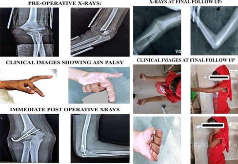
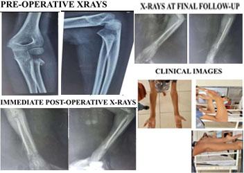
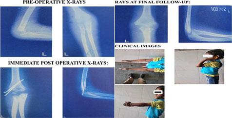
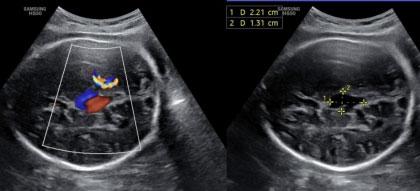
Background : Elbow fractures in children are common and may have serious complications. The aim of our study is to estimate the morphology and outcome of fractures around Elbow joint in paediatric age group of patients.
Materials and Methods : Patients in the age group of 3-12 years with unilateral fracture around the Elbow joint were included in our study. Management protocol was decided after detailed history, examination and radiological investigations depending upon the type of fracture, displacement of the fracture and classification whether to go for conservative or operative intervention. The patients were followed up for a period of 12 months and the final outcome of patient for Elbow injury was assessed on the basis of MAYO ELBOW SCORE.
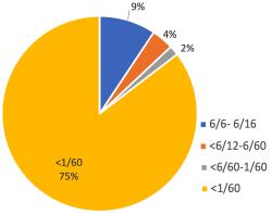
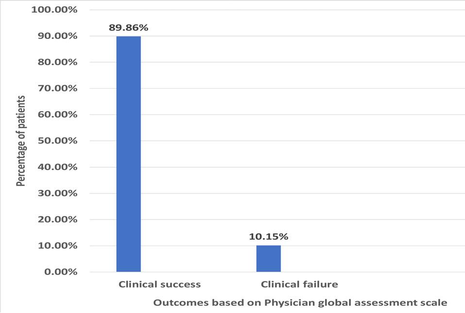
Results : Majority of our patients presented with Supracondylar Humerus Fracture (77%) while Medial Condyle Humerus Fractures were least common (3%). We did not have any patients of Olecranon Fracture or Epiphyseal separation injuries. Majority of the patients (75%) were taken for operative intervention whereas rest were managed conservatively. Overall it was observed that out of 52 patients with Elbow injuries excellent outcome was recorded in 46 patients. Good and fair outcomes were recorded in 3 patients each.
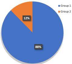
Conclusion : Excellent outcomes as per “MAYO ELBOW SCORE” were recorded in majority (88%) of patients and poor outcomes were not recorded in any of the patients. Minimal complications were recorded in our study : Cubitus varus deformity in 9% and restricted range of motion in 3% of patients.
Key words :Adolescent, Child, Elbow Injuries, Elbow Joint.
Fractures around Elbow joint are common in paediatric population. Sometimes children having Elbow injuries may have serious complications like neurovascular injuries, cubitus varus deformity and Volkmann’s contracture in supracondylar fracture, while nonunion, valgus deformity and late ulnar nerve palsy in lateral condyle fracture1.
Supracondylar Humerus Fractures account for 60% of all Pediatric Elbow Fractures, classically occurring as a result of fall on an outstretched hand2-4. Lateral Condyle Humerus Fractures are the second most common elbow fracture after the supracondylar Humerus Fracture 5 . Radial Neck Fractures are relatively rare in children, accounting for 1% of all fractures in children6,7. Olecranon and distal humeral epipyseal seperations are relatively uncommon injuries around the Elbow joint. The aim of our study is to estimate the morphology and outcome of fractures around Elbow joint in paediatric age group of patients.
MATERIAL AND METHODS
This prospective observational cohort study of 52
Department of Orthopaedics, Medical Collegte Baroda & Sir Sayajirao General Hospital, Vadodara, Gujarat 390001
1MS (Ortho), Senior Resident
2MS (Ortho), Associate Professor and Corresponding Author
3MS (Ortho), Associate Professor
Received on : 28/12/2023
Accepted on : 23/02/2024
[J Indian Med Assoc 2025; 123(1): 15-8]
Editor's Comment :
Supracondylar humerus fracture is the commenest while medial condyle humerus fracture is the least common fracture around the elbow joint in paediatric age group. Excellent outcome can be achieved in these fractures with proper treatment protocol.
patients with Elbow injuries was undertaken at the Department of Orthopaedics of a state run Government Tertiary Care Institute from November, 2021 to September, 2023 after getting approval from Institutional Ethics Committee for Biomedical and Health Research. All the cases were treated between 15-11-2021 & 30-09-2022. Patients in the age group of 3-12 years with unilateral fracture around the Elbow joint managed conservatively or with operative intervention were included. The patients with concomitant head injuries, pathological fractures or polytrauma patients were excluded. The morphology of fracture was assesed as per classification or type of fractures. Management protocol was decided after detailed history, examination and radiological investigations depending upon the type of fracture, displacement of the fracture and classification. Majority of the undisplaced or minimally displaced stable fractures of Supracondylar Humerus, Medial Condyle Humerus were managed conservatively. Radial Neck Fractures with <30 degrees of angulation
123, No 01, January 2025Journal
were managed conservatively whereas fractures with 30-60 degrees of angulation were managed with manipulative closed reduction mainly with Patterson or Israeli maneuver followed by immobilization. Patients with displaced fractures or Radial Neck Fractures with >60 degrees angulation were taken for operative intervention. Conservatively managed fractures were kept immobilized in long arm cast or a posterior splint for atleast 3 weeks followed by active elbow range of motion exercises.
Patients with displaced Supracondylar Humerus Fractures were treated with closed reduction and kwire fixation with mini-open ulnar nerve exploration for introduction of medial k-wire. Patients with Lateral Condyle Humerus Fractures underwent open reduction and internal fixation with k-wire or 4 mm cannulated screws. K- wires were removed after atleast 4 weeks when signs of union were seen radiologically which was followed by active Elbow range of motion exercises.
The follow-up protocol was to be started from first week post injury. Then second follow up was done after 4 weeks and third follow up after 8 weeks. Then patients were called for follow up at every three months. Any progressive deformity, union status and neurovascular abnormalities were looked for during follow up period. The final outcome was assessed following one year of injury using MAYO ELBOW SCORE. As per MAYO ELBOW SCORE results were graded as excellent with score more than 90, good outcome with score of 75-89, fair outcome with score of 60-74 and poor outcome with scores less than 60.
OBSERVATIONS AND RESULTS
This prospective observational cohort study of 52 paediatric patients with Elbow injuries was carried out at Department of Orthpaedics from November, 2021 to September, 2023. Our study has patients with age of 3-12 years with mean age of 7 years. Majority of patients (48) had injury due to low velocity trauma mainly due to fall while playing whereas rest 4 patients had history of Road Traffic Accident. All the patients presented with closed fractures. Majority of our patients presented with supracondylar Humerus Fracture (77%) while Medial Condyle
Humerus Fractures were least common (3%). We did not have any patients of Olecranon Fracture or Epiphyseal Separation injuries. 40 patients presented with Supracondylar Humerus Fracture out of which 26 patients had Gartland Type 3, 11 patients had Type 2 whereas 3 patients had Type 1 fracture. There were no patients with Flexion Type Supracondylar Humerus Fracture. One patient with Type 3 Supracondylar Humerus Fracture had Anterior Interosseous Nerve Palsy which recovered after fixation within a duration of 4 weeks. Other patient with same kind of Type 3 supracondylar humerus fracture had pink pulseless hand which recovered after fixation with cross pinning.
Next common fracture in our study was Radial Neck Fracture. Six patients presented with Radial Neck Fractures out of which 5 patients with Judet Type 2 and 1 with Judet Type 3. Out of 4 patients of Lateral Condyle Humerus Fracture 3 had Weiss type 3 whereas 1 patient had Weiss Type 2. Two patients with medial condyle humerus fracture were managed conservatively with excellent outcome in both patients. Majority of the patients (75%) were taken for operative intervention whereas rest were managed conservatively. Overall it was observed that out of 52 patients with Elbow injuries excellent outcome was recorded in 46 patients. Good and fair outcomes were recorded in 3 patients each (Tables 1-4).
The most common complication observed in our study was cubitus varus deformity following
Table 1 — Outcomes of Patients Treated for Fracture Supracondylar Humerus Outcomes Gartland Type IGartland Type IIGartland Type III ConservativeOperativeConservativeOperativeConservativeOperative Excellent030002060024 Good000001000001 Fair000002000100 Poor000000000000 Total030005060125
Table 2 — Outcomes of Patients Treated for Fracture Lateral Condyle Humerus OutcomesWeiss Type IWeiss Type IIWeiss Type III ConservativeOperativeConservativeOperativeConservativeOperative Excellent000000010002 Good000000000001 Fair000000000000 Poor000000000000 Total000000010003
Table 3 — Outcomes Of Patients Treated For Radial Neck Fractures Outcomes Judet Type IJudet Type IIJudet Type III ConservativeOperativeConservativeOperativeConservativeOperative Excellent000002030001 Good000000000000 Fair000000000000 Poor000000000000 Total000002030001
Table 4 — Outcomes of Patients with Elbow Injuries
Supracondylar Humerus Fracture (5 patients). Other complications like restricted range of motion was observed in 2 patients with Supracondylar and Lateral Condyle Humerus Fracture. One patient had nonunion following lateral condyle humerus fracture.
DISCUSSION
The fractures around Elbow in children should be given special attention by the treating surgeon as such fractures can result into immediate complications as well as late deformities. The present study is comparable to the study done by Fahey in Chicago, and a study in Hong8-11 and also can be compare to the study done in East Africa by Wamisho12. In our study it was found that Elbow injuries were found with Male:Female ratio of 3:1. This is comparable with the findings in other studies done by Fahey in Chicago where M:F ratio was 2:1 and a study in Hong (M:F = 2.7:1)8-11
The most frequent type of fracture encountered in our study was Supracondylar Humerus (77%) followed by Radial Neck Fractures (12%) and Lateral Condyle Humerus Fractures (8%). These findings are comparable to the study done by Wamisho12 where 69% Supracondylar Humerus Fractures were encountered.
Excellent outcomes were obtained in all Type 1 fractures which were managed conservatively. Excellent outcomes were also obtained in the patients who were operated for Type 3 fractures (92%) in our study. The outcomes were variable in patients who were managed conservatively for Type 2 fractures but excellent outcomes were seen in all patients who were operated for Type 2 fractures. In one of the studies it was found that Type 2 fractures resulted in both satisfactory (57.7%) and unsatisfactory outcomes (42.3%), regardless of the treatment13. It was found from our study that cubitus varus was one of the most common complications which developed after Supracondylar Humerus Fractures mainly among the patients with Type 2 and Type 3 fractures which were managed conservatively. On regular follow up of the patients with Lateral Condyle Humerus fractures it was found that the patients who underwent
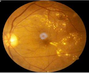
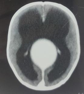
Fig 1 — A/C/O Close Fracture Supracondylar Humerus Left with Anterior Interosseous
123, No 01, January 2025Journal
CLOSED reduction with internal fixation went on to develop NONUNION suggesting that OPEN reduction followed by internal fixation should be preferred in such patients. Excellent outcome was obtained in all the patients of Radial Neck Fractures either managed conservatively or taken for operative intervention depending on fracture type, angulation and classification.
CONCLUSION
This study was done on 52 paediatric patients who presented with Elbow injuries. Overall 75% of these patients were taken for operative intervention whereas 25% patients were managed conservatively which was decided as per fracture morphology, displacement and classification. Excellent outcomes as per “MAYO ELBOW SCORE” were recorded in majority (88%) of patients and poor outcomes were not recorded in any of the patients. Minimal complications were recorded in our study: Cubitus varus deformity in 9% and restricted range of motion in 3% of patients (Figs 1-8).
Conflict of Interests : The authors declare no conflict of interests.
Support Financial : There was no financial support from public, commercial or non-profit sources.
REFERENCES
1Okubo H, Nakasone M, Kinjo M, Onaka K, Futenma C, Kanaya F — Epidemiology of paediatric elbow fractures: a retrospective multi-centre study of 488 fractures. J Child Orthop 2019; 13(5): 516-21.
2Cheng JC, Shen WY — Limb fracture pattern in different pediatric age groups: a study of 3,350 children. J Orthop Trauma 1993; 7(1): 15-22
3Wilkins KE — Fractures and dislocations of the elbow region. In: Rockwood CA, King RE, Wilkins KE, editors. Fractures in children. 3rd ed. New York: JB Lippincott; 1991: 526-617.
5Badelon O, Bensahel H, Mazda K, Vie P — Lateral humeral condylar fractures in children. J Pediatr Orthop 1988; 8(1): 31-4
6Brandão GF, Soares CB, Teixeira LE, Boechat Lde C — Displaced radial neck fractures in children: association of the Métaizeau and Böhler surgical techniques. J Pediatr Orthop 2010; 30(2): 110-4.
7Robert M, Moulies D, Longis B, Alain JL — Fractures of the
upper part of the radius in children. Chir Pediatr 1986; 27(6): 318-21.
8Smith FM — Medial epicondyle injuries. J Am Med Assoc 1950; 142(6): 396-402.
9Maylahn DJ, Fahey JJ — Fractures of the elbow in children; review of three hundred consecutive cases. J Am Med Assoc 1958; 166(3): 220-8.
10Landin LA, Danielsson LG — Elbow fractures in children. An epidemiological analysis of 589 cases. Acta Orthop Scand 1986; 57(4): 309-12.
11Cheng JC, Shen WY — Limb fracture pattern in different pediatric age groups: a study of 3,350 children. J Orthop Trauma 1993; 7(1): 15-22
12Wamisho BL, Admassie, Daniel, Banchiamlak A — Fractures around child’s elbow-Radiological patterns. East and Central African Journal of Surgery 2007; 13(2): 13. (ISSN: 1024297X).
13Sinikumpu J, Victorzon S, Pokka T, Lindholm E, Peljo T, Serlo W — The long-term outcome of childhood supracondylar humeral fractures. Bone Joint J 2016; 98-B(10): 1410-7. doi:10.1302/0301-620X.98B10.35923.
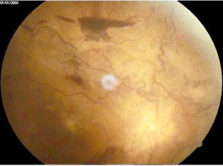
Fig 2 — A/C/O Close Fracture Radial Neck Right Without NVD (Judet Type 3) Operative Intervention : Cr + Tens Nailing
Fig 3 — A/C/O Close Fracture Lateral Condyle Humerus Left Without NVD (Weiss Type 2) Operative Intervention : Open Reduction + K-wire Fixation
Original Article
Correlation of Serum Vitamin D with Serum Calcium Level in Hypothyroid Patients
Background : Vitamin D is necessary for the normal functioning of many organs, including the thyroid gland. It's deficiency is considered a risk factor for the development of many thyroid disorders, including autoimmune thyroid diseases and thyroid cancer. However, the interaction between Vitamin D and thyroid function is still not fully understood.
Aims and Objectives : The aim of the study was to evaluate the association between hypothyroidism and serum Vitamin D3 levels in the Indian population and its association with ionized calcium.
Materials and Methods : A cross-sectional analytical study was carried out among 100 patients with sub-clinical hypothyroidism or overt primary hypothyroidism who met the inclusion criteria from November, 2019 to November, 2020 in a tertiary care centre in Salem. Basic demographic details were obtained and the estimation of serum calcium, Vitamin D and thyroid profile was done by an automated analyzer.
Results : In this study, two-thirds of the patients were female. The bulk of the patients (82%) had overt hypothyroidism. Though, Vitamin D and TSH levels had a significant negative correlation (r = -0.333, P value = 0.001), there is a positive relationship between Vitamin D levels and serum calcium (r = 0.047, P value = 0.644). Serum calcium levels were negatively correlated with TSH levels but this finding was statistically not significant (r = -0.141, P value = 0.162).
Conclusion : This study indicates that patients with hypothyroidism suffer from low serum levels of Vitamin D, which correlate with TSH. These findings may suggest a potential role for 25-OH Vitamin D in the development of hypothyroidism. This study recommends that patients with hypothyroid disorders be regularly checked for levels of Vitamin D and serum calcium.
[J Indian Med Assoc 2025; 123(1): 19-23]
Key words :Hypothyroidism, Thyroid Function Tests, Thyrotropin, Vitamin D Deficiency.
Hypothyroidism, the clinical condition of thyroid hormone deficiency, is a common disorder in the general population 1 . It is defined mainly by biochemical criteria due to the wide diversity in clinical presentation and the overall lack of a specific symptom2. TSH levels above the reference range suggest clinical or overt primary hypothyroidism. On the other hand, free thyroxine levels below the reference range suggest primary hyperthyroidism. TSH levels above the reference range and free thyroxine concentrations within the normal range indicate mild or sub-clinical hypothyroidism, which is frequently encountered as an early indication of thyroid dysfunction3. It is a common condition in India, with a prevalence of 1.9 percent in women by 2020 and the prevalence increases with age4. Thyroid hormones are universal determinants of organ function. Hence, there may be a multiplicity of symptoms. Especially in the elderly, clinical presentations may be atypical and go undiagnosed4
Department of General Medicine, Vinayaka Missions Kirupananda Variyar Medical College and Hospital, Salem, Tamil Nadu
636308
1MD (General Medicine), Professor
2MD (General Medicine), Assistant Professor
3MBBS, Postgraduate Student and Corresponding Author
Received on : 24/09/2023
Accepted on : 04/01/2024
Editor's Comment :
Vitamin D is necessary for the normal functioning of many organs, including the thyroid gland. Its deficiency is considered a risk factor for the development of many thyroid disorders.
This study indicates that patients with hypothyroidism suffer from low serum levels of Vitamin D, which correlate with TSH. These findings may suggest a potential role for 25-OH Vitamin D in the development of hypothyroidism.
Vitamin D plays a major role in physiological processes that modulate mineral metabolism and immune function with probable link to several chronic and infectious conditions 5. Vitamin D is a steroid molecule, mainly produced in the skin, that regulates the expression of a large number of genes6. Calcium and phosphate levels are two minerals that Vitamin D aids in controlling. Healthy bones, teeth and muscles require these nutrients. Deformities of the bones, such as rickets in children and bone pain in adults due to Osteomalacia, can result from insufficient Vitamin D in the body7.
Vitamin D deficiency is a global health problem8. Over one billion people Worldwide are Vitamin D deficient or insufficient9. Vitamin D deficiency prevails in epidemic proportion all over the Indian subcontinent. With a prevalence of 70%-90% in the general
123, No 01, January 2025Journal
population in India10. This prevalence is remarkably high. Several studies have shown that Vitamin D may play a role in many biochemical mechanisms, in addition to bone and calcium metabolism11. The role of Vitamin D as an immune modulator has been emphasized in recent years and low levels of the hormone were observed in several autoimmune diseases, including multiple sclerosis12, SLE13, Type 1 diabetes mellitus and rheumatoid arthritis. Most effects of Vitamin D are mediated via the Vitamin D3 Receptor (VDR). The immune modulator properties of Vitamin D are attributed to its effect on T and B lymphocytes, all of which harbors VDRS. Low Vitamin D may increase the degree of auto-immunity and subsequently increase the prevalence of Auto-immune Thyroid Diseases (AITDs)14. Thyroid diseases are among the most common endocrine abnormalities. The pathogenesis of AITDs, like other auto-immune diseases, is multifactorial, combining genetic, immune, environmental and hormonal influences such as Vitamin D.
Few past studies have reported the impact of Vitamin D deficiency on thyroid diseases. Currently, there is no consensus regarding the optimal role of Vitamin D deficiency in hypothyroid patients or its association with hypothyroidism. In the present study, we explored the probable interaction between Vitamin D status and hypothyroidism and also tried to find any correlation between serum calcium levels and hypothyroidism.
MATERIALS AND METHODS
Study Settings :
Salem District is one of the 38 districts of Tamil Nadu State in Southern India. A hospital-based crosssectional analytical study was carried out among patients diagnosed with sub-clinical hypothyroidism and overt primary hypothyroidism in a tertiary care centre in Salem. The Tertiary Care Centre is located in the rural area of Salem and the hospital has 560 beds. The Department of General Medicine normally provides 24-hour service to more than One Lakh Outpatients per year, mainly to the people of Salem and partly to the neighboring districts of Erode and Namakkal.
Study Period and Study Population :
This study was conducted for a period of 1 year, ie, from November, 2019 to November, 2020. All patients 18 years of age and older, of both genders, diagnosed with sub-clinical hypothyroidism and overt primary hypothyroidism who fulfilled the inclusion criteria were recruited for the study.
Ethical consideration : The approval for this study was obtained through the ethical clearance of the Institutional Ethics Committee on Human Subjects
(Approval No. VMKVMC&H/IEC/19/65). After obtaining approval from the Institutional Ethics Committee, the data was collected from the patient who fulfilled the inclusion criteria and protocol for enrolling in the study after receiving their signed informed consent.
Selection of Study Participants :
Inclusion Criteria :
•Patients with sub-clinical hypothyroidism aged over 18 years with a TSH level greater than 5.1 and less than 10 mIU/ml
•Patients with overt primary hypothyroidism aged over 18 years with a TSH level greater than 10 mIU/ml
Exclusion Criteria :
Patients on Vitamin D supplementation or calcium supplements; patients with central hypothyroidism; hepatic dysfunction; renal dysfunction and patients on anti-epileptic medications
Sample Size Determination and Sampling Method :
In the cross-sectional study by Amer et al. in India by 2019, the prevalence of Vitamin D deficiency in sub-clinical and clinical hypothyroidism cases was 90%15. Considering this prevalence, the minimum sample size of 96 was calculated using the formula 3.84*pq/d2, where prevalence (p) = 90, q (1-p) = 10, and precision (d) = 6, with a 95% confidence interval. All consecutive patients (100 in number) who fit the inclusion criteria were enrolled in the study.
Data Collection Procedure :
A semi-structured questionnaire was used by the principal investigator using a one-on-one interview method to collect data. Each individual was given a thorough clinical examination, a history was obtained, and a clinical diagnosis was determined. After overnight fasting, 3 ml of venous blood was withdrawn from the patients in the plain vacutainer. Blood was allowed to clot, and a centrifuge was used to separate serum. Serum thus separated was stored at 4-8°C until the analysis was done. Estimation of serum T3, T4 and TSH was done by a fully automated analyzer, Minividas and serum calcium was analyzed by the Arsenezo method in a semi-automated analyzer. Serum concentrations of Vitamin D3 in patients were measured by the immuno-metric assay method (competitive principle). The quantitative determination of 25-OH Vitamin D was carried out by a direct, competitive chemiluminescence immunoassay (direct chemiluminescent reactions).
Operational Definition :
(1)Vitamin D deficiency :
In our study, Vitamin D deficiency was defined, in accordance with the manufacturer, as serum levels
123, No 01, January 2025Journal
of Vitamin D3 less than 20 ng/mL in adults. Vitamin D insufficiency was defined as serum levels of Vitamin D3 between 20 and 29 ng/mL in adults. Vitamin D sufficiency was defined as serum levels of Vitamin D3 greater than 30 ng/mL in adults16
(2)Hypothyroidism :
Hypothyroidism cases were classified into two subgroups according to their thyroid function status measured by the ELIZA technique17.
Sub-clinical hypothyroidism describes a situation in which no overt clinical feature of hypothyroidism is present, with serum levels of T4 and T3 still in the normal range but with higher levels of TSH.
Clinical hypothyroidism describes the condition in which an overt clinical feature of hypothyroidism is present, with lower levels of T4 and T3 and higher levels of TSH.
Thyroid Stimulating Hormone (TSH)
Normal : 0.5-5.0 mIU/l
Sub-clinical Hypothyroidism : 5.1 to <10 mIU/l
Overt Hypothyroidism : >10 mIU/l
Triiodothyronine (T3) – Normal level: 1.2 – 4.4 pg/ml
Thyroxine (T4) – Normal Level: 0.8-2.0 ng/dl
(3)Serum Calcium : Calcium sufficiency was defined as serum levels of calcium between 8.6 mg/dl and 10.3 mg/dl in adults.
Data Processing and Analysis :
Data analysis was performed using IBM-SPSS version 21.0 (IBM-SPSS Science Inc, Chicago, IL). Descriptive statistics like mean and Standard Deviation were used for continuous variables like age, TSH, T3, T4, etc, while frequency and percentage were used for gender variables. An independent sample T-test was used for comparing two means and Pearson’s correlation was done to see the correlation between two continuous variables. A P value of 0.05 was considered significant.
RESULTS
Almost half (45%) of the patients were 36-45 years of age and more than one-third were 26-35 years of age. 67% of the patients were female and 33% were male. Most (82%) of the patients had overt hypothyroidism. Almost 73% of patients were deficient in Vitamin D and 27% of patients had an insufficient amount of Vitamin D. Table 1 describes the basic characteristics of the study population.
Table 2 describes the gender-based clinical characteristics and their association as expressed by the independent T test. No statistical difference (P <0.05) was seen in all variables, except for the age variable, where a difference in the mean age of Males is 37.18±7.23 and Females is 33.44 ±7.56 (P value =
0.02). The mean concentration of Vitamin D is higher in Females compared to Males, but this difference was statistically not significant (P value = 0.42). The mean concentration of serum calcium is almost equal between Females and Males. Thus, this difference was statistically not significant (P value = 0.92).
Table 3 describes the association of thyroid status with vitamin D and serum calcium by an independent T test. The mean concentration of Vitamin D is higher in sub-clinical hypothyroidism (20.66 ± 4.68) in comparison to overt hypothyroidism with a mean of 16.67 ± 4.60 and this difference was statistically significant (P value = 0.001). Conversely, the mean concentration of calcium is higher in sub-clinical hypothyroidism (9.21 ± 0.62) in comparison to overt hypothyroidism with a mean of 9.01 ± 0.62 and this difference was not statistically significant (P value = 0.235).
Table 4 describes the correlation between Vitamin D, serum calcium and thyroid hormone levels. Thyroid Stimulating Hormone (TSH) levels correlate negatively with Vitamin D levels (r = -0.333, P value = 0.001). Vitamin D concentrations increase along with calcium levels in the blood. There is a positive correlation between Vitamin D levels and serum calcium, but there is no statistical significance (r = 0.047, P value = 0.644). T3 and T4 levels had no statistically significant change (P values = 0.644, 0.466, and 0.650, respectively). TSH levels were
Table 1 — Basic Characteristics of the Study Population Patients CharacteristicsFrequencyPercentage (N = 100)
Table 2 — Gender-based Clinical Characteristics
Table 3 — Association of Thyroid Status with Vitamin D and Serum Calcium Thyroid StatusSub-ClinicalOvert P value HypothyroidismHypothyroidism (N=18)(N=82)
123, No 01, January 2025Journal
Table 4 — Correlation Co-efficients of Vitamin D and Calcium with Thyroid Function Tests
VariablesVitamin DSerumTSHT3T4 Calcium
Vitamin D :
Pearson Correlation10.047*-0.3330.0740.046*
P value-0.6440.001*0.4660.650
Serum Calcium :
Pearson Correlation0.0471-0.1410.1010.003*
P value0.644-0.1620.3180.977
negatively linked with serum calcium levels, but this finding was not statistically insignificant (r = -0.141, P value = 0.162).
DISCUSSION
Various previous studies have documented a relationship between the occurrence of hypothyroidism and serum concentrations of Vitamin D8,18-20. A literature review has reported that Vitamin D deficiency plays a critical role in thyroid disease development, including thyroid cancer21,22
We observed in this study that more than twothirds (67%) of the patients were Female. In addition, almost half of them fall under the age group of 36 to 45. These findings suggest that hypothyroidism is more common among Females than Males. Similar findings were also observed in India by Sinha R, et al in 2019. They found that 67.1% of the patients were female and more than half of the patients fell under the 40-59 age group 23 . A study in India by Unnikrishnan, et al in 2013 also found a greater number of female patients with hypothyroidism 24 Similar observations were also noted in a study by Mackawy, et al which had a greater number of female subjects with hypothyroidism8
In this study, patients with clinical or overt hypothyroidism had significantly lower serum levels of Vitamin D than those with sub-clinical hypothyroidism (P value 0.001). It may be associated with insufficient intestinal absorption of Vitamin D or the body’s inability to activate Vitamin D correctly. Parallel to our findings in India, Amer AH, et al noticed in 2019 that patients with overt hypothyroidism had lower levels of Vitamin D than those with sub-clinical hypothyroidism (P value 0.001)15. Furthermore, a study in India by Lohokare R, et al (2016) discovered that hypothyroid individuals had significantly lower serum levels of Vitamin D 25 (OH) than euthyroid patients (P value 0.001)25. Kim reported in the Korean population by 2016 that Vitamin D deficiency is more prevalent in auto-immune Hashimoto’s thyroiditis presenting with overt hypothyroidism than sub-clinical hypothyroid variants26. In the present study, Thyroidstimulating Hormone (TSH) levels correlate negatively with Vitamin D levels. Similar studies have shown that
a reciprocal relationship exists between serum TSH and Vitamin D levels in hypothyroid subjects26,27
According to a study done by Talaei A, et al in Iran by 2018 where 12 weeks supplement of 50,000 IU Vitamin D was given to one group and one group received placebo, the supplemented group had significant decrease in the TSH levels. They also found the prevalence of Vitamin D deficiency to be high in hypothyroid patients. They, thus, suggested a significant relationship between Vitamin-D deficiency and hypothyroidism28
We observed a lower level of serum calcium in patients suffering from overt hypothyroidism (9.01 ± 0.62) when compared with sub-clinical hypothyroidism (9.21±0.62), but it was not statistically insignificant (P value = 0.235). In contrast to our findings, a study by Mackawy, et al in Saudi Arabia discovered that serum calcium levels recorded a significant decrease in hypothyroid patients when compared to controls8 In India, a study by Sinha R, et al in 2019 observed that hypothyroid people had significantly lower blood calcium levels than the control group23. Disturbance in calcium homeostasis is frequently observed with dysfunction in the thyroid gland29. Thyroxine, which normally controls blood calcium levels through cellular calcium release, is reduced in hypothyroidism. As a result, there is less thyroxine in the circulation, which leads to less thyroxin entrance into cells and hence less extracellular calcium release30
These findings point towards the role of Vitamin D as a potential modifiable risk factor for hypothyroidism. The effect of Vitamin D is mediated through its binding to the Vitamin D Receptor (VDR) and activation of VDR-responsive genes. VDR is found in several cell types, including the thyroid gland 29 . So, probably, Vitamin D plays a role in maintaining an euthyroid state by interacting with its receptor in the thyroid gland. Similar findings were reported in other studies as well8,25
This study indicates that patients with hypothyroidism suffer from low serum levels of Vitamin D and calcium and these levels are associated with the degree and severity of hypothyroidism. Screening of newly diagnosed hypothyroid patients for 25-OH Vitamin D and serum calcium levels and supplementation of Vitamin D at an early stage of diagnosis are strongly recommended.
Limitations :
Our study population of 100 samples is comparatively small and the study can be extended to a larger population. The levels of phosphorous, parathyroid hormone and thyroid antibodies were not investigated. No genetic workup was done. The
123, No 01, January 2025Journal
current study was conducted as a hospital-based study due to a lack of resources. As this was a crosssectional study, the association was found to lack a temporal association between hypothyroidism and the level of Vitamin D. Due to resource constraints, certain important variables related to Vitamin D, such as indoor versus outdoor physical activity, seasonal changes and geographical coordinates, could not be ascertained, which could have led to some biases. In addition, it was not possible to identify whether or not Vitamin D levels were affected by pathological disorders such as non-alcoholic fatty liver disease.
CONCLUSION
In this study, clinical or overt hypothyroidism was associated with considerably lower serum Vitamin D levels than sub-clinical hypothyroidism. This study indicates that patients with hypothyroidism suffer from low serum levels of Vitamin D, which correlate with TSH. These findings may suggest a potential role for 25-OH Vitamin D in the development of hypothyroidism. This study recommends that patients with hypothyroid disorders be regularly checked for levels of Vitamin D and serum calcium. There may be a rationale for the recommendation of Vitamin D and calcium supplementation for hypothyroid patients. Ongoing and future long-term randomized control trials are required to determine the role of Vitamin D in the pathogenesis of hypothyroidism.
Acknowledgment : NIL.
Funding : NIL.
Conflicts of Interest : The authors declare no Conflicts of Interest.
2D’Aurizio F, Villalta D, Metus P, Doretto P, Tozzoli R. Is vitamin D a player or not in the pathophysiology of autoimmune thyroid diseases? Autoimmun Rev 2015; 14(5): 363-9.
3Wang J, Lv S, Chen G, Gao C, He J, Zhong H, et al — Metaanalysis of the association between vitamin D and autoimmune thyroid disease. Nutrients 2015; 7(4): 2485-98.
4Shivakumar, Bhargavi SK, Prasad Naidu M — Study of Serum Calcium, Magnesium and Phosphorous Levels in Hypothyroidism. SIJB 2020; 3(2): 22-6.
5DeLuca HF — Evolution of our understanding of vitamin D. Nutr Rev 2008; 66(10 Suppl 2): S73-87.
6Holick MF, Chen TC— Vitamin D deficiency: a worldwide problem with health consequences. Am J Clin Nutr 2008; 87(4): 1080S-6S.
7Musa IR, Gasim GI, Khan S, Ibrahim IA, Abo-Alazm H, Adam I — No Association between 25 (OH) Vitamin D Level And Hypothyroidism among Females. Open Access Maced J Med Sci 2017; 5(2): 126-30.
8Mackawy AMH, Al-ayed BM, Al-rashidi BM — Vitamin D Deficiency and Its Association with Thyroid Disease. Int J Health Sci (Qassim) 2013; 7(3): 267-75.
9Sharma V, Gupta A, Showkat R — Vitamin D Deficiency & Low Serum Calcium Levels in Hypothyroid Patient. International
Journal of Contemporary Medical Research, ISSN (Online): 2393-915X; (Print): 2454-7379 | ICV: 77.83, January 2018; 5(1)
10Gupta A — Vitamin D deficiency in India: prevalence, causalities and interventions. Nutrients 2014; 6(2): 729-75.
11Holick MF — Vitamin D: extraskeletal health. Endocrinol Metab Clin North Am 2010; 39(2): 381-400, table of contents.
12Munger KL, Zhang SM, O’Reilly E, Hernán MA, Olek MJ, Willett WC, et al — Vitamin D intake and incidence of multiple sclerosis. Neurology 2004; 62(1): 60-5.
13Cutolo M, Otsa K — Review: vitamin D, immunity and lupus. Lupus 2008; 17(1): 6-10.
14Unal AD, Tarcin O, Parildar H, Cigerli O, Eroglu H, Demirag NG — Vitamin D deficiency is related to thyroid antibodies in autoimmune thyroiditis. Cent Eur J Immunol 2014; 39(4): 493-7.
15Amer AH, Chaudhari K, Trivedi R, Patel R — Study of the serum levels of Vitamin d and Calcium ionized in thyroid disorders. International Journal of Medical and Biomedical Studies 2019; 3(7): 93-9.
17Fatourechi V — Subclinical Hypothyroidism: An Update for Primary Care Physicians. Mayo Clin Proc 2009; 84(1): 65-71.
18Ke W, Sun T, Zhang Y, He L, Wu Q, Liu J, et al — 25Hydroxyvitamin D serum level in Hashimoto’s thyroiditis, but not Graves’ disease is relatively deficient. Endocr J 2017; 64(6): 581-7.
19Ahi S, Dehdar MR, Hatami N — Vitamin D deficiency in nonautoimmune hypothyroidism: a case-control study. BMC Endocr Disord 2020; 20(1): 41.
20Metwalley KA, Farghaly HS, Sherief T, Hussein A — Vitamin D status in children and adolescents with autoimmune thyroiditis. J Endocrinol Invest 2016; 39(7): 793-7.
21Aktas HS — Vitamin B12 and Vitamin D Levels in Patients with Autoimmune Hypothyroidism and Their Correlation with AntiThyroid Peroxidase Antibodies. Med Princ Pract 2020; 29(4): 364-70.
22Muhammad H, Ahmed I, Muhammad A, Laique T — Vitamin D and Calcium Levels in Female Hypothyroid Patients Presenting to OPD in Sialkot: Cross Sectional Study. PJMHS 2021; 15(9): 2708-10.
23Sinha R, Bhushan I — The Study of Serum Calcium and 25OH Vitamin D Levels in Newly Diagnosed Hypothyroid Patients.
24Unnikrishnan AG, Kalra S, Sahay RK, Bantwal G, John M, Tewari N — Prevalence of hypothyroidism in adults: An epidemiological study in eight cities of India. Indian J Endocrinol Metab 2013; 17(4): 647-52.
25Lohokare R, Gupta A, Jain A — A study of vitamin D levels in hypothyroid patients, a case control study in a tertiary care hospital of central India. National Journal of Medical and Dental Research 2016; 4(2): 89-92.
26Kim D — Low vitamin D status is associated with hypothyroid Hashimoto’s thyroiditis. Hormones (Athens) 2016; 15(3): 385-93.
27ElRawi HA, Ghanem NS, ElSayed NM, Ali HM, Rashed LA, Mansour MM — Study of Vitamin D Level and Vitamin D Receptor Polymorphism in Hypothyroid Egyptian Patients. J Thyroid Res 2019; 2019: 3583250.
28Talaei A, Ghorbani F, Asemi Z — The Effects of Vitamin D Supplementation on Thyroid Function in Hypothyroid Patients: A Randomized, Double-blind, Placebo-controlled Trial. Indian J Endocrinol Metab 2018; 22(5): 584-8.
29Sato K, Han DC, Fujii Y, Tsushima T, Shizume K — Thyroid hormone stimulates alkaline phosphatase activity in cultured rat osteoblastic cells (ROS 17/2.8) through 3,5,3’-triiodo-L-thyronine nuclear receptors. Endocrinology 1987; 120(5): 1873-81.
30Sridevi D, Dambal AA, Sidrah, Challa AS, Padaki SK — A Study of Serum Magnesium, Calcium and Phosphorus in Hypothyroidism. International Journal of Clinical Biochemistry and Research 2021; 3(2): 236-9.
Original Article
A Study of Disease Outcome
following Tyrosine Kinase Inhibitor Therapy in Patients with Chronic Myeloid Leukemia, from a Tertiary Care Center of North Bengal
Debasis Chakrabarti1, Arka Mukhopadhyay2, Pasang Lahmu Sherpa3, Dipanjan Bandyopadhyay4
Background and Objectives : Chronic Myeloid Leukemia (CML) remains one of the very few malignancies with excellent prognosis following the advent of Tyrosine Kinase Inhibitors (TKIs). Apart from Imatinib, there are several novel TKIs which can be used as first line therapy with even better clinical outcomes like faster clinical response, less evidences of recurrence and lesser adverse effects like cytopenia.
Materials and Methods : A hospital based observational, descriptive and prospective study was designed to follow up patients being diagnosed with CML in chronic phase and were started on TKIs, Imatinib or Nilotinib and they were followed up with records of baseline blood counts, bone marrow examination, BCR-ABL studies and their blood counts were recorded over 3rd and 6th months of therapy.
Results : Most of the patients had an excellent clinical response by 3rd month. Still, 14.89% (n=7) patients had leukocyte count >12000/mm3 whereas 4.25% (n=2) patients had leukocytopenia with counts <4000/mm3. Hemoglobin trend was steadily increasing whereas platelet count remained within normal limits. For those patients treated with Nilotinib had comparatively better cytological response by 3rd and 6th months compared to Imatinib, with a slightly higher increase in hemoglobin level and lesser evidence of cytopenia over the course of 6 months.
Interpretation : TKIs have resulted excellent cytological response although issues like cytopenia remains an important concern.
Conclusion : TKIs remain the cornerstone while treating CML patients. Among the TKIs, Nilotinib can garner faster cytological response while have lesser chance of having cytopenia compared to Imatinib. [J Indian Med Assoc 2025; 123(1): 24-8]
Key words :Chronic Myeloid Leukemia, TKI in CML, Cytopenia, Imatinib versus Nilotinib, Tyrosine Kinase Inhibitors.
Chronic Myeloid Leukemia (CML), one of the most common type of hematological malignancies1, is also among the best treatable malignancies2. Since the advent of Tyrosine Kinase Inhibitors (TKI), treatment of CML has been revolutionized with excellent prognosis altogether increasing the life expectancy similar to that of general population3,4 Imatinib has been used as a first line TKI for treating CML for almost 2 decades. In recent years, Nilotinib is also being considered as a first line therapy4
Although several studies compared Imatinib to Nilotinib for treatment efficacy and adverse effects from around the world in recent days, there are very few Indian literature for the comparison5-7. We aim at analyzing the baseline blood parameters to that of
Department of Medicine, North Bengal Medical College & Hospital, Sushruta Nagar, Siliguri, West Bengal 734012
1MD (Medicine), Associate Professor and Corresponding Author 2MD (Medicine), Senior Resident 3MD (Medicine), Associate Professor 4MD, Professor and Head
Received on : 31/03/2023
Accepted on : 12/05/2023
Editor's Comment :
CML can be present at a very young age, even in the second decade.
TKI therapy reduces the blood count dramatically, but they are prone to cause cytopenia which remains a major concern. Nilotinib, compared to Imatinib, has a faster response, yet less propensity to cause cytopenia, whereas Imatinib remains better tolerated, at least as a first line therapy.
3rd month and 6th month changes through this prospective study.
AIMS AND OBJECTIVES
(1)To observe the baseline hematological parameters in patients of CML
(2)To document the disease outcomes over 3 and 6 months following treatment with TKIs
(3)To compare the blood count changes over 3rd and 6th month between Imatinib and Nilotinib
MATERIALS AND METHODS
This is an observational, prospective and a descriptive hospital-based study. Place of study were Medicine Outpatient Departments (OPD) and
Inpatient Departments (IPD) of North Bengal Medical College and Hospital situated in the Darjeeling district in the state of West Bengal. Study population includes all the patients attending medicine OPD and IPD with the diagnosis of CML. The duration of this study was January, 2018 to December, 2019, a total of 2 years. Ethical committee approval has been taken before starting the study. Patients were chosen in to the study based on the inclusion and exclusion criteria, described as follows:
Inclusion Criteria :
All the newly diagnosed patients with CML.
Exclusion Criteria :
(1)Morbidly ill patients not eligible for chemotherapy and follow up.
(2)Patients not giving consent to this study. History of the chosen patients were taken relevant to the illness. Data collected from the history were demographic data like age, sex, address, presenting complaints. Clinical examinations were done for signs like pallor, jaundice, pedal edema, ascites, hepatosplenomegaly, any sign of hemorrhage. Complete blood count recorded at baseline, then repeated at 3rd and 6th month of follow up. CML was confirmed by BCR-ABL (FISH) study. A case record form was used to keep all these data charted.
Statistical Analysis :
All the data were collected and tabulated in a master chart, followed by assortment with standard statistical tools. Standard statistical analyses made using dedicated computer software SPSS, version 27.
RESULT AND ANALYSIS
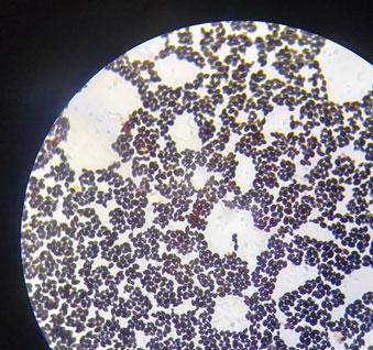
We recorded total of 47 patients’ data during the study.
Baseline counts: Hemoglobin level at presentation mostly was below 10 g/dL with (81.25%) with a mean of 8.738 g/dL. Three patients had hemoglobin level below 6 g/dL at presentation.
All the patients presented with a high leucocyte count as the mean value was 268.43 thousand/dL. 72.92% (n=35) of the patients having a count ranging from 1.5-3.5 x 10³ /dL.
Neutrophil with band form count was distributed maximally within 40-50% of total cells in 77.78% of male patients and 61.9% of female patients in this study with a cumulative total 70.83% patients (n=34). All the patients were found to have lymphocyte count under 10% among which a maximum of 19 patients (39.58%) were having lymphocyte count of 3%.
Platelet count was distributed ranging from 165 to
800 thousand/dL with a high mean count of 425.44 thousand/dL. Most of the patients have platelet count between 300-600 thousand per dL in 34 patients (70.83%).
Blood Counts after 3 Months of TKI Therapy :
Hemoglobin at 3rd month was placed mostly within the range of 8-12 g/dL with a total 76.6% patients (n=36) and mean hemoglobin increased to 10.513 g/ dL overall.
Leucocyte count after 3 months of TKI treatment ranged 6000-10000/dL for 53.19% of patients (n=25), although 14.89% patients (n=7) had a leucocyte count >12000/dL whereas 4.25% patients (n=2) had a leucocyte count <4000/dL.
Platelet count at 3rd month was mostly distributed across the range of 200-400 thousand/dL of patients whereas 17.02% patients (n=8) had platelet count below 150 thousand/dL.
Blood Counts after 6 Months of TKI Therapy :
Hemoglobin level at 6th month was placed mostly within the range of 12-14 g/dL with 72.34% of patients (n=34) and the mean value was 11.294 g/dL.
Total WBC count was found to be ranged between 4000-6000 cells/dL in maximum of 31 patients (65.95%). However, 4 patients (8.51%) had total WBC count below 4000 cells/dL among which 2 had a count <2000 cells/dL.
Platelet count was found with a range of 250-300 thousand/dL in 14 patients (29.78%). However, 11 patients (23.4%) were found to have platelet count below 150 thousand/dL at 6th month.
Comparison among Mean Counts over Baseline, 3rd and 6th Months:
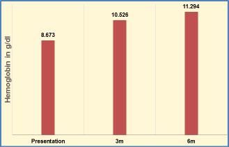
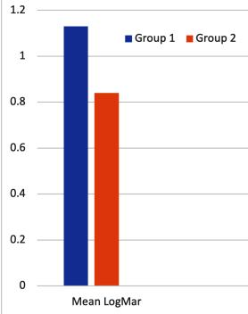
Mean hemoglobin had an increasing trend from a baseline mean of 8.738 g/dL to 10.513 d/dL at 3rd month, followed by 11.294 d/dL in the 6th month (Fig 1).
Fig 1 — Mean Hemoglobin in g/dL Trend
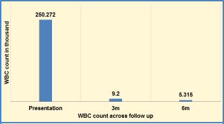
While extrapolating leucocyte count over 6 months of TKI therapy, average WBC count was found to be 268.678 thousand cells/dL in the baseline, which reduced to 9.2 thousand cells/dL in 3rd month, followed by 5.315 thousand cells/dL in 6th month (Fig 2).
Mean platelet count at the time of diagnosis recorded at 427.170 thousand/dL, which was down to 256.936 thousand/dL after 3 months of TKI, a slight reduction further after 6 months at 227.489 thousand/ dL (Fig 3).
Additionally, in comparative analysis of differential counts between 3rd and 6th month, it was found that neutrophil count was reduced from 64% at 3rd month to 58% in 6th month, whereas lymphocyte count was increased from 30% at 3rd month to 35% in 6th month. Eosinophil count increased from 3% at 3rd month to 5% in 6th month. And monocyte count was reduced from 3% at 3rd month to 2% at 6th month. Mean basophil count remained at 0% in both 3rd and 6th month data (Table 1).
Comparison between Imatinib versus Nilotinib :
On the basis of Blood Counts over baseline, after 3 months and after 6 months (Table 2).
DISCUSSION
This study was conducted to observe the hematological parameters in CML patients at baseline and at 3 and 6 months of receiving standard care as per international protocols at a Tertiary Care Hospital in Sub-Himalayan West Bengal, India. 41.67% of patients presented with a hemoglobin <8 g/dL and the mean hemoglobin at presentation remained 8.74 g/dL. Mean leucocyte count at presentation remained at 268.678 thousand/dL. In the differential leucocyte count most dominant type was neutrophil with band form with mean count of 42.73%. Basophil count requires particular mention as it ranged from 2% to 25% where the mean count was 9.42% which establishes the trend of higher basophil count in CML patients8. Platelet count was mostly on the higher side with mean being 425.44 thousand/dL.
After 3 months of TKI therapy, mean hemoglobin was raised to 10.513 g/dL. Total leukocyte count was reduced to a mean count of 9200/dL with only 14.89% patients having a count more than 11000/dL. None of the patients had blast cell in the peripheral smear. Mean platelet count was reduced to 256.94 thousand/ dL with only 17.02% patients having a count <150 thousand/dL. As we already know, TKI therapy does reduce cell count, but predisposes the patient to a cytopenia as early as 3 months, as for our study, it manifested as thrombocytopenia.
By 6 months of treatment, hemoglobin further increased to a mean of 11.294 g/dL. But what drew
Table 1 — Comparison of differential counts at 3rd and 6th month (n=47)
Table 2 — Imatinib versus Nilotinib on the basis of Blood Cell Counts
Fig 2 — Mean Leucocyte Count in thousand/dL Trend
Fig 3 — Mean Platelet Count in thousand/dL Trend
123, No 01, January 2025Journal
our attention was 2 patients with hemoglobin of 7.8 g/dL. Mean total leukocyte count further dropped to 5314.89/dL. Again, 8.51% patients had a count of <4000/dL. Similarly, mean platelet count dropped to 227.49 thousand/dL, and more importantly 23.4% patients experienced a platelet count below 150 thousand/dL. Although our patients did not have any hemorrhagic complication during the study, thrombocytopenia is quite usual and hemorrhagic manifestations have been recorded in the literature8
We treated our patients Imatinib (n=37) and Nilotinib (n=10) (as feasible as per hospital supply). It has been proven in literature that Nilotinib was more effective to bring the total leukocyte count to a normal range10 Mean leukocyte count at 3 months was 8350 /dL for Nilotinib compared to 9429.73 /dL for Imatinib. But when we compared leukocyte count on baseline, 3rd month and 6th month in between Imatinib and Nilotinib treated patients, this reduction in leukocyte count was statistically insignificant. What drew our attention was the fact of Imatinib having more propensity to cause cytopenia both for leukocyte and platelets. Even though mean leukocyte count was lower in Nilotinib treated patients, still there were 10.81% Imatinib treated patients who had leukocyte count less than 4000/dL compared to none for Nilotinib. Even after 6 months of TKI therapy, there were no patient with leukopenia in Nilotinib treated patients. Platelet count reduction was higher for Imatinib, and there were 18.92% patients with platelet count <150 thousand/ dL for Imatinib compared to 10% for Nilotinib at 3 months of TKI therapy and 24.32% for Imatinib compared to 20% for Nilotinib at the end of 6 months of TKI therapy which was reflected as a statistically significant data while comparing platelet counts on 6th month (paired 't' test, one sided p=0.009) as reduction in platelet count was higher in Imatinib treated patients. When we look for hemoglobin trends, it was increasing over 6 months. Again, for Nilotinib mean hemoglobin was higher at 10.6g/dL at 3 months, 12.07 g/dL at 6 months compared to 10.49 g/dL at 3 months and 11.08 g/dL at 6 months for Imatinib. Change in hemoglobin trend did not yield any statistical significance.
Although there are published literature comparing between Imatinib and Nilotinib elsewhere in the World, to best of our knowledge, there is no such study in Indian context and we believe our study is a first of its kind in this country for comparing these two TKIs10,11
CONCLUSION
With significant progress made over last 2 decades, CML remains arguably the blood cancer with
best prognosis. TKIs have revolutionized the CML treatment and it was quite evident. Most dramatic was the reduction of leukocyte count mostly to normal counts within 3 months. Hemoglobin level consistently increased and reached a normal population level by 6 months. What drew our attention was the cytopenia both for leukocytes and platelets. None of the patients had any cytopenia related complications.
The most interesting finding was found while comparing Imatinib to Nilotinib as first line therapy. Nilotinib treated patients reached a normal leukocyte count faster than Imatinib and they had higher increase in hemoglobin level as well. On the contrary, none of the Nilotinib treated patients had leukopenia even after 6 months of treatment compared to Imatinib where one tenth of the patients had leukopenia. Reduction in the platelet count was roughly similar both for Nilotinib and Imatinib but higher proportion of Imatinib treated patients reported a thrombocytopenia. There is a tendency of having cytopenia with TKI therapy and it remains the major concern globally flagging up to stop the treatment. A longer follow up and blood count monitoring is warranted to further address the issue in a wider prospect.
Despite having better clinical response, during the course of treatment we found that Nilotinib was less tolerated for about one fifth of the patients, mostly as restlessness, headache, palpitation, anxiety which are similar to the already reported publications11. Imatinib was way better tolerated with not a single adverse effect complained by the patients. Lack of adequate literatures, along with being more expensive hold back Nilotinib for wider uses as a first line therapy for CML.
There are very few studies Worldwide comparing Imatinib and Nilotinib as first line treatment modality. We hope this study highlighted key factors and would pioneer further larger and multi-centric studies as we progress to superior treatment options in the future.
Limitations :
(1)Smaller study population may have resulted hospital bias.
(2)This hospital serves a smaller geographic area which may not represent the overall epidemiology.
(3)This was a single centre study. Multicentre study should delineate the parameters better.
(4)Shorter follow up of only 6 months leaves us hankering after a way longer follow up, at least for years.
(5)Molecular analysis was not performed either at baseline or follow up.
Vol 123, No 01, January 2025Journal of the Indian Medical Association
(6)Mutational analysis was not performed before selection of TKI.
(7)Higher treatment expenses and limited hospital supplies
Conflict of Interest : None.
REFERENCES
1Smith A, Howell D, Patmore R, Jack A, Roman E — Incidence of haematological malignancy by sub-type: a report from the Haematological Malignancy Research Network. Br J Cancer [Internet] 2011 [cited 2022 Feb 11]; 105(11): 1684-92. Available from: https://www.ncbi.nlm.nih.gov/labs/pmc/articles/ PMC3242607/
2Pulte D, Jansen L, Brenner H — Changes in long term survival after diagnosis with common hematologic malignancies in the early 21st century. Blood Cancer J [Internet] 2020 [cited 2022 Feb 11]; 10(5): 56. Available from: https:// www.nature.com/articles/s41408-020-0323-4
3Bower H, Björkholm M, Dickman PW, Höglund M, Lambert PC, Andersson TM — Life expectancy of patients with chronic myeloid leukemia approaches the life expectancy of the general population. J Clin Oncol 2016; 34: 2851-7.
4Hochhaus A, Baccarani M, Silver RT, Schiffer C, Apperley JF, Cervantes F, et al — European LeukemiaNet 2020 recommendations for treating chronic myeloid leukemia. Leukemia 2020; 34: 966-84.
5Kantarjian HM, Hughes TP, Larson RA, Kim D-W, Issaragrisil S, le Coutre P, et al — Long-term outcomes with frontline nilotinib versus imatinib in newly diagnosed chronic myeloid leukemia in chronic phase: ENESTnd 10-year analysis. Leukemia [Internet] 2021 [cited 2022 Feb 12]; 35(2): 440-53. Available from: https://www.nature.com/articles/s41375-020-01111-2.
6Nakamae H, Fukuda T, Nakaseko C, Kanda Y, Ohmine K, Ono T, et al — Nilotinib vs. imatinib in Japanese patients with newly diagnosed chronic myeloid leukemia in chronic phase: longterm follow-up of the Japanese subgroup of the randomized ENESTnd trial. Int J Hematol [Internet] 2018; 107(3): 327-36. Available from: http://dx.doi.org/10.1007/s12185-017-2353-7.
7Saglio G, Kim DW, Issaragrisil S, Le Coutre P, Etienne G, Lobo C, et al — Nilotinib versus imatinib for newly diagnosed chronic myeloid leukemia. New England Journal of Medicine 2010; 362(24): 2251-9.
9Song KW, Rifkind J, Al-Beirouti B, Yee K, McCrae J, Messner HA, et al — Subdural hematomas during CML therapy with imatinib mesylate. Leukemia & lymphoma. 2004 Aug 1;45(8):1633-6.
10Saglio G, Kim DW, Issaragrisil S, Le Coutre P, Etienne G, Lobo C, et al — Nilotinib versus imatinib for newly diagnosed chronic myeloid leukemia. New England Journal of Medicine 2010; 362(24): 2251-9.
11Larson RA, Hochhaus A, Hughes TP, Clark RE, Etienne G, Kim DW, et al — Nilotinib vs imatinib in patients with newly diagnosed Philadelphia chromosome-positive chronic myeloid leukemia in chronic phase: ENESTnd 3-year follow-up. Leukemia 2012; 26(10): 2197-203.
Original Article
Impact of Revised Basic Course Workshop on Medical Educators : An Expedition of NMC Mandate to Formal Praxis !!!
Jarina Begum1, Syed Irfan Ali2, D Lakshmi Lalitha3
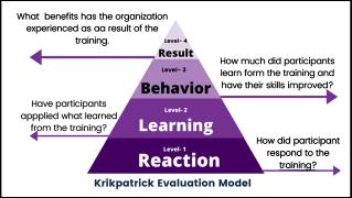
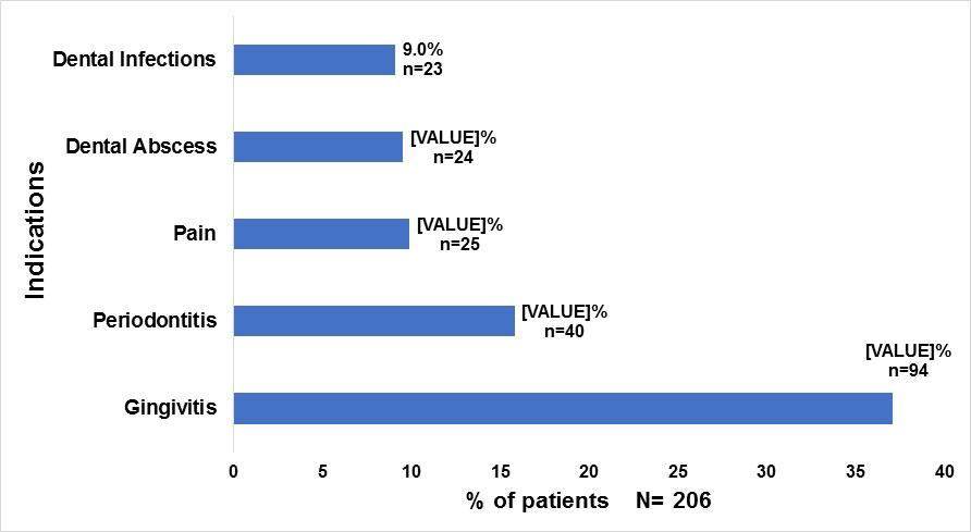
Background : Kirkpatrick’s model is one of the suitable methods for assessing educational programs at different levels. The present study is planned to evaluate the impact of the Revised Basic Course Workshop (rBCW) on medical educators.
Aims and Objectives : (1) To evaluate the effects of rBCW on response, knowledge, and self-perceived behavior of faculties as per Kirkpatrick’s program evaluation model. (2) To identify the perceived challenges and suggest solutions to bridge the gap.
Materials and Methods : An educational intervention was carried out among 28 faculties through the Faculty Development Program followed by a structured follow-up questionnaire. The data was analyzed in terms of percentage, proportions, paired t-test and thematic analysis.
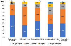
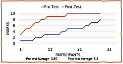
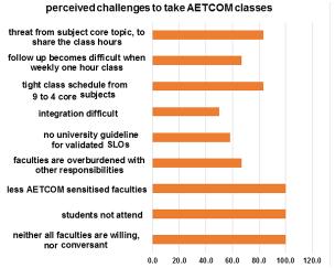
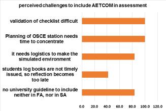
Result : The majority (71.4%) were male with a mean age of 36.21 years. All faculties were satisfied, 86.4% either agreed or strongly agreed with quality. Thematic analysis of reflections by participants highlighted a few important aspects of medical education system in terms of themes & subthemes. The increase in knowledge was evaluated by pre and post-test, which was found to be statistically significant (P<0.05). The behavior change was perceived positively by the participants. A few challenges were encountered like pandemic effects, lack of motivation & co-ordination, mismatched resources for which the suggested solutions were refresher training, more aligned resources, etc.
Conclusion : All the faculties were satisfied with an increased knowledge & positive change in behavior after rBCW. Yet, it was perceived as inadequate in terms of various challenges during implementation which necessitated the need for the implementation of suggested solutions.
[J Indian Med Assoc 2025; 123(1): 29-33]
Key words :Revised Basic Course Workshop, Kirkpatrick's Programme evaluation, Faculty Development Programme, Feedback, Competency-based Medical Education, National Medical Commission, Indian Medical Graduate.
Faculty Development Programme (FDP) is a focused term that covers a range of activities designed to improve student learning and to help faculty improve their competence as teachers (Eble & McKeachie, 1985)1.
FDPs are an important aspect of medical education and in the efficient delivery of medical curriculum, however, it has been subjected to major changes recently in the context of the new Competency-based Medical Education (CBME) curriculum. There is a large gap between the demand for medical education training and the supply of resources especially trained faculties in South East Asia regions, the regulatory medical councils thus recommend FDP to enhance the quality of medical education2
Department of Community Medicine, Manipal Tata Medical College, Jamshedpur, Jharkhand 831017
1MD (Community Medicine), Professor and Head, FAIMER Fellow, Faculty and Corresponding Author
2MD (Community Medicine) Professor
3 MD (Biochemistry), Professor & Dean, Department of Biochemistry, Great Eastern Medical School & Hospital, Srikakulam, Andhra Pradesh 532484
Received on : 11/12/2023
Accepted on : 04/01/2024
Editor's Comment :
rBCW is mandatory as per NMC. Although it is evaluated, it does not go beyond level 2 of Kirkpatrick's Programme evaluation model. For successful implementation and effective outcome, we need to go beyond level 2 in terms of self, peer assessment, and an independent system to observe the behavior change in the long run through an observational checklist to bring the intended outcome and create the impact through rBCW in the medical education system.
India has adopted outcome-based medical education and has rolled out CBME from the year 2019 with a goal of Indian Medical Graduates (IMG) as the output3
FDP for medical teachers started long back as the National Teacher’s Training Centre (NTTC), however, the 1997 regulations and further 2009 GMER guidelines made medical education Units/ Departments in Colleges and Medical Education Training for each faculty a mandate. This in turn necessitated the establishment of various regional, nodal centres along with Advanced Courses in Medical Education (ACME), Fellowship in Foundation
Vol 123, No 01, January 2025Journal
for Advancement of International Medical Education and Research (FAIMER) courses. Although the CBME curriculum has provided all the competencies, there was a need to prepare the faculties for the sudden change in the medical education system4,5.
The basic level faculty training was named Basic Medical Education Technology Workshop (BMET) which had a three-day intensive training format. Later this was renamed as a revised Basic Medical Education Technology Workshop (rBMET) with a revised curriculum aligned to CBME. In 2014, MCI introduced an ACME course delivered through regional and nodal centers. Later from 2019 onwards Curriculum Implementation Support Program Workshops (CISPWs) were rolled out to orient the faculties to the changes in the curriculum. Recently in 2023, the rBMET became BCME, with various reforms. Faculty Development Program planning and implementation is crucial and critical at the same time. A systematic approach can help, however, a longitudinal program, instead of a cross-sectional bolus will be more impactful. Direct observation of teaching and giving feedback will help in the true acquisition of teaching competencies and the faculty can appreciate their progress from novice level to expert level in implementing different components of CBME6-8
Kirkpatrick’s model is one of the suitable methods for assessing educational programs such as a Faculty Development Program at four levels- response, learning, change in behavior & impact of training. It provides one technique for appraisal of the evidence for any reported training program and could be used to evaluate whether a training program is likely to meet the needs and requirements of both the organization implementing the training and the staff who will participate9
The present study is planned to evaluate the impact of FDP ie, Revised Basic Course Workshop on Medical educators.
AIMS AND OBJECTIVES
(1) To evaluate the effects of rBCW on response, knowledge, and self-perceived behavior of faculties as per Kirkpatrick’s program evaluation model.
(2) To identify the perceived challenges and suggest solutions to bridge the gap.
MATERIALS AND METHODS
An educational Intervention was carried out in a Tertiary Health Care Institution for a 6-month duration, among a total of 28 participants who were faculties of various Departments enrolled in rBCW and gave their consent. The study duration was 6 months.
Ethical approval was taken from the Institutional Ethical Committee (20/IEC/GEMS&H/2021). The data was collected through a pre-designed, pre-validated questionnaire which included a pre-post-test, and a feedback survey, along with the reflections by participants’ postworkshop. A follow-up questionnaire was circulated online after 6 months of the workshop to analyze the self-perceived change of behavior & to identify the challenges along with solutions to address them. The follow-up questionnaire consisted of demographic details, practice of concepts learned during rBCW with evidence in the last 6 months, change in teaching/learning style & open-ended questions for challenges and possible solutions.
The pre & post test, feedback & follow-up questionnaire were prepared and validated by Medical Education Experts. After getting ethical approval, the faculties were approached for rBCW enrolment. Out of 30, only 28 showed up and attended both the sessions (morning & afternoon) on all the 3 days of the workshop, thus included in the study. To measure the learning pre-test was conducted at the beginning and a post-test at the end of the workshop to evaluate the improvement in knowledge. Likewise, a feedback survey form was circulated among all the participants to measure their perception of the program. The participants were then instructed to write up their reflections in brief based on Rolfe’s reflective model and submit them within 2 days via WhatsApp in Microsoft Word format. After 6 months of the workshop, a follow-up online questionnaire was shared with them to fill along with a request to attach the evidence wherever necessary.
The quantitative data thus collected was summarised in the form of Percentage, Mean and Standard Deviation. Students paired t-test was used to assess the difference between the mean knowledge scores of the participants before and after the intervention. Thematic analysis was done for the qualitative data obtained from the feedback survey, reflections & open-ended questions in the follow-up questionnaire.
RESULTS
Out of all participants 71.4% were male. The mean age of the study population was 36.21 years. The majority (78%) belonged to clinical subjects followed by para-clinical & pre-clinical subject faculties. Likewise majority (57.14%) were assistant professors followed by Associate Professors and Professors.
The FDP (RBCW) was evaluated at various levels based on Kirkpatrick’s program evaluation model (Fig 1). Level-1: reaction through a feedback survey,
level-2: learning through a pre & post-test analysis, level-3 (partially): behavior change through a followup questionnaire and level-4: Impact which was not included in the current research.
Reaction to the program was evaluated through feedback analysis of the participants at the end of rBCW along with the reflective writings of participants. All the faculties were satisfied with the Workshop and 86.4% perceived it useful in the context of the current Medical Education System. The majority either agreed or strongly agreed with the appropriateness of content (92%) of the workshop, adequacy of pace (86%), appropriate presentation style (82%), adequacy of time (64%), and use of interactive T/L methods (79%) during the sessions of the workshop (Fig 2).
The qualitative data obtained from the feedback survey summed up with a few verbatim by the participants which are mentioned below:
•Needed more time for the AETCOM session, it was not sufficient.
•Need revision of the given schedule for rBCW as per the need!
•Inclusion of more concepts and more breaks in between sessions.
•Handouts or pre-reading materials could be made available.
•A shorter duration of sessions and more group activities are required.
The learning of the participants was measured using a pre-test, before and post-test, after the intervention consisting of 10 MCQs based on the content of the workshop. Thus, the knowledge gain was evaluated by comparing pre and post-test scores through paired t-tests, which was found to be statistically significant (P<0.05)(Fig 3).
The change in behavior in level 3 was measured partially through a self-perceived online follow-up questionnaire through a Google survey form, with the provision of the required evidence to be uploaded, wherever needed. This was a simple and easy way to keep a check on the progress of the participants by self-observation. However, complete evaluation at level 3 was a difficult task in terms of the unavailability of a uniform observational checklist for the same along with possible subjective bias of the observers. Analysis of the responses to the follow-up questionnaire showed a positive self-perceived change in behavior among the participants in terms of the practice of the concepts learned during FDP in real-time (Table 1).
The qualitative data received from open-ended questions in the follow-up questionnaire was analyzed by thematic analysis. A few important themes emerged under challenges encountered while practicing concepts taught during rBCW and possible solutions to overcome them (Fig 4).
The thematic analysis of the reflections by the study participants unveiled a few pertinent themes and sub-themes under each category of what happened, so what & what next related to rBCW (Table 2)
DISCUSSION
The current study observed that all of the participants were satisfied with the workshop & perceived it useful in the context of the current Medical Education System. Likewise, another study by Heydari, et al concluded that the workshop on new teaching
Fig 1 — Kirkpatrick Evaluation model
Fig 2 — Level-1 Reaction: Feedback of the participants towards the FDP (RBCW)
Fig
Table 1 — Self-Perceived Practice of Concepts Learned in RBCW (N=28) Practice
and learning methods significantly improved the satisfaction, knowledge and behavior of healthcare staff10.
The learning of the participants after rBCW showed significant improvement in terms of increased test scores and increased awareness towards various terminologies, tools & technology related to CBME for producing a competent Indian Medical Graduate. However current study emphasized self-perceived behavior change in terms of an online follow-up questionnaire with the provision of evidence, wherever needed, through self-observation. Similar studies like in Dorri et al. it was found that CPR in-service training had a favorable effect on all four levels of the Kirkpatrick model for nurses and nurse's aides. However, they found behavior levels more challenging and sensitive attributed to the right opportunity to change behaviors, the unpredictability of required time for a change of behavior, the appropriate working environment for any change, etc11
The present study aimed to evaluate the effectiveness of a training program following
Kirkpatrick's four-level model, however evaluation of the 4th level was not attempted for various reasons like time constraints, limited resources and lack of standardized outcome indicators. Similarly, studies have highlighted the challenges of evaluation of a training program at 4th level. It was concluded that creating long-term positive changes requires repetition and the utilization of multiple educational strategies through a longitudinal study12.
The present study highlighted challenges to the implementation of CBME concepts after the rBCW such as pandemic effects, unawareness, lack of motivation among faculties, mismatched resources, lack of coordination between various departments, improper planning & execution & unavailability of any existing system for reward or reinforcement, lost to follow up of faculties pertaining change of institution, etc. Likewise, solutions offered were refresher training every year, student involvement in planning, more aligned resources (4M) as per demand, interdepartmental collaborations’, strategies for regular follow-ups, ensuring quality, increasing accountability, Effective feedback system. Similar challenges were encountered by medical educators for implementation of CBME which were suggested to be tackled by incremental changes to the old curriculum rather than the overhaul revision, more faculty input, appropriate pace of training & adequate time for preparation13
The immediate impact of RBCW was good in enhancing the knowledge of participants; however, periodic vigilance and frequent refresher training are required to ensure proper implementation of the fundamentals of rBCW by trained teachers14
Table 2 — Thematic Analysis of Reflections by Participants
CategoriesThemes Sub Themes
What Happened?TeamworkGroup activities Sharing opinions
Adult learning.CBME curriculum highlights Clarity on new T/L & assessment methods Interaction
So What? KnowledgeConfidence Communication Support
It might be premature to add, but this may be an appropriate time to create a Resource Centre for Faculty Development which might function as a focal center for educational innovations and reforms. Medical Council of India, now the National Medical Commission as the regulator of Medical Education in India, has taken this unique step to add science to the art of teaching. Although it’s worth a try to restructure Medical Education, its relevance in the long term as an art should not be undermined through micromanagement. Moreover, an established system must be there to evaluate change at all levels and apprise accordingly. Initial results of FDPs are encouraging and so we must continue our efforts, particularly for the Colleges that are newly established. Motivated faculties
Vol 123, No 01, January 2025Journal
needed routine improvisation in FDP curriculum as per the advancing technology and current trends in medical education. However, the frequent changes may create confusion among faculties. A longer period might be needed to see the impact of FDPs on medical education and thereby better health status of the population15
CONCLUSION
All the faculties were satisfied. An increase in knowledge & positive behavior change was observed after the Revised Basic Course Workshop. Yet, it was perceived as inadequate in terms of various challenges during implementation which necessitated the need for the suggested solutions.
Recommendations :
education-regulations-1997. Accessed on 2019 May 25.
(1)Availability of a standardized observational checklist for evaluating behavior change.
(2)Effective system for 360-degree feedback by multiple sources.
(3)Establishing learning & developmental portfolios for faculties.
(4)Provision of flexibility to both trainers & trainees as per the need and context.
(5)Establishment of the continuum of support to each medical educator.
Limitations :
A small sample size was selected and it was not possible to measure the 4th-level results as per Kirkpatrick’s model of Programme evaluation attributed to time constraints.
REFERENCES
1Lotti A (nd) — A brief history of Faculty Development for Medical Education in United States. Breve storia del Faculty Development in Medicina negli Stati Uniti. Unito.It. Retrieved May 27, 2023, from https://www.ojs.unito.it/index.php/tutor/article/ download/5252/4719/
2Tenzin K, Dorji T, Choeda T — Impact of faculty development programme on self-efficacy, competency and attitude towards medical education in Bhutan: a mixed-methods study. BMC Med Educ 2019; 19: 468. https://doi.org/10.1186/s12909-0191904-4.
3Medical Council of India — Competency Based Undergraduate Curriculum. Medical Council of India; 2018. Available at: https://www.mciindia org/CMS/information-desk/for-colleges/ ug-curriculum. Accessed on 2023 May 29.
4Zodpey S, Sharma A, Zahiruddin QS, Gaidhane A, Shrikhande S — Faculty development programs for medical teachers in India. J Adv Med Educ Prof 2016; 4(2): 97-101.
5Medical Council of India — Regulations on Graduate Medical Education, 1997; May, 2018. Available at:https:// www.mciindia.org/CMS/rules-regulations/graduate-medical-
7Medical Council of India. Curriculum Implementation Support Program of the Competency Based Undergraduate Medical Education Curriculum. New Delhi: Medical Council of India. 2019.
8Nagaraja MS, ML RD — Faculty development programs for implementing competency based medical education in India: challenges and opportunities. International Journal Of Community Medicine And Public Health 2021; 8(6): 3163-66. https:/ /doi.org/10.18203/2394-6040.ijcmph20212028.
9Balandin SA, Sigafoos S, Reed J, Vicki — The Kirkpatrick model: A useful tool for evaluating training outcomes. J Intellectual Develop Disability 2009; 34: 266-74.
10Heydari MR, Taghva F, Amini M, Delavari S — Using Kirkpatrick's model to measure the effect of a new teaching and learning methods workshop for health care staff. BMC Research Notes 2019; 12(1): 388. https://doi.org/10.1186/ s13104-019-4421-y.
11Dorri S, Akbari M, Dorri Sedeh M — Kirkpatrick evaluation model for in-service training on cardiopulmonary resuscitation. Iranian Journal of Nursing and Midwifery Research 2016; 21(5): 493-7. https://doi.org/10.4103/1735-9066.193396.
12Momennasab M, Mohammadi F, DehghanRad F, Jaberi A — Evaluation of the effectiveness of a training programme for nurses regarding augmentative and alternative communication with intubated patients using Kirkpatrick's model: A pilot study. Nursing Open 2023; 10(5): 2895-903. https://doi.org/ 10.1002/nop2.1531.
13Ramanathan R, Shanmugam J, Gopalakrishnan SM, Palanisamy KT, Narayanan S — Challenges in the implementation of competency-based medical curriculum: Perspectives of prospective academicians. Cureus [Internet] 2022; 14(12): e32838. Available from: http://dx.doi.org/10.7759/ cureus.32838.
14Jankhwala MS, Nagar S, Thomas E, Mahajan S — Assessment of Impact of Revised Basic Course Workshop (RBCW) in Medical Education Technology on the Trained Teachers at a Tertiary Care Teaching Hospital. Journal of Research in Medical Education & Ethics 2018; 8(Special Issue): S94-S100.
15How KKHS — National faculty development program [Internet]. Org.in. [cited 2023 Sep 2]. Available from: https:// www.nmc.org.in/wp-content/uploads/2020/09/FDP-initiativeUG-Med-Education.pdf.
Fig 4 — Challenges & Solutions to practice CBME concepts learned in rBCW
Original Article
Assessment of Effect of Nicotine on Severity of Diabetic Retinopathy
Background : Diabetic Retinopathy (DR) is a microvascular complication of Diabetes, caused by high blood sugar levels that damages the vascular endothelial pericytes. DR is one of the most frequent causes of new cases of blindness among adults. Nicotine is a known risk factor for development of Diabetes and progression of DR. The aim of this study is to find the association of Nicotine with severity of DR.
Materials and Methods : 100 type 2 diabetic patients, between 20 and 75 years of age, visiting the Department of Ophthalmology from July, 2021 to December, 2022, were divided into two Groups : Group 1 included Diabetics consuming nicotine (>2 years) and Group 2 had Diabetics without Nicotine addiction. Best corrected visual acuity was recorded followed by dilated fundus examination to look for DR. Visual acuity and severity of DR were compared in two groups.
Results : The mean LogMar visual acuity in Group 1 was 1.13 and 0.84 in Group 2 (p<0.05). DR changes were found to be more severe in Group 1 as compared to Group 2 (p= 0.031). Proliferative DR was more common in nicotine users as compared to non-users (p=0.028). Increased duration of nicotine use resulted more severe DR (p= 0.0289).
Conclusion : Nicotine has a deleterious effect on visual acuity and it causes more severe DR. It is recommended that physicians must counsel diabetic patients regarding abstinence from Nicotine besides doing routine Diabetes counselling.
Key words :Diabetes, Nicotine, Diabetic Retinopathy.
Diabetes is a chronic illness characterized by elevated blood glucose levels, accompanied by disturbed metabolism of fats and proteins. Over time, a persistently high blood sugar level can damage pericytes of these blood vessels. Diabetic Retinopathy (DR) is a microvascular complication of Diabetes, caused by high blood sugar levels damaging the Retina. With uncontrolled Diabetes, it can cause blindness if left undiagnosed or untreated1
Diabetic Retinopathy is one of the frequent cause of new cases of blindness among adults aged 20-74 years2. There are many risk factors associated with Diabetes Mellitus (DM) and development of DR such as quality of diabetic control, duration of disease, age of onset and age of patient3 Diabetic retinopathy progresses from mild non-proliferative abnormalities, characterized by increased vascular permeability, to moderate and severe Non-proliferative Diabetic Retinopathy (NPDR), characterized by vascular closure, to Proliferative Diabetic Retinopathy (PDR),
Department of Ophthalmology, Sri Aurobindo Institute of Medical Sciences, Indore, Madhya Pradesh 453555
1MS, Senior Resident
2MS, Associate Professor
3MBBS, Resident
4MS, Assistant Professor
5 MS, DNB, Fellowship Vitreoretina, Professor and Corresponding Author
Received on : 03/07/2023
Accepted on : 08/09/2024
[J Indian Med Assoc 2025; 123(1): 34-8]
Editor's Comment :
“India has an estimate of 77 million cases of diabetics with an additional 25 million cases being pre-diabetic.”
Diabetes mellitus can lead to various sight threatening complications affecting quality of life.
There are many risk factors which are modifiable and evaluating them may help live a better life.
With a rising trend in consumption of tobacco, it is important to understand that nicotine has a deleterious effect on vision as it worsens diabetic retinopathy and may even lead to blindness.
We as health care providers must counsel every patient and serve for better mankind.
characterized by the growth of new blood vessels on the Retina and posterior surface of the vitreous, haemorrhages and tractional retinal detachment. Macular oedema, characterized by retinal thickening from leaky new blood vessels, can develop at all stages of Retinopathy4
Nicotine consumption is also a very important risk factor associated with various systemic illnesses including Diabetes. Nicotine is a dangerous and highly addictive chemical. It can cause narrowing of the arteries and hardening of the arterial walls, which in turn, decreases the blood flow 5 . Tobacco Optic Neuropathy is another rare disorder of optic nerve function related to the toxic effects of an unidentified constituent of Tobacco6
Generally, awareness of the association between smoking and cancer, cardiovascular diseases and respiratory diseases is higher7 A study concluded
123, No 01, January 2025Journal
that close to two thirds (64%) of Diabetic patients reported that smoking will not affect the disease and only 10% reported that smoking causes a lot of aggravation of Diabetes8. This shows the lack of awareness regarding nicotine and its side effects and its association with diabetes among general population including many health professionals.
The aim of this study is to find the association of nicotine use with severity of Diabetic Retinopathy. To assess this, patients with history of diabetes were selected and analysed for visual acuity and grade of Diabetic Retinopathy and compared with their Nicotine usage.
This study was prompted by the need of evaluating such individuals (both Diabetics and Nicotine users) clinically, in a developing nation like India where the prevalence of Nicotine consumption and Diabetes is high but the resources as well as awareness is quite low so as to assess the risk factors regarding development of Diabetic Retinopathy and thereby counselling the patient to refrain from Nicotine consumption.
MATERIALS AND METHODS
The present study was conducted after approval from the Institutional Ethical Committee (IEC NO: SAIMS/IEC/2021/189) and in adherence to the tenets of declaration of Helsinki. It is an observational study of 100 cases which was conducted over a period of 1.5 years starting from July, 2021 to December, 2022.
A clinical history was obtained from patients presenting to the Ophthalmology out-patient department at a Tertiary Care Centre with regard to their status of DM and Nicotine use. Patients with age between 20-75 years with known history of Diabetes Mellitus type 2 with and without history of nicotine consumption were included in the study. Patients were selected on the basis of history of duration of type 2 diabetes of 5-10 years with random blood sugar less than 150 mg/dl, only patients with HbA1c less than 8 gm% were included confirmed by respective blood tests. Exclusion criteria consisted of patients with any systemic co-morbidity other than diabetes mellitus type 2, patients on insulin therapy, patients with any other ocular co-morbidity apart from Diabetic Retinopathy and patients who had already quit nicotine. Total 100 patients were selected which were divided into two groups. Group 1 consisted of 50 patients with additional history of Nicotine consumption of at least 2 years. The second group included 50 diabetic patients with no history of consumption or exposure to nicotine. Nicotine users were considered to be patients with any habitual use
of tobacco plant leaf and form of nicotine was products for a minimum of two years. Commonly used form of nicotine was smoke inhalation of cigarettes, bidi and cigars, as well as consumed in smokeless way as in products that were either sniffed or chewed.
Best corrected visual acuity was assessed using Snellen’s alphabet chart (converted into LogMar values for the purpose of statistical analysis) and preliminary torch light examination was done followed by detailed anterior segment examination using slit lamp bio microscopy and detailed fundus examination by indirect Ophthalmoscope.
On indirect Ophthalmoscopy clinical features of diabetic patients were noted and classified for objective diagnosis by Early Treatment Diabetic Retinopathy System (ETDRS) Classification9
Data thus obtained was subjected to statistical analysis, chi square test and p value less than 0.05 was considered significant.
OBSERVATIONS AND RESULTS
Our study included patients lying in age group 2074 years. We found that Group 1 consisted of Diabetic patients who were nicotine consumers and had significantly higher number of Males (76%) than Females (24%) while vice versa in group 2 with 60% female preponderance.
Table 1 shows the distribution of grades of diabetic retinopathy in Group 1 and Group 2. Changes of diabetic retinopathy were present in both groups with a significantly higher number of proliferative Diabetic Retinopathy in nicotine users (85.7 %) as compared to non- users (Figs 1-3).
Visual acuity measured was converted to LogMar values for the purpose of statistical analysis. Mean LogMar value for visual acuity in Group 1 was 1.13 and in Group 2 was 0.84. The difference of visual acuity was significant in both Groups. The visual acuity was significantly lower in Group 1.
Table 2 compares the visual acuity with fundus changes in the nicotine users. There were no Diabetic Retinopathy changes in fundus of nicotine users with visual acuity between 6/6 and 6/12. There was significant deterioration of vision as the fundus changes progressed. Thirty percent of nicotine users with history of diabetes having no signs of clinical
diabetic retinopathy had visual acuity 6/6-6/12. Sixteen percent of Group 1 patients had visual acuity between 6/12- 6/60, out of these 16%, 25% had mild NPDR changes, while 6.25% each belonged to patients with no changes, moderate NPDR, severe NPDR and PDR changes. Twenty two percent of Group 1 patients had visual acuity less than 6/60-1/60 among which 27.27% patients had mild NPDR changes, 36.36 % had moderate NPDR changes while severe NPDR and PDR changes were 18.18% each. Total 32% of
patients in Group 1 had visual acuity less than 1/60. 18.75 % of patients with visual acuity less than 1/60 had Moderate NPDR changes while 25 % and 56.25% of patients had severe NPDR and PDR respectively. It is important to note that 75% of patients having PDR changes in Group 1 had visual acuity less than 1/60 (Fig 5).
6/6-6/1215----15 (30%)
<6/12- 6/60141118 (16%)
<6/60- 1/60-342211 (22%)
<1/60--34916 (32%)
Total16 (32%)7 (14%)8(16%)7 (14%)12 (24%)50(100%)
Table 3 compares the fundus changes with respect to duration of Nicotine use. There is significant progression of Diabetic Retinopathy with increase in duration of nicotine use. The proliferative changes in Diabetic Retinopathy increases as duration of Nicotine consumption increases. However, there is no association of duration
Fig 1 — Showing Moderate to severe Non-proliferative Diabetic Retinopathy Changes
Table 2 — Visual Acuity with respect to Fundus changes of Nicotine users
Fig 3 — Shows majority of Proliferative Diabetic Retinopathy Patients belonged to Group 1.
Fig 4 — Shows significant mean LogMar value to be higher in Group 1 signifying deterioration of visual acuity in Group 1
Table 3 — Fundus changes with respect to duration of nicotine use
Duration ofNoneMildModerateSeverePDRTotalp value
Nicotine useNPDRNPDRNPDR (years)
<1010-42-16 (32%)
11-20231118 (16%)
21-30121228 (16%)0.014
31-403121310 (20%)
41-50-1-168 (16%)
Total16 (32%)7 (14%)8(16%)7(14%)12 (24%)50 (100%
of Nicotine use in patients with no Diabetic Retinopathy changes. The severity of Diabetic Retinopathy increases as the duration of nicotine use increases (p value <0.05).
DISCUSSION
In India, there are estimated 77 million people above the age of 18 years who are suffering from diabetes (type 2) and nearly 25 million are prediabetics8 Diabetic Retinopathy is an important cause of blindness and occurs as a result of longterm cumulative damage to the small blood vessels in the Retina. Apart from good control over modifiable risk factors of Diabetes, there are various nonmodifiable risk factors responsible for the progression of Diabetic Retinopathy like duration, age of onset and current age of patient 8 The present study correlated group 1 and 2 on the basis of duration of diabetes and HbA1c less than 8 gm%, thereby removing such confounding factors.
Nicotine has long known to be a major risk factor for many systemic co-morbidities viz cardio vascular diseases, cerebro-vascular accidents, among which is severity of diabetic retinopathy. Our study had similar observations, 87.5% of patients with proliferative Diabetic Retinopathy were Nicotine users.
According to Global Adult Tobacco Survey (GATS) 2016- 17, the prevalence of smoking tobacco use was 10.38% and smokeless tobacco use was 21.38% in India. Of all adults, 28.6% currently consume tobacco either in smoke or smokeless form, including 42.4% of men and 14.2% of women9. Similarly, in our study we found male pre-ponderance in Group 1 with nicotine users.
In Stockholm diabetes intervention study, smoking habits were correlated with the progression of Retinopathy and the total number of complications, deteriorating overall health of diabetic smokers during the 5-years study period. The patients who smoked had a three-fold increase of the risk of progression of retinopathy compared with the non-smokers, independent of blood glucose control10. Similarly, the present study suggests clinical changes of proliferative Diabetic Retinopathy to be more in nicotine users as compared to Diabetics. These findings also support the pathophysiological changes of Nicotine on Retina.
Nicotine has vasoconstrictive effect which is mediated through sympathetic activation which in turn reduces retinal blood flow and the ability of retinal vessels to autoregulate to hyperoxia. Smoking reduces oxygen carrying capacity of blood which when associated with decreased retinal blood flow leads to hypoxia. Hypoxia is major factor contributing to worsening of Diabetic Retinopathy 11 On the contrary, study by Yanagi, et al conclude that smoking is associated with wider retinal calibre in Japanese women, which may be vasodilation in response to long term hypoxia, however these changes are reversible on long term cessation of smoking12 Author could find limited evidence of reversibility of parafoveal microvasculature changes after nicotine abstinence, so, it is highly recommended area for further research considering the prevalence of Nicotine consumption in India.
Another effect of Nicotine consumption is Toxic Optic Neuropathy. In our study we found no cases with such changes in the fundus of patients clinically, though imaging or Electrophysiological tests can be of help to diagnose sub clinical cases13
The effect of Nicotine on visual acuity and severity of Diabetic Retinopathy is found to be significant (p<0.05). India being a developing nation has high prevalence of both Diabetics and Nicotine users. As per National Family Health Survey (NFHS-5) the Tobacco users in India are more in working group population. The visual impairment caused by the
Fig 5 — Shows distribution of visual acuity among patients with proliferative diabetic retinopathy in Group 1
Vol 123, No 01, January 2025Journal of the Indian Medical Association
combined effect of diabetes and nicotine poses socioeconomical burden on the society due to loss of jobs and deficit in manpower. Meta-analysis by Pan, et al has contributed to the growing evidence that both active and passive smoking are significant modifiable factors for risk of type 2 diabetes14 Prevention is always a better strategy than to cure the illness. Although risk of continued damage due to diabetes remains high in the short-term after smoking cessation, it decreases substantially among abstinent in the long run15
The limitation of the study is small sample size and non-quantified Nicotine usage and no use of OCT and OCT-A was done.
CONCLUSION
Nicotine has a deleterious effect on visual acuity and it causes more severe DR. Due to high burden of Diabetics in India as well as high prevalence of nicotine consumption especially among young population, it is recommended that physicians must counsel Diabetic patients regarding abstinence from Nicotine besides doing routine Diabetes counselling.
REFERENCES
1Fong DS, Aiello L, Gardner TW, King GL, Blankenship G, Cavallerano JD, Ferris FL, et al — Retinopathy in diabetes. Diabetes Care 2004; 27(suppl 1): 84-7.
2Joussen AM, Poulaki V, Le ML, Koizumi K, Esser C, Janicki H, et al — A central role for inflammation in the pathogenesis of diabetic retinopathy. The FASEB Journal 2004; 18(12): 1450-2.
3Patel V, Rassam S, Newsom R, Wiek J, Kohner E — Retinal blood flow in diabetic retinopathy. British Medical Journal 1992; 305(6855): 678-83.
4Hammes HP — Pericytes and the pathogenesis of diabetic retinopathy. Hormone and Metabolic Research 2005; 37(S 1): 39-43.
5Morgado PB, Chen HC, Patel V, Herbert L, Kohner EM — The acute effect of smoking on retinal blood flow in subjects with and without diabetes. Ophthalmology 1994; 101(7): 12206.
6Nentwich MM, Ulbig MW — Diabetic retinopathy-ocular complications of diabetes mellitus. World Journal of Diabetes 2015; 6(3): 489.
7Pokhrel BR, Thankappan KR, Mini GK, Sarma PS — Tobacco use among health professionals and their role in tobacco cessation in Nepal. Prev Control 2006; 2: 117-25. 10.1016/ j.precon.2007.03.003.
8Thankappan KR, Thresia CU — Tobacco use and social status in Kerala. Indian J Med Res 2007; 126: 300-8.
9Bowling B — (2015). Kanski’s clinical ophthalmology (8th ed.). W B Saunders.
10Mumbai and Ministry of Health and Family Welfare, Government of India, World Health Organization, Centers for Disease Control and Prevention, Tata Institute of Social Sciences GATS 2: Global Adult Tobacco Survey -India 2016-17
11Omae T, Nagaoka T, Yoshida A — Effects of habitual cigarette smoking on retinal circulation in patients with type 2 diabetes. Investigative Ophthalmology & Visual Science 2016; 57(3): 1345-51.
12Yanagi M, Misumi M, Kawasaki R, Takahashi I, Itakura K, Fujiwara S, et al — Is the association between smoking and the retinal venular diameter reversible following smoking cessation? Invest Ophthalmol Vis Sci 2014; 55(1): 405-11. doi: 10.1167/iovs.13-12512. PMID: 24302587.
13Reichard P, Britz A, Cars I, Nilsson BY, Sobocinsky-Olsson B, Rosenqvist U — The Stockholm Diabetes Intervention Study (SDIS): 18 months’ results. Acta Med Scand 1988; 224(2): 115-22. doi: 10.1111/j.0954-6820.1988.tb16748.
14Pan A, Wang Y, Talaei M, Hu FB, Wu T — Relation of active, passive, and quitting smoking with incident type 2 diabetes: a systematic review and meta-analysis. Lancet Diabetes Endocrinol 2015; 3(12): 958-67. doi: 10.1016/S22138587(15)00316-2. Epub 2015 Sep 18.
15Bhatia G — Tobacco Endgame: Can India Share the Dream? Indian J Psychol Med 2023; 45(2): 189-92. doi: 10.1177/ 02537176221139144. Epub 2022 Dec 12.
Original Article
Serum Procalcitonin and CRP Level as Severity Marker of Dengue Fever : An Observational Study in Medical College, Kolkata
Background : Among different markers of inflammation and sepsis, Procalcitonin (PCT) and C-reactive Protein (CRP) are being studied to investigate their accuracy for the diagnosis of bacterial infections. However, their role in viral diseases like Dengue is less explored.
Aims and Objectives : To find relationship between serum Procalcitonin and CRP with severity of Dengue Fever.
Materials and Methods : An Observational study conducted among 100 Dengue patients (IgM Positive) and were subjected to Tests for Serum Procalcitonin and CRP along with other routine blood tests, results were compared between three groups Dengue Fever (DF), Dengue Hemorrhagic Fever (DHF) and Dengue Shock Syndrome (DSS).
Results : We found that mean CRP in DF, DHF, DSS are respectively 16.52, 42.59 and 77.20 and mean Procalcitonin in DF, DHF, DSS are respectively 0.0519, 0.2574 and 0.7800.
Conclusion : We found that Abnormal CRP was more significant with DHF and DSS patients and Procalcitonin is more elevated in DSS, though abnormal Procalcitonin in DHF also statistically significant.
Key words :Dengue Fever, CRP, Procalcitonin.
Dengue Is a Vector Borne Disease caused by four dengue virus serotypes (DENV 1-4), is the most important arboviral infection worldwide. The virus is transmitted to humans by the bites of infected female Aedes aegypti mosquitoes. Following an incubation period of 4-10 days within the mosquito, an infected mosquito is capable of transmitting the virus for the rest of its life. Dengue virus causes Dengue Fever (DF) and its more severe form, Dengue Hemorrhagic Fever (DHF) & Dengue Shock Syndrome (DSS). The major symptoms of Dengue are high grade fever, headache, retro-orbital pain, myalgia, arthralgia and rash. Most of the symptomatic infections will result in a benign disease course recover within 1 week of onset fever. The prevalence of Dengue Shock Syndrome (DSS) among adults is approximately 18%, it is the most common cause of death from Dengue. The occurrence and progression of DHF are similar to those of DF in the acute phase, eventually become
1MD, Assistant Professor, Department of Medicine, IPGME&R and SSKM Hospital, Kolkata 700020
2MD, Associate Professor, Department of Medicine, Medical College & Hospital, Kolkata 700073
3MD, Assistant Professor, Department of Medicine, Nil Ratan Sarkar Medical College and Hospital, Kolkata 700014
4MD, Senior Resident, Department of Medicine, Medical College & Hospital, Kolkata 700073 and Corresponding Author
5MD, Professor, Department of Medicine, Medical College & Hospital, Kolkata 700073
Received on : 14/04/2023
Accepted on : 09/07/2023
[J Indian Med Assoc 2025; 123(1): 39-43]
Editor's Comment :
Elevated level of C-reactive Protein (CRP) is significantly associated in patient with Dengue Hemorrhagic Fever (DHF) and Dengue Shock Syndrome (DSS) and Procalcitonin is more elevated in DSS.
Procalcitonin and C-reactive Protein along with clinical judgement and hemodynamic parameters can be used as cost-effective severity marker of Dengue Fever. Though till more study and discovery of other parameters to determine or assess severity of Dengue is needed.
more severe and may lead to vascular leakage and shock. To identify patients who are at high risk for detoriation, likely to be benefitted from early intervention with supportive therapy, has now become the focus of intense research in recent years1,2. CRP is an acute-phase reactant, used to aid in the diagnosis of bacterial infections, is synthesized by the liver, primary in response to IL-6, which is produce during infection and also in many types of inflammation. Studies have shown higher levels of C-reactive Protein (CRP) in Severe Dengue versus Non-severe Dengue, with a CRP cutoff level of 30.1 mg/L (AUC, 0.938; 100% sensitivity, 76.3% specificity)3. In a study on adult patients in Indonesia on the third day of fever, CRP was higher in those who developed plasma leakage, 10.1 (IQR 4.3-36.5) versus 6.3 (IQR 3.0-21.6) mg/L (p = 0.014)4. Though other studies using highly sensitive (hs) CRP did not find a difference between the severity grades 5 Procalcitonin is the pre-hormone of calcitonin,
123, No 01, January 2025Journal
normally secreted by the C cells of the thyroid in response to hypercalcemia. It is also produced by the Hepatocytes and peripheral blood mononuclear cells6, modulated by lipopolysaccharides and sepsisassociated cytokines mechanism of its production after inflammation still not completely understood. Study shows, In the patients with Dengue, 72% had a PCT level >0.1 ng/mL and 25% had a PCT level >0.5 ng/mL, which was higher than that of patients with influenza (34% at a PCT level >0.1 ng/mL and 16% at a PCT level >0.5 ng/mL7-9
MATERIALS AND METHODS
This is a Hospital based Observational Study conducted at Medical College, Kolkata from January, 2020 to July, 2021, Purposive sampling done with study population 100. Fever for 5 days or more with Dengue IgM positive patient are included in the study while age less than 12 and more than 60 years, pregnant female patient of any age group and patients suffering from any Chronic Inflammatory Diseases are excluded from the study. The study was commence after obtaining permission from Institutional Ethics Committee was followed standard ethical guidelines. Permission was also taken from concerned HODs and Sister in Charge of the respective wards. Consent form to participate in a research study (in Bengali or English or Hindi) was used to take consent from the patients or their relatives (if patient cannot communicate). Dengue IgM antibody, Serum CRP, Serum Procalcitonin, Complete Hemogram (Includes platelet count, PCV), SGOT, SGPT, Creatinine and other relevant investigations are taken as Laboratory parameters. Serum CRP and Procalcitonin are main variables of the study and Socio-demographic parameters (Level of education, occupation, family income, marital status, Residence, BMI) and Severity of Dengue illness in terms of Dengue Fever, Dengue Hemorrhagic Fever and Dengue Shock Syndrome act as co-variables.
Statistical Analysis :
For statistical analysis data were entered into a Microsoft Excel and analyzed by SPSS (version 27.0; SPSS Inc, Chicago, IL, USA) and Graph Pad Prism version-5. Paired t-tests and Chi-square test, oneway ANOVA analysis done as appropriate. Once a 't' value is determined, a p-value can be found using a table of values from Student’s t-distribution. If the calculated p-value is below the threshold chosen for statistical significance then the null hypothesis is rejected in favor of the alternative hypothesis. p-value <0.05 was considered for statistically significant.
RESULTS
In our study 42(42.0%) patients were Female and 58(58.0%) patients were Male with mean age 35.65±14 and 22 (22.0%) patients were from Rural area and 78(78.0%) patients were from Urban area. Among them 58(58.0%) patients had DF, 27(27.0%) patients had DHF and 15(15.0%) patients had DSS. In 49% patient complaints of retro- orbital pain, and 95% of Bodyache, while 59% patients developed rash. Respiratory complications in form of ARDS and Plural effusion seen in 3% and 12 % patients respectively. 13 patients develops Heart Failure, ACS seen in 6 patients and Arrthymia in form of Atrial fibrillation in 2 patients. Transaminitis observed in 54% of patients and 2(2%) patients develops encephalopathy. In our study 78(78.0%) patients had Abnormal CRP and 22(22.0%) patients had normal, with mean valve 32.66±27.29, and 8(8.0%) patients had abnormal Procalcitonin and 92(92.0%) patients had normal Procalcitonin, while mean value was 0.216±0.578. On analysis we found that In DF Group, 18(31.0%) patients had Rash, In DHF Group 27(100.0%) patients had Rash, In DSS Group, 14(93.3%) patients had Rash, Association of Rash versus Severity Grade was statistically significant (p<0.0001). In DF Group, 36(62.1%) patients had Abnormal CRP and 22(37.9%) patients had Normal CRP, In DHF Group 27(100.0%) patients had Abnormal CRP, In DSS Group, 15(100.0%) patients had Abnormal CRP. Association of CRP group vs Severity Grade was statistically significant (p<0.0001). In DF Group, 58(100.0%) patients had normal Procalcitonin, In DHF Group 4(14.8%) patients had Abnormal Procalcitonin and 23(85.2%) patients had normal Procalcitonin, In DSS Group, 4(26.7%) patients had Abnormal Procalcitonin and 11(73.3%) patients had normal Procalcitonin. Association of Procalcitonin versus Severity Grade was statistically significant (p=0.0010). In DF Group, 51(87.9%) patients had PCV <45 and 7(12.1%) patients had PCV 45 to 50. In DHF Group 2(7.4%) patients had PCV <45, 23(85.2%) patients had PCV 45 to 50 and 2(7.4%) patients had PCV >50. In DSS Group, 14(93.3%) patients had PCV 45 to 50 and 1(6.7%) patients had PCV>50. Association of PCV versus Severity Grade was statistically significant (p<0.0001). In Normal CRP Group, 8(36.4%) patients had normal Transaminitis, In Abnormal CRP Group, 8(36.4%) patients had abnormal Transaminitis, Association of Transaminitis versus CRP was not statistically significant (p=0.0602). In Transaminitis, 8 (14.8%) patients had Abnormal Procalcitonin and 46(85.2%) patients had
Vol 123, No 01, January 2025Journal
Normal Procalcitonin. Association of Procalcitonin versus Transaminitis was statistically significant (p=0.0064).
In DF Group, the mean CRP (Mean±SD) of patients was 16.52±14.24, In DHF Group, the mean CRP (Mean±SD) of patients was 42.59±13.04, In DSS Group, the mean CRP (Mean±SD) of patients was 77.20± 27.02. Difference of mean CRP with both Group was statistically significant (p<0.0001). In DF Group, the mean Procalcitonin (Mean±SD) of patients was 0.0519±0.0547, In DHF Group, the mean Procalcitonin (Mean±SD) of patients was 0.257± 0.208. In DSS Group, the mean Procalcitonin (Mean±SD) of patients was 0.780±1.346. Difference of mean Procalcitonin with both Group was statistically significant (p<0.0001). In DF Group, the mean Platelet count (Mean±SD) of patients was 59268.9655± 25772.5422, In DHF Group, the mean Platelet count (Mean±SD) of patients was 15092.5926± 11157.9887, In DSS Group, the mean Platelet count (Mean±SD) of patients was 28733.3333±26053.4250. Difference of mean Platelet count with both Group was statistically significant (p<0.0001). In DF Group, the mean PCV (Mean±SD) of patients was 41.98±2.25, In DHF Group, the mean PCV (Mean±SD) of patients was 47.89±2.12. In DSS Group, the mean PCV (Mean±SD) of patients was 47.91±1.43. Difference of mean PCV with both Group was statistically significant (p<0.0001). In DF Group, the mean Creatinine (Mean±SD) of patients was 0.86±0.30, In DHF Group, the mean Creatinine (Mean±SD) of patients was 1.09±0.30. In DSS Group, the mean Creatinine (Mean±SD) of patients was 1.49±0.84. Difference of mean Creatinine with both Group was statistically significant (p<0.0001). In DF Group, the mean SGOT (Mean±SD) of patients was 48.79± 104.37, In DHF Group, the mean SGOT (Mean±SD) of patients was 113.81±88.68. In DSS Group, the mean SGOT (Mean±SD) of patients was 147.06±78.75. Difference of mean SGOT with both Group was statistically significant (p=0.0005). In DF Group, the
mean SGPT (Mean±SD) of patients was 32.34±48.29. In DHF Group, the mean SGPT (Mean±SD) of patients was 87.48±57.23. In DSS Group, the mean SGPT (Mean±SD) of patients was 112.86±61.15.Difference of mean SGPT with both Group was statistically significant (p<0.0001)(Tables 1-3).
DISCUSSION
In our study among total 100 study population 58(58.0%) patients had DF, 27(27.0%) patients had DHF and 15(15.0%) patients had DSS. Among them 42(42.0%) patients were Female and 58(58.0%) patients were Male with mean age 35.65±14 and 22 (22.0%) patients were from Rural area and 78(78.0%) patients were from Urban area. Similar to study by Pan ST, et al10(2014), Retro-orbital pain observed in 49(49%), rash in 59(59%) patients, 3(3.0%) patients had ARDS and 12(12.0%) patients had Plural effusion. 6(6.0%) patients had ACS, 2(2.0%) patients had Arrhythmia, 13(13.0%) patients had Heart failure, 2(2.0%) patients had Encephalopathy, 40(40.0%) patients had Nausea/Vomiting. And 15(15.0%) patients had diarrhea also 15(15.0%) patients had Pain abdomen.
In our study 78(78.0%) patients had Abnormal CRP and 22(22.0%) patients had Normal CRP, 8(8.0%) patients had abnormal Procalcitonin and 92(92.0%) patients had normal Procalcitonin. It was
Table 1 — Association between CRP and Procalcitonin versus Severity Grade SEVERITY GRADE
Table 2 — Distribution of mean CRP : Severity Grade
Table 3 — Distribution of mean Procalcitonin : Severity Grade
Vol 123, No 01, January 2025Journal
found that 28(28.0%) patients had platelet count under <20,000, 35(35.0%) patients had 20,000 to 50,000 Platelet count, 33(33.0%) patients had 50,000 to 1,00,000 Platelet count and 28(28.0%) patients were >1,00,000 Platelet count.
It was found that in DF Group, 18(31.0%) patients had Rash, In DHF Group 27(100.0%) patients had Rash and in DSS Group, 14(93.3%) patients had Rash which was statistically significant (p<0.0001).
Present study showed that in DF Group, 8(13.8%) patients had Pain abdomen, in DHF Group 5(18.5%) patients had Pain abdomen and in DSS Group, 2(13.3%) patients had Pain abdomen which was not statistically significant (p=0.8348).
Simon L, et al11 (2004) found that Procalcitonin (PCT) level was more sensitive and more specific than CRP level for differentiating bacterial from noninfective causes of inflammation. We found that in DF Group, 36(62.1%) patients had Abnormal CRP and 22(37.9%) patients had Normal CRP. In DHF Group 27(100.0%) patients had Abnormal CRP. In DSS Group, 15(100.0%) patients had Abnormal CRP. This was statistically significant (p<0.0001). It was found that in DF Group, 58(100.0%) patients had normal Procalcitonin, in DHF Group 4(14.8%) patients had Abnormal Procalcitonin and 23(85.2%) patients had normal Procalcitonin and In DSS Group, 4(26.7%) patients had Abnormal Procalcitonin and 11(73.3%) patients had normal Procalcitonin. This was statistically significant (p=0.0010).
It was found that in DF Group, the mean CRP (Mean±SD) of patients was 16.5224±14.2457, in DHF Group, the mean CRP (Mean±SD) of patients was 42.5926± 13.0414 and in DSS Group, the mean CRP (Mean±SD) of patients was 77.2000±27.0283 which was statistically significant (p<0.0001).
Thanachartwet V, et al 9 (2016) found that Procalcitonin >0.7 ng/mL and Pereipheral Venous Lactate (PVL) >2.5 mmol/L were independently associated with dengue shock and/or organ failure. A combination of Procalcitonin >0.7 ng/mL and PVL >2.5 mmol/L provided good prognostic value for predicting dengue shock and/or organ failure . Our study showed that in DF Group, the mean Procalcitonin (Mean±SD) of patients was 0.0519± 0.0547, In DHF Group, the mean Procalcitonin (Mean±SD) of patients was 0.2574±0.2087 and in DSS Group, the mean Procalcitonin (Mean±SD) of patients was 0.7800±1.3467 which was statistically significant (p<0.0001).
Present study showed that in DF Group, the mean Platelet count (Mean±SD) of patients was 59268.9655±25772.5422, in DHF Group, the mean
Platelet count (Mean±SD) of patients was 15092.5926±11157.9887 and in DSS Group, the mean Platelet count (Mean±SD) of patients was 28733.3333±26053.4250 which was statistically significant (p<0.0001).
We observed that in DF Group, the mean PCV (Mean±SD) of patients was 41.9845±2.2536, in DHF Group, the mean PCV (Mean±SD) of patients was 47.8963±2.1272 and in DSS Group, the mean PCV (Mean±SD) of patients was 47.9133±1.4337 which was statistically significant (p<0.0001).
In our study 54(54.0%) patients had Transaminitis. It was found that in Normal CRP Group, 8(36.4%) patients had Transaminitis and in Abnormal CRP Group, 8(36.4%) patients had Transaminitis which was not statistically significant (p=0.0602).
We found that among the patients having Transaminitis, 8 (14.8%) patients had Abnormal Procalcitonin and 46(85.2%) patients had Normal Procalcitonin which was statistically significant (p=0.0064).
We examined that in DF Group, the mean SGOT (Mean±SD) of patients was 48.79±104.37. In DHF Group, the mean SGOT (Mean±SD) of patients was 113.81±88.68 and in DSS Group, the mean SGOT (Mean±SD) of patients was 147.06± 78.75 which was statistically significant (p=0.0005). It was found that in DF Group, the mean SGPT (Mean±SD) of patients was 32.34± 48.29, In DHF Group, the mean SGPT (Mean±SD) of patients was 87.48±57.23 and in DSS Group, the mean SGPT (Mean±SD) of patients was 112.86±61.15 which was statistically significant (p<0.0001).
CONCLUSION
We concluded that both Procalcitonin and Creactive Protein can be used as cost-effective severity marker of Dengue Fever. Diagnostic accuracy could be enhanced by combining these tests with bedside clinical judgment. The results improve their knowledge of the pathogenesis of DSS by identifying the association between the epidemiology, clinical signs and biomarkers involved in DSS.
REFERENCES
1Yacoub S, Wills B — Predicting outcome from Dengue. BMC Med 2014; 12: 147. [PubMed] [Ref list]
2Huy NT, Van Giang T, Thuy DH — Factors associated with dengue shock syndrome: a systematic review and metaanalysis. PLoS Negl Trop Dis 2013; 7(9): e2412. Published 2013 Sep 26. doi:10.1371/journal.pntd.0002412
3Chen CC, Lee IK, Liu JW, Huang SY, Wang L — Utility of Creactive protein levels for early prediction of dengue severity in adults. Biomed Res Int 2015; 2015: 936062.
Vol 123, No 01, January 2025Journal of
4Eppy S, Nainggolan L, Rumende CM — The differences between interleukin-6 and c-reactive protein levels among adult patients of dengue infection with and without plasma leakage. Acta Med Indones 2016; 48(1): 3-9.
5Durán A, González A, Delgado L, Mosquera J, Valero N — Serum level of Creactive protein is not a parameter to determine the difference between viral and atypical bacterial infections. J Med Virol 2016; 88(2): 351-5. 8.
6Oberhoffer M, Stonans I, Russwurm S, Stonane E, Vogelsang H, Junker U, et al — Procalcitonin expression in human peripheral blood mononuclear cells and its modulation by lipopolysaccharides and sepsis-related cytokines in vitro. J Lab Clin Med 1999; 134(1): 49-55. doi: 10.1016/s00222143(99)90053-7. PMID: 10402059. Anonymous. Dengue hemorrhagic fever, diagnosis, treatment and control. Geneva, Switzerland: World Health Organization; 1986. [Google Scholar]
7Lubell, Yoel & Blacksell, Stuart & Dunachie, Susanna & Tanganuchitcharnchai, Ampai & Althaus, Thomas & Watthanaworawit, Wanitda & Paris, Daniel & Mayxay, Mayfong & Peto, Thomas & Dondorp, Arjen & White, Nicholas & Day, Nicholas & Nosten, Francois & Newton, Paul & Turner, Paul.
(2015). Performance of C-reactive protein and procalcitonin to distinguish viral from bacterial and malarial causes of fever in Southeast Asia. BMC Infectious Diseases 15. 10.1186/ s12879-015-1272-6.
8Ingram PR, Inglis T, Moxon D, Speers D — Procalcitonin and C-reactive protein in severe 2009 H1N1 influenza infection. Intensive Care Med 2010; 36(3): 528-32. doi:10.1007/s00134-009-1746-3
9Thanachartwet V, Desakorn V, Sahassananda D, Jittmittraphap A, Oer-Areemitr N, Osothsomboon S, et al — Serum procalcitonin and peripheral venous lactate for predicting dengue shock and/or organ failure: a prospective observational study. PLoS Neglected Tropical Diseases 2016; 10(8): e0004961.
10Pan ST, Su PA, Chen KT, Lin HJ, Lai WP — Comparison of the clinical manifestations exhibited by dengue and nondengue patients among children in a medical center in southern Taiwan. Journal of Acute Medicine 2014; 4(2): 53-6.
11Simon L, Gauvin F, Amre DK, Saint-Louis P, Lacroix J — Serum procalcitonin and C-reactive protein levels as markers of bacterial infection: a systematic review and meta-analysis. Clinical Infectious Diseases 2004; 39(2): 206-17.
If you want to send your queries and receive the response on any subject from JIMA, please use the E-mail or Mobile facility.
Know Your JIMA
Website:https://onlinejima.com www.ejima.in
For Reception:Mobile : +919477493033 For Editorial :jima1930@rediffmail.com
Mobile : +919477493027
For Circulation :jimacir@gmail.com
Mobile : +919477493037
For Marketing :jimamkt@gmail.com
Mobile : +919477493036 For Accounts:journalaccts@gmail.com
Mobile : +919432211112
For Guideline:https://onlinejima.com
Original Article
Role of Interleukin-6 and Bells Adjustment Inventory Scoring in Evaluating Stress on Surgeons during Surgery
Hariprasad Kanakapura Veerendranath1, Preethi B L2
Background and Objectives : Stress is a major public health problem in our society and Health Care Workers are subject to high levels of stress at the workplace1. Interleukin-6 (IL-6) is a pleiotropic cytokine and has been shown to be stress responsive. Our objective was to evaluate IL-6 as a marker of stress in surgeons when measured before and after performing a surgery and corelate the same with their individual adjustment score using Bells Adjustment Inventory (BAI) questionnaire.
Materials and Methods : In 40 Subjects were enrolled in our study, 20 surgeons and 20 of the study subjects in control group were non-health care workers performing clerical work. BAI questionnaire was administered to both groups and their morning blood sample collected and IL-6 measured as a baseline. In addition, surgeons blood sample was collected a second time after they performed a surgical procedure.
Results : It was observed that IL-6 (pg/ml) levels at baseline in Surgeons was found to be higher in comparison to control subjects, possibly indicating higher stress levels among surgeons even at rest during routine work period. Our study revealed that surgeons with greater than 5 years of experience had a lesser increase in their postsurgery IL-6 levels in comparison to junior surgeons. Regarding BAI scores, in the health domain surgeons had a better and statistically significant adjustment in comparison to controls. In the overall total adjustment scores, controls, control subjects had significantly better scores.
Conclusion : We found that the baseline IL-6 values were higher in surgeons in comparison to controls, in addition, our study demonstrated an increase in the IL-6 levels postsurgery. Therefore, we can conclude that IL-6 can be an effective marker of acute stress for further research in larger studies.
Key words :Interleukin-6, Stress, Surgeons.
Stress is considered a major public health problem in our society and Health Care Workers are subject to high levels of stress at the workplace. Virtually anything that places special psychological or physical demands upon a person, anything that can potentially cause imbalance in their equilibrium can be considered Stress2
Work-related Stress is a potential cause of concern in Health Care Workers and is associated with poor job satisfaction, increase in sick days, anxiety, poor sleep which in turn could lead to medical errors and near misses3.
Among European Health Care Workers stress represents one of the biggest challenges for health and safety at work affecting 22%4
Burnout can affect physicians’ satisfaction with their work and the quality of medical care they provide. Increasing evidence suggests that physician burnout
M S Ramaiah Medical College, Bengaluru, Karnataka 560054
1MD (Emergency Medicine), Assistant Professor, Department of Emergency Medicine and Corresponding Author
2MD, Professor, Department of Physiology
Received on : 30/11/2023
Accepted on : 04/12/2023
[J Indian Med Assoc 2025; 123(1): 44-9]
Editor's Comment :
Interleukin-6 (IL-6) has been identified as a potential biomarker of stress of all kinds. Further research into IL-6 could provide insights into the physiological impact of stress, potentially informing strategies to mitigate burnout and improve overall well-being in high-pressure environments.
can adversely affect patient safety and quality of patient care and contribute to medical errors5-9
Surgeons appear to suffer higher levels of stress. This can potentially lead to anxiety, depression, problems in interpersonal relationships, alcohol dependency and in rare cases self-harm10
IL-6 has been shown to rise following acute stressors such as a speech task, mirror tracing and exercise. Physiological mechanisms underlying stress-related alterations in IL-6 levels involve the interdependent relationship of IL-6 and the Hypothalamic-pituitary-adrenal (HPA) axis. Central and peripheral catecholaminergic systems may also be involved in the regulation of IL-611
Mouse models, have shown that Interleukin-6 (IL6) is the dominant cytokine inducible upon acute Stress alone. Stress-inducible IL-6 is produced from brown adipocytes in a beta-3-adrenergic-receptordependent fashion12
123, No 01, January 2025Journal
IL-6 has a broad effect on cells of the immune system and beyond and often displays hormone-like characteristics that affect homeostatic processes13 Elevated levels of pro-inflammatory cytokines, such as IL-6, have been noted in the development of common mental health problems14 O’Donovan, et al suggested that clinically anxious individuals have lower morning cortisol and elevated IL-6 levels in comparison to non-anxious individuals, highlighting a potential pathway by which anxiety may increase risk of inflammatory diseases and found a relationship between negative emotions and biological responses15.
Several clinical evaluation questionnaire scales have been developed to assess a patient’s degree of Anxiety and Depression. The Bell Adjustment Inventory (BAI) is a test to assess an individual’s adjustment or coping skills and by logical inference, his/her coping mechanisms for stress. Adjustment is the main component of human life. It is the process of finding and adopting modes of behavior suitable to the environment or to the changes in the environment. Identification and evaluation of stress amongst surgeons will help in mitigating Stress related burden on health care workers. Surgeons working Operation Theatres are subjected to high Stress and anxiety environment, their Stress levels before surgery and after surgery can be evaluated. We planned to evaluate the impact of acute Stress in Surgeons and see if IL-6 levels change during this acute Stress task in a presumably high anxiety environment. We hypothesized that increased IL-6 may be seen in Surgeons when subjected to acute Stress condition like the one experienced by a surgeon during surgery. The IL-6 levels and BAI scores in age matched control group of non-health care workers can act as a reference for comparison.
AIMS AND OBJECTIVES
The primary objective of the study was to evaluate IL-6 levels before and after surgical stress in Surgeons. The secondary objective was to assess personality of individual’s adjustment score using BAI questionnaire & IL-6 in surgeons and Non-surgeons (Control).
MATERIALS AND METHODS
Our study has a case-control design with data acquired prospectively and was conducted at MS Ramaiah Medical College and Hospitals, Bengaluru. The study was approved by the Institute Scientific and Ethical Committees with due considerations to face the potential ethical challenges, a written
45
informed consent was obtained for study participation. After setting significance level at 5% or 0.05, we considered logistical and ethical issues and as ours was a pilot study, sample size needed was 40 and they were enrolled for the study after a written informed consent was obtained. The study participants were 20 Volunteer Surgeons from the same institute and controls were 20 non-healthcare workers in the same hospital. At base line, all the healthy study subjects’ clinical parameters were evaluated for inclusion and exclusion criteria. Bells Adjustment Inventory (BAI) questionnaire was administered to all participants and their morning blood sample collected and baseline IL-6 levels measured. The type of surgery scheduled was blinded to investigators to avoid bias. In addition, Surgeons’ blood sample was collected a second time after they performed a surgical procedure. The blood sample (3ml) were drawn and collected in EDTA vaccutainers and Plasma stored at 4oC and IL-6 levels measured using standard ELISA kit (EIA IL-6, Immunotech). The BAI questionnaire was received back and analysed whilst maintaining confidentiality. Statistical methods, the collected data was entered in Microsoft Office Excel sheet and data were analysed using the Statistical Package Python software (version 3.7). Student t test was used to know the difference Surgeons versus Control with respect to age and Preop IL-6. Paired t-test was used to analyse the Pre-op IL-6 before and after intervention. Mann Whitney test was used to analyse the BAI Total Adjustment score.
Test Description :
Bell published his Adjustment Inventory in 1934 with an aim to measure adjustment of students. It is suitable for the use of both genders. He calculated the reliability by the “odd-even” technique and testretest method. It was validated against Bereuter Personality Inventory. The present Adjustment Inventory was prepared in 1968. This inventory includes four parts-Home, Health, Social and Emotional. Each part has 35 statements, which are answered in ‘Yes’ and ‘No’16.
The Adjustment Inventory has four parts. Each part has 35 statements. For each Yes’ response 1 score is to be given. The total number of ‘Yes’ scores thus making the total score of the individual in the part. The inventory is a totally negative inventory. When an individual answers in Yes it indicates his/ her difficulties. If he/she answers in ‘No,’ it indicates that the individual has no such difficulty. It culminates in a total score reflective of total adjustment score (Table 1).
Table 1 — BAI Reference Ranges and Interpretation
Ares ofDescription
Score Range
AdjustmentMen Women
HomeExcellent0-10-1
Good2-32-3
Average4-114-12
Unsatisfactory12-1613-17
Very Unsatisfactory>16>17
HealthExcellent0-10-1
Good2-32-4
Average4-85-9
Unsatisfactory8-13 10-14
Very Unsatisfactory>13>14
SocialVery Aggressive0-20-4
Aggressive3-65-8
Average7-159-19
Retiring16-2020-24
Very Retiring>20>24
EmotionalExcellent 0-10-2
Good2-33-6
Average4-117-15
Unsatisfactory12-1516-20
Very Unsatisfactory>15>20
Total ScoreExcellent0-80-16
Good9-2117-30
Average22-4731-58
Unsatisfactory48-6059-79
Very unsatisfactory>60>79
RESULTS
—
SurgeonsIL-6 before SurgeryIL-6 after Surgeryp-value IL-6 (pg/ml)28.6±17.129.5±11.50.693
In our study the mean age of control subjects was 43.45±8.60 years and Surgeons’ 43.70±8.79 years respectively. Male to Female ratio was 2.3:1. The ratio of controls to Surgeons was 1.5:1. It was observed that IL-6 (pg/ml) at baseline was 28.57±17.07 in Surgeons which significantly higher (p<0.024) in comparison to control subjects, indicating higher Stress levels among surgeons even at rest during routine work period (Table 2 & Fig 1). Further, upon measuring the IL-6 levels in the Surgeons after they performed surgery, revealed that IL-6 increased after surgery to 29.50±11.46 from pre-surgery values of 28.57±17.08 (p<0.693) (Table 3 & Fig 2). The Types of Surgery performed involved one above knee Amputation, one Nephrectomy, Vaginal Hysterectomy, Haemorriods, breast lump, Hernia, Appendicitis etc. When we looked at IL-6 level variation in relation to Stress among Surgeons, we found our study revealed that Surgeons with greater than 5 years of experience had a lesser increase in their postsurgery IL-6 levels ie, 28.59±12.33 and 30.86±10.66 in comparison Surgeons with less than 5 years of experience whose IL-6 levels showed a greater spike from a baseline of 25.76±13.64 to 32.78±21.57 postsurgery. Demonstrating perhaps that Surgeons with more than 5 years’ experience had better coping capabilities17
Note : Student 't' test was conducted to analyse the significance difference between Two groups (Surgeons versus Control) with 95% Confidence level Pre-op IL-6 found to be significant (p-value 0.024)
Table 2 — Observed difference in IL-6 at Baseline Control versus Surgeons
Fig 1 — Observed difference in IL-6 at baseline control versus Surgeons
Fig 2 — Comparison of baseline IL-6 and Postoperative IL-6 in Surgeons
Table 3
Comparison of baseline IL-6 and Postoperative IL-6 in Surgeons (Mean±SD)
BAI personality Scores assessing the individual’s adjustment in various domains were as follows. In the domain of home both Surgeons and controls had good adjustment (3.60±3.90 versus 2.85±3.91). In the health domain Surgeons had a better and statistically significant adjustment in comparison to controls (4.00±3.52 versus 4.85±5.16 p<0.001). However, in the social domain a trend towards higher aggressiveness in Surgeons was observed (6.55±5.32 versus 6.30±7.06 p<0.035). Surgeons had better scores in the emotional domain which was statistically significant (4.55±4.48 versus 5.55±6.08 p<0.008). In the overall total adjustment scores, control subjects had significantly better scores (25.30±10.29 versus 17.05±20.44 p<0.000) (Table 4 and Fig 3).
The BAI scores among Heath, Emotion and total Adjustment were significantly different between Surgeons and Controls. Overall Surgeons faring significantly better ie, moderate Anxiety levels were much lower compared to controls. In terms of health, emotional adjustment capabilities and overall better coping capabilities (Table 4).
DISCUSSION
Chronic low-grade inflammation, in particular increased concentrations of proinflammatory cytokines such as IL-6 in the circulation, is observed with increasing age. We must note that IL-6 levels increase because of various medical, psychological conditions and life-style choices as well. Research showing that acute as well as Chronic Psychological Stress also increase concentrations of IL-6 supports
Note : Mann Whitney test was used to analyse the significance difference between two groups (Surgeons versus Control) for above variable with 95% Confidence level Other than BAI Home all the above variables found significant. Interpretation of BAI scores: Grand sum between 0-21 indicates Very Low Anxiety. A grand sum between 22-35 indicates moderate anxiety.
the notion of a close link between an organism’s response to physiological and psychological perturbations. In summary we would further underscore the particular importance of IL-6 as a messenger molecule that connects peripheral regulatory processes with the CNS18
Psychological Stresses were associated with the physiological response such as changes in immunological response and inflammation. Anxiety and Depression contribute to increased risk for disorders with an inflammatory etiology and elevated inflammatory activity may be a significant moderator of emotion-disease interactions19.
Stress has long been recognised as a significant factor contributing to performance of individuals in aviation, military and competitive sports. In all these fields, specific training interventions have been established. Surgery being a human safety critical domain in which the Surgeon’s performance is a crucial determinant for patients’ outcome, yet the effects of Stress on medical professionals are seldom acknowledged and formal training to cope with Stress is rarely offered20
In a study of Stress in women who reported greater Depression, Anger, Fatigue, or Total Mood disturbance had significantly higher levels of IL-6, and women with greater vigour had lower IL-6 levels. It was observed that Levels of IL-6 were not related to Anxiety or confusion21,22
Surgical expertise being complex, Surgeons at large are resilient and are adapted to high Stress
Fig 3 — BAI scores in various domains
123, No 01, January 2025Journal
levels compared to other specialities and normal population. Association of Surgeons of Great Britain and Ireland evaluated 1000 members with a postal questionnaire related to occupational stressors, it was observed that major individual stressors were, interference of the job with personal life, general administration and burden of higher number of patients in the clinics. Surgeons showed mean Stress scores significantly higher than the general population on two subscales of the mental health index23,24
In our study the Surgeons demonstrated better coping capabilities in comparison to Non-health care Workers, in terms of IL-6 levels and BAI scores. The Surgeons were also seen to cope with the stress of surgery with no significant raise in IL-6 levels before and after surgery. Surgery is a highly pressured field with specific demands. Surgeons characteristically enjoy the stimulating features of their work. The issue of intraoperative stress elicited strong responses in Surgeons, seems particularly to affect non-technical performance: judgment, decision making and Communication. The coping strategies identified were highly specific to intraoperative Stress management. Decision-making pathways and team leadership under acute Stress appear especially important. Our study involved medical and psychological components. Taking cognizance of this, the authors considered some of the potential ethical issues associated. BAI questionnaire dwells into personal lives of the study subjects which means maintaining confidentiality is paramount. This also meant that the data will not be shared with the hospital administration. The IL-6 levels measurement was done in the research lab using numbered samples to preserve confidentiality. We addressed these challenges by full approval from Institutional Review Board (IRB) obtaining informed consent, implementing confidentiality measures with all data acquired.
CONCLUSION
Our study was able to demonstrate that IL-6 can be used for evaluation of acute Stress. We found that at baseline IL-6 values were higher in Surgeons in comparison to controls, likely owing to Stress prior to starting work in the Operating Room. In addition, our study demonstrated an increase in the IL-6 levels postsurgery. Therefore, we can conclude that IL-6 can be an effective marker of acute Stress for further research in larger studies. BAI scores also suggests that Surgeons have good coping skills and with greater experience in the surgical field possibly Stress is better managed physiologically. Surgeons develop such coping strategies individually, during their training
and subsequent practice through years of Observation and by trial and error. Through this study we would advocate for more structured formal training to develop and instil stress-management strategies among Health Care Workers in general and particularly Surgeons.
ACKNOWLEDGMENTS
The authors would acknowledge all the Surgeons and control study participants, lab personnel and college administration who encourage research activities.
Presentation at a Meeting : Nil
Conflicting Interest : Nil
REFERENCES
1Fugelli P — The burned-out physician. Nord Med 1987; 102(12): 360-2.
2Phipps L — Stress among doctors and nurses in the emergency department of a general hospital. CMAJ 1988; 139(5): 375-6. PMID: 3409116; PMCID: PMC1268150.
3Mehta M, Singh MM, Gupta SK, Kushal A — Study of Stress among Health Care Professionals: A Systemic Review. Int J Res Foundation Hosp Healthc Adm 2018; 6(1): 6-11.
4Guglielmi, Dina & Simbula, Silvia & Vignoli, Michela & Bruni, Ilaria & Depolo, Marco & Bonfiglioli, Roberta & Violante, Francesco. (2013). Solving a methodological challenge in work stress evaluation with the Stress Assessment and Research Toolkit (StART): A study protocol. Journal of occupational medicine and toxicology (London, England). 8. 18. 10.1186/ 1745-6673-8-18.
5Spickard A Jr, Gabbe SG, Christensen JF — Mid-career burnout in generalist and specialist physicians. JAMA 2002; 288(12): 1447-50. doi: 10.1001/ jama.288.12.1447.PMID:12243624.
6Shanafelt TD, Bradley KA, Wipf JE, Back AL — Burnout and self-reported patient care in an internal medicine residency program. Ann Intern Med 2002; 136(5): 358-67. doi: 10.7326/ 0003-4819-136-5-200203050-00008. PMID: 11874308.
7West CP, Huschka MM, Novotny PJ, Sloan JA, Kolars JC, Habermann TM, et al — Association of perceived medical errors with resident distress and empathy: a prospective longitudinal study. JAMA 2006; 296(9): 1071-8. doi: 10.1001/ jama.296.9.1071.PMID:16954486.
8Firth-Cozens J, Greenhalgh J — Doctors’ perceptions of the links between stress and lowered clinical care. Soc Sci Med 1997; 44(7): 1017-22. doi: 10.1016/s0277-9536(96)00227-4. PMID: 9089922.
9Haas JS, Cook EF, Puopolo AL, Burstin HR, Cleary PD, Brennan TA — Is the professional satisfaction of general internists associated with patient satisfaction? J Gen Intern Med 2000; 15(2): 122-8. doi: 10.1046/j.1525-1497.2000.02219.x. PMID: 10672116; PMCID: PMC1495336.
10Balch CM, Freischlag JA, Shanafelt TD — Stress and burnout among surgeons: understanding and managing the syndrome and avoiding the adverse consequences. Arch Surg
Vol 123, No 01, January 2025Journal of the Indian Medical Association
11Koelsch S, Boehlig A, Hohenadel M, Nitsche I, Bauer K, Sack U — The impact of acute stress on hormones and cytokines and how their recovery is affected by music-evoked positive mood. Scientific Reports 2016; 6(1): 1-1.
12Qing H, Desrouleaux R, Israni-Winger K, Mineur YS, Fogelman N, Zhang C, et al — Origin and function of stress-induced IL6 in murine models. Cell 2020; 182(2): 372-87.
13Hunter CA, Jones SA — IL-6 as a keystone cytokine in health and disease. Nat Immunol 2015; 16(5): 448-57. doi: 10.1038/ ni.3153. Erratum in: Nat Immunol 2017; 18(11): 1271. PMID: 25898198.
14Valkanova V, Ebmeier KP, Allan CL — CRP, IL-6, and depression: a systematic review and meta-analysis of longitudinal studies. J Affect Disord 2013; 150(3): 736-44.
15O’Donovan A, Hughes BM, Slavich GM — Clinical anxiety, cortisol and interleukin-6: evidence for specificity in emotion–biology relationships. Brain Behav Immun 2010; 24(7): 10747.
16Acona Nina —• A Study of the Bell Adjustment Inventory. (1937).
17Rohleder N, Aringer M, Boentert M — Role of interleukin-6 in stress, sleep, and fatigue. Ann N Y Acad Sci 2012; 1261: 8896. doi: 10.1111/j.1749-6632.2012.06634.x. PMID: 22823398.
18Marsland AL, Walsh C, Lockwood K, John-Henderson NA — The effects of acute psychological stress on circulating and stimulated inflammatory markers: A systematic review and
19Dantzer R, O’Connor JC, Freund GG, Johnson RW, Kelley KW — From inflammation to sickness and depression: when the immune system subjugates the brain. Nat Rev Neurosci 2008; 9(1): 46-56. doi: 10.1038/nrn2297. PMID: 18073775; PMCID: PMC2919277.
20Wetzel CM, Kneebone RL, Woloshynowych M, Nestel D, Moorthy K, Kidd J, et al — The effects of stress on surgical performance. The American Journal of Surgery 2006; 191(1): 5-10.
21Lutgendorf SK, Garand L, Buckwalter KC, Reimer TT, Hong SY, Lubaroff DM — Life stress, mood disturbance, and elevated interleukin-6 in healthy older women. Journals of Gerontology Series A: Biomedical Sciences and Medical Sciences 1999; 54(9): M434-9.
22Starkweather AR — The effects of exercise on perceived stress and IL-6 levels among older adults. Biological Research for Nursing 2007; 8(3): 186-94.
23Green A, Duthie HL, Young HL, Peters TJ — Stress in surgeons. Br J Surg 1990; 77(10): 1154-8. doi: 10.1002/ bjs.1800771024. PMID: 2224465.
24Rosiek A, Kornatowski T, Rosiek-Kryszewska A, Leksowski £, Leksowski K — Evaluation of Stress Intensity and Anxiety Level in Preoperative Period of Cardiac Patients. Biomed Res Int 2016; 2016:1248396. doi: 10.1155/2016/1248396. Epub 2016 Mar 2. PMID: 27042655; PMCID: PMC4793098.
Original Article
The Practice and Faculties
Perception of
Challenges
on Implementing AETCOM Session : A Mixed Method Study
Hironmoy Roy1, Alka Rawekar2, Kuntala Ray3
Background : CBME curriculum has been made implemented since 2019 entrant batch. Since then as an integral part of CBME curriculum, AETCOM training has also been commenced throughout India in all Medical Colleges, with the NMC published AETCOM booklet as the guiding module for AETCOM training. But, so far the literature has been searched, faculties feedback on AETCOM teaching and assessment, not much explored.
Aims and Objectives : (1) To explore the teaching learning methods as practiced, for AETCOM modules, (2) To explore the assessment methods, as practiced, for AETCOM, (3) To determine the perception of faculties, for challenges in implementing the AETCOM modules in their phase.
Materials and Methods : The faculties of Phase 1 & Phase 2 disciplines have been chosen as they have conducted AETCOM classes and assessments for at least two times besides the COVID period. After the proper administrative approval, the Head of the Department(s) and the faculties of the Phase 1 & Phase 2 disciplines have been interviewed with a prestructured questionnaire having both close ended and open ended questions. The responses of the open ended questions was recorded during interview and later on analysed thematically.
Result : All the faculties, who have been interviewed, were sensitized in faculty development programme (RBCW/ BCME). While revealing the department wise practice of AETCOM teaching, it was evident that, majority of the departments has taken the responsibility of teaching AETCOM modules except one. Commonly for each modules two hours get spend to teach. Only two departments (29%) used to take the small group sessions to teach AETCOM, whereas other uses lecture sessions for teaching AETCOM. None of the department has ever attempted for any integration for teaching AETCOM. Besides the NMC mandated 5 marks theory question only 37% have practice of evaluation of reflective writing. 75% departments also assess AETCOM in practical examination either in form of separate OSCE station (42%), or, as a part of the psychomotor station in OSCE carrying AETCOM items in checklist (42%). One department (12.5%) use to assess the AETCOM of the student by global assessment in oral-viva table. Whatever checklist they use in AETCOM assessment, are usually set in and validated in departmental meeting only. In COVID times AETCOM has been assessed theory exam, online viva as well as incorporating OSCE in summative examination. Faculties perceived ‘lack of willingness by all departmental faculties’ ‘low attendance of students’, ‘lack of validated SLOs’, the ‘difficulty in validation of checklists’, ‘less time for planning of OSCE stations’ perceived as the challenges for AETCOM assessment.
Conclusion : Till today, when almost four years have passed since the commencement of AETCOM sessions, neither the teaching of AETCOM module nor the assessment of AETCOM is getting its proper way as was suggested in the module booklet; where the cause may be multifactorial. This study likes to explore the causes within its limitation.
Key words :AETCOM, Assessment, Module, Challenges.
Competency-based Medical Education (CBME) is being implemented across India in Medical Colleges from the 2019 entrant batch. Several new components are introduced which require focused faculty training and handholding at times. The Medical Council of India had prepared a meticulous roadmap for this and dedicated faculty development programs
1MD, Professor, Department of Anatomy, IPGME&R and SSKM Hospital, Kolkata 700020 and Corresponding Author
2MD, Professor and Head, Department of Physiology, Jawaharlal Nehru Medical College, Karnataka 590010, Datta Meghe Institute of Higher Education and Research, Sawangi Meghe, Wardha, Maharashtra 442107
3MD, Professor, Department of Community Medicine, IPGME&R and SSKM Hospital, Kolkata 700020
Received on : 18/09/2023
Accepted on : 30/10/2023
[J Indian Med Assoc 2025; 123(1): 50-6]
Editor's Comment :
Aligning to directives of ongoing CBME curriculum AETCOM sessions to be taught and to be assessed to the UG medical students by all the departments. This present study explores that neither all the departments are practicing it in equity nor assessment is going on in equitable way. Secondly this study attempts to explore the challenges perceived by the faculties for such, so that in future it may get streamlined.
were initiated for the smooth and effective transition into CBME. For teaching AETCOM sessions the NMC (erstwhile MCI) has proposed an AETCOM module1 where vividly the module based teaching plans are written. Alongwith the teaching learning methods, the assessment plans are also mentioned in each
123, No 01, January 2025Journal
module. In Phase 1 there are five modules with dedicated 34 teaching hours, in Phase 2 there are eight modules to teach in dedicated 35 hours. For each module, the desired competencies, their level in Millar’s pyramid, the case scenarios (where needed) as well as the teaching methods (eg, The exploratory large group, the small group discussion, the tag-along sessions, the panel discussions etc.) have been mentioned in detailed. In many of the modules the competencies have been set in such so that the “shows how” level has been addressed.
Now, the 2019 entrant students are in the Phase 3 MBBS. So it’s quiet obvious that the faculties of Phase 1, Phase 2 MBBS have already implemented the AETCOM modules for the students and the faculties of Phase 3 MBBS are presently implementing the module for the said students. Moreover in the interim, lockdown prevailed for the COVID-19 pandemic. So it can be presumed that Phase 1 disciplines have taught & assessed AETCOM for atleast three times in full course (2019 entrant batch, 2020 entrant batch, 2021 entrant batch) whereas, the Phase 2 disciplines have taught it for two times in full course. Whatever the literatures have been searched, there the authors either worked before the proper implementation of module, or in the COVID lockdown times, when the module could not be properly implemented nationwide. Nowhere the feedback and perception of faculties, who are factually accustomed with AETCOM classes and assessment for atleast of two subsequent sessions; been found.
As till date no evaluation has been done on implementation of AETCOM curriculum said programme so far has been searched in the existing literature. So this study aims to explore the existing practice of teaching and assessing AETCOM as well as the faculties’ perception for challenges to implement it; in regards to Phase 1 & Phase 2 MBBS disciplines.
Research Question :
(1)Whether the AETCOM sessions have been taught and assessed in Phase 1 & Phase 2 MBBS courses?
(2)Whether the faculties perceives any challenges to implement the AETCOM curriculum in their respective phases, or not ?
AIMS AND OBJECTIVES
Aims :
•This study aims to explore how the AETCOM sessions are being in practice for the MBBS students and
•secondly what are the perceived challenges by faculties to implement AETCOM sessions for MBBS students
Objectives :
(1)To explore the teaching learning methods as practiced, for AETCOM modules, in Phase 1& Phase 2 MBBS
(2)To explore the assessment methods, as practiced, for AETCOM in Phase 1 & Phase 2 MBBS
(3)To determine the perception of faculties of Phase 1 & Phase 2 MBBS, for challenges in implementing the AETCOM modules in their phase
MATERIALS AND METHODS
Study Design : Exploratory- purpose based fundamental study- a mixed method study
Study Period : 06 months.
Period Required for Data Collection : Two months
Study Setting : The study has been carried on in the Institute of Post Graduate Medical Education & Research, a Government Medical College of Kolkata incepted for undergraduate admission since 2008. For data collection all the departments involved in teaching of Phase 1 & Phase 2 MBBS students were visited and faculties were approached.
Sample Size : At present academic year, disciplines of first, second and third Professor part 1 are participating in AETCOM teaching and assessment. But, excluding the COVID lockdown times, the departments of first & second Professor MBBS have already taught AETCOM for atleast two times to the students of their respective professional courses. So, the departments participating in teaching of Phase 1 & Phase 2 MBBS courses have been included in this study.
Study Population : Eight departments were visited- Anatomy, Physiology, Biochemistry, Pathology, Microbiology, Pharmacology, FMT & Community Medicine for the data collection.
Inclusion Criteria : The faculties, who provided the informed consents, will be incorporated in the project. Fortunately all of them have provided consent.
Exclusion Criteria : The clinical departments, though participates in teaching to the Phase 2 students, but, as not participates in teaching of AETCOM module for Phase 1 & Phase 2 (rather they teaches the modules for Phase 3) are not been included in this study.
Study Tool :
(1) A pre-designed, semi structured, interview schedule was used. After initial framing the
123, No 01, January 2025Journal
questionnaire has been validated by the MEU and CC members of the institute. The questionnaire contains five parts, as —
Part 1 : General information
Part 2 : Close ended questions in regards to the departmental practice of teaching AETCOM
Part 3 : Close ended questions of faculties’ practice of teaching AETCOM
Part 4 : Close ended questions of practice of assessment of AETCOM
Part 5 : Open ended questions for perceived challenges to implement the class & assessment of AETCOM
(2) A smartphone of Samsung Galaxy S10 model was used for recording their responses to open ended questions
Methods of Data Collection :
As this project is a part of the course of PG Diploma ME under Dutta Meghe Institute of Medical Sciences (JNMC Wardha), so initial approval has been obtained from the respected guide. After the approval, proposal was submitted for accordance from Institutional Ethics Committee and the respective permission was obtained vide memo no IPGMER/ IEC/2023/223 dt. 13/4/2023. As a necessary tool a pre-designed information collection sheet and faculty interview guide have been made and validated by the MEU faculty members of the institute.
Individual departments (Anatomy, Physiology, Biochemistry from Phase 1; Pathology, Pharmacology, Microbiology, FMT from Phase 2 & Community Medicine) were visited. The concerned Head of the Department and one faculty, who is used to take the AETCOM classes for students, as selected by HOD; were interviewed and their responses were recorded. The responses to open ended questions were recorded in Smartphone Voice Recorder system with permission from the faculty and later on analysed.
Statistics : The data collected will be checked for completeness and consistency. Collected quantitative responses were analysed in Microsoft excel tool-sheet to gather the result. The recorded narrations were coded in common used phrases and accordingly analysed using the concept of thematic analysis.
RESULT
Out of total 16 faculties interviewed, there were six (6) Professors, four (4) Associate Professors & six (6) Assistant Professors. All the faculties, who have been interviewed, were sensitized in Faculty Development Programme (RBCW/BCME). Out of the eight (8) departments, three departments have taken
AETCOM classes for three subsequent batches of students. Four (4) departments have taken classes AETCOM classes for two batches. One department has not yet participated in any of AETCOM classes for students (Table 1 / Fig 1, Issue 1, Q 1-3).
Departmental Practice of Teaching AETCOM
:
While revealing the department wise practice of AETCOM teaching, it was evident that, Anatomy Department has taught 2 modules, Physiology Department has taught 2 modules and Biochemistry Department is used to teach one AETCOM modules for Phase 1 students. Pathology Department yet to teach any module, Microbiology Department used to teach 2 modules, Pharmacology Department used to teach one modules, Community Medicine department used to take classes on two modules and rest of the three modules are used to be taken by FMT Department. Although 28.5% respondents have told that they needed for more than 3 hours classes for one AETCOM module but majority dedicates for two hours or even less to coven one module of AETCOM. Similarly, only two departments (29%) has told that they used to take the small group sessions to teach AETCOM, whereas other uses lecture sessions for teaching AETCOM. None of the department has ever attempted for any integration for teaching AETCOM. During the COVID-19 lockdown times, faculties have taken online classes by powerpoint, to teach AETCOM (Table 1 / Fig 1, Issue 2, Q 4-8).
Faculties’ Practice of Teaching AETCOM :
While collecting faculties practice on teaching AETCOM it came out that only 28.5% of them ask the students to write the reflection after the AETCOM classes. Even 7.14% has never guided students on writing reflections. Even 21% faculties never described specific learning objective from the competencies given. Only 50% faculties “often” tells the objectives in class. During the classes almost all faculties used to introduce interactivity by probing questions (100%), followed by brain storming (64%). Only 21% faculties follow the practice of arranging debates among students (Table 1 / Fig 1, Issue 3, Q 9-11).
Departmental Practice of Assessment of AETCOM:
Although all the departments assess AETCOM in both formative & summative examination in especially in format of 5-marks theory question but only 37% have practice of evaluation of reflective writing. 75% departments also assess AETCOM in practical examination either in form of separate OSCE station
123, No 01, January 2025Journal
Table 1
Responses% age
Issue 1 : Demographic distribution of the participants
Q1For how many years you are03 batches0337.5% taking AETCOM classes?02 batches0225% [n=8, 8 departments]01 batch000 None0112.5%
Q2Have you undergoneYes16100% RBCW/BCW training?No000 [n=16, 16 faculties]
Issue 2 : Frequency distribution of responses on Departmental practice of AETCOM teaching
Q4How many AETCOM modulesnone0112.5% have been taught in yourOne module0225% department in last two years?Two modules0450% [n=8, 8 departments]Three modules0112.5% More than 3 modules000
Q5.On average how many01 hour0228.5% teaching hours get dedicated02 hours0228.5% for teaching of one module?03 hours0114.2% [n=7, as one dept hasn’tMore than 3 hours0228.5% taught AETCOM till date]
Q6What is the usual teachingLarge group, didactic lecture00 learning method get followedLarge group- interactive lecture0228.5% in teaching AETCOM?Large group- role play [n=7, as one dept hasn’t with interactive lecture0228.5% taught AETCOM till date]Large group- Video demonstration with interactive lecture0114.2% Small group teaching0228.5%
Q7Have the AETCOM classesAlways000 were arranged by integratingOften000 with other departments?Seldom000 [n=7, as one dept hasn’tnever07100% taught AETCOM till date]
Q8During COVID times howOnline PPT0266% you taken AETCOM classes?WhatsApp video demonstration [n=3, as only Phase 1 disciplines followed by online discussion0133% had students at that time]No class happened000 others000
Issue 3 : Frequency distribution of responses on faculties of AETCOM teaching
Q9Do you guide students forAlways0428.5% how to write reflection?Often0857.1% [n=14, as one department Seldom017.14% yet to teach AETCOMnever017.14% so two faculties excluded]
Q10Do you practice to narrateAlways000 Specific Learning Objective (SLO)Often0750% in all classes of AETCOM? Seldom0428.5% [n=14, as one department yetnever0321.4% to teach AETCOM so two faculties excluded]
Q11During teaching how youProbing question14100% make the session interactive?Brain storming & think-pair-share0964.2% (multiple option)Ask to write reflection0321.4% [n=14, as one department yetArranging debates0321.4% to teach AETCOM soothers000 two faculties excluded]
(42%), or, as a part of the psychomotor station in OSCE carrying AETCOM items in checklist (42%). One department (12.5%)use to assess the AETCOM of the student by global assessment in oral-viva table. Whatever checklist they use in AETCOM assessment, are usually set in and validated in departmental meeting only. In COVID times AETCOM has been assessed theory exam, online viva as well as incorporating OSCE in summative examination (Table 1 / Fig 1, Issue 4, Q 1217).
Faculties’ Perception of Challenges to Implement AETCOM Classes & Assessment :
Lack of willingness by all departmental faculties to participate the AETCOM classes, low attendance of students, lack of validated SLOs, other institutional assignments of the faculties, difficulty in integration with other departments, whole day packed up class schedule etc. were perceived as the hindrances to implement the AETCOM classes. Even if the faculties wishes to make the AETCOM assessments, the difficulty in validation of checklists, less time for planning of OSCE stations perceived as the challenges for AETCOM assessment. Even they have mentioned that late issuing of logbook makes the effect of reflection writing less for the students (Table 2&3 / Fig 2&3).
DISCUSSION
AETCOM has been mandated since 2019 entrant batch in the MBBS curriculum.
123, No 01, January 2025Journal
Table 1 Contd.....
Responses% age Issue 4 : Frequency distribution of responses on Departmental practice of AETCOM assessment
Q12In which exam you used toIn formative assessment only000 assess AETCOM?In summative assessment only000 [n=8, 8 departments]In both08100%
Q13How you assess AETCOM5 marks theory question08100% in theory exam? (one option)Reflection analysis only000 [n=8, 8 departments]Both0337.5%
Q14Do you assess AETCOMYes0675% in Practical exam?No0125% [n=8, 8 departments]
Q15Which tool you use to assessA full AETCOM OSPE station0342.8% AETCOM in practical exam?As a part of other psychomotor (best option) station with AETCOM checkpoints0342.8% [n=7, as one departmentPeer evaluation000 has not assessedMultisource feedback000 AETCOM in practical exam]Kalamazoo checklist000 Global assessment in Viva table0112.5%
Q16If checklist is used, how it getsDepartmental decision06100% validated?University000 [n=6, as sixMEU/CC members000 departments use checklist]No validation done000
Q17During COVID period howOnly theory03100% you have assessed AETCOM? Online viva0133.3% (multiple option)OSPE in formative assessment 0133.3% [n=3, as during COVID times only departments of Phase 1 have participated]
Table 2 — Faculties’ perception of challenges to take AETCOM classes (n=14*) (*As one department has never taken AETCOM classes, so excluded) Challenges to continue AETCOM classNumbers of% age of Respondentrespondent
Code 1“neither all faculties are willing, nor conversant” 14100.0%
Code 2“students not attend” 14100.0%
Code 3“faculties are overburdened with other responsibilities” 966.7%
Code 4“no university guideline for validated SLOs” 858.3%
Code 5“integration difficult”7 50.0%
Code 6“tight class schedule from 9 to 4 core subjects”1183.3%
Code 7“follow up becomes difficult when weekly one hour class”9 66.7%
Code 8“threat from subject core topic, to share the class hours”1183.3%
Although the interim COVID pandemic has created a mess in the educational forum all over India. In our institution too it took time to cope up with the normal pace of teaching learning after the pandemic. Whatsoever, the AETCOM teaching and assessment has been initiated from the very 2019 entrant batch, who are at present in clinical disciplines. So this study was carried on to explore the practice of the AETCOM.
Zayapragassarazan Z, Kumar S, Kadambari D in 2019 2, have reviewed the records of feedback of 200 participant faculties of subsequent nine (9) ATCOM sensitization programmes and documented that according to them learning of AETCOM was more preferred in self-directed learning mode rather than the didactic lecture. Moreover it also came out that assessment would to be standardized and for the sustainability of the ATCOM module, it would to be fostered by the administration. Since it was 2019, means when the module have just launched, so obviously, authors could not enlighten the perception of the faculties who have already participated for the AETCOM classes and assessment for atleast subsequent two years.
In subsequent year, Srivastava SR, Srivastava PS3 has published their review of 28 published articles for the anticipated challenges of sustainability of AETCOM teaching, where they have pointed out collaborative efforts (integration) among the departments to sustain the AETCOM classes to undergraduates.
In 2021, Ghosh A, Bir A4 in their work of faculties’ perception of written assessment of AETCOM, with 96 participant faculties, have mentioned that majority of the faculties went in against of written assessment of AETCOM. In this context it would to mention that their COVID lockdown prevailed during their period of work.
Table 3 — Faculties’ perception of challenges to take AETCOM assessment in Practical (n=14*)
(*As one department has never taken AETCOM assessment in Practical, so excluded)
Challenges to include AETCOM in assessmentNumbers of% age of respondentrespondent
Code 1“No university guideline to include neither in FA, nor in SA”1183.3%
Code 2“Students log books are not timely issued, so reflection becomes too late”641.7%
Code 3“It needs logistics to make the simulated environment”1183.3%
Code 4“Planning of OSCE station needs time to concentrate”14100.0%
Code 5“validation of checklist difficult”14100.0%
Recently, in 2022, Ganguly B, D Souza R, Nunes R 5 have published their work to explore the challenges in teaching learning process of bioethics module. In the period of one year of 2020-21, they initially have interviewed 357 undergraduate students and 47 teachers with structured questionnaire, and later conducted FGD with 32 faculties. Majority of the students (54%) preferred the teacher-directed learning, whereas they expressed their comfortability for assessment even by non-teaching staffs (74%). But they mentioned that assessment would to be restricted in formative part only. 25-34% teachers preferred debates, case based discussions in village setting as the teaching method. They opined that AETCOM to be assessed in both formative and summative examinations, but they opined against the OSCE as the tool. Incidentally, at that time (2020-21), both the teachers and students were very first time exposed to AETCOM module with the prevailed COVID scenario.
Jain T, Mohan Y (2022)6 has mentioned in their article has proved the significant changes in communication skill in the medical interns posted in rural/urban training centres during their internship, when they got sensitized with AETCOM by using the AETCOM module. Whatsoever, since the 2019 entrat students in our institution has not been promoted to internship till now, so this could not been evaluated. In 2023, at present, even when this study has been conducted, the disciplines of Phase 1, Phase 2 &
Phase 3 MBBS are teaching AETCOM following the module prescribed by NMC. But their practices reflects that still there are hindrances, challenges to implement the AETCOM teaching as well as assessment in the desired mode. Even some of the departments’ don’t know whether they would need to teach it or not. Possibly the suggestion of Kapoor A (2017)7 for implementing the concept of ‘facilitator’s handbook’ felt essential to run the AETCOM teaching and assessment in standardized way.
Scope of this Study :
By the study, the present practice of AETCOM module implementations has been explored out. Moreover the challenges perceived by the faculties are mapped out to make the recommendation. This study, if being carried out with more time period, can be extended to the other phases of MBBS curriculum and focused group discussion also can be carried out with the faculties to explore the challenges. Moreover this study may be further extended to other medical institutions to map out their practice and challenges.
Limitations of this Study :
For the time constrain, this study has been carried out for only two months’ time, in limited numbers of faculties. This limitation may be overcome when more time would been allowed.
123, No 01, January 2025Journal
ACKNOWLEDGEMENT
The authors like to acknowledge the authorities of the School of Heath Professional Educators & Research of Jawaharlal Nehru Medical College, Datta Meghe Institute of Higher Education & Research, Sawangi (Meghe), Wardha, Maharashtra conducting the course of Post Graduate Diploma in Medical Education (Health Professional’s educators); for approving the proposal of the project. The authors sincerely convey their thanks to the Member secretary & Chairperson of the Institutional Ethics Committee, the Director, the Head of the Department (s) of Anatomy, Physiology, Biochemistry, Pathology, Microbiology, Pharmacology, Forensic Medicine, Community Medicine of IPGME&R, Kolkata for their intense guidance, permission & cooperation for the project. The authors are grateful to all the faculties who have kindly participated in the project.
CONCLUSION
Teaching & assessment of AETCOM is now mandatory by NMC since 2019 GMER and in recent 2024 guideline also it is pertinent for every undergraduate department. Every challenges and hurdles would to be smoothen by administrative policies so that it gets implemented in all spheres.
REFERENCES
1NMC AETCOM module as available in link https:// www.nmc.org.in/wp-content/uploads/2020/01/ AETCOM_book.pdf
2Zayapragassarazan Z, Kumar S, Kadambari D — Record review of feedback of participants on attitude, ethics and communication module (AETCOM) proposed by Medical Council of India (MCI). Education in Medicine Journal 2019; 11(1): 43-8. https://doi.org/10.21315/ eimj2019.11.1.5
3Srivastava SR, Srivastava PS — Implementation of AETCOM module in Indian settings: Points to ponder.Review article. Medical Journal of Babylon 2021; 18(4): 287-90.
4Ghosh A, Bir A — Role of written examination in the assessment of attitude ethics and communication in medical students: Perceptions of medical faculties. J Edu Health Promot [serial online] 2021 [cited 2023 Aug 1];10:23. Available from: https://www.jehp.net//text.asp?2021/10/1/23/308139
5Ganguly B, D’Souza R, Nunes R — Challenges in the Teaching–Learning Process of the Newly Implemented Module on Bioethics in the Undergraduate Medical Curriculum in India. ABR (2022). https://doi.org/10.1007/s41649-022-00225-2
6Jain T, Mohan Y, Maiya GR, Nesan GSCQ, Boominathan C, Eashwar AVM — Evaluating the effectiveness of ‘AETCOM Module’ on the medical interns posted in peripheral health centres of a tertiary care medical college in Kanchipuram, Tamil Nadu. J Family Med Prim Care 2022; 11(6): 2828-33. doi: 10.4103/jfmpc.jfmpc_1647_21. Epub 2022 Jun 30. PMID: 36119158; PMCID: PMC9480674.
7Kapoor A — ATCOM-Challenges in Implementation and Possible Way Forward. Journal of Research in Medical Education & Ethics 2017; 7: 85. 10.5958/2231- 6728.2017.0001
Original Article
The Study of Thyroid Profile in Patients with Chronic Kidney Disease — A Hospital Based Observational Study
Daemi Nongtdu1, Polok Das2
Background : Chronic Kidney Disease encompasses a spectrum of different pathophysiologic processes associated with abnormal kidney function and a progressive decline in glomerular filtration rate.
Materials and Methods : A hospital based observational study of one year duration was conducted in the Department of General Medicine, Silchar Medical College and Hospital with 100 Chronic Kidney Disease patients. The thyroid profile of the 100 patients with Chronic Kidney Disease patients was conducted .
Results and Observation : Age ranges from 30-59 years which were sub-grouped into three age groups [30-39 years, 40-49 years,50-59 years]. Female patients were 52 in number constituting 52% whereas males were 48 in number constituting 48%. The study range of serum T3 is 11-171 ng/dl and the mean being 79.80 ng/dl (normal range is 80-180ng/dl ). In our study 65 patients had low T3 syndrome, 50 patients had low T4 syndrome and 3 patients had Hypothyroidism.
Conclusion : As age increases the incidence of low T3 syndrome increases in our study. According to our study as age increases the severity of Chronic Kidney Diseases increases.
Key words :Low T3, Chronic Kidney Disease, Thyroid Function.
Chronic Kidney Disease encompasses a spectrum of different pathophysiologic processes associated with abnormal kidney function and a progressive decline in glomerular filtration rate1. CKD is a clinical syndrome that arises due to the loss of irreversible renal function and contributes to endocrine, excretory, metabolic, synthetic and excretory activity resulting in the accumulation of substances such as Non-protein Nitrogen products, resulting in metabolic disturbances resulting in some distinct clinical manifestations.
Chronic Kidney Disease is defined according to the presence or absence of markers of kidney damage and the level of Kidney Function (GFR), irrespective of Kidney Disease (the specific diagnosis).
(1) Kidney damage for >3months, as defined by structural or functional abnormalities of the kidney, with or without decreased GFR, manifest by either, (a) Pathological abnormalities or (b) Markers of Kidney damage, including abnormalities in the composition of the blood or urine or abnormalities in imaging tests.
Department of General Medicine, Silchar Medical College and Hospital, Assam 788014
1MBBS, Postgraduate Trainee
2MD, Associate Professor and Corresponding Author
Received on : 19/03/2022
Accepted on : 04/04/2022
[J Indian Med Assoc 2025; 123(1): 57-9]
Editor's Comment : Thyroid dysfunction is common in CKD patients and the severity of dysfunction increases with the progression of CKD.
AIMS AND OBJECTIVES
(1) Study of prevalence of Thyroid dysfunction in patients with Chronic Renal Failure.
(2) To correlate severity of Chronic Renal Failure and alteration of Thyroid indices.
MATERIALS AND METHODS
Study Setting :
The study was conducted in Department of General Medicine, Silchar Medical College and Hospital, Assam.
Study Period :
The current study was conducted among the Indoor patients in the Department of Medicine, Silchar Medical College over a period of one year from 1st June, 2019 to 31st May, 2020.
Study Design :
The present study is a Hospital based Observational Study.
Source of Data :
Patients with Chronic Kidney Disease admitted in Silchar Medical College and Hospital, Silchar, Assam
123, No 01, January 2025Journal
who were on conservative management as well as hemodialysis fulfilling the criteria of Chronic Kidney Disease.
Sample Size :
All patients admitted with Chronic Kidney Disease in the Department of General Medicine as inpatient of Silchar Medical College over a period of one year from 1st June, 2019 to 31st May, 2020 meeting the inclusion criteria and exclusion criteria and are willing to participate in the study.
RESULTS
In our study of 100 patients of CKD who were on conservative management including Hemodialysis fulfilling the criteria were studied, among them 48 were males and 52 were females and their age varied from 30 -59 years (Tables 1&2).
Table 1 — Age Distribution of Cases
Table 2 — Sex Distribution of Cases
Among the 100 patients in our study, 52 patients were females and 48 were males constituting respectively to 52% females and 48% males (Table 3).
In our study of 100 patients,65 patients have low serum T3 levels (65%) in the different stages of CKD and 35 patients have normal serum T3 levels (35%) and patients of stage 5 disease have the highest number of patients with low serum T3 values with a statistically significant p value of <0.05. So in this study, majority of the population studied have low T3 syndrome with the mean value being 79.80.
DISCUSSION
The present study was aimed at to assess the prevalence of Thyroid Dysfunction in CKD and severity of Renal Disease. A large number of Hormonal Systems are affected by CRF, yet it remains unclear to what extent these changes are responsible for manifestations of Uremic Syndrome.
In our study, CKD patients on conservative management including Hemodialysis were studied. This is because thyroid profile undergoes changes due to dialysis independent of that due to Chronic Kidney Disease. Dialysis also changes the previous serum thyroid hormone status in patients with renal failure. Various studies have been studied by comparing CKD patients on conservative management and patients on Hemodialysis by Ramirez2 and Kayima, et al3 .
In our study of 100 patients who were on conservative management including Hemodialysis fulfilling the criteria of CKD were studied, among these 100 patients 52 were females and 48 were males and their age varied from 30-39 years. Among these 100 patients, patients of age group 30-39 years were 16, 40-49 years were 51, 50-59 years were 33 in number which constitutes a total of 100 patients.
Among the 100 patients, 52 were females constituting 52% and 48 were males constituting 48%.
In our study of 100 patients, 65 patients have Low serum T3 levels (65%) in the different stages of CKD, and 35 patients have Normal serum T3 levels (35%) and patients of stage 5 disease have the highest number of patients with Low serum T3 values with a statistically significant p value of <0.05.
So, in this study, majority of the population studied have Low T3 syndrome with the mean value being 79.80. So 65 patients had Low T3 syndrome in our study. The prevalence of Low T3 in stage 3A&B is 33% whereas in stage 4 is 51% and in stage 5 is 78.95%. This observation is consistent with Sang Heon Song, et al4 in which the prevalence of low T3 will be increased according to the increase in stage of CKD.
Table 3 — Distribution of Low T3 in various stages of CKD
As stated previously, HD and continuous ambulatory peritoneal dialysis have shown to affect the Thyroid profile independently of CKD. Also drugs like heparin, furosemide used during dialysis will affect the Thyroid profile. Kayima, et al3 and Giordano, et al5 have shown studies regarding effect of dialysis on CKD patients with Thyroid dysfunction. These studies showed no significant improvement in thyroid profile after repeated hemodialysis. But in the patients who have undergone Renal Transplant Surgery, most of the Thyroid function parameters returned to normal with TSH below normal.
CONCLUSION
It is concluded from the present study that the prevalence of Low T3 increases with age and the severity of Chronic Kidney Disease also increases as age increases.
REFERENCES
1Harrison’s principles of internal medicine ,20th edition volume 2,Braunwald E, Fauci AS, Kasper DL, Hauser SL, Longo DL, Jameson JL. Harrison’s principles of internal medicine. McGraw Hill; 2001.
2Ramírez G, Jubiz W, Gutch Cf, Bloomer Ha, Siegler R, Kolff WJ — Thyroid abnormalities in renal failure: A study of 53 patients on chronic hemodialysis. Annals of Internal Medicine 1973; 79(4): 500-4.
3Kayima JK, Otieno LS, Gitau W, Mwai S — Thyroid hormone profiles in patients with chronic renal failure on conservative management and regular haemodialysis. East African Medical Journal 1992; 69(6): 333-6.
4Song SH, Kwak IS, Lee DW, Kang YH, Seong EY, Park JS — The prevalence of low triiodothyronine according to the stage of chronic kidney disease in subjects with a normal thyroidstimulating hormone. Nephrology Dialysis Transplantation 2009; 24(5): 1534-8.
5Giordano C, De Santo NG, Carella C, Capodicasa G, Amato G, Nuzzi F, Mioli V, et al — Thyroid status and nephron loss-A study in patients with chronic renal failure, end stage renal disease and/or on hemodialysis. The International Journal of Artificial Organs 1984; 7(3): 119-22.
Original Article
Influence
of Hypothyroidism on Serum Calcium Levels in Postmenopausal Women
M Monisha1, R Swetha2, G Veeraraghavan3, C Ananda Vayaravel4
Background : Hypothyroidism, is a common endocrine disorder manifesting with altered mineral metabolism. Hence, we designed this research to compare the levels of total and ionised calcium in postmenopausal women with and without thyroid dysfunction.
Materials and Methods : This is a case control study with 75 hypothyroid postmenopausal women and 25 euthyroids. Triiodothyronine (T3), Thyroxine (T4) level, Thyroid Stimulating Hormone (TSH) and serum total and ionised calcium were analysed. Statistical analysis was done using SPSS software.
Results : Postmenopausal women with hypothyroidism showed a significant reduction in total and ionised calcium levels with increase in duration of disorder. Correlation analysis with calcium (Total and Ionised) showed a significant negative correlation for Thyroid stimulating hormone and positive correlation for Triiodothyronine and Thyroxine.
Conclusion : Reduction in total and ionised calcium level was observed in postmenopausal women with hypothyroidism. It is due to reduced intestinal absorption and increased renal loss which is worsened by dysfunctional thyroid.
[J Indian Med Assoc 2025; 123(1): 60-2]
Key words :Hypothyroidism, Ionised calcium, Osteoporosis, Postmenopausal Women.
Thyroid hormone is a central regulator of body’s, hemodynamic system, thermoregulation as well as metabolism which includes regulation of lipid, carbohydrate, protein, electrolytes and minerals1 India accounts for 42 million individuals with thyroid disease 2 . Hypothyroidism is a common thyroid disorder among postmenopausal women over the age of 50 years. This could be attributed to the physiological negative feedback by Thyroxine during menopause3,4. Calcium ion is the fifth element and most prevalent cation found in body with vital roles. Some of these include skeletal mineralisation, neuromuscular conduction, blood coagulation, maintenance of normal tone and excitability of skeletal and cardiac muscles5. Hypothyroidism is receiving greater attention as it causes a wide range of clinical manifestations ranging from metabolic to cardiovascular disorders 6 , deteriorating renal function 7 and derangement in bone mineral
1MBBS, MD, Assistant Professor, Department of Biochemistry, Trichy SRM Medical College Hospital and Research Centre, Trichy, Tamilnadu 621105 and Corresponding Author
2MBBS, Junior Resident, Department of Neurology, Christian Medical College, Vellore, Tamilnadu
3 MBBS, MD, Assistant Professor, Department of Radiodiagnosis, Trichy SRM Medical College Hospital and Research Centre, Trichy, Tamilnadu 621105
4MSc, MBA, PhD, Professor & Principal, Sri Venkateswara College of Paramedical Sciences, Puducherry 605102
Received on : 08/08/2023
Accepted on : 26/08/2023
Editor's Comment :
Calcium reduction was observed in postmenopausal women with hypothyroidism. Moreover, menopausal oestrogen deficiency induces calcium loss influencing extra skeletal calcium homeostasis. So, evaluation of mineral status in hypothyroidism may support early prediction and prevention of osteoporosis and neuromuscular complications in postmenopausal women.
metabolism. It has an altered mineral metabolism by its direct or indirect action on bone turnover8 and exacerbates the risk of secondary osteoporosis. Oestrogens take a role in bone re-modelling by retarding interleukin (IL)-6 productions that reduces bone resorption and influences osteoclast apoptosis. Menopause, therefore results in longevity of osteoclasts; induction of calcium loss by reduction in intestinal calcium absorption and renal calcium conservation 9. The ionised calcium, owing to its biological active property, has been chosen as a best indicator of calcium status. In our study, serum total and ionised calcium and its correlation with thyroid profile (Triiodothyronine [T3], Thyroxine [T4] and Thyroid Stimulating Hormone [TSH]) in postmenopausal hypothyroid women were assessed.
MATERIALS AND METHODS
It is a prospective, case control study carried out for a period of 2 months. It included 100 postmenopausal women and they were divided into 4 groups.
Vol 123, No 01, January 2025Journal
Group I – 25 Euthyroid postmenopausal women (controls).
Group II – 25 Hypothyroid postmenopausal women (duration less than 2 years).
Group III – 25 postmenopausal women (duration of 2 -5 years).
Group IV – 25 postmenopausal women (duration of more than 5 years).
Inclusion Criteria :
Postmenopausal women with TSH levels >5.50 (mIu/ml).
Exclusion Criteria :
Postmenopausal women with hepatic, renal disease or any other critical illness, on mineral supplementation, anti-thyroid drugs or any other medications known to affect the calcium metabolism or postmenopausal women who undergone total or subtotal thyroidectomy were excluded from the study. Subjects were selected based on inclusion and exclusion criteria. After obtaining informed consent, under aseptic precautions, 5ml of fasting venous blood sample is collected from each patient and analysed for thyroid profile, calcium and albumin. Thyroid profile was done by ELISA; Total Calcium by photometric Arsenazo-III dye binding method and albumin by BCG dye method. Ionised calcium was calculated by the formula:
The data was analysed by SPSS software. Descriptive statistics were expressed as mean and Standard Deviation. Analysis of Variance (ANOVA) was performed to determine the significant differences between the means of independent groups. Pearson’s correlation coefficient was used to establish the correlation between Total thyroid profile and calcium. P value <0.05 was considered statistically significant.
OBSERVATIONS AND RESULTS
Among 100 postmenopausal women, 25 were grouped as controls with no underlying thyroid disorder. Remaining 75 were grouped based on their duration of the disorder. The mean age of the control group was 49.5 years. The mean age of II, III & IV groups were 49.1, 51.8 and 53.6 years respectively. We observed a significant decrease in mean of total (F test = 8.14) and ionised calcium (F test = 8.78) in hypothyroidism with a highly significant p value of <0.001 when compared with
controls (Table 1). Decease in serum Calcium (Total and Ionised) was noted with increase in duration of thyroid dysfunction. Hypothyroidism for more than 5 years, had a significant decrease in calcium concentration. Pearson’s correlation analysis showed a significant negative correlation between TSH and total and ionised calcium with ‘r values' of 0.52 and 0.48 respectively; non-significant positive correlation between thyroid hormones and calcium (Table 2).
DISCUSSION
We observed a reduced concentration of calcium (total and ionised) in hypothyroidism when compared to subjects with normal thyroid function. Our findings were in accordance to Neelakkhi Kalita, et al11 and Susanna, et al study12. Decreasing levels of serum calcium level after thyroidectomy as endorsed in MS Islam study also proved the fact of mineral derangement in thyroid disorder13. T3, T4 and TSH levels had a significant difference between the study groups. The reduction in serum calcium is in proportion with the duration of hypothyroidism establishing a negative effect on the mineral and associated with degree and severity of the disorder. Further, a negative significance between TSH and calcium and a non-significant positive correlation with the thyroid hormones and calcium are in accordance with Mackawy AM, et al study14
Serum calcium level and its rate of turnover were lowered reflecting the risk for progression to osteoporosis during endocrine abnormalities. Hypothyroidism confers to approximately 2% to 3% of bone loss over the following 5 to 10 years of postmenopausal period15
Bone loss due to extraction of calcitonin from the gland facilitates tubular retention of phosphate and tubular discharge of calcium from kidneys16.
*p<0.05-Significant; **p<0.001-Highly significant
Table 2 — Correlation of Calcium (Total and Ionised) with Thyroid Profile in Hypothyroid Postmenopausal Women
Correlation coefficient (r-value)T3T4TSH
Table 1 — Distribution of Serum Total, Ionized Calcium and Thyroid Profile among Various Groups
Vol 123, No 01, January 2025Journal
The mechanisms of calcium reduction in hypothyroid postmenopausal women is attributed to (a) dysfunctional thyroid associated with an increased excretion of calcium and phosphorous17, (b) Further, oestrogen deficiency after menopause induces loss of calcium by indirect effects on extra skeletal calcium homeostasis as well as decreased intestinal calcium absorption18
Altered mineral metabolism emphasizes for the evaluation and supplementation of minerals in hypothyroid postmenopausal women. In addition to osteoporosis, it has been recommended that early diagnosis of hypocalcaemia and its replacement would prevent life-threatening complications like laryngospasm, tetany, seizures and cardiac abnormalities19
Limitations :
•The complaints pertaining to osteoporosis and neuromuscular disorders including aesthesia, weakness or tetany accompanied with hypocalcaemia were not incorporated into this study.
•Dietary history was not considered.
CONCLUSION
Serum ionised and total calcium levels were significantly altered in hypothyroidism. The effect of hypothyroidism on the blood levels of total and ionised calcium affects various metabolisms and results in clinical manifestations in these patients. Hence it signifies the need for routine screening and treatment among postmenopausal hypothyroid women. Supplementation of minerals should be carried out early in these patients to prevent severe bone and neuromuscular complications.
Conflict of Interest : None
Funding Sources : None
REFERENCES
1Mariani LH, Berns JS — The Renal Manifestations of Thyroid Disease. Journal of the American Society of Nephrology 2011; 23(1): 22-6. https://doi.org/10.1681/ASN.2010070766.
2Murgod R, Soans G — Changes in electrolyte and lipid profile in hypothyroidism. International Journal of Life science and Pharma Research 2012; 2(3): 185-94. https://www.ijlpr.com/ admin/php/uploads/116_pdf.pdf.
3Aoki Y, Belin RM, Clickner R, Jeffries R, Phillips L, Mahaffey KR — Serum TSH and total T4 in the United States population and their association with participant characteristics: National Health and Nutrition Examination Survey (NHANES 1999-2002). Thyroid 2007; 17(12): 1211-23. https://doi.org/ 10.1089/thy.2006.0235.
4Hollowell JG, Staehling NW, Flanders WD — Serum TSH, T4, and thyroid antibodies in the United States population (1988 to 1994): National Health and Nutrition Examination Survey (NHANES III). The Journal of Clinical Endocrinology & Me-
5Alan HG, Janet RM, Donald MM — Varley’s Practical Clinical Biochemistry, Page 601.
6Ladenson PW — Recognition and management of cardiovascular disease related to thyroid dysfunction. The American Journal of Medicine 1990; 88(6): 638-41. https://doi.org/ 10.1016/0002-9343(90)90532-I.
7Monisha M, Merriwin D — Renal insufficiency in overt hypothyroidism. International Journal of Research in Pharmaceutical Sciences 2020; 11(SPL2): 13-5. https://doi.org/10.26452/ ijrps.v11iSPL2.2053.
8Shivaleela MB, Poornima RT, Jayaprakash Murthy DS — Serum calcium and phosphorous levels in thyroid dysfunction. Age (Years) 2012; 42(1.56): 43-23. https://pdfs.semanticscholar.org/ 0f5f/1ab8d8664c6e075800728bca3a03a46b4766.pdf.
9Uebelhart D, Schlemmer A, Johansen JS, Gineyts E — Effect of menopause and hormone replacement therapy on the urinary excretion of pyridinium cross-links. The Journal of Clinical Endocrinology & Metabolism 1991; 72(2): 367-73. https:/ /doi.org/10.1210/jcem-72-2-367.
10Forster JA, Querusio LU, Burchard KW, Gann DS — Hypercalcemia in critically ill surgical patients. Annals of Surgery 1985; 202(4): 512. https://doi.org/10.1097/00000658198510000-00011.
11Kalita N, Dutta Choudhury B — A cross sectional study evaluating the association of serum calcium, serum magnesium, and body mass index in premenopausal and postmenopausal women. Int J Res Med Sci 2017; 5(5): 1953-8. https://doi.org/ 10.18203/2320-6012.ijrms20171824.
12Susanna TY, Sagayaraj A, Shashidhar KN, Gomathi M, Mahesh V — A correlative study of thyroid profile and mineral status in patients with hypothyroidism-a hospital-based case control study. Asian J Pharm Clin Res 2016; 9(3): 292-4. https:/ /innovareacademics.in/journals/index.php/ajpcr/article/view/ 11343.
13Islam MS, Paul D, Sultana T, Rahman MQ, Rehena Z, Ahmed AN — Evaluation of serum calcium level measurement in total thyroidectomy patients-a prospective study in tertiary hospitals. Bangladesh Journal of Medical Biochemistry 2011; 4(1): 4-9. https://doi.org/10.3329/bjmb.v4i1.13775
14Mackawy AM, Al-Ayed BM, Al-Rashidi BM — Vitamin D deficiency and its association with thyroid disease. International Journal of Health Sciences 2013; 7(3): 267. https://doi.org/ 10.12816/0006054.
15Gupta A — Osteoporosis in India—the nutritional hypothesis. Natl Med J India 1996; 9(6): 268-74. http://europepmc.org/ article/med/9111786.
16Suneel B, Nagendra DR, Aparna RR, Balakrishna D, Naidu JN — Mineral Status in Thyroid Disorder (Hypo & Hyper). Int J Appl Biol Pharm 2011; 2(4): 423-9. http://ijabpt.com/pdf/ 19058-B[2].Suneel.pdf.
17Gohel MG, Shah AM, Makadia JS — A study of serum calcium, magnesium and phosphorous level in hypothyroidism patients. International Journal of Medical and Health Sciences 2014; 3(4): 308-12. https://www.ijmhs.net/journals-aid229.html
18Kumar KS, Bhaskar P — Osteoporosis-An Emerging Disease of the 21st Century, Part 1: An Overview. Journal of family Medicine and Primary Care 2012; 1(1): 66. https://doi.org/ 10.4103/2249-4863.94457
Food-drug Interactions : Clinical Significance and Management
Arjun Dhall1, Rajiv Dhall2
The type of food consumed and the time interval between food and oral drug intake are both crucial in the genesis of some significant Food-drug Interactions (FDIs). Such interactions (usually detrimental but sometimes advantageous) are on the rise because of an increasingly elderly population in which they are more frequent. This article presents a review of the literature focussing on various clinically relevant FDIs. Emphasising on the prescriber’s perspective, prevention and management have been highlighted. [J Indian Med Assoc 2025; 123(1): 63-5]
Key words :Food-drug Interactions, Bio-availability, Pharmacokinetics, Pharmacodynamics.
Healthcare Providers need to be very wary of the type (and timing) of food the patient may be consuming (regularly or intermittently) when prescribing oral medication. There are common pathways between food and oral drugs with regard to utilisation by the body and this may lead to interactions in several ways.
Pathophysiology and Pharmacological Basis :
FDIs may be Pharmacokinetic (involving drug absorption, distribution, metabolism and elimination) or Pharmacodynamic (involving drug effects at the action-site) 1 or a combination of both, different outcomes may thus be expected in terms of drug effectiveness and toxicity 2,3 . Pharmacokinetic interactions may involve interference with metabolising enzymes and transporters. CYP3A4 is part of the metabolising enzyme superfamily Cytochrome P450 which destroys drugs presystemically. Transporters in the Gastrointestinal enterocytes include the efflux transporter Pglycoprotein (P-gp) which extrudes unknown substances and the influx transporters Organic Anion Transporting Polypeptides (OATPs) which facilitate uptake of drugs4. These interactions may raise or lower drug blood levels and consequently their effects and toxicities. Increased absorption is particularly concerning in drugs with a narrow therapeutic index.
Physicochemical properties of food such as protein, fat, carbohydrate or fibre content are important pharmacokinetically4. Common parameters altered are area under the concentration-time curve, Bioavailability and the Maximum plasma concentration2. FDIs are of particular concern in
Peerless Hospitex Hospital and Research Center Ltd, Kolkata, West Bengal 700094
1MBBS, MBA, House Officer Oncology, Department of Surgical Oncology
2MBBS, DGO, DNB, MD, FRCOG, Consultant, Department of Obstetrics and Gynaecology and Corresponding Author
Received on : 21/11/2023
Accepted on : 04/12/2023
Editor's Comment :
Food-drug interactions are a distinct possibility in clinical practice. They may significantly impact the desired outcome. Specific advice regarding their avoidance and prevention must be given when prescribing.
elderly patients because of age-related changes in Pharmacokinetic function, diminished drug binding to Plasma Proteins and diminished Renal and Hepatic function and also because they are commonly on multiple medications for chronic co-morbidities5. FDI effects cannot be generalised because of unknown phytochemicals in some food items and inter-personal differences among individuals 6 . The hepatic metabolism of several drugs is accelerated by High Protein, Low Carbohydrate Diets.
Practical Examples and Clinical Significance :
From the prescriber’s perspective, several food items such as Fruits, Fruit Juices, Dairy Products, Fermented Foods, Aged Cheeses, Alcoholic Beverages and Caffeine based beverages can potentially interact with many drugs Pharmacokinetically. The nutritious Grapefruit is rich in flavonoids which have many health benefits7. However, Grapefruit Juice (GFJ) is notorious for causing FDIs by significantly decreasing intestinal metabolism (and hence significantly increasing bioavalability) of drugs which are substrates of the CYP3A enzyme8. GFJ may also affect the activity of OATP and P-gp transporters9 GFJ has been shown to significantly diminish the bioavailability of fexofenadine (a widely used anti-allergic antihistamine) likely by direct inhibition of intestinal OATP-A10. This may be relevant to Apple and Orange Juices also. GFJ increases the bio-availability of some drugs such as the immuno-suppressant cyclosporine and the antimalarial artemether. This is a ‘drug sparing’ effect allowing lowering of drug doses thereby minimising side effects. Oral nitrofurantoin, cefuroxime, itraconazole and griseofulvin are increasingly absorbed when taken along with food. This beneficial interaction may be utilized judiciously. Conversely, concomitant
Vol 123, No 01, January 2025Journal
intake of food diminishes absorption of azithromycin, isoniazid, amoxicillin, levothyroxine and ketoconazole. This is particularly significant in case of tetracyclines and fluoro-quinolones interacting with dietary Calcium and Iron. There is complete non-absorption of oral bisphosphonates when given along with food. A high protein diet raises serum albumin levels with increased binding (and decreased efficacy) of Warfarin11
Pharmacodynamically, the action of the anticoagulant Warfarin may be opposed by vegetables rich in Vitamin K (such as spinach, broccoli and asparagus) and summative hyperkalaemia may occur with hyperkalaemia causing drugs like the antihyperensives telmisartan and ramipril when potassium rich foods like bananas and oranges are consumed alongwith. Tyramine (a precursor of catecholamines) is a migraine trigger and may cause hypertensive crisis when a patient on MAOIs (Mono Amine Oxidase Inhibitors) consumes tyramine rich foods 4 . MAOIs include the anti-depressants moclobemide, phenelzine and tranylcypromine, the antibacterial linezolid and the anti-tuberculosis agent isoniazid. Fermented and smoked foods, aged and mature cheeses, cured meats (such as pepperoni and salami), fermented cabbage (such as sauerkraut and kimchee) and soy sauce may contain significant amounts of tyramine12
Clinical manifestations result from decreased therapeutic effect, increased drug toxicity or the appearance of an unexpected adverse effect. Many anti-neoplastic agents suppress appetite causing diminished food intake and nutritional compromise13
Reduced antimicrobial absorption may lead to subtherapeutic blood levels with potential therapeutic failure and subsequent bacterial resistance. Bradycardia, hypotension and bronchoconstriction may result from the increased bio-availability of the non-selective beta-blocker propranolol when taken with a high protein diet. Such a diet can, however, decrease the concentration and efficacy of carbidopa/levodopa and theophylline. Jitteriness, Insomnia and Cardiac arrhythmias may present as adverse theophylline related effects when taken along with Caffeine containing Beverages14. Conversely, ciprofloxacin which affects liver function may increase blood caffeine levels when taken with such beverages12
Summative hyperkalaemia as an FDI may cause Nausea, Vomiting or even Cardiac Arrest. Intake of alcohol can increase or decrease the effects of many drugs12. Facial flushing and Vomiting (a ‘disulfiram reaction’) may occur when Alcohol is taken concomitantly with metronidazole or isoniazid14
Sudden rise of blood tyramine as an FDI may cause
hypertensive crisis with headache, nausea, sweating, chest pain, breathlessness and cardiovascular events.
GFJ can lead to increased absorption (and accentuated toxicity) of some statins (like simvastatin and atorvastatin), some antihypertensives like nifedipine and some antiarrhythmics such as amiodarone. Interestingly, not all the drugs in a given class of drugs are affected. Statin related side effects may be myalgia, myopathy or rhabdomyolysis14.
The bio-availability of the anti-neoplastic drug mercaptopurine (a purine analogue inactivated by xanthine oxidase) may be reduced by concurrent intake of cow’s milk which contains Xanthine Oxidase15. Concomitant use of Pomegranate fruit juice (rich in flavonoids) and sildenafil (a drug used for erectile dysfunction) has been reported to cause acute painful priapism (which is an emergency and may cause impotence).
Diagnosis :
A carefully taken history is the most important pointer to the diagnosis. The age, gender, Body Mass Index, cognitive function, presence of other comorbidities, full list of regular medications, typical daily meal plan, appetite, preponderance of fats or proteins in the diet, awareness regarding timing of medications with regard to food intake, awareness regarding which food items to avoid and whether there is a competent carer are all important determinants of FDIs. Specific symptoms and signs attributable to known side effects of (and treatment failure of) commonly used drugs are additionally important.
When not confirmed by history alone, blood levels of the drug in question may lead to the diagnosis.
Awareness and Prevention :
Health Education and counselling of the patient (and carers) is the key in raising awareness. Medication labels and package inserts must be checked particularly for interaction warnings. The physician and the pharmacist must be consulted if there is a concern regarding taking a particular food or beverage whenever one is on long term medication12
Management : The Presciber’s Perspective
FDIs have to be avoided and prevented. Once they do happen, the immediate management will depend on the type of interaction. Therapeutic failure of an important drug like Warfarin, levothyroxine or fexofenadine would necessitate temporary dose increment. Drug toxicity like hypertensive crisis, cardiac arrhythmias and Orthostatic Hypotension have to be treated in their own right. Ultimately, however, management hinges on preventive measures to avoid further episodes.
Even seemingly simple dietary manipulations can improve the therapeutic outcome with several drugs.
123, No 01, January 2025Journal
Avoiding dietary fibre can improve the absorption of digoxin. Metformin absorption is decreased with large amounts of dietary fibre and high sodium intake can lower Lithium blood levels5. Fat-rich foods improve the absorption of lipid-soluble drugs such as some antiretrovirals like saquinavir and atazanivir and also of griseofulvin. Presence of food in the digestive tract may reduce absorption of many drugs and this may be avoided by taking the drug 1 hour before or 2 hours after food intake 11 . NSAIDs (Non Steroidal Anti Inflammatory Drugs) like ibuprofen, naproxen and ketoprofen, however, can cause gastric irritation and should be taken with food or milk. The main absorption site of drugs and food components may be separated by using enteric coated tablets which disintegrate in the lower part of the small intestine.
It is prudent to limit drug prescription to only essential medications for as short a duration as possible with periodic reviews16
Clinically Significant Newer Frontiers :
For mitigating FDIs, New Chemical Entities (NCEs) are now assessed for the bio-pharmaceutical performance risk for Food Effect (FE) by experimental models (in vitro and in vivo). The ability of these tools to predict human FE depends on building an in vitro in vivo relationship (IVIVR). Fed/fasted dissolution studies show a reasonable correlation to human FE making them useful tools in flagging (and preventing) high risk NCEs entering clinical development. In silico (computer) modelling can also enable studying FE mechanism 17. Software vendors and knowledge providers play an important part in providing interaction alerts (which should be difficult to override)18
Discussion and Key Practice Points :
Drug interactions may often be merely theoretical but are sometimes clinically very relevant.
Factors to be considered while advising the timing of drug administration with regard to meal timing include drug Pharmacokinetics, optimizing drug efficacy and minimizing Gastrointestinal (GI) intolerance 19 Nitrofurantoin causes adverse GI effects and should be taken after meals. Erythromycin absorption is less affected by food and it may be given with low fat meals if GI upset occurs. Food may alter absorption and may also improve gastric tolerance. Proper understanding of Pharmacokinetic and Pharmacodynamic properties and their judicious clinical utilization is essential for obtaining optimal clinical benefit20.
It is the prescriber’s responsibility to ensure (1)Avoidance of known and possible FDIs (2)Awareness of FDIs among patients and their carers
(3)That the patient’s nutritional status is not hampered by FDIs and
(4)Regular review of prescription and over the counter medicines in relation to the patient’s dietary habits.
Funding : None.
Conflict of Interest : None.
REFERENCES
1Thanacoody R — Drug interactions. In: Whittlesea C, Hodson K. editors. Clinical Pharmacy and Therapeutics. Elsevier; Amsterdam, The Netherlands: 2019. 53-65.
2Amadi CN, Mgbahurike AA — Selected food/herb–drug interactions. Am J Ther 2018; 25: 423-33.
3Mouly S, Lloret-Linares C, Sellier P-O, Sene D, Bergmann JF — Is the clinical relevance of drug-food and drug-herb interactions limited to grapefruit juice and Saint-John’s wort? Pharmacol Res 2017; 118: 82-92.
4Choi JH, Ko CM — Food and Drug Interactions J Lifestyle Med 2017; 7(1): 1-9.
5Leibovitch ER, Deamer RL, Sanderson LA — Food-drug interactions: Careful drug selection and patient counseling can reduce the risk in older patients. Geriatrics 2004; 59(3): 19-33.
6Petric Z, Zuntar I, Putnak P, Kovacevic DB – Food-Drug Interactions with Fruit Juices. Foods 2021; 10(1): 33.
7Mertens-Talcott SU, Zadezensky I, De Castro WV, Derendorf H, Butterweck V — Grapefruit drug interactions: can interactions with drugs be avoided? J Clin Pharmacol 2006; 46(12): 1390-416.
8Kirby BJ, Unadkat JD — Grapefruit Juice, a Glass Full of Drug Interactions? Clin Pharmacol Ther 2007; 81(5): 631-3.
9Glaeser H, Bailey DG, Dresser GK, Gregor JC, Schwarz UI, McGrath JS, et al — Intestinal drug transporter expression and the impact of grapefruit juice in humans. Clin Pharmacol Ther 2007; 81(3): 362-70.
10Dresser GK, Kim RB, Bailey DG — Effect of grapefruit juice volume on the reduction of fexofenadine bioavailability: possible role of organic anion transporting polypeptides. Clin Pharmacol Ther 2005; 77(3): 170-7.
11Bushra R, Aslam N, Khan AY — Food-Drug Interactions. Oman Med J 2011; 26(2): 77-83.
12http s://www.drugof fice.gov.hk/eps/do/en/consumer/ news_informations/knowledge_on_medicines/ food_drug_interaction.html Drug Office, Department of Health, Feb 2020 (accessed 15th August, 2023).
13Yamreudeewong W, Henann NE, Fazio A, Lower DL, Cassidy TG — Drug-food interactions in clinical practice J Fam Pract 1995; 40(4): 376-84.
14Hulisz D, Jakab J — Food–Drug Interactions Which Ones Really Matter? US Pharm 2007; 32(3): 93-98.
15de Lemos ML, Hamata L, Jennings S, Leduc T. - Interaction between mercaptopurine and milk J Oncol Pharm Pract 2007; 13(4): 237-40.
16Genser D — Food and drug interaction: consequences for the nutrition/health status. Ann Nutr Metab 2008; 52(Suppl 1): 29-32. doi: 10.1159/000115345.
17Mathias N, Xu Y, Vig B, Kestur U, Saari A, Crison J, et al — Food Effect in Humans: Predicting the Risk Through In Vitro Dissolution and In Vivo Pharmacokinetic Models. AAPS J 2015; 17(4): 988-98.
18Yu KH, Sweidan M, Williamson M, Fraser A — Drug interaction alerts in software—what do general practitioners and pharmacists want? Med J Aust 2011 2019; 195(11-12): 676-80.
19Samajdar SS, Mukherjee S, Tripathi SK — Antimicrobial drug administration and food timings: clinico-pharmacological considerations. Explor Anim Med Res 2021; 11(1): 10-3. DOI : 10.52635/EAMR/11.1. 10-13
20Grannell L — When should I take my medicines? Aust Prescr 2019; 42(3): 86-9. doi: 10.18773/austprescr.2019.025.
Case Report
A Rare Case of Aneurysm of Vein of Galen in 32-33 weeks Foetus
Nalin G Patel1, Anil K Patel2, Rajya Hitesh Jivabhai3, Shaiv N Patel4, Himani S Sharma4, Shachi A Patel5
An aneurysm of vein of Galen is a rare congenital arteriovenous malformation of the Central Nervous System with reported incidence of less than 1% of cerebral vascular malformations with prevalence of 1 in 25,000. We report a case of aneurysm of Vein of Galen diagnosed in intrauterine life at 32-33 weeks of gestation. [J Indian Med Assoc 2025; 123(1): 66-7]
Key words :Vein of Galen, Sonography, Colour doppler, “Keyhole sign”.
Vein of Galen aneurysm occurs at 6th to 11th weeks gestation, however, its diagnosis is made in third trimester or postnatal period. Third trimester Ultrasound scan & Colour Doppler have a very important role in evaluation of this anomaly as there is evidence of approximately 50% mortality in the neonatal period, due to shunting lead to complications such as hydropsfetalis or fetal cardiomegaly1
CASE REPORT
A 23-year-old woman, primigravida with 32-33 weeks of pregnancy came for an tenatal ultrasound to Radiology Department of SMS Hospital.
On sonography examination of the foetal head, a midline, deeply situated,interhemispheric, oval shaped anechoic cyst was seen. This intracranial cystic lesion showed high frequency Doppler signals on colour doppler.
No other intracranial defect seen.
Patient’s delivery was normal at 36 weeks 2 days of gestation, birth weight was 1830 gms. All the findings were confirmed with postnatal sonography & CT scan. Size of aneurysm of vein of Galen has increased with development of hydrocephalus in 6 month infant. Infant is malnourished & cachexic. No operation is still carried out.
DISCUSSION
The “Keyhole sign” is a gray scale sonography finding. A midline, intracranial, interhemispheric, spherical, anechoic cyst extending from the thalami to atubular channel was seen, resemblinga “Keyhole”
Most cases present in neonatal life with Congestive Cardiac Failure. We report a case of aneurysm of vein of Galen diagnosed in intrauterine life at 32-33 weeks of
Department of Radiology, Dr M K Shah Medical College and Research Centre, Gujarat 382424
1MD, Professor and Head
2MD, Assistant Professor
3MBBS, Resident and Corresponding Author
4MBBS Final Year Student
5MBBS, Intern
Received on : 15/05/2023
Accepted on : 13/11/2023
Editor's Comment :
Ultrasound & colour Doppler are important imaging modalities for prenatal diagnosis of aneurysm of vein of Galen. Which present with congestive cardiac failure in neonatal life.
gestation. Third trimester Sonography & Colour Doppler are important imaging modality for prenatal diagnosis 3 Foetal manifestations have included non-immune hydrops, hydrocephalus and intracranial haemorrhage. A cystic cranial mass was identified by Ultrasound in a foetus at 32-33 weeks of gestation. Both pulsed-wave Doppler and colour-velocity imaging studies suggested aneurysm of the vein of Galen2,4. The foetus demonstrated - no evidence of hydrops on serial Ultrasound examinations.
The presence of this malformation should prompt close follow-up for the remainder of the pregnancy. Careful obstetric management and early postnatal intervention may lead to a favourable outcome (Figs 1-4).
CONCLUSION
The prenatal diagnosis of aneurysm of vein of Galen
Fig 1 — Aneurysm of Galen
is usually made during thethird trimester by Ultrasound & Colour Doppler 5 . The cerebral shunt created by the aneurysmc an increase the cardiac preload and lead to Congestive Heart failure.
Intrauterine ultrasound signs of Heart failure, such as cardiomegaly, tricuspidin sufficiency, polyhydramnios, pericardial and pleural effusion, oedema and ascites carry a poor prognosis and indicate an intractable high-flow anomaly.
REFERENCES
1Sepulveda W, Platt CC, Fisk NM — Prenatal diagnosis of cerebral arteriovenous mal for mation using colour Doppler Ultrasonography : case reportand review of the literature. Ultrasound Obstet Gynecol 1995; 6(4): 282-6.doi:10.1046/ j.1469-0705.1995.06040282.x.PMID:8590192 Review.
3Evans AJ, Twining P — Case report : in utero diagnosis of a vein of Galen aneurysm using colour flow Doppler. Clin Radiol 1991; 44(4): 281-2. doi: 10.1016/s0009-9260(05)80200-4.
4Bahlmann F — Three-dimensional color power angiography of an aneurysm of the vein of Galen. Ultrasound Obstet Gynecol 2000; 15(4): 341. doi: 10.1046/j.14690705.2000.00103.x.
5Sepulveda W, Platt CC, Fisk NM — Prenatal diagnosis of cerebral arteriovenous malformation using color Doppler ultrasonography: case report and review of the literature. Ultrasound Obstet Gynecol 1995; 6(4): 282-6. doi: 10.1046/j.14690705.1995.06040282.x.
Fig 2 — Vein of Galen
Fig 3 — Antenatal ultrasound image of vein of galen “Keyhole sign”
Fig 4 — CT image of 6 month infant - increase in size of Aneurysm of vein of Galen & dilated lateral ventricles
Case Report
Hypokalemic Paralysis during Pregnancy with Rhabdomyolysis — A Case Report
Hypokalemic Paralysis in pregnancy is very rare and its aetiology may be congenital or acquired. Manifestations may range from numbness, weakness to complete Paralysis. We present a case of a 38-year-old Primigravida at 24 weeks of gestation presenting with acute pain in both lower limbs and progressive difficulty in walking. She had no other co-morbidities. On evaluation her lower limb doppler study was normal and serum potassium was low (2.1meq/l). Her total CK was elevated. So, a diagnosis of Hypokalemia in pregnancy with rhabdomyolysis subsequent to Hypokalemia was made. She was started on intravenous potassium replacement followed by oral supplements. She improved dramatically and discharged after 3 days. Hypokalemia should be suspected in any patient with muscular weakness and early recognition can prevent more serious complications like respiratory paralysis or fatal arrhythmias.
[J Indian Med Assoc 2025; 123(1): 68-9]
Key words :Hypokalemic Periodic Paralysis, Pregnancy, Familial, Paralysis, Rhabdomyolysis.
Hypokalemic Periodic Paralysis is a disorder of muscle in which voltage-gated ion channels (typically calcium or sodium and less frequently potassium) are mutated, resulting in abnormalities of sarcolemmal excitation. It typically first manifests in adolescence as bouts of mild to severe muscle weakness lasting for hours and sometimes days, triggered most commonly by exercise or high carbohydrate meals 1 . Hypokalemic paralysis during pregnancy is a rare manifestation. Here, we report a rare case of Hypokalemic Paralysis during pregnancy who presented with acute onset of lower limb pain with progressive difficulty in walking.
CASE REPORT
A 38 years old elderly Primigravida at 24weeks of gestation attended our emergency obstetric unit with complaints of acute pain in both lower limbs and progressive difficulty in walking for 1 day. Pregnancy was spontaneous conception. She gave history of flight travel for 6 hours the previous day. She had history of high carbohydrate intake.
She did not give history of Vomiting or Diarrhea. There was no history of any drug intake for other ailments. She had no significant medical or surgical history.
Examination — On examination she was conscious and well oriented for time, place and person. She was
Department of Obstetrics and Gynaecology, SRM Medical College Hospital and Research Centre, Tamilnadu 603211, SRM Institute of science and Technology, Kattankulathur, Tamilnadu 603203
1MD, Professor and Corresponding Author
2MD, Professor
3MD, Professor, Consultant, Department of Obstetrics and Gynaecology, Apollo Hospitals, Greams Road, Chennai, Tamil Nadu 600006
4MS, Consultant, Department of Obstetrics and Gynaecology, GEMS Hospital, Perungudi, Chennai, Tamil Nadu 600096
Received on : 18/07/2022
Accepted on : 07/08/2023
Editor's Comment :
Hypokalemic periodic paralysis has heterogeneous causesboth acquired and genetic.
Early recognition of hypokalemic periodic paralysis in pregnancy is crucial to prevent serious complications.
haemodynamically stable. Her blood pressure was 120/70 mmHg. Her fetal growth was corresponding to 24 weeks. On CNS examination sensory system was intact and power of both lower limbs was 3/5 and upper limbs were normal. All reflexes were present. No calf muscle tenderness was present.
Investigations — With a suspicion of Deep Vein Thrombosis, lower limb doppler study was done and found to be normal. Neurologist opined to do serum electrolytes and MRI spine. Thyroid function test, serum magnesium and serum calcium were normal but serum potassium was low(2.1 meq/L), ECG showed U wave characteristic of hypokalemia. Her total Creatinine Kinase (CK) was very much elevated (3999 U/L) and CK-MB was marginally increased (69 IU/L), urine spot potassium was 11mmol/L and there was no metabolic acidosis or alkalosis. Patient was diagnosed with Hypokalemic paralysis in pregnancy with hypokalemic induced rhabdomyolysis as CK levels were elevated
Treatment — With physician consultation she was started on intravenous potassium infusion (30meq in 500ml ringer lactate over 12 hours) for 2 days and was also started on oral supplementation. Patient responded well and was able to walk by 3rd day of starting treatment. Patient’s serum potassium levels came back to normal (3.5meq/L) and was discharged . Her rest of the antepartum period was uneventful.
DISCUSSION
The aetiology of Hypokalemic Paralysis may be varied, ranging from congenital to acquired causes1. A careful past
Vol 123, No 01, January 2025Journal
history elicitation regarding the age of onset, repeated episodes of weakness ,precipitating factors like exercise, carbohydrate load, increased salt intake etc, may be helpful to make a diagnosis of congenital cause2,3. History of any other members in the family suffering from similar condition will help us to diagnose familial conditions like Familial Hypokalemic Periodic Paralysis (FHPP), Thyrotoxic Periodic Paralysis and Anderson Tawill Syndrome 4,5. There are two distinct forms of muscle involvement observed in Hypokalemic Paralysis - Paralytic episodes and Myopathy. Paralytic is more common compared to myopathy and both together are very rare. Hypokalemia can occur in pregnancy due to excessive vomiting causing imbalance of fluids and electrolytes, resulting in potassium loss6 Symptoms of Hypokalemia can be fatigue, muscle pain, muscle weakness, abnormal heart rhythms, abdominal cramps. Extremely low levels of potassium can cause temporary paralysis. The index case had history of high carbohydrate intake.
The probable mechanism by which hypokalemia causes rhabdomyolysis is that it causes vasoconstriction, ischemia, necrosis and rhabdomyolysis.7 Rhabdomyolysis is a potentially life-threatening syndrome resulting from the breakdown of skeletal muscle fibers. An increased predilection for hypokalemia-induced rhabdomyolysis has been reported in pregnancy with unknown pathophysiology8. In our present case as CK total was very high, she was diagnosed to have Hypokalemic Paralysis in pregnancy with hypokalemia induced rhabdomyolysis. Rhabdomyolysis was the cause for her acute onset of pain. Probable trigger might be her high carbohydrate intake.
Maitri Kulkarni has reported a case where use of steroids in a pre-eclamptic women induced Hypokalemic Paralysis.9 Ukaonu reported hypokalemic myopathy in pregnancy caused by clay ingestion 10. Hypokalemiainduced rhabdomyolysis as a result of distal renal tubular acidosis in a pregnant woman has been reported by Srisuttayasathien 11 . Frappoala reported familial Hypokalemic Periodic Paralysis in pregnancy 12 Hernandez, et al (2009 ) reported 2 cases - a case of quadriplegia and another case requiring ventilatory support, later diagnosed as Bartter Syndrome13. Hence, any case with Hypokalemic Paralysis should be considered as high risk pregnancy and should be monitored Treatment includes correction of Hypokalemia and treating the precipitating cause like hydration, correction of the metabolic acidosis with alkali therapy in patients with renal tubular acidosis and potassium supplementation 11. In our patient she was advised low carbohydrate diet with potassium supplementation.
CONCLUSIONS
The heterogeneity of the causes for Hypokalemia makes it more difficult to diagnose. Whatever may be the cause of Hypokalemic Paralysis, the condition is easily reversible with potassium administration. Hypokalemia
should be suspected in any patient with muscular weakness and early recognition of this condition can prevent more serious complications like respiratory Paralysis or fatal Arrhythmias.
ACKNOWLEDEMENT
We thank the faculties of Department of Neurology, SRM Medical College Hospital and Research Centre, for helping us to manage this patient.
REFERENCES
1Mount David B — 18th Edition. Vol. 1. Philadelphia: Graw; 2013. Electrolytes/Acid Base Balance. In: D L Longo, A S Fauci, D L Kasper, SL Hauser, JL Jameson, JLoscalzo. editors . Harrison’s principles of internal medicine; pp. 351-57.
2Venance SL, Cannon SC, Fialho D, Fontaine B, Hanna MG, Ptacek LJ, et al — The primary periodic paralyses: diagnosis, pathogenesis and treatment. Brain 2006; 129(Pt 1): 8-17.
3Statland JM, Fontaine B, Hanna MG, Johnson NE, Kissel JT, Sansone VA, et al — Review of the Diagnosis and Treatment of Periodic Paralysis. Muscle Nerve 2018; 57(4): 522-30.
4Parant O, Grandjean S, Castagno R, Letourneur B, Desprats R, Larrue V — Hypokaliemic periodic paralysis and pregnancy: perinatal management. A case report. J Gynecol Obstet Biol Reprod (Paris) 2007; 36(6): 607-10.
5Subbiah RN, Gula LJ, Skanes AC, Krahn AD — AndersenTawil syndrome: management challenges during pregnancy, labor, and delivery. Journal of Cardiovascular Electrophysiology 2008; 19(9): 987-9.
6Walch A, Duke M, Auty T, Wong A — Profound Hypokalaemia Resulting in Maternal Cardiac Arrest: A Catastrophic Complication of Hyperemesis Gravidarum? Case Rep Obstet Gynecol 2018; 2018: 4687587. doi: 10.1155/2018/4687587.
7Efstratiadis G, Voulgaridou A, Nikiforou D, Kyventidis A, Kourkouni E, Vergoulas G — Rhabdomyolysis updated. Hippokratia 2007; 11(3): 129-37.
8Muthukrishnan J, Harikumar K, Jha R, Modi K — Pregnancy predisposes to rhabdomyolysis due to hypokalemia. Saudi Journal of Kidney Diseases and Transplantation 2010; 21(6): 1127-8.
9Kulkarni M, Tv S, Gopal N — Hypokalemic paraplegia in pregnancy. J Clin Diagn Res 2014; 8(6): OD03-OD4.
10Ukaonu C, Hill DA, Christensen F — Hypokalemic myopathy in pregnancy caused by clay ingestion. Obstet Gynecol 2003; 102(5 Pt 2): 1169-71.
11Srisuttayasathien M — Hypokalemia-Induced Rhabdomyolysis as a result of Distal Renal Tubular Acidosis in a Pregnant Woman: A Case Report and Literature Review. Case Rep Obstet Gynecol 2015; 2015: 947617.
12Frappaolo A, Vadnais M — Familial hypokalemic periodic paralysis in pregnancy: A case report. The Journal of Obstetrics And Gynaecology Research 2019; 45(8): 1608-12.
13Hernández Pacheco JA, Estrada Altamirano A, Pérez Borbón GM, Torres Torres C — Hypokalemic paralysis during pregnancy: a report of two cases. Ginecol Obstet Mex 2009; 77(12): 589-96.
Short Communication
Candida Auris : An Emerging Fungal Pathogen in Health-care Associated Infections
Healthcare-associated Infections (HAIs) are those infections that patients acquire while receiving health care. All HAIs should be taken care of to keep the hospital environment sterile and healthy. There are various HAIs like Surgical Site Infections, Ventilator Associated Pneumonia, Catheter Associated Urinary Tract Infections, Central Line Associated Blood Stream Infections and also all multidrug resistance pathogens added recently. So knowing and identifying all HAIs is very important step in hospital infection and prevention. Various standard precautions and of cohorting of recommended infections should be done immediately will help to keep overall microbial flora sterile. In multidrug resistance newly added is the resistance offered against Candida auris. So, identifying the infections caused by Candida auris and following proper treatment protocols and isolation strategies is the need of time.
In the last decades, fungal infections have been growing faster, presenting a serious threat to the global population, due to the antimicrobial resistance issues, but also the fact that there are much fewer drug classes available than bacterial diseases Additionally, with the ongoing corona virus disease 2019 (COVID-19) pandemic situation concerning millions of cases Worldwide, nosocomial infections including Candida auris may contribute to worsening of healthcare settings. Chowdhary (2020) discussed the situation in India, where for four months there was a 60% case-fatality rate of patients with diagnostic coronavirus disease, while two-thirds of them had confirmed Candida auris infection1 Candida auris, this species of Candida was first described after isolation from external ear discharge of a patient in Japan. Candida auris has since been reported from a wide spectrum of clinical manifestations, ranging from colonization through deep-seated infections and candidemia. (Jagdish Chandra)2. Candida auris is a recently identified Multidrug Resistant (MDR) emerging nosocomial pathogen. It poses a great challenge for hospital infection control practices, posing a major threat in Critical Care Units globally3. Healthcare-associated Infections (HAIs) are those
Department of Microbiology, Dr D Y Patil Medical College, Hospital & Research Center, Dr D Y Patil Vidyapeeth, Pimpri, Pune
411018
1MBBS, MD (Microbiology), Assistant Professor
2MBBS, MD (Microbiology), Professor
3MBBS, MD (Microbiology), Professor & Head
Received on : 06/01/2023
Accepted on : 23/02/2024
Editor's Comment :
Candida auris is one of the emerging nosocomial pathogens. Therefore, understanding this pathogen is crucial for infection control procedures in order to address the difficulties it presents.
Source identification is of utmost importance in order to initiate and regulate transmission prevention strategies through frequent source management.
infections that patients acquire while receiving health care.Candida species are one of the most common. Out of every 100 hospitalized patients, seven patients in advanced countries and ten patients in emerging countries acquire HAI4. All nosocomial infections should be taken care of to keep the hospital environment sterile and healthy. In the context of nosocomial candidaemia, Candida auris is overrepresented and has become endemic in South Africa and India where it accounts for 15% and 5-30% of the national reported candidaemia figures, respectively. The clinical spectrum of C auris-related infections ranges from mild, superficial infections such as otitis media to invasive diseases similar in spectrum to invasive candidiasis due to other species. The epidemiology for candidaemia is similar to that for other Candida spp. At-risk groups include those at extremes of age, ICU patients and patients with underlying immunosuppression or chronic diseases, especially following healthcare exposure. Crude mortality rates of Candida auris invasive fungal disease remain high (30-60%) Screening for colonisation is recommended for patients with inpatient healthcare contact in settings where Candida auris transmission has occurred or close contacts of
123, No 01, January 2025Journal
confirmed Candida auris cases. Public health laboratories commonly recommend superficial swabs of the axillae and groin for patient screening and environmental sponge samples for mycological culture. This is based on the ability of Candida auris to colonise multiple body sites including nares, mouth, external ear canals, urine, wounds and rectum. Swabs immersed in Amies transport medium are preferred over dry swabs as they promote the viability of the organism, and use of flocked swabs is likely to improve yield5
So knowing this can add onto the preventing strategies for hospital prevention from infection. Unlike other Candida infections, which are generally thought to result from autoinfection from host flora, Candida auris can be transmitted between patients. Controlling Candida auris is challenging for several reasons: (1) it is resistant to multiple classes of antifungals, (2) it can be misidentified as other yeasts by commonly available identification methods and (3) because of its ability to colonize patients perhaps indefinitely and persist in the healthcare environment, it can spread between patients in healthcare settings. Candida auris can infect people of all ages; Candida auris candidiasis is most common in older persons, and infections in neonates and children have occurred6 Candida auris is often misidentified as C haemulonii using conventional methods7 Candida auris is a budding yeast, which almost never forms short pseudohyphae and does not form germ tubes. Some strains form aggregates of cells, whereas others do not. Unlike most other Candida species, it grows well at 40-42ºC on CHRO M agar. Candida auris colonies appear white, pink, red or purple8 C auris grows on routine laboratory and mycological media such as Sabouraud Dextrose Agar (SDA) and chromogenic media and has an optimum growth temperature of 37-40oC. A prolonged incubation time of up to 10 days may be required for screening of primary clinical samples9. For patients and residents requiring testing for Candida auris colonization, collection of following specimens is recommended: minimum of (a) a nasal swab plus a combined bilateral axillary and groin swab, (b) other sites as indicated : swab from wound, urine, line exit site. When testing patients or residents at high risk of Candida auris, testing should be repeated if initial results are negative as the sensitivity of a single screen is limited. One approach is to perform additional testing at 7 and 14 days to maximize sensitivity10. Potentially effective enhanced measures to control Candida auris outbreaks include regular active surveillance cultures for Candida auris carriage of all patients in affected
Fig 3 — Candida auris on corn-meal agar
Fig 1 — Candida auris Gram stain
Fig 2 — Candida auris on hi-chrom agar
Vol 123, No 01, January 2025Journal
wards, cohorting of Candida auris -positive patients with dedicated nursing staff in separate areas, as well as rigorous environmental cleaning and disinfection11 In vitro data suggest that both sodium hypochlorite and improved hydrogen peroxide (0.5%, 1.4%) are effective agents against Candida auris while quaternary ammonium compounds are not. Therefore, quaternary ammonium compounds should not be used for disinfection of the environment or medical equipment potentially exposed to Candida auris10 Non-albicans Candida spp. infections are progressively emerging in hospitals and ICUs’ settings. Candida auris with high mortality rates, multidrug resistance, environmental resilience and horizontal transmission has become an issue in clinical practice. Candida auris MDR strains may continue to emerge independently and simultaneously throughout the world in next few years. High level of knowledge and alertness by Physicians and Healthcare Workers, especially in critical care settings, would help to control the spread and improve diagnostic and therapeutic strategies12 Candida auris has now become the leading cause or among the leading causes of invasive fungal infections in many healthcare centres, mostly due to its potential to present or develop resistance to multiple classes of antifungal drugs and due to its ability to persist in healthcare settings. Timely diagnosis by rapid and reliable identification methods and diligence in infection control measures can help to contain the spread of Candida auris and prevent and control outbreaks 13 The discovery and emergence of Candida auris, however, have significantly changed the way clinicians need to consider antifungalresistant Candida, and it represents new challenges not previously associated with this genus of fungi1
CONCLUSION
Candida auris is an important transmissible, emerging nosocomial pathogen. So, in infection control practices, awareness about this pathogen is of great importance to deal with the challenges offered by it. As epidemiologically the identification features also changes so collecting epidemiological data will be of great help to curtail with the management of
associated diseases. As source identification will help the control officer to start and regulate the methods for prevention of transmission by regularising the source control.
REFERENCES
1Tafaj S, Rodrigues CF — Candida auris: A Quick Review on Identification, Current Treatments, and Challenges. Int J Mol Sci 2021; 22: 4470. https://doi.org/10.3390/ ijms22094470
2Textbook Of Medical Mycology By Jagdish Chandra 4th edition
3Bhat KS, Kisku KH, Kunungo R — Candida auris candidemia an emerging threat: A case report and mini review of the literature. J Curr Res Sci Med 2019; 5: 110-3.
4Haque M, Sartelli M, McKimm J, Abu Bakar M — Infect Drug Resist 2018; 11: 2321-33. Published online 2018 Nov 15. doi: 10.2147/IDR.S177247
5Keighley C, Garnham K, Harch SKJ, Robertson M, Chaw K Teng JC, et al — Candida auris: Diagnostic Challenges and Emerging Opportunities for the Clinical Microbiology Laboratory. Curr Fungal Infect Rep 2021; 15: 116-26.
6Forsberg — Candida auris: The recent emergence of a multidrug-resistant fungal pathogen. Medical Mycology 2019; 57: 1-12.
7Wang X, Bing J, Zheng Q, Zhang F, Liu J, Yue H, et al — The first isolate of Candida auris in China: clinical and biological aspects, Emerging Microbes & Infections. 2018; 7: 1, 1-9, DOI: 10.1038/s41426-018-0095-
9Keighley C, Garnham K, Harch SAJ, Robertson M, Chaw K, Teng JC, et al — Candida auris: Diagnostic Challenges and Emerging Opportunities for the Clinical Microbiology Laboratory. Curr Fungal Infect Rep 2021; 15(3): 116-26. doi: 10.1007/ s12281-021-00420-y. Epub 2021 Jun 23.
10Ontario Agency for Health Protection and Promotion (Public Health Ontario), Provincial Infectious Diseases Advisory Committee. Interim guide for infection prevention and control of Candida auris. Toronto, ON: Queen’s Printer for Ontario; 2019.
11European Centre for Disease Prevention and Control. Candida auris in healthcare settings – Europe – first update, 23 April 2018. Stockholm: ECDC; 2018
12Cortegiani A — Epidemiology, clinical characteristics, resistance, and treatment of infections by Candida auris Cortegiani. Journal of Intensive Care 2018; 6: 69.
13Ahmad S, Alfouzan W — Candida auris: Epidemiology, Diagnosis, Pathogenesis, Antifungal Susceptibility, and Infection Control Measures to Combat the Spread of Infections in Healthcare Facilities. Microorganisms 2021; 9: 807.
Drug Corner
Effectiveness and Safety of Lincomycin in Dental Practice
Aims and Objectives : The retrospective observational study aimed to assess the safety and effectiveness of a 1000 mg lincomycin hydrochloride in managing odontogenic conditions caused by susceptible pathogens.
Materials and Methods : The study included 253 participants aged 11 to 80 years diagnosed with dental conditions such as periodontitis, dental abscess, and gingivitis. Lincomycin hydrochloride 1000 mg was administered once daily for 5 days. Outcome measures included adverse events, Total Symptom Severity (TSS) Score, Physician’s Global Assessment Scale (PGA) and Causality Assessment.
Results : After lincomycin treatment, a significant reduction in TSS score was observed (p=0.0000027108), indicating effectiveness in alleviating symptoms. Adverse events were rare (5%), primarily gastrointestinal disturbances, vomiting, headache, diarrhoea, skin rashes or mouth ulcers.
Conclusion : Lincomycin demonstrated effectiveness in reducing symptom severity in the treatment of odontogenic conditions. The study suggests a favourable safety profile for lincomycin, making it a potentially well-tolerated intervention for odontogenic infections. Further research, including comparative studies and extended follow-ups, is warranted for a comprehensive evaluation.
Key words :Lincomycin, Effectiveness, Safety, Dental infections.
Oral health significantly influences overall systemic well-being. The mouth is often regarded as a gateway to general health, with certain oral signs and symptoms potentially indicating conditions such as osteoporosis, diabetes, Human Immunodeficiency Virus (HIV) and specific endocrine issues. The repercussions of inadequate oral health extend beyond the mouth, impacting our daily wellness and overall quality of life1. One such factor that significantly impacts poor oral health is dental infection.
Dental infections typically stem from the tooth or its supportive structures and can potentially extend to neighbouring tissues. In situations where facial structures are compromised, the infection commonly arises from factors such as necrotic pulp, periodontal pockets, or pericoronitis 2 . Dental caries and periodontal diseases have long been recognized as predominant challenges to oral health, impacting populations in both developed and developing nations, affecting approximately 20-50% of the global
5BDS, Executive - Medical Affairs, Intellimed Healthcare Solutions, Mumbai 400071
Received on : 08/01/2025
Accepted on : 14/01/2025
[J Indian Med Assoc 2025; 123(1): 73-8]
population and serving as a leading cause of tooth loss. In India, particularly among individuals aged 30 and above, periodontal disease has been identified as the primary factor, contributing to nearly 80% of tooth loss.
Antibiotics find extensive use in the management of dental caries and other related dental issues, serving both therapeutic and prophylactic purposes. Dental practitioners often prescribe antibiotics with the awareness that the oral cavity naturally harbours a substantial number of microorganisms as part of its normal flora, potentially leading to infections in patients. Inappropriate utilization of antimicrobials fosters the development of antibiotic-resistant microbial strains, heightens the risk of antibioticrelated adverse reactions and represents a misuse of healthcare resources. Past studies have highlighted instances where dental practitioners frequently prescribed inappropriate antibiotics, contributing to the escalation of antimicrobial resistance 3 . The systematic review by Bhuvaraghan, et al, 2021 highlighted that antibiotics are being misused extensively in the context of dental diseases. Additionally, there was a prevalent trend of selfmedication among the general population too. Addressing this issue requires the immediate implementation of focused stewardship programs in the dental care domain4
The World Health Organization reports that numerous bacterial microorganisms linked to dental
123, No 01, January 2025Journal
and oral-maxillofacial infections exhibit antibiotic resistance. Although these infections are clinically significant and prevalent, recent information is lacking regarding the range of clinical pathogens and the corresponding antimicrobial resistance. Up-to-date data is essential for formulating clinical recommendations and guidelines for the treatment of dental and oral-maxillofacial infections5
Lincomycin, an antibiotic derived from Streptomyces lincolnensis. Lincomycin is a primary therapeutic option for bacterial infections affecting the respiratory system, soft tissues, bones, joints and oral health 6 . Lincomycin’s spectrum includes Grampositive bacteria such as Staphylococcus and Streptococcus (pyogenes, viridans, pneumoniae), Corynebacterium diphtheriae and anaerobes like Clostridium tetani, Clostridium perfringens and Propionibacterium. It has demonstrated efficacy in treating orodental and circumoral infections caused by Staphylococcus and anaerobes. Its chemical composition is distinct from all other antibiotics available for clinical use. Hnatko’s, et al research, particularly in the treatment of osteomyelitis and softtissue infections, carries significant implications for dentistry. The study demonstrated that prolonged use of Lincomycin over 1 to 3 months was effective and relatively safe in patients with osteomyelitis and soft tissue infections7. Lincomycin’s mode of action is believed to stem from its ability to inhibit protein synthesis rather than interfering with cell wall formation. Lincomycin effectiveness may well be ascribed to its capacity to penetrate diseased bone and severely infected soft tissue7
Based on a study by Khosla, et al, lincomycin hydrochloride 500mg capsules is a traditional antibiotic proven effective for alleviating dental infections associated with gingivitis, periodontitis, and pre/post-surgical dental procedures 7. A placebocontrolled study by Ariaudo AA, highlighted that Lincomycin is an effective adjunct in post-periodontal surgery. The study also favours prophylactic use of Lincomycin before and after periodontal surgery8
Although there is research that validates the effectiveness of Lincomycin in dental infections, studies focussing on the safety perspective of Lincomycin in dental procedures remain obscure. This study was conducted to evaluate the safety and effectiveness of Lincomycin in the management of dental infections.
MATERIALS AND METHODS
Study Design :
This was an open-label, single arm, retrospective,
observational study. The study aimed to evaluate the safety and effectiveness of a 1000 mg sustainedrelease tablet of lincomycin hydrochloride for managing odontogenic conditions caused by susceptible pathogens. Patients willing to follow the procedures as per the study protocol voluntarily signed an informed consent form.
Patient Criteria :
The study included participants aged between 11 and 80 years who had been diagnosed with odontogenic conditions like periodontitis, dental abscess, gingivitis, etc. The participants who had not been a part of a similar investigation within the preceding four weeks were included in the study.
Participants who were pregnant, lactating, allergic to lincomycin antibiotics, had pre-existing renal, liver, or cardiac conditions, or other conditions as determined by the investigator (such as uncontrolled diabetes, uncontrolled hypertension, etc), or who were already on antibiotic treatment or unable to follow the study procedures and protocol were excluded from the study.
Study Intervention :
Lincomycin hydrochloride 1000 mg sustained release tablet was administered once daily for 5 days.
Outcome Measures :
The primary outcome measures involved adverse events and causality assessment. The secondary outcomes involved the Total Symptom Severity (TSS) score and the Physician’s Global Assessment (PGA) scale. The TSS scale evaluated the effectiveness of Lincomycin treatment, while the PGA was used to assess the successful treatment outcome at the end of the lincomycin treatment. The TSS score and PGA scale are widely used tools to assess disease severity and treatment response. The TSS score quantifies overall symptom severity by summing ratings of multiple symptoms, providing a composite measure that tracks changes over time. It has been validated and shown to be reliable in clinical trials. The PGA scale, a clinician-reported outcome, evaluates the overall severity of a patient’s condition based on clinical judgment. Both tools are considered reliable and valid, with the TSS showing strong inter-rater reliability and responsiveness, and the PGA consistently used across various diseases to track disease progression or improvement.
Data Analysis :
A sample size of 253 patients was studied. Categorical data were expressed in numbers and percentages. The effectiveness was tested using the 74
Wilcoxon signed rank test. All adverse events were recorded and evaluated using the causality assessment tool; causality was categorized as definite, probably, possible or unlikely.
RESULTS
Demographic Details :
A total of 253 patients, comprising 156 (61.66%) males and 97 (38.34%) females, were enrolled in the study. The patients belonged to an age group ranging from 11 to 80 years old who had been diagnosed with dental conditions like periodontitis, dental abscess, gingivitis, etc. Although there were no potential biases in patient selection, the study participants mostly belonged to 31-40 years age group (Table 1).
Indications for Lincomycin :
The study included 18 dental conditions for which Lincomycin was recommended. Patients in each group with dental abscess, dental infection, gingivitis, pain and periodontitis accounted for more than 10% of these indications (Fig 1). The remaining indications are summarized in Table 2.
Effectiveness of Lincomycin based on Symptom Severity Score :
The effectiveness of Lincomycin was evaluated using the TSS score. The symptoms assessed were
acute pain, gum pain, gingivitis, periodontitis, bleeding gum, and tooth mobility. At baseline, 83(32.81%) and 163(64.43%) patients suffered from moderate and severe symptoms, respectively (Fig 2). After 5 days of lincomycin treatment, the number of patients with moderate and severe symptoms was substantially less (Fig 2), with a significant reduction in the TSS score (p=0.0000027108) (Table 3). There was a substantial decrease in the average TSS score at day 5 versus baseline (Fig 3).
Overall Treatment Outcome of Lincomycin :
The overall treatment outcome of Lincomycin was assessed using the PGA scale. A total of 164 (87.70%) patients recovered from their clinical conditions (Fig 4). Notably, no fatal outcomes were recorded throughout the study.
Safety Outcomes of Lincomycin Treatment :
A total of 240 (95%) patients did not experience any adverse reactions post-lincomycin treatment. However, 13 (5%) of patients experienced adverse drug reactions primarily related to Gastrointestinal (GI)
Fig 1 — Indications for lincomycin use in the study population accounting for more than 10% of the dental conditions
Table 1 — Age distribution of patients involved in the study
Table 2 — Indications for lincomycin use in the study population accounting for less than 10% of the dental conditions
Fig 2 — Total symptom severity score of patients at baseline versus day 5 of lincomycin treatment
123, No 01, January 2025Journal
Table 3 — Summary of total symptom score of patients at baseline versus day 5 of lincomycin treatment
TSS : Total Symptom Score; N : number of patients; SD : Standard Deviation; SE : Standard Error; Sig: Significant; P : Probability value
system like GI disturbance (n=3, 23.08%), vomiting or headache (n=3, 23.08%), diarrhoea (n=3, 23.08%), abdominal pain (n=1, 7.69%) and skin rashes or mouth ulcers (n=3, 23.08%)(Figs 5,6) (Table 4). All adverse events were mild in nature and did not require medications. All 13 adverse events were completely resolved.
Causality Assessment of Lincomycin Treatment :
Table 4 — System-Wise Adverse Events System / Eventn%
Total Patients with No Adverse Events24095.00%
Total Patients with Adverse Events135.00% Gastrointestinal
Dermatological Skin Rashes or Mouth Ulcers323.08%
The causality assessment scale used in this study aimed to determine the probability of an adverse event being linked to Lincomycin treatment. Six (50%) patients out of a total of 12 individuals with adverse reactions reported a possible causal relationship between Lincomycin and adverse reactions, whereas only one patient (8.33%) reported no possible causal relationship between Lincomycin and drug reactions.
Confounding Factors :
Potential confounding factors that may influence the outcomes of this study on lincomycin use in dental infections warrant careful consideration. Patient adherence to the prescribed treatment regimen is a significant variable, as inconsistent medication use could lead to suboptimal outcomes or skewed efficacy assessments. Variability in dental hygiene practices among participants is another critical factor; individuals with better oral hygiene may experience faster resolution of infections, independent of the antibiotic therapy. Additionally, the use of concomitant treatments, such as pain relievers, antiinflammatory medications, or adjunctive dental procedures like drainage or debridement, may influence the perceived effectiveness of Lincomycin. These factors, if not adequately controlled or accounted for, could confound the results and limit the ability to draw definitive conclusions about the efficacy of Lincomycin.
Fig 3 — Average total symptom score of patients at baseline versus day 5 of lincomycin treatment
Fig 4 — Patient recovery rate after 5 days of lincomycin treatment
DISCUSSION
Antibiotic therapy in the management of dental infections becomes essential in cases involving systemic symptoms, fascial space infections, and the spread of infections to the bony cortex and surrounding soft tissue. Dental infections commonly involve Gram-negative organisms, facultative anaerobes, and strict anaerobes, with anaerobes surpassing aerobic bacteria by a factor of three2. This study was carried out to evaluate the safety and effectiveness of Lincomycin in dental infections. The study demonstrated a significant decrease in symptom severity on Day 5 versus baseline levels, suggesting its potential as an effective intervention for addressing dental infections. The significant improvement in symptom severity underscores the positive impact of Lincomycin on dental treatment outcomes.
In the private sector, dentists prescribe antibiotics in approximately 10% of cases, making them the primary prescribers among various specialties. Although standardized metrics like antibiotic days are beneficial for guiding stewardship efforts, there is a lack of reported data on antibiotic consumption in dentistry. In 2013, a cross-sectional study Suda KJ,
et al showed that more than one antibiotic dose per day was prescribed to 16.2% of the Veteran dental patients from the Veteran Affairs dental care in the United States, totaling 476,451 patients. Aminopenicillins were the most commonly prescribed class (69.4%), followed by Lincomycin (21.9%)9
The impact of systemic antibiotics on oral microbiota composition remains relatively unexplored, with a predominant research focus on the gut microbiota despite the oral cavity serving as the initial segment of the gastrointestinal tract. A study conducted by Kopra, et al aimed to investigate potential associations between prescribed antibiotics, periodontal status, and oral microbiota, among other objectives. Out of 505 participants, 261 individuals (51.7%) had received antibiotics. The breakdown of prescriptions included 29.4% cephalosporins, 25.7% penicillin, 14.3% quinolones, 12.7% macrolides or Lincomycin, 12.0% tetracycline, and 5.8% trimethoprim or sulphonamide. Notably, the use of Lincomycin correlated with lower levels of specific bacterial groups, suggesting potential benefits in reducing periodontal inflammation. Individual analysis revealed that changes in bacterial levels were most consistently associated with cephalosporins, macrolides/ Lincomycin, or quinolones, while penicillin and trimethoprim showed no significant impact on bacterial phyla10
Prioritizing safety is crucial in any medical intervention, and our study suggests a reassuring safety profile for Lincomycin. Among the total study population, only 5% suffered from adverse reactions using Lincomycin. The adverse drug reactions were primarily related to GI disturbance, vomiting or headache, diarrhoea, and skin rashes or mouth ulcers. Our findings were similar to the findings reported by Khosla VM, et al , where adverse reactions reported were found to be rare with Lincomycin administration and were in the form of either diarrhoea, skin rashes, or mouth ulcers7
The paucity of available data on lincomycin has limited the scope of an elaborate discussion in this manuscript. Nevertheless, the current study contributes valuable evidence to the existing body of knowledge on lincomycin, paving the way for further research in this area.This study has certain limitations, including its single-center design, which may affect the generalizability of the findings. The retrospective design and short follow-up period limit the ability to assess long-term outcomes. The single-arm approach which lacks a comparator group, limits the ability of the study to assess the true treatment effect and control for biases, such as the placebo effect.
Fig 6 — Incidence of Adverse Drug Reaction with Lincomycin Treatment
Fig 5 — Adverse events Distribution
123, No 01, January 2025Journal
While this study supports Lincomycin’s effectiveness, its relative efficacy in comparison to other antibiotics used for similar infections remains unexplored. The retrospective design and short follow-up period limit the ability to assess long-term outcomes, such as recurrence or sustained effectiveness. Lastly, the study primarily represents patients aged 31-40, which might limit the generalizability to other age groups. Future research should include comparative studies with other antibiotics and incorporate longer followup periods to evaluate safety and effectiveness comprehensively. The limited data on lincomycin restricts an in-depth analysis, yet the study offers significant contributions to the existing literature, warranting further investigation. The long-term impact of antibiotic use in dental infections requires further investigation, particularly concerning antibiotic resistance and effects on the oral microbiota 3 Prolonged use of antibiotics may contribute to the emergence of resistant bacterial strains, complicating future treatment options11. Additionally, antibiotics could disrupt the balance of oral microbiota, potentially causing dysbiosis, secondary infections, or other oral health issues12. Longitudinal studies with extended follow-up, including microbial and resistance profiling, are needed to assess these risks and inform safe, effective clinical use and antimicrobial stewardship.
CONCLUSION
In summary, this study confirms the effectiveness of Lincomycin in the treatment of odontogenic conditions and highlights its generally positive treatment outcomes. The favorable safety profile indicates the potential of Lincomycin as a welltolerated intervention for odontogenic infections, suggesting its consideration as a viable treatment option in managing such conditions.
Declaration : Article is not published / submitted in any other journal.
Acknowledgments : The authors thank all the study investigators, study coordinators, and other study personnel who participated in the study, for their contributions.
Conflict of Interest : The study was supported by Intellimed Healthcare Solutions, which collected data, provided writing support, and facilitated publication support. The healthcare company was engaged by Wallace Pharmaceuticals, which provided financial and publication assistance for the study. The
authors have ensured transparency and the integrity of the research process by following rigorous protocols. To minimize investigator bias, independent data analysis was conducted by an external statistician, and a blinded peer review process was employed. All authors are employed by Intellimed Healthcare Solutions and have disclosed any potential conflicts of interest in accordance with institutional guideline.
Fundings : The study and publication were financially supported by Wallace Pharmaceuticals Pvt Ltd.
REFERENCES
1Batra P, Saini P, Yadav V — Oral health concerns in India. J Oral Biol Craniofac Res 2020; 10(2): 171-4.
2Erazo D, Whetstone DR. Dental Infections. In: StatPearls [Internet]. Treasure Island (FL): StatPearls Publishing; 2023 [cited 2023 Nov 10]. Available from: http:// www.ncbi.nlm.nih.gov/books/NBK542165/
3Haque M, Sartelli M, Haque SZ — Dental Infection and Resistance—Global Health Consequences. Dentistry Journal 2019; 7(1): 22.
4Bhuvaraghan A, King R, Larvin H, Aggarwal VR — Antibiotic Use and Misuse in Dentistry in India-A Systematic Review. Antibiotics (Basel) 2021; 10(12): 1459.
5Meinen A, Reuss A, Willrich N, Feig M, Noll I, Eckmanns T, et al — Antimicrobial Resistance and the Spectrum of Pathogens in Dental and Oral-Maxillofacial Infections in Hospitals and Dental Practices in Germany. Frontiers in Microbiology [Internet]. 2021 [cited 2023 Nov 17];12. Available from: https:/ /www.frontiersin.org/articles/10.3389/fmicb.2021.676108
6JIMA [Internet]. [cited 2023 Nov 17]. Available from: https:// onlinejima.com/read_journals.php?article=1242
7Khosla VM — Lincomycin in oral surgery. Oral Surgery, Oral Medicine. Oral Pathology 1970; 29(4): 485-90.
8Ariaudo AA — The Efficacy of Antibiotics in Periodontal Surgery: A Controlled Study with Lincomycin and Placebo in 68 Patients. Journal of Periodontology Periodontics 1969; 40(3): 150-4.
9Suda K, Fitzpatrick M, Gibson G, Jurasic M, Miskevics S, McGregor JC, et al — Antibiotic Use Metrics to Guide Antibiotic Stewardship Priorities: Dental Antibiotic Prescribing in the U.S. Veterans Affairs (VA) System. Open Forum Infectious Diseases 2017; 4(suppl_1): S252-3.
10Kopra E, Lahdentausta L, Pietiäinen M, Buhlin K, Mäntylä P, Hörkkö S, et al — Systemic Antibiotics Influence Periodontal Parameters and Oral Microbiota, But Not Serological Markers. Frontiers in Cellular and Infection Microbiology [Internet] 2021 [cited 2023 Nov 28]; 11. Available from: https:// www.frontiersin.org/articles/10.3389/fcimb.2021.774665
11Systemic antibiotics increase microbiota pathogenicity and oral bone loss | International Journal of Oral Science [Internet]. [cited 2025 Jan 13]. Available from: https://www.nature.com/ articles/s41368-022-00212-1
12Oral Microbiome: A Review of Its Impact on Oral and Systemic Health [Internet]. [cited 2025 Jan 13]. Available from: https://www.mdpi.com/2076-2607/12/9/1797
Drug Corner
Assessment of Effectiveness and Safety of Lincomycin in Surgical Site Infections
Background: Surgical Site Infections (SSIs), often caused by Staphylococcus, pose a significant health challenge. Lincomycin, which is effective against gram-positive and anaerobic bacteria, lacks sufficient safety data in India, especially in SSIs. This study aimed to address this gap by investigating the effectiveness and safety of Lincomycin in various SSIs.
Method: This open-label, retrospective, observational, single arm study focused on evaluating the safety and effectiveness of a 1000 mg sustained-release Lincomycin hydrochloride tablet for treating SSIs caused by susceptible pathogens. Lincomycin was administered for 5 days and primary outcomes included adverse events and causality assessment, whereas secondary outcomes included Total Symptom Severity (TSS) and Physician’s Global Assessment scale (PGA).
Results: A significant reduction in the TSS score (p=0.0000001256) was observed after 5 days of Lincomycin treatment compared to the baseline. Clinical success with Lincomycin was achieved in 89.86% of patients, and 65.38% of the patients successfully recovered from their clinical condition. Adverse drug reactions were reported by only 3.5% of the patients and were mainly associated with Gastrointestinal (GI) disturbances. Among these cases, 54.90% deemed the causal relationship between Lincomycin and GI disturbances unlikely.
Conclusion: This study confirms the effectiveness of Lincomycin hydrochloride (1000 mg) for the treatment of SSIs, with positive overall outcomes. Its favourable safety profile suggests that Lincomycin is a well-tolerated pharmacological intervention, making it a viable treatment option for managing SSIs in India.
[J Indian Med Assoc 2025; 123(1): 79-83]
Key words :Lincomycin, Surgical Site Infection, Effectiveness, Tolerability.
The most common organisms implicated as causes of Surgical Site Infections (SSIs) include Staphylococcus aureus, Staphylococcus epidermidis, Aerobic streptococci, and Anaerobic cocci 1 The global pooled incidence of SSI was found to be 2.5% (95% CI: 1.6, 3.7). Based on the subgroup analysis by WHO region and survey period, the incidence of SSI was 2.7% (95% CI: 2.2, 3.3%) and 2.5% (95% CI: 1.8, 3.5%), respectively2. Surgical-site infections remain one of the foremost causes of morbidity and mortality in India. The SSI rate in India varies widely and ranges from 1.6% to 38%, depending on the setting 3 . The mortality associated with SSIs represents just the visible aspect of a more significant issue. Beneath lies additional challenges, including extended hospital stays, the necessity for repeat surgeries, readmission to the hospital, reduced
4Doctor of Pharmacy, Executive- Medical Affairs, Intellimed Healthcare Solutions, Mumbai 400071
Received on : 14/01/2025
Accepted on : 14/01/2025
Quality of Life, and the financial and social hardships endured by affected families due to loss of daily income3
Several patient-related factors, such as advanced age, malnutrition, low levels of serum albumin, obesity, smoking, pre-existing infections and immunosuppression (eg, due to Diabetes Mellitus or irradiation), elevate the risk of SSIs. On the surgical front, various factors, including contaminated surgical procedures, emergency surgeries, lengthy operations, suboptimal sterilization, improper instrument handling and inadequate antiseptic preparation of the surgical site, may contribute to the onset of infection. Certain physiological conditions like multiple traumas, hemodynamic instability, shock, extensive blood transfusions during surgery and postoperative occurrences of hypothermia, hypoxia, and hyperglycaemia can also predispose individuals to SSIs4
The choice of antibiotics for Surgical Antibiotic Prophylaxis (SAP) relies on local resistance patterns and institutional guidelines, although, in most cases, SAP regimens are selected based on the antibiotics’ spectrum of activity in alignment with the surgical procedure performed5. The guidelines provided by the Centers for Disease Control and Prevention suggest
123, No 01, January 2025Journal
the administration of the selected antibiotic approximately 60 minutes before making the surgical incision6. Addressing the challenges associated with SAP in today’s era of antibiotic resistance is crucial for enhancing patient care and surgical outcomes. The growing problem of antibiotic-resistant bacteria and the strategies needed to ensure effective SAP pose escalating challenges for healthcare professionals within the surgical setting. Enhancing existing practices and integrating antibiotic stewardship programs through collaborative multidisciplinary teams are expected to continue playing a pivotal role in mitigating antibiotic resistance (AR) and reducing the incidence of SSIs caused by resistant pathogens 5 . The initial timing of drug administration, redosing of the drug, if required, the length of prophylactic therapy and dosage in obese patients are critical elements in avoiding SSIs and contributing to antibiotic stewardship. Reducing unnecessary antibiotic use is essential for minimizing adverse effects and preventing the development of AR. It is also essential to regularly review antibiotic selection to avoid contributing to emerging resistance patterns identified on the antibiogram1
Lincomycin is an antibiotic produced by actinomycete Streptomyces lincolnensis, belonging to the lincosamide group, initially discovered in 1964. It demonstrates a range of antibacterial properties, mainly targeting anaerobic bacteria such as Clostridium (including tetani and perfringens) and Propionibacterium, as well as gram-positive bacteria, including Staphylococcus, Streptococcus (such as Pyogenes, Viridans, and Pneumonia), and Corynebacterium diphtheriae7,8 Lincomycin serves as a treatment choice for bacterial infections affecting the respiratory system, soft tissues, bones, joints, and oral health9. The primary benefit of lincomycin lies in its ability to be utilized across a significantly broader range of clinically effective therapeutic doses8
Several studies have shown significant effectiveness in employing lincomycin to prevent SSIs10-12. Moreover, the safety aspect of lincomycin in several studies indicated no toxicity, with only mild adverse effects, such as allergic contact dermatitis, anaphylaxis, bowel upset, occasional diarrhea or other non-bothersome side effects 10,13. A study involving both in-vivo and in-vitro assessments of Lincomycin’s effectiveness against Staphylococcus aureus revealed no relapse and side effects, and the drug was well tolerated by 95 children with pyoderma14. Though Lincomycin has been on the market for many decades, there are few clinical
studies on Lincomycin safety outcomes in India. This study has been undertaken to understand the use of Lincomycin and its safety outcomes in combating various SSIs.
MATERIALS AND METHODS
Study design
This was an open-label, retrospective, observational, single arm study. The study aimed to evaluate the safety and effectiveness of a 1000mg sustained-release tablet of Lincomycin hydrochloride in the treatment of SSIs caused by susceptible pathogens. The patients willing to follow the procedures per the study protocol voluntarily signed an informed consent form.
Patient criteria
The study included individuals aged between 11 and 80 years who had been diagnosed with SSIs, such as Skin and Soft Tissue Infections (SSTIs), postoperative infections, boils, furuncles, abscesses, cellulitis, etc. The participants who had not been a part of a similar investigation within the preceding four weeks were included in the study.
Individuals who were pregnant, lactating, allergic to Lincomycin antibiotics, had pre-existing renal, liver, or cardiac conditions, or other conditions as determined by the investigator (such as uncontrolled diabetes, uncontrolled hypertension, etc.), or who were already on antibiotic treatment or unable to follow the study procedures and protocol were excluded from the study.
Study intervention
Lincomycin hydrochloride 1000 mg sustained release tablet was administered once daily for 5 days. No comparator group was used.
Outcome measures
The primary outcome measures involved adverse events and causality assessment. The secondary outcomes involved the Total Symptom Severity (TSS) score and the Physician’s Global Assessment Scale (PGA). The TSS scale evaluated the clinical cure, while the PGA was used to assess the successful treatment outcome at the end of the Lincomycin treatment.
Data analysis
A sample size of 286 patients was considered appropriate. Categorical data were expressed in numbers and percentages. The effectiveness was tested using the Wilcoxon signed rank test. All adverse events were recorded and their causality was
123, No 01, January 2025Journal
evaluated using the causality assessment tool; causality was categorized as definite, probably, possible or unlikely.
RESULTS
Demographic details
A total of 286 patients were enrolled in the study. The patients belonged to an age group ranging from 11 to 80 years old who underwent surgical intervention. Most patients who underwent surgical interventions were male 167(58.39%). The individuals in the 31-40 age group contributed to the highest percentage of patients (Table 1).
Indications for Lincomycin
The study included 48 surgical procedures for which Lincomycin was recommended. Patients in each group with abscesses, boils, cellulitis, diabetic foot, infected wounds, postoperative infection, and skin and soft tissue infection accounted for more than 5% of the indications. (Figure 1). The remaining interventions are summarized in Table 2.
Effectiveness of Lincomycin based on symptom severity score
The effectiveness of Lincomycin was evaluated using the TSS score. The symptoms assessed were abdominal pain, pus, burning micturition, swelling, fever, and induration. At baseline, an estimated 63(22.03%) and 102(35.66%) patients suffered from moderate and severe symptoms, respectively (Figure 2). After 5 days of Lincomycin treatment, the number of patients with moderate and severe symptoms was substantially less, with a significant reduction in the TSS score (p=0.0000001256) (Table 3). There was a significant decrease in the average TSS score at day 5 versus baseline (Figure 3).
Table 1 — Age distribution of patients involved in the study
Years82.80%
Years6121.33%
Years6422.38%
Overall treatment outcome of Lincomycin
The overall treatment outcome of Lincomycin was assessed using the Physician Global Assessment (PGA) scale. A significantly high percentage of patients, 257(89.86%), achieved clinical success with Lincomycin. Importantly, no fatal outcomes were recorded throughout the study.
Safety outcomes of Lincomycin treatment
A total of 276 (96.5%) patients did not experience any adverse reactions Post-Lincomycin treatment.
Figure 3: Average total symptom score of patients at baseline versus day 5 of Lincomycin treatment
Figure 1: Each group of surgery-dependent indications contributing to more than 5% of the total population.
Figure 2: Total symptom severity score of patients at baseline versus 5 days of lincomycin treatment.
(Figure 6) Only 10(3.5%) of patients experienced adverse drug reactions primarily related to gastrointestinal (GI) disturbance (10%), GI intolerance (30%), diarrhea (30%), nausea (20%) and vomiting (10%).
Causality assessment of lincomycin treatment
The causality assessment scale used in this study aimed to determine the probability of an adverse event being linked to Lincomycin treatment. The majority of patients, accounting for 28 (54.90%), reported the causal relationship between Lincomycin and the GI disturbances as unlikely. Further, only 6(11.76%) patients reported a possible causal relationship between Lincomycin and drug reactions.
DISCUSSION
SSIs are becoming increasingly common among hospital-acquired infections as surgeries become more frequent, leading to longer hospital stays, higher costs, and increased morbidity and mortality rates. The use of antibiotic prophylaxis method surgery has been an evidence-based practice, significantly reducing the incidence of SSIs by up to four times. Therefore, this study aimed to assess the safety and effectiveness of Lincomycin in the treatment of SSIs. The results of the study showed a significant reduction in symptoms on Day 5 versus baseline levels in patients receiving Lincomycin therapy, indicating its potential as a therapeutic intervention for managing SSIs. The significant improvement from baseline symptoms highlights the positive impact of Lincomycin on patient outcomes.
The results of the overall treatment outcome showed that the majority of patients achieved positive treatment outcomes, indicating the potential clinical benefits associated with Lincomycin administration in SSIs. Additionally, a study conducted by Keighley MRB, et al concluded that 24-hour prophylaxis with lincomycin is as effective as 5-day therapy in reducing complications caused by anaerobic organisms 15
Table 2 — Each group of surgery-dependant indications contributing to less than 5% of the total population.
Breast lump, calcified lump in the neck, otitis media, dilation and curettage, ectopic, oedema, granuloma, injury sutured wound, laparoscopic cholecystectomy, foot gangrene, lipoma, mastitis, oozing wounds, operate wound part site infusion, gastrointestinal tract, orchitis, perianal fistula, sebaceous cyst, skin ulcer and SSI (7%)Mild diarrhoea (0.35%)
Folliculitis (3.15%) Pain and swelling (4.9%)
Foot ulcer, right axillary hidradenitis, sepsis (4.2%)
Infected cyst, wound infection, and carbuncle over the back (5.25%) Leg ulcer and multiple furuncles (4.2%)
Table 3 — Summary of total symptom score of patients at baseline versus 5 days of lincomycin treatment. TSSMeanNMedianSDSEWilcoxon WP-Value% EffectResult
TSS: Total symptom score; N: number of patients; SD: Standard Deviation; SE: Standard error; P: Probability value
Figure 5: Incidence of adverse drug reactions due to Lincomycin treatment
Figure 4: Outcome assessment of Lincomycin treatment using PGA scale
123, No 01, January 2025Journal
Similarly, in another research, Alaa A, et al studied the effect of peritoneal lavage with a Lincomycingentamycin mixture compared to normal saline lavage on postoperative infections in 40 patients undergoing colorectal cancer surgery. The normal saline lavage did not significantly reduce the number of positive cultures, while antibiotic lavage resulted in negative cultures in 90% of cases. The antibiotic lavage was associated with a reduced occurrence of intraabdominal abscesses and wound infections16
Safety considerations are paramount in any medical intervention and our study indicates a reassuring safety profile for Lincomycin. Minor Gastrointestinal disturbances emerged as the primary adverse reactions, affecting only a small population of patients. Our study findings support the findings reported by ACC, et al . The study reported that prophylactic use of Lincomycin in patients undergoing appendicectomy was associated with no side effects17. In another study by Moodley J, et al patients undergoing cesarean section showed no adverse events due to lincomycin in either mothers or babies18 Causality assessments further support the safety profile of Lincomycin, with the majority of cases indicating that lincomycin administration did not lead to the onset of Gastrointestinal disturbances. This suggests a broad applicability of Lincomycin across diverse surgical scenarios without compromising safety.
This study has certain limitations, including the absence of a comparator group and a short 5-day assessment period, which may not capture long-term effects or complications. While conducted across multiple centers, broader patient diversity and settings are needed to improve external validity. Future research should conduct comparative studies with other antibiotics and implementing extended followup studies would be essential for evaluating safety.
CONCLUSION
In conclusion, the study affirms the effectiveness of Lincomycin hydrochloride (1000mg) sustained release tablet in treating SSIs and highlights its overall positive treatment outcomes. The favourable safety profile emphasizes Lincomycin’s potential as a welltolerated pharmacological intervention in SSIs, supporting the consideration of Lincomycin as a viable treatment option for managing SSIs in India.
REFERENCES
1Crader MF, Varacallo M. Preoperative Antibiotic Prophylaxis. In: StatPearls [Internet]. Treasure Island (FL): StatPearls Publishing; 2023 [cited 2023 Oct 23]. Available from: http:// www.ncbi.nlm.nih.gov/books/NBK442032/
2Mengistu DA, Alemu A, Abdukadir AA, Mohammed Husen A, Ahmed F, Mohammed B, et al. Global Incidence of Surgical Site Infection Among Patients: Systematic Review and MetaAnalysis. Inquiry. 2023;60:469580231162549.
3Mekhla, Borle FR. Determinants of superficial surgical site infections in abdominal surgeries at a Rural Teaching Hospital in Central India: A prospective study. J Family Med Prim Care. 2019 Jul;8(7):2258–63.
4Ansari S, Hassan M, Barry HD, Bhatti TA, Hussain SZM, Jabeen S, et al. Risk Factors Associated with Surgical Site Infections: A Retrospective Report from a Developing Country. Cureus. 11(6):e4801.
5Menz BD, Charani E, Gordon DL, Leather AJ, Moonesinghe SR, Phillips CJ. <p>Surgical Antibiotic Prophylaxis in an Era of Antibiotic Resistance: Common Resistant Bacteria and Wider Considerations for Practice</p>. IDR. 2021 Dec 7;14:5235–52.
6Baseel D, Kim J, Mohammed S, Lowe A, Siddiqi J. The Ideal Time to Administer Pre-operative Antibiotics: Current and Future Practices. Cureus. 14(5):e24979.
7Kaplan K, Weinstein L. Lincomycin. Pediatric Clinics of North America. 1968 Feb 1;15(1):131–9.
8Spízek J, Novotná J, Rezanka T. Lincosamides: chemical structure, biosynthesis, mechanism of action, resistance, and applications. Adv Appl Microbiol. 2004;56:121–54.
9JIMA [Internet]. [cited 2023 Oct 25]. Available from: https:// onlinejima.com/read_journals.php?article=1242
10Hagen R. Osteomyelitis after operative fracture treatment. A report of 62 cases treated with radical surgery and Lincomycin (Lincocin). Acta Orthop Scand. 1978 Dec;49(6):542–8.
11Leigh DA, Pease R, Henderson H, Simmons K, Russ R. Prophylactic lincomycin in the prevention of wound infection following appendicectomy: a double blind study. Br J Surg. 1976 Dec;63(12):973–7.
12Wright GT, Harper J. FUSIDIC ACID AND LINCOMYCIN THERAPY IN STAPHYLOCOCCAL INFECTIONS IN CYSTIC FIBROSIS. The Lancet. 1970 Jan 3;295(7636):9–14.
13Guslits SS. Lincomycin in commonly encountered infections. Can Fam Physician. 1968 Sep;14(9):65–9.
14Rao RR, Padmavathi K, Ramani TV, Jyothi PA, Krishna Mohan K. Efficacy of Lincomycin against the strains of Staphylococcus aureus isolated from various types of pyodermas in children/ : in vitro and in vivo study. Indian J Dermatol Venereol Leprol. 2000;66(4):185–7.
15Short-Term Prophylaxis with Tobramycin and Lincomycin in Bowel Surgery - M. R. B. Keighley, A. R. Crapp, 1976 [Internet]. [cited 2023 Nov 8]. Available from: https:// journals.sagepub.com/doi/10.1177/003693307602100211
16The effect of peritoneal lavage with a mixture of lincomycin–gentamicin on postoperative infection in cases of colorectal cancer surgery - Document - Gale Academic OneFile [Internet]. [cited 2023 Jun 26]. Available from: https://go.gale.com/ps/ i.do?id=GALE%7CA506512573&sid=googleScholar&v= 2.1&it=r&linkaccess=abs&issn=111020 98&p=AONE&sw= w&userGroupName=anon%7E6da8586&aty=open+web+entry
17Leigh DA, Pease R, Henderson H, Simmons K, Russ R. Prophylactic lincomycin in the prevention of wound infection following appendicectomy: A double blind study. British Journal of Surgery. 2005 Dec 7;63(12):973–7.
18Moodley J, Zeeman DJ. Prophylactic and antimicrobial therapy using lincomycin in patients undergoing emergency caesarean section. S Afr Med J. 1981 Jun 13;59(25):911–3.
Letter to the Editor
[The Editor is not responsible for the views expressed by the correspondents]
Fascinating Journey of Confocal Microscopy
SIR, — The letter describes the fascinating progression of the confocal microscope from its discovery to its most recent developments. Marvin Minsky came up with the first idea for a confocal scanning microscope in 1957, which involved moving the stage to scan the illumination point in the focus plane. Due to the most likely lack of the powerful light sources required for imaging and the computing capacity needed to handle vast volumes of data, Minsky’s idea went mostly undetected. His ambitions to view biological processes as they take place in living tissue (in vivo) was a major factor in the creation of the confocal technique, and Minsky wanted to image neural networks in unstained preparations of living brains. Several researchers developed functional laser scanning confocal microscope designs in the years that followed Minsky’s discovery. In 1979, Dutch physicist G Fred Brakenhoff created a scanning confocal microscope, and almost simultaneously, Colin Sheppard added an image creation theory to the technology. The idea was cultivated by Tony Wilson, Brad Amos, and John White, who later (during the late 1980s) showed the value of confocal imaging in the analysis of fluorescent biological material. In 1987, the first commercial instruments were released. More powerful and stable lasers, high-efficiency scanning mirror systems, high-throughput fiber optics, better thin film dielectric coatings, and detectors with improved noise characteristics were made possible by improvements in optics and electronics during the 1990s. One of the most important developments in optical microscopy is laser scanning confocal microscopy. Visual sections of small structures that would be challenging to physically segment can be seen, and the images obtained can be used to create 3D structures. In order to create a 3D reconstruction, the technique essentially scans specimen point-by-point with a focussed laser beam.
Modern confocal microscopes can be thought of as fully integrated electronic systems where the optical microscope plays a central role in a configuration that includes several laser systems combined with wavelength selection devices and a beam scanning assembly, one or more electronic detectors, and a computer (for image display, processing, output, and storage). Modern confocal microscopes can be thought of as fully integrated electronic systems, with the optical microscope serving as the primary component of a setup that includes one or more electronic detectors, a computer (for image display, processing, output, and storage), and a number of laser systems coupled with wavelength selection devices and a beam scanning assembly.
With the rapid development of precision instrument manufacturing and semiconductor processing industry, the observation and measurement of micro-structure surface profile has become an important orientation of scientific research. Confocal microscopy has become a crucial tool for biological research across many areas during the past 30 years. Even with the most recent developments in light sheet and field synthesis microscopy, confocal microscopy will continue to play a significant role in biological imaging for many years to come due to its ease of use and universal accessibility. Confocal microscopy-based approaches continue to be the most straightforward way for biologists with little familiarity with imaging to address fundamental concerns because these more advanced technologies still require significant expertise to establish and follow through to analysis.
ACKNOWLEDGEMENT
We acknowledge the image source for second figure taken from Dr D Y Patil Medical College, Hospital and Research Centre, Pune India and first figure adapted from google source. https://doi.org/
Preliminary confocal Microscope
10.1002/sca.4950100403
Zeiss Confocal Laser Scanning Microscope 710
REFERENCES
1Minsky M — Memoir on inventing the confocal scanning microscope. Scanning 1988; 10: 128-38. https://doi.org/10.1002/ sca.4950100403
2Song L, Varma CA, Verhoeven JW, Tanke HJ — Influence of the Triplet Excited State on the Photobleaching Kinetics of Fluorescein in Microscopy. Biophys J 1996; 70: 2959-68.
3Methods in Cell Biology Cell Biological Applications of Confocal Microscopy VOLUME 70 Second Edition. Edited by Brian Matsumoto.
4Pawley J B (ed), Handbook of Biological Confocal Microscopy, New York: Plenum Press, [1995].
5Bagc C, Sever M, Belder N, Adam P Bennett, Erdener S and Dalkaraa T — Overview of extracellular vesicle characterization techniques and introduction to combined reflectance and fluorescence confocal microscopy to distinguish extracellular vesicle subpopulations Neurophotonics 2022; 9(2): 021903.
6Zeidan, Adel, Dvir Yelin — Reflectance confocal microscopy of red blood cells: simulation and experiment. Biomedical optics express 2015; 6(11): 4335-43.
7Skvara H, Plut U, Schmid JA, Jonak C — Combining in vivo reflectance with fluorescence confocal microscopy provides additive information on skin morphology. Dermatol Pract Concept 2012; 2(1): 3-12.
8Paddock SW (ed.) Confocal Microscopy: Methods and Protocols, Totowa, New Jersey: Humana Press, [1999].
9Chiu LD, Su L, Reichelt S, Amos WB — Use of a white light supercontinuum laser for confocal interference-reflection microscopy. J Microsc 2012; 246(2): 153-9.
10Centonze V and Pawley J — Tutorial on Practical Confocal Microscopy and use of the Confocal Test Specimen. Handbook of Biological Confocal Microscopy, New York: Plenum Press 1995; 549-70.
11Lopez J, Fujii S, Doi A, Utsunomiya H — Microscopy/Image Processing: A deconvolution revolution for confocal image enhancement. Optics Jan 2019; 2.
12Foltánková V, Matula P, Sorokin D, Kozubek S, Bártová E — Hybrid Detectors Improved Time-Lapse Confocal Microscopy of PML and 53BP1 Nuclear Body Colocalization in DNA Lesions Microsc. Microanal 2013; 19: 360-9.
13Bruschini AC, Charbon E — Dynamic range extension for photon counting arrays. Opt Express 2018; 26: 22234-48.
14Becker W, Su B, Holub O, Weisshart K — FLIM and FCS detection in laser-scanning microscopes: Increased efficiency by GaAsP hybrid detectors. Microsc Res Tech 2011; 74: 804-11.
Dr DY Patil Medical College, Bhagyashri Rupesh Takbhate1 Hospital and Research Centre, Srikanth Prasad Tripathy2 Pimpri, Pune 411018
1MSc, Medical Microbiology, Research Fellow, Central Research Facility
2MD (Medicine), Director Medical Research, Central Research Facility