JOURNAL OF THE INDIAN MEDICAL ASSOCIATION, VOL 118, NO 06, JUNE 2020
Student's Corner ECG : The Other Face M Chenniappan1, A Kader Sahib2 of the heart (Fig.3) Non progression of R wave is defined as R wave less than 3mm in V3 when chest electrodes are correctly placed. In this situation, taking ECG one space below or above may increase the R wave voltage in V3 in which case anterior MI as the cause of non-progression of R wave is unlikely
S
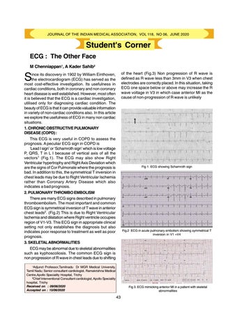
ince its discovery in 1902 by William Einthoven, the electrocardiogram (ECG) has served as the most cost-effective investigation. Its usefulness in cardiac conditions, both in coronary and non coronary heart disease is well established. However, most often it is believed that the ECG is a cardiac investigation, utilised only for diagnosing cardiac condition. The beauty of ECG is that it can provide valuable information in variety of non-cardiac conditions also. In this article we explore the usefulness of ECG in many non cardiac situations. 1. CHRONIC OBSTRUCTIVE PULMONARY DISEASE (COPD) : This ECG is very useful in COPD to assess the prognosis. A peculiar ECG sign in COPD is ‘Lead I sign’ or ‘Schamroth sign’ which is low voltage P, QRS, T in L I because of vertical axis of all the vectors1 (Fig.1). The ECG may also show Right Ventricular hypertrophy and Right Axis Deviation which are the signs of Cor Pulmonale where the prognosis is bad. In addition to this, the symmetrical T inversion in chest leads may be due to Right Ventricular Ischemia rather than Coronary Artery Disease which also indicates a bad prognosis. 2. PULMONARY THROMBO EMBOLISM There are many ECG signs described in pulmonary thromboembolism. The most important and common ECG sign is symmetrical inversion of T wave in anterior chest leads2. (Fig.2) This is due to Right Ventricular Ischemia and dilatation where Right ventricle occupies region of V1-V3. This ECG sign in appropriate clinical setting not only establishes the diagnosis but also indicates poor response to treatment as well as poor prognosis. 3. SKELETAL ABNORMALITIES ECG may be abnormal due to skeletal abnormalities such as kyphoscoliosis. The common ECG sign is non progression of R wave in chest leads due to shifting
Fig 1. ECG showing Schamroth sign
Fig.2 ECG in acute pulmonary embolism showing symmetrical T inversion in V1 –V4
1
Adjunct Professor,Tamilnadu Dr MGR Medical University, Tamil Nadu; Senior consultant cardiologist, Ramakrishna Medical Centre,Apollo Speciality Hospital, Trichy 2 Chief Interventional Consultant cardiologist, Apollo Speciality hospital, Trichy Received on : 09/06/2020 Accepted on : 15/06/2020
Fig 3. ECG mimicking anterior MI in a patient with skeletal abnormalities
43
