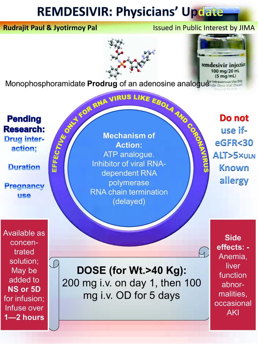
JOURNAL OF THE INDIAN MEDICAL ASSOCIATION, VOL 118, NO 06, JUNE 2020
Imaging in Medicine Computed Tomography (CT) Chest imaging in Diagnosis and Management of patients with COVID-19 Vimal Raj1, Jagalpathy Jagdish2 Over the last few weeks, there is an increase in the number of cases of Corona VIrus Disease- 2019 (COVID -19) reported in India. Every clinician is likely to manage one or other form of presentation of the disease in his or her practice. Therefore, every clinician needs to be aware of the imaging appearances of the COVID-19. Chest radiograph is not a good diagnostic tool and higher imaging modality such as Computed Tomography (CT) may be appropriate. The risk of spread of infection amongst patients and to the healthcare workers can be a challenge in performing CT studies for patients with COVID-19. In this review, we describe common CT appearances of COVID-19 infection and its complications. We also discuss the role of CT in assessing disease severity, prognosis and its utility in monitoring of disease progression. National and international guidelines relating to the use of CT imaging in COVID-19 are also highlighted. [J Indian Med Assoc 2020; 118(6): 35-42]
Key words : COVID-19, CT, MDCT, Imaging, Chest CT, Novel Corona Virus
N
ovel coronavirus was identified, in January 2020, as a cause of pneumonia in many patients since December 2019 in China. The infection spread rapidly within China and in the rest of the world over the next few weeks. The World Health Organisation (WHO) termed the illness as Corona Virus Disease 2019 (COVID-19) and declared it as a global pandemic on 11th of March 20201,2. By 12th of June 2020, more than 74 lakh confirmed cases have been reported to WHO worldwide with a more than 4 lakh patients succumbing to the disease. diseased patients. India with more than 3 lakh cases was the fourth-worst effected country behind the United States of America, Brazil and Russia3. These numbers are likely to further increase in India as travel and lockdown restrictions are eased. COVID-19 presents with symptoms of lower respiratory tract infection and many patients remain asymptomatic or with mild symptoms4,5.Isolation of virus in the respiratory samples using real-time reverse transcriptase-polymerase chain reaction (RT-PCR) is the testing of choice for detection of COVID-19. RTPCR has variable sensitivity ranging from 37% to 91%6-11. RT-PCR results can be falsely negative initially
Editor's Comment :
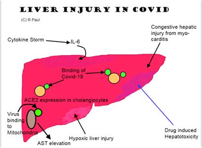
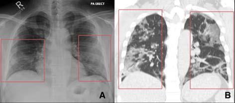
CT scans have variable sensitivity in detecting patients with Covid-19 but is specially helpful for areas with limited availability of RT-PCR. Ground glass opacities, consolidation, reticular changes are most common finding. Protection of healthcare workers of Radiology Department is of prime importance.
and often require repeating of test. RT-PCR tests are also not easily available in different parts of the country and there can be significant delay in getting the results of these tests. Chest imaging tests can be useful in the detection and management of patients with suspected or known COVID-19. Chest radiographs (CXR) are widely available and cost-effective but can also have variable sensitivity, with nearly 60% of patients having a normal CXR in one of the recent studies10 12-14. Computed Tomography (CT) imaging of the chest has a better resolution than CXR and may allow improved detection and assessment of patients with known or suspected COVID-19 (Figure 1). In this review, we describe common CT appearances of COVID-19 infection and its complications. We also discuss the role of CT in assessing disease severity, prognosis and its utility in monitoring of disease progression. National and international guidelines relating to the use of CT imaging in COVID-19 are also highlighted.
1 FRCR,CCT (UK), EDM, PGDMLS, Department of Radiology, Narayana Hrudayalaya, Bangalore and Corresponding Author 2 Department of Radiology, Al Rahba Hospital, Seha, Abudhabi, UAE Received on : 16/06/2020 Accepted on : 19/06/2020
35