“Thehardestbrainmalformationtofindisthesecondone!” (Satisfaction
of Search Errors)
Multiple anomalies
Same anomaly, different causes (insult, genetic, metabolic)
Single genetic defect, multiple anomalies over time


Multiple anomalies
Same anomaly, different causes (insult, genetic, metabolic)
Single genetic defect, multiple anomalies over time


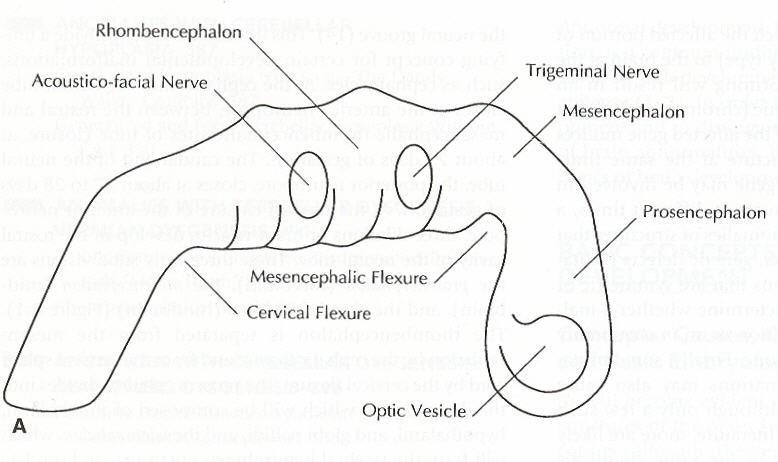
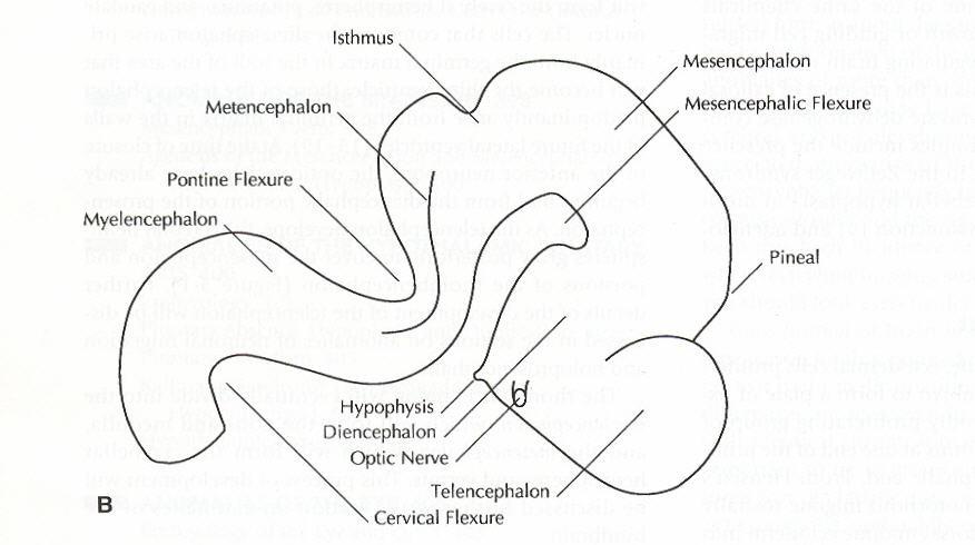
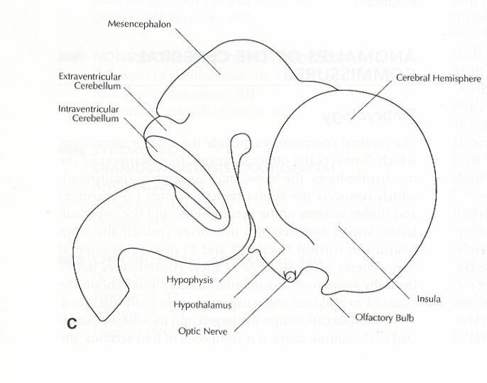
Prosencephalon
Diencephalon
Thalami, hypothalami, globi pallidi
Telencephalon
Putamin, caudate
Neocortex Mesencephalon Midbrain
Rhombencephalon
Myelencephalon
Pons, medulla
Metencephalon
Cerebellum, vermis
Anomalies of Dorsal Induction
Commissuration Anomalies
Cerebral Cortex Anomalies
Stem Cell Proliferation or Apoptosis
Neuronal Migration
Post Migrational Cortical Organization
Anomalies of Ventral Induction
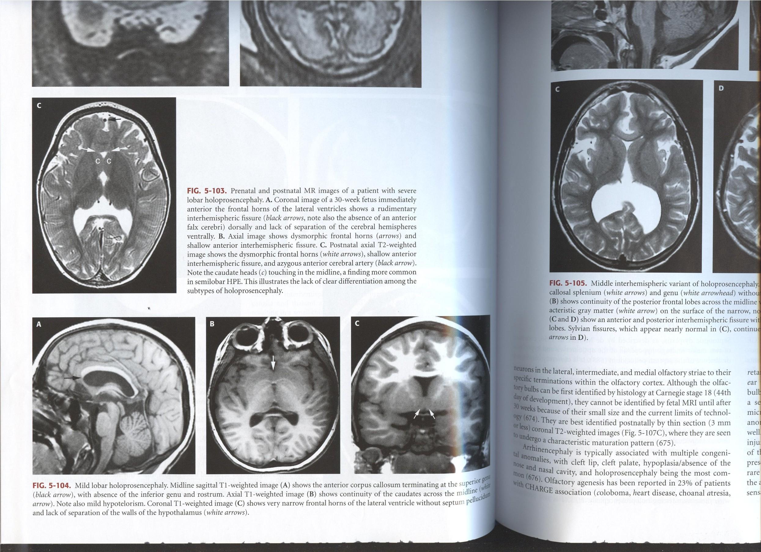
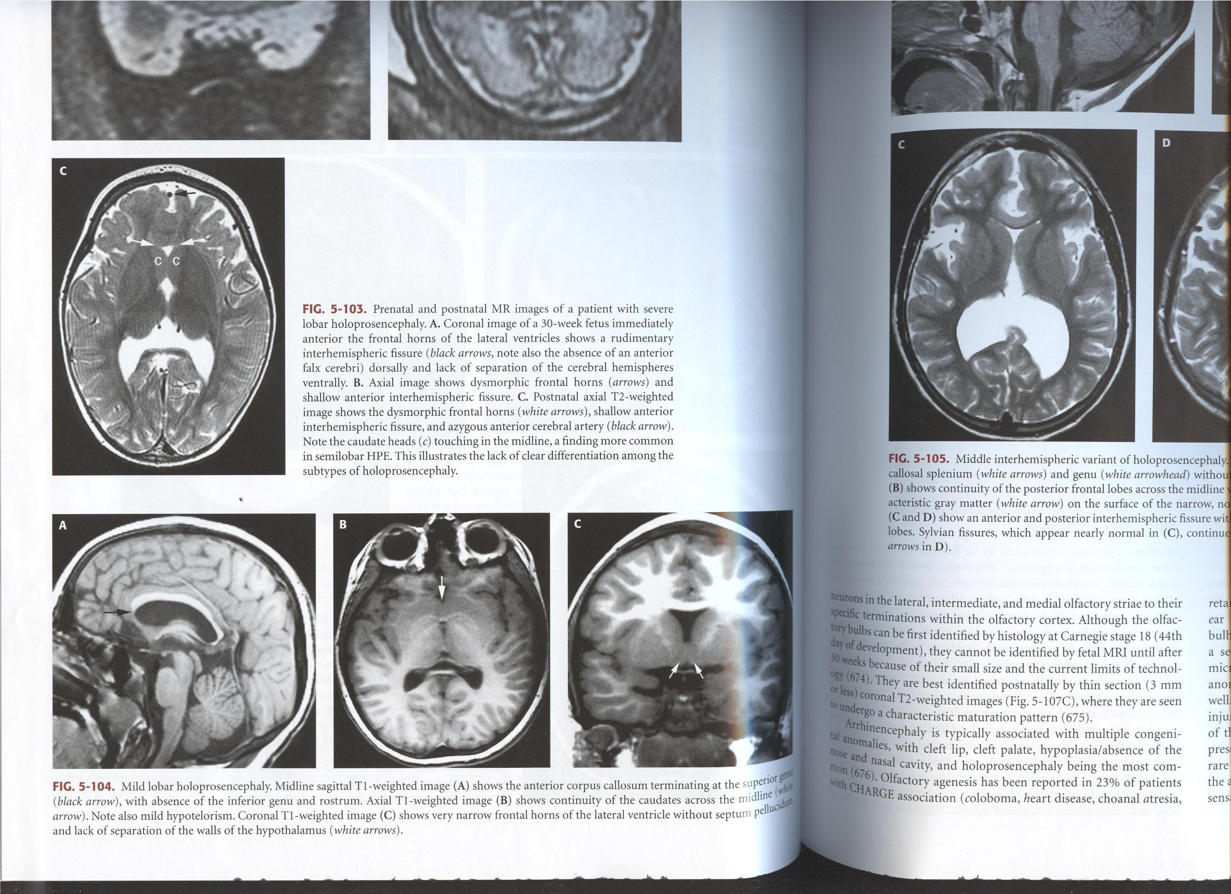
Holoprosencephaly
Septooptic Dysplasia
Arrhinia/Arrhinencephaly
Hypothalamic-Pit Axis
Eye and Orbit
Dorsal Induction
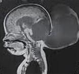
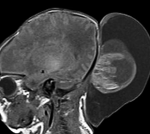
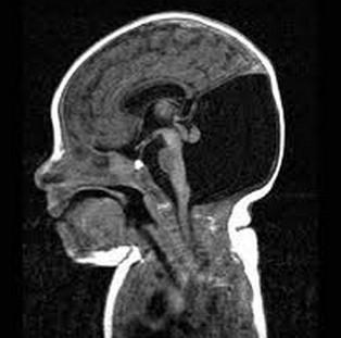
Occipital Encephalocele
Ventral Induction
Rhombencephalosynapsis
Isolated Vermian Hypoplasia (IVH)
Dandy Walker malformation
Unilateral Cerebellar Dysplasia/Hypoplasia
May be single disruptive event
Pontocerebellar Hypoplasia Spectrum, non progressive
But what is normal?
Use the Sweep & Run approach to developmental pediatric brain MRI…
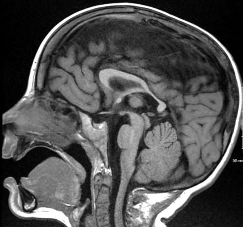
Sweep the Midline
Map the Myelin
Run the Rim
Ice the Cake
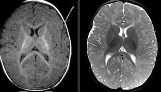
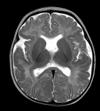

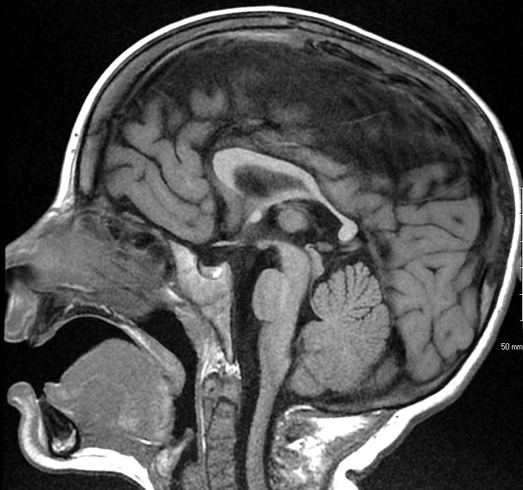

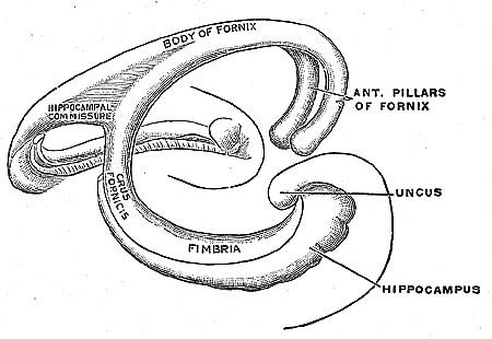
Three midline commissures involved in abnormalities of the Corpus Callosum:
Anterior Commissure
Corpus Callosum
Hippocampal Commissure: typically rudimentary, fibers between the body of the fornix
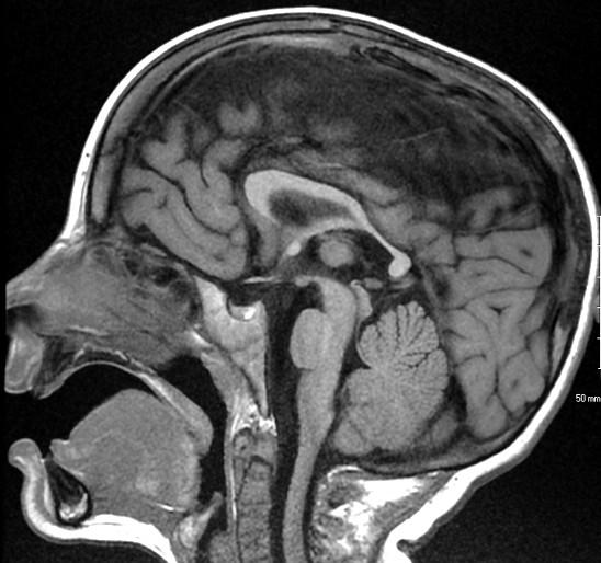
Anterior commissure
Corpus Callosum
Rostrum, genu, body, splenium

Anterior commissure
Corpus Callosum
Rostrum, genu, body, splenium
Sella/Suprasellar region
optic nerve & chiasm size, posterior pituitary “bright spot”, stalk

Anterior commissure
Corpus Callosum
Rostrum, genu, body, splenium
Sella/Suprasella
ON, post pit “bright spot”, stalk
Midbrain
Patency aqueduct
Pons
Relative size to midbrain and medulla, the Goldilocks principle: “not too big, not too small, just right…”

Anterior commissure
Corpus Callosum
rostrum, genu, body, splenium
Sella/Suprasella
ON, post pit bright spot, stalk
Midbrain
Patency aqueduct
Pons
Relative size to midbrain and medulla,“just right”
Vermis – 3 lobes
3 lobes: Anterior (40%) , Posterior (60%) , small Flocculonodular lobe
Ant and Post lobes separated by the Primary fissure
closed 4th ventricle, closed fastigial point

Anterior commissure
Corpus Callosum
rostrum, genu, body, splenium
Sella/Suprasella
ON, Post pit bright spot, stalk
Midbrain
Patency aqueduct
Pons
“just right”
Vermis
3 lobes, closed fastigial point
Cerebellar Tonsils terminate normally at or above the foramen magnum
Sweep the Midline…Name the parts…where do we start?
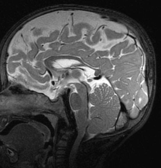
Sag T2 Cube nongated Normal

Sag T2 Cube nongated Normal

Normal
Sag T2 Cube
Nongated: CSF dephasing in the aqueduct on this sequence

Sag T2 Cube nongated Normal

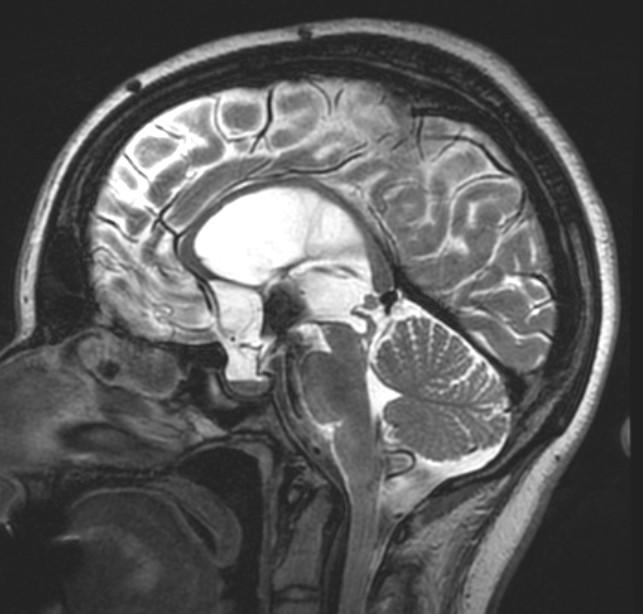
Primary fissure 3 lobes Closed fastigial point
Sag T2 Cube Normal

Anterior commissure
Corpus Callosum
Rostrum, genu, body, splenium
Sella/Suprasella
ON, post pit bright spot, stalk
Midbrain
Patent aqueduct
Pons
“just right…”
Vermis
3 lobes
Cerebellar Tonsils
Map the Myelin:
Use both T1 and T2 to evaluate myelin.
Run the Rim:
Start at noon and evaluate the cortex clockwise, should be 2-33 mm thick.
Ice the Cake: Use remaining sequences (GRE, DWI, contrast) to refine your diagnosis.
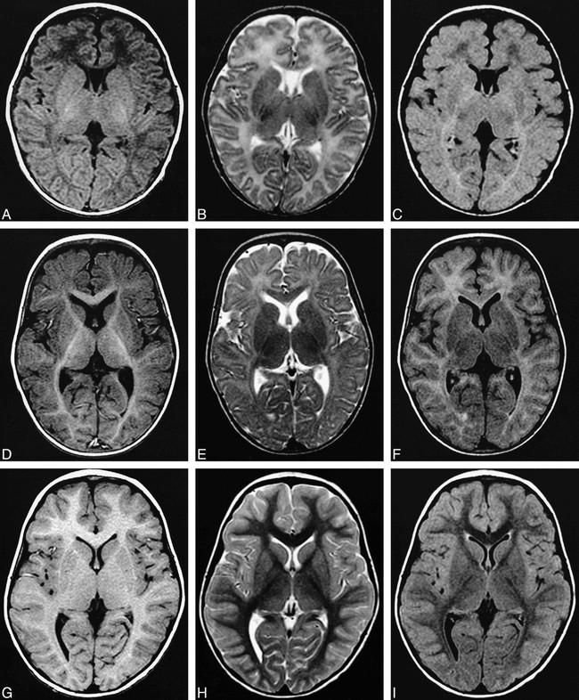

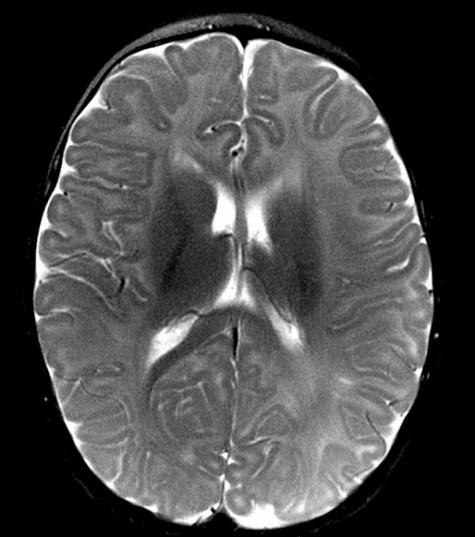

Remember to start with Sweep the Midline… Where is the anterior commissure?
Always finish the sweep looking for second anomaly…
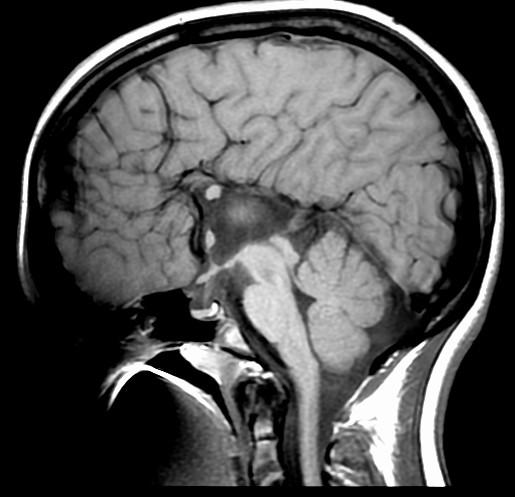
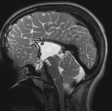

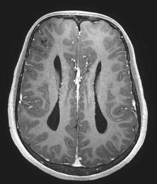
Parallel ventricles
Radial or pallisading gyri in sagittal plane
Colpocephaly
Longhorn or Viking helmit frontal horns
High riding 3rd vent
“Keyhole” temporal horns
Vascular anomalies: “wandering ACAs”
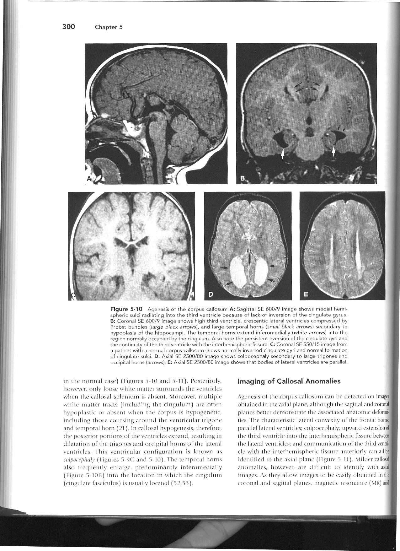
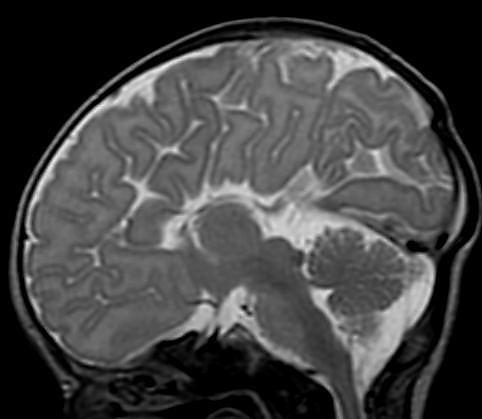
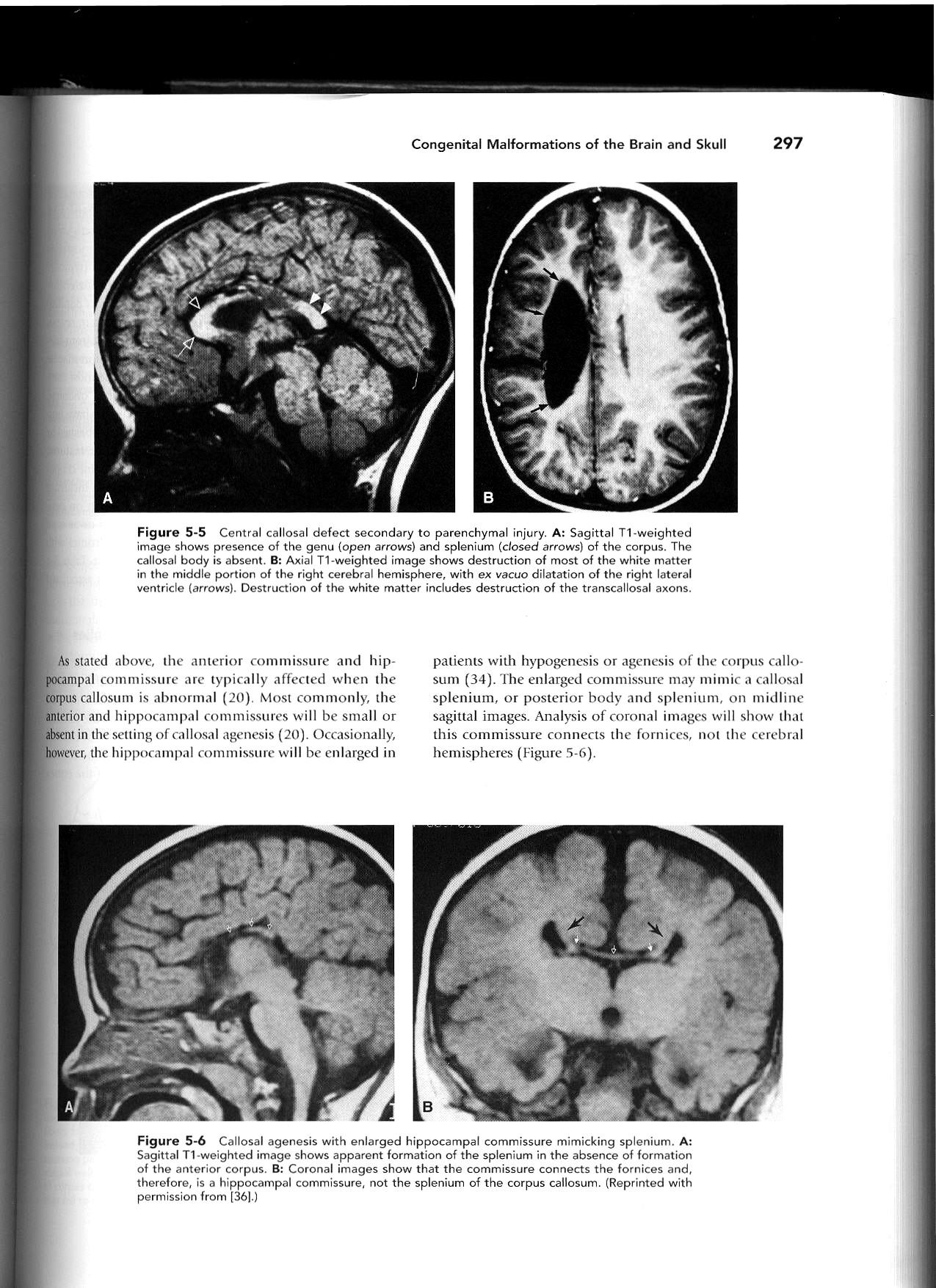
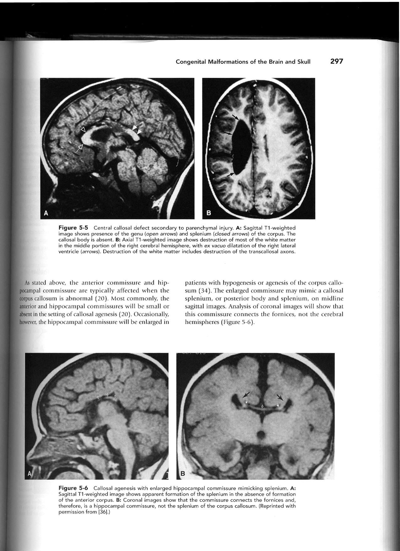
All 3 commissures are absent.
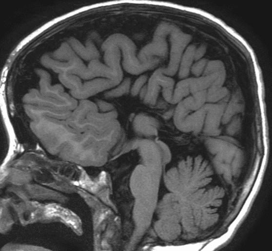
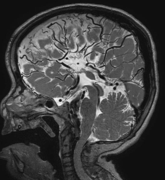
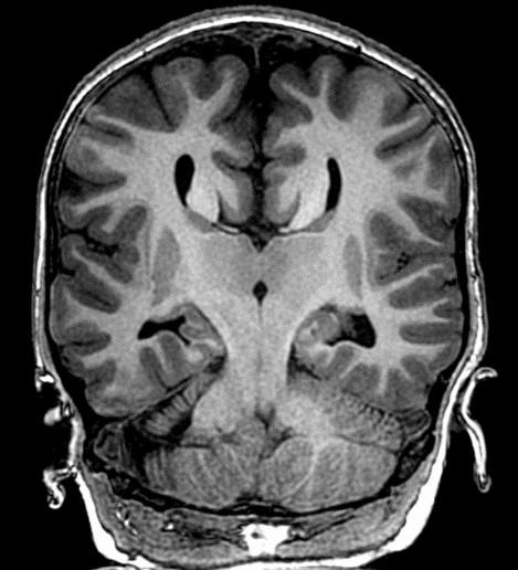
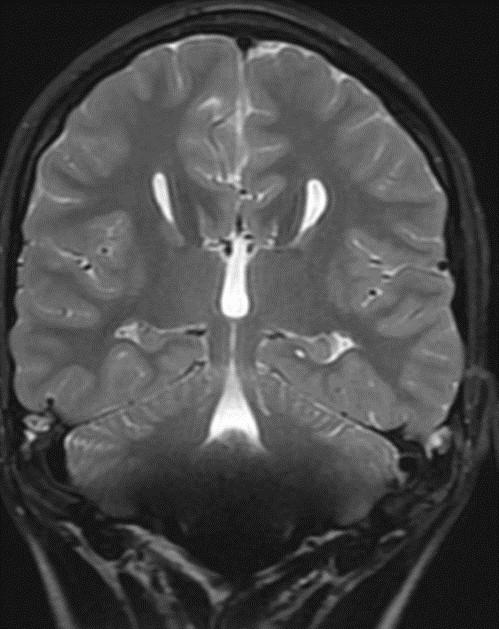
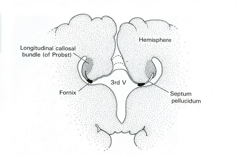
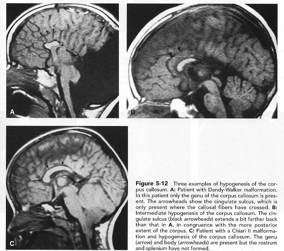
Cingulate gyrus (black arrows) “mirrors” the development of the corpus callosum.
Enlarged HC connects fornices, not cerebral hemispheres
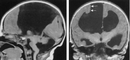
Type I Communicating Cyst
Cyst communicates with the third ventricle.

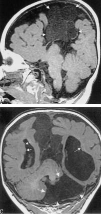
Type II Complex
Noncommunicating Cyst(s) Cysts demonstrate differing signal characteristics than CSF.

Sweep the Midline: What parts are missing?
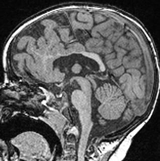
Run the Rim…where is the cortex “too thick?”

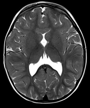


“Missing”=disorganized rostrum, genu, anterior body of the corpus callosum Cortex of the anterior medial frontal lobes “too thick” with fused grey matter across the anterior midline where the “genu” should be.
Alobar
Most severe, complete lack of “cleavage”, fused thalami.
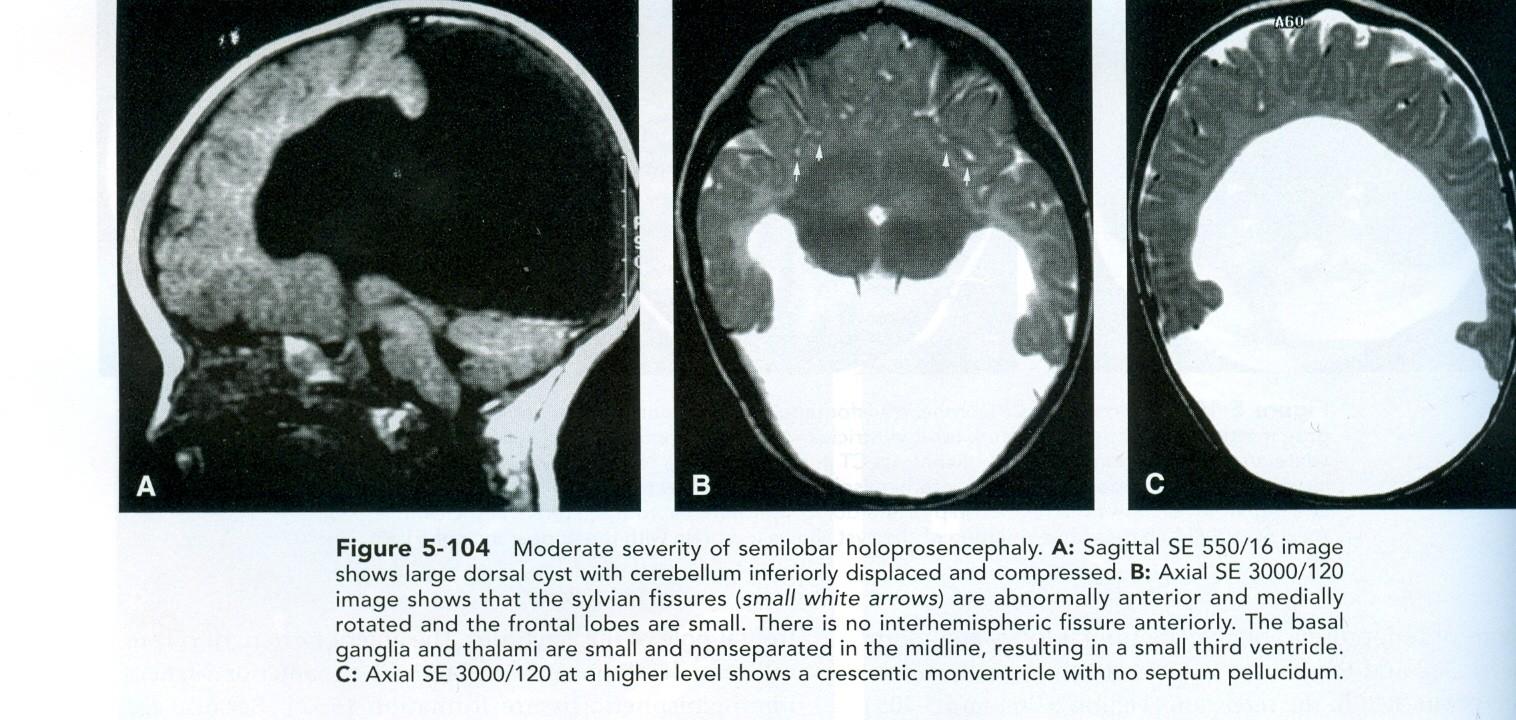
Semilobar
“ in between” with separated occipital lobes
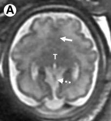
Lobar Mild, often only anterior frontal lobe fused, with separate thalami

Sweep the Midline:
“Comma shaped Corpus Callosum with abnormal mid body…”
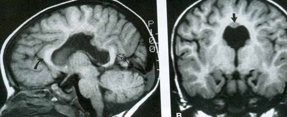

Syntelencephaly: Midline CC “lack of cleavage”
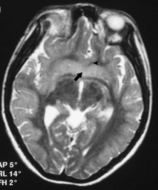
Middle Interhemispheric “MIH” Variant of Holoprosencephaly
Dorsal Induction Anomaly
Presentation
Motor defects
Oromotor dysfunction
Associated with:
Azygous ACA
Polymicrogyria PMG
Cortical dysplasias
GM heterotopia
Absent septum pellucidi
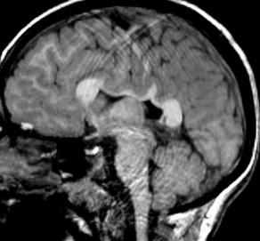
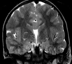
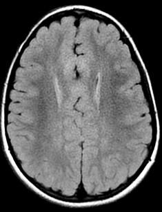
Blunted anterior Corpus, with subtle fusion at the level of the anterior commissure…?

Fusion (lack of cleavage) is just anterior /superior to the anterior commissure.


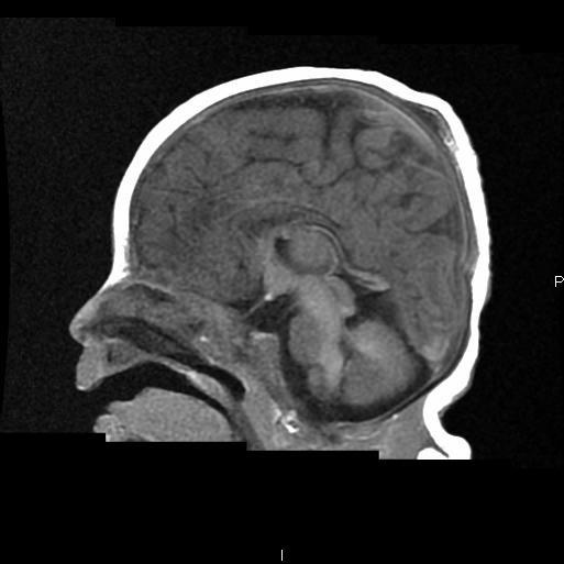
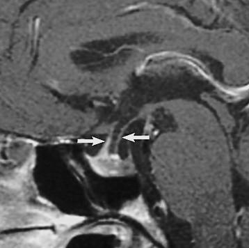
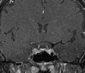
Ectopic posterior pituitary “bright spot.” Missing infundibular stalk.


Contrast or ultra thin T2 needed to confirm.
Thin or truncated stalk
IGHD – Growth Harmone Deficiency if stalk is present but abnormal
PanHypoPit Deficiencies if stalk is completely absent.
Need repeated endocrine evaluations if normal at first imaging.
Neonate: Hypoglycemia, jaundice, Failure to Thrive
Normal or shallow sella
Abscent or hypoplastic stalk Ectopic posterior pituitary
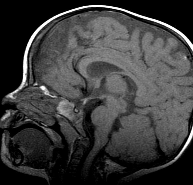
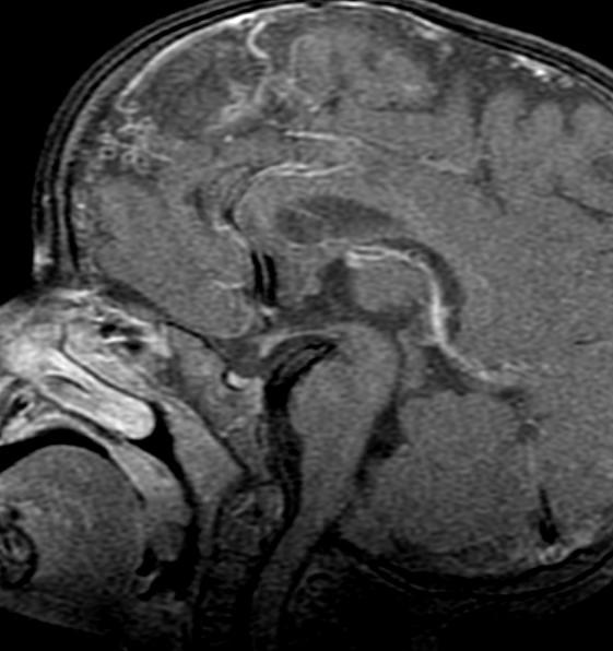


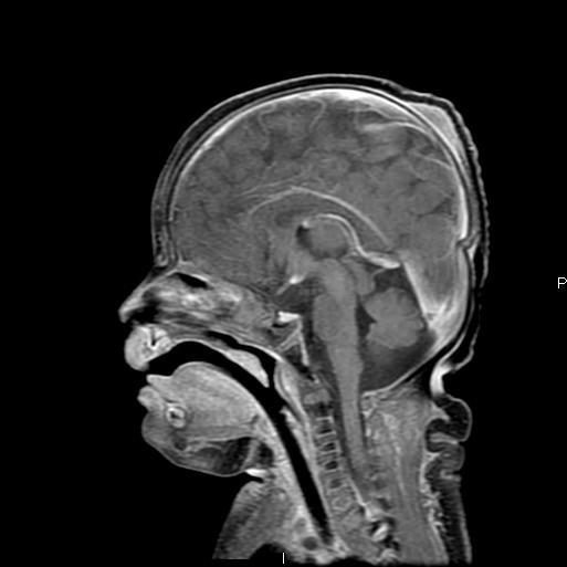
Abnormal notocord integration, related to lack of fusion of the initial two notochord “enlages” at gastrulation, results in notocord “duplication” with double ventral induction structures…can be seen with split tongue, palate, cranial base encephaloceles with splite dens, vertebrobasilar vascular anomalies.
What did this patient present with?
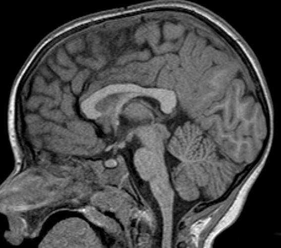
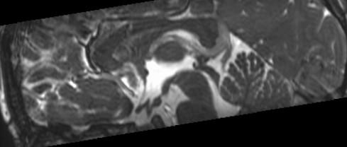
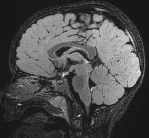
Gelastic “Laughing” Seizures
Precocious puberty
Seizures
Rare calcification
Normal
Rarely increased T2 Flair signal
No enhancement
Trigger Biopsy:
Enhancement
Growth

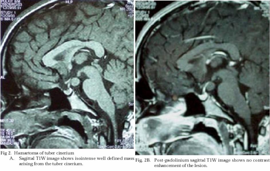

Different patient, same finding…
What procedure did this patient have?
What is the MRI sequence called?

Note the CSF flow dephasing through the defect in the floor of the third ventricle.

Differential for “cyst in the posterior fossa” starts with whether the vermis is normal or not.
Is the vermis normal? Can you identify the normal structures of the Vermis?
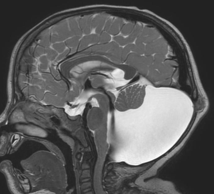

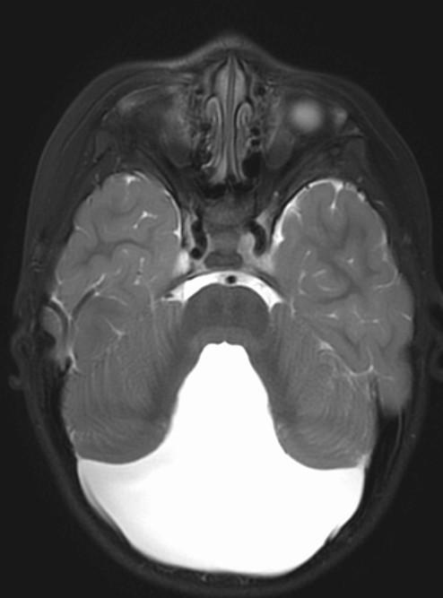
Malrotated hypoplastic vermis, with vertical primary fissure.
“OPEN” fastigial point of the fourth ventricle. Hypoplastic cerebellar hemispheres.
Enlarged posterior fossa
Cystic dilatation 4th ventricle
Uplifted tentorium, TSV sinus, torcula
“torcula-lambdoid inversion” with torcula above the lambdoid suture
Agenetic or hypogenetic vermis with “vermian tail, pushed upward
Cerebellar hypoplasia
CC anomalies 32%
Hydrocephalus up to 90%
Aqueductal stenosis
4th ventricle outlet obstruction
Polymicrogyria, heteropia 5-10%
Occipital Meningo-Encephaloceles 16%
Syndromic = Extracranial anomalies 50%
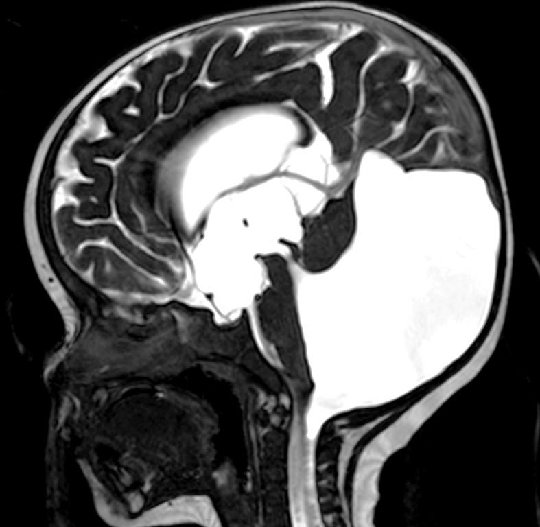
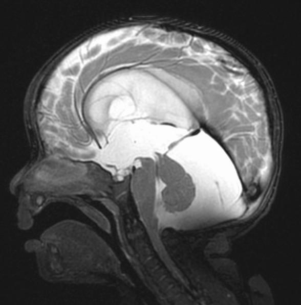
Lobes?
Primary Fissure? Fastigial Point?
Hint: Normally formed Vermis which is pushed forward DDX: mega cisterna magna (no mass effect on the vermis) versus Blake’s pouch cyst (mass effect on inferior vermis pushing vermis superiorly.)

Are the structures of the vermis normal? What is wrong with the fourth ventricle? What is the diagnosis?

Are the structures of the vermis normal? What is wrong with the fourth ventricle? What is the diagnosis?

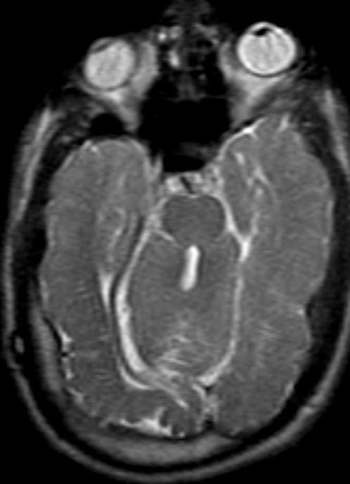
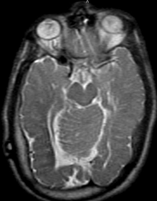

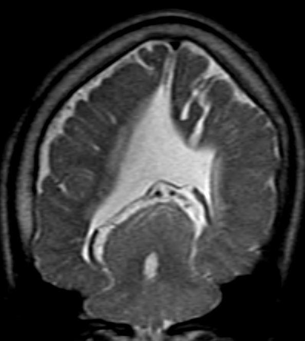

Vermis appears “large and rounded”
Rounded fastigial point of the fourth ventricle
Cerebellar dysgenesis
Fused hemispheres, absent or hypoplastic vermis, and superior cerebellar peduncles
Needs to have fusion of the dentate nuclei
Assn: hydrocephalus, limbic anomalies, cortical malformations, absent septum pellucidum, multiples suture synostosis
Where is the vermis? Big or small?
Do you have a normal fastigial point?
What is the abnormal horizontally oriented structure? What is the diagnosis?
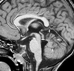
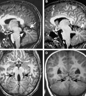
Where is the vermis? Big or small? Too small…
Do you have a normal fastigial point? Yes, pointed… What is the abnormal horizontally oriented structure? the superior cerebellar peducle…
What is the diagnosis?


What is the yellow arrow pointing to?
What is the diagnosis?


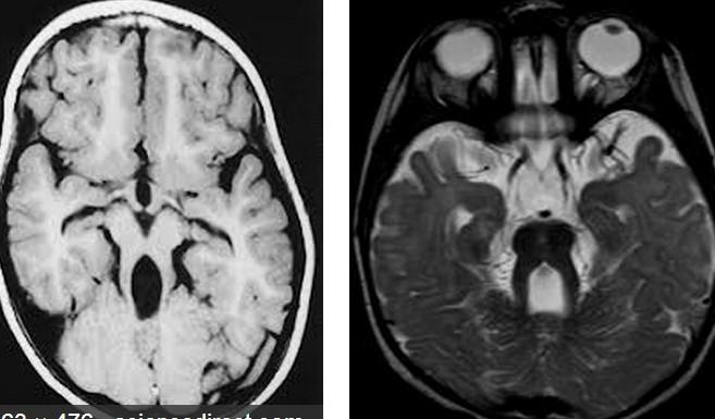

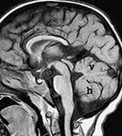
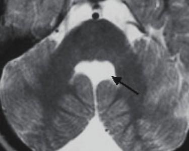
“molar tooth” midbrain
Large horizontally oriented superior cerebellar peduncles
Small, dysplastic vermis
“bat wing” 4th ventricle configuration
Associated with:
Absence of decussation of WM pathways in brain stem on DTI
Supratentorial:
Absent septum pellucidum
Fused fornices
Ventriculomegaly
Polymicrogyria
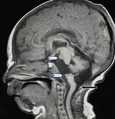
White arrows: Small pons remnant with nonformation of the midportion of the brainstem
Clinical: cranial neuropathies
Associated with small cerebellum
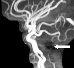
Abnormal vertebrobasilar vasculature
Etiology?
very early vascular insult?
No gliosis to suggest hypoxia or ischemia
In animals: seen with hox gene deletions leading to lack of single rhombomere development, so brainstem “short” but typically not fully disconnected

