(Inherited Disorders of White Matter) (Inherited Disorders of White Matter)
Aaron Field, MD PhD
Aaron Field, MD PhD
afield@uwhealth.org afield@uwhealth.org
Learning Objectives Learning Objectives
Review the most “commonly encountered” leukodystrophies
• Review the most “commonly encountered” leukodystrophies
• Learn broad categories of underlying mechanisms Learn broad categories of underlying mechanisms
• Learn what imaging features are most relevant to ddx Learn what imaging features are most relevant to ddx
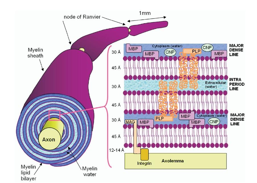
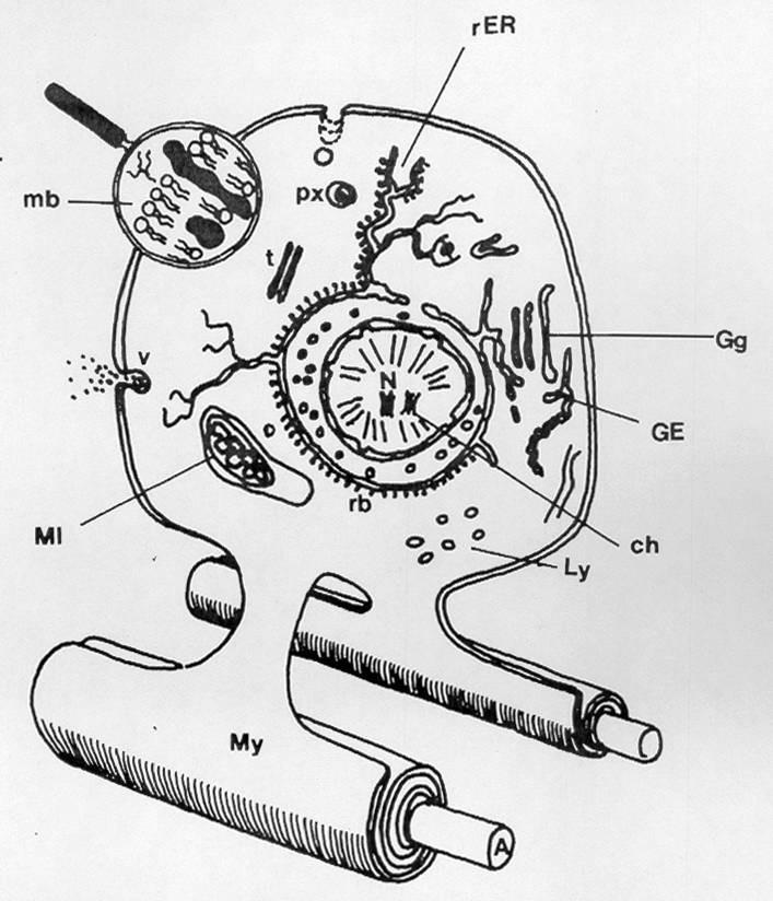
White Matter Microstructure White Matter Microstructure
High lipids High lipids
T1-bright T1-bright
Low free water
Low free water
T2-dark T2-dark
Laule et al, Neurotherapeutics 2007;4:460-484
Myelin Myelin
• Sensory before motor Sensory before motor
• Back to front Back to front
• Central to peripheral Central to peripheral
Normal Myelination by MRI Normal Myelination by MRI
Medulla Medulla
Dorsal pons
Dorsal pons
Cerebellar peduncles
Cerebellar peduncles
PLIC PLIC
Perirolandic WM Perirolandic WM
Normal Myelination by MRI Normal Myelination by MRI
Peri-atrial / subcortical “terminal zones” may persist into adulthood
JA Phelan and LH Lowe, Univ. of Missouri - KC
Obstacles to Learning and Obstacles to Learning and Recognizing Leukodystrophies Recognizing Leukodystrophies
Various forms of a given disease
• Various forms of a given disease
e.g. single vs. multiple enzyme defects
e.g. single vs. multiple enzyme defects
Various and non-specific clinical presentations
• Various and non-specific clinical presentations
e.g. infantile, juvenile, adult forms
e.g. infantile, juvenile, adult forms
• White matter MR signal changes with age White matter MR signal changes with age
• Overlapping imaging features (especially Overlapping imaging features (especially late late) )
• 60% never get specific diagnosis 60% never get specific diagnosis
JA Phelan and LH Lowe, Univ. of Missouri - KC
Delayed vs. Hypo-/Dys-/De-myelination
Delayed vs. Hypo-/Dys-/De-myelination
• Delayed myelination Delayed myelination
• Non-specific feature observed in almost all children with Non-specific feature observed in almost all children with delayed development of any cause – the myelin ultimately delayed development of any cause – the myelin ultimately formed often formed often normal normal
• Dysmyelination Dysmyelination
• Myelin formation is fundamentally disordered Myelin formation is fundamentally disordered
• Hypomyelination Hypomyelination
• Myelin is more Myelin is more deficient deficient than than disordered disordered
• Demyelination Demyelination
• Myelin is formed (sometimes normally) but breaks down Myelin is formed (sometimes normally) but breaks down
• There is overlap! There is overlap!
• Disordered myelin ( Disordered myelin (dysdysmyelination) more apt to break down myelination) more apt to break down ((dedemyelinate) than normal myelin myelinate) than normal myelin
Delayed vs. Hypo-/Dys-/De-myelination
Delayed vs. Hypo-/Dys-/De-myelination
• Delayed myelination Delayed myelination
• Non-specific feature observed in almost all children with Non-specific feature observed in almost all children with delayed development of any cause – the myelin ultimately delayed development of any cause – the myelin ultimately formed often formed often normal normal
• Dysmyelination Dysmyelination
• Myelin formation is fundamentally disordered Myelin formation is fundamentally disordered
• Hypomyelination Hypomyelination
• Myelin is more Myelin is more deficient deficient than than disordered disordered
• Hard to diagnose before 1 year (paucity of myelin is normal) Hard to diagnose before 1 year (paucity of myelin is normal)
• Rule of thumb: Rule of thumb:
Deficient myelin unchanged over 6 mos in child > 1 y/o
Deficient myelin unchanged over 6 mos in child > 1 y/o
• AR lysosomal deficiency in galactocerebroside AR lysosomal deficiency in galactocerebroside ββ-galactosidase galactosidase
• Presents 3-6 mos w/ irritability, episodic fever, increased muscle Presents 3-6 mos w/ irritability, episodic fever, increased muscle tone and developmental delay tone and developmental delay
• Eventually hyperacusis, myoclonus, optic atrophy, early death Eventually hyperacusis, myoclonus, optic atrophy, early death
Barkovich JE. Pediatric
Neuroimaging 2005
JA Phelan and LH Lowe
Univ. of Missouri - KC
Cheon JE et al. Radiographics
2002;22:461-476
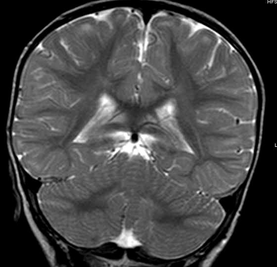
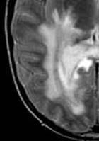
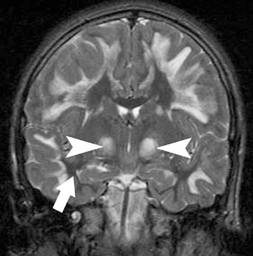
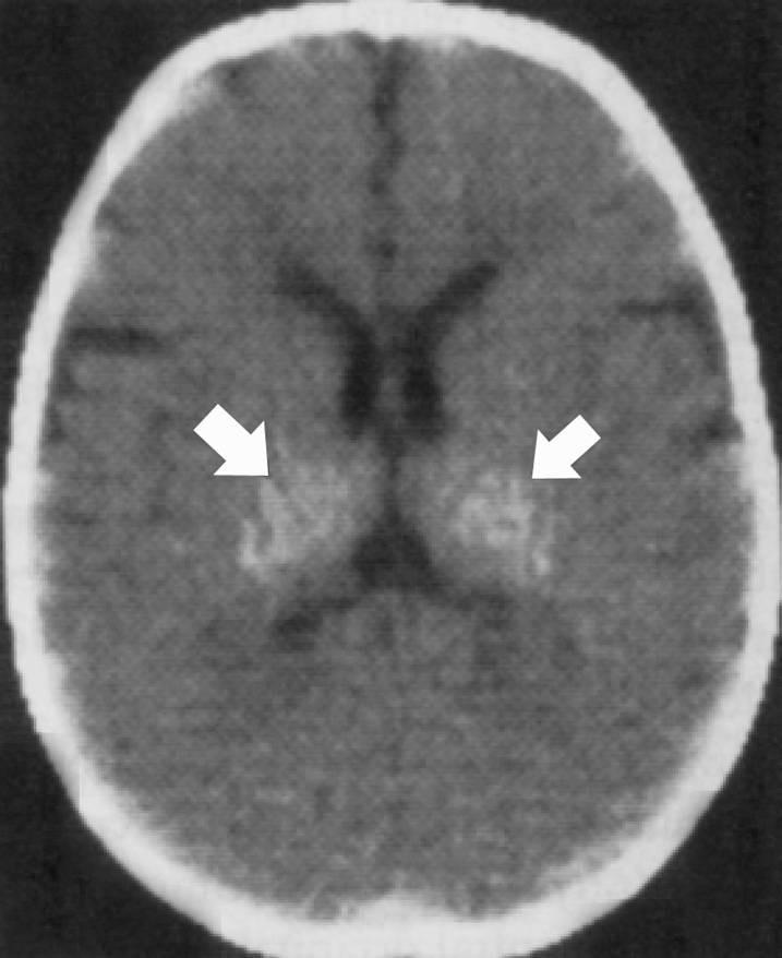
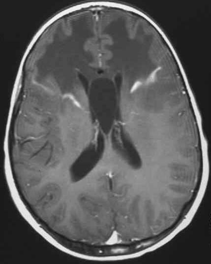
Krabbe Krabbe
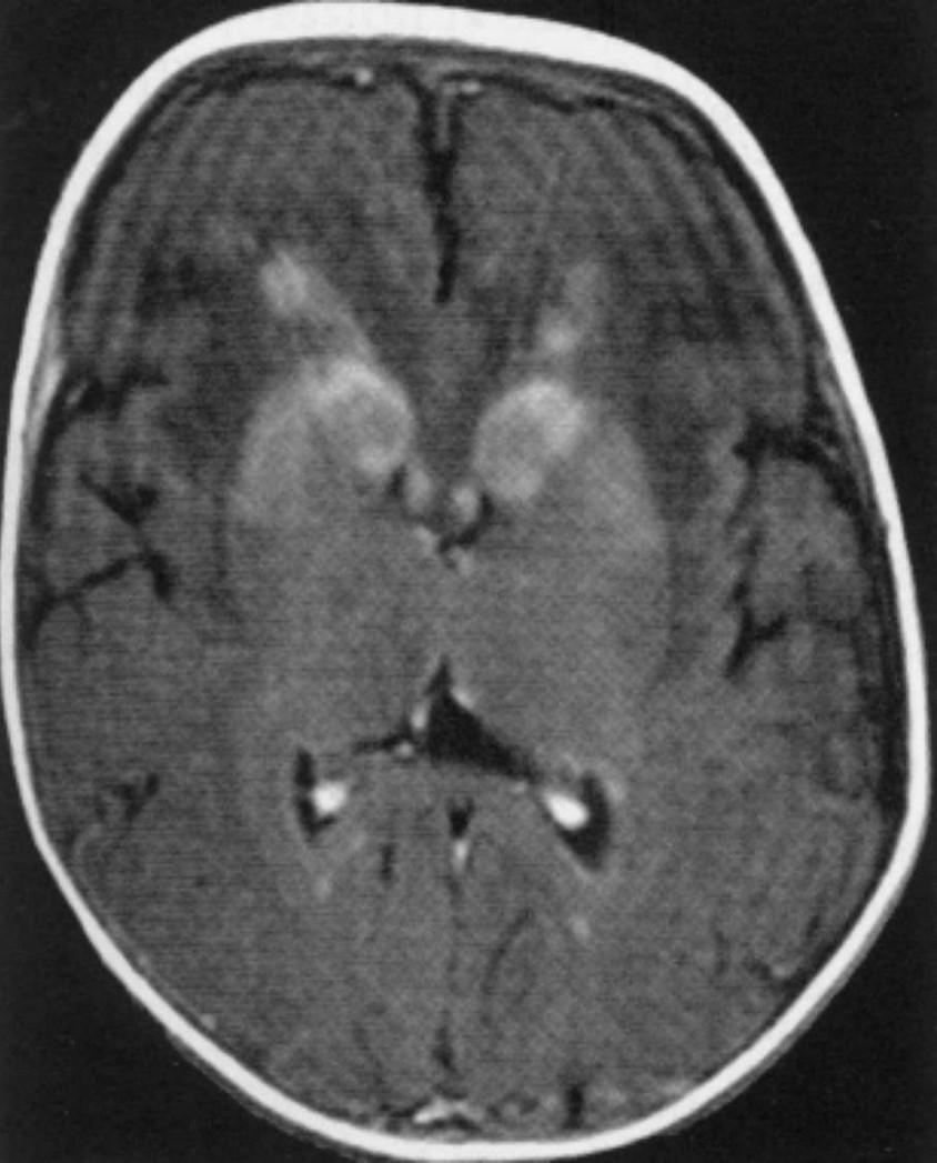
• CT: CT: Hyperdense thalami Hyperdense thalami
• MR: Deep WM, cerebellar WM, eventually thalami MR: Deep WM, cerebellar WM, eventually thalami
Barkovich JE. Pediatric Neuroimaging 2005
JA Phelan and LH Lowe Univ. of Missouri - KC
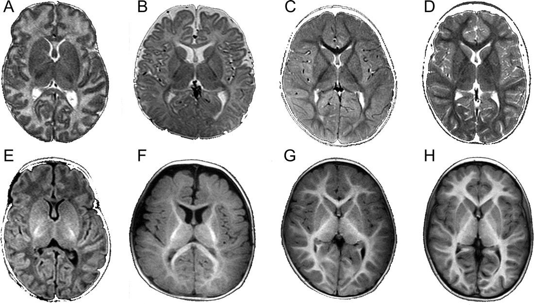
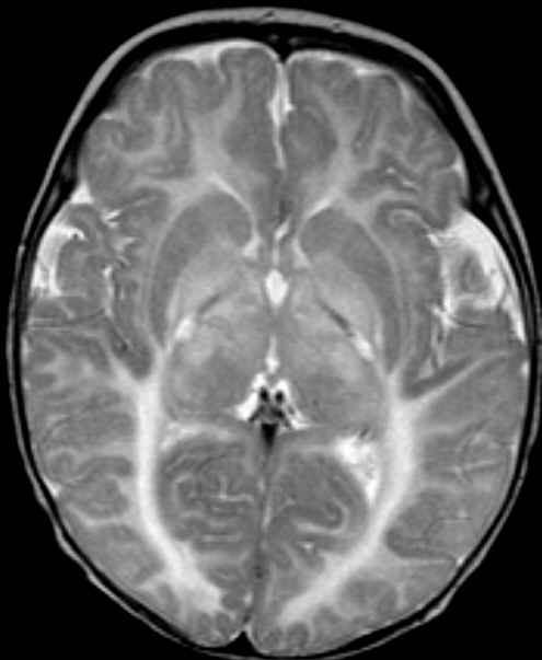
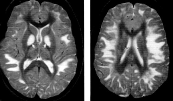
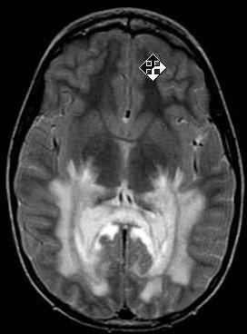
Deep WM Predominant Deep WM Predominant (Subcortical U-Fibers Spared) (Subcortical
• Infant with hypotonia, psychomotor retardation Infant with hypotonia, psychomotor retardation
• Late first year: spasticity, weakness, dystonia, ataxia, then Late first year: spasticity, weakness, dystonia, ataxia, then macrocephaly, seizures macrocephaly, seizures
• Imaging similar to Krabbe Imaging similar to Krabbe
Deep WM Predominant Deep WM Predominant (Subcortical U-Fibers Spared) (Subcortical
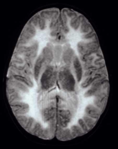
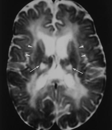
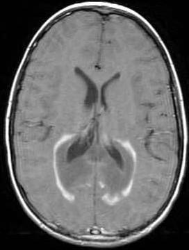
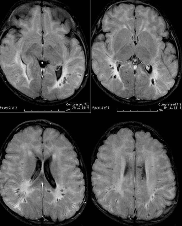
• Most common Most common inherited leukodystrophy! inherited leukodystrophy!
• AR lysosomal disorder – arylsulfatase deficiency AR lysosomal disorder – arylsulfatase deficiency
• Sulfatides accumulate in brain, kidneys, liver, GB, peripheral Sulfatides accumulate in brain, kidneys, liver, GB, peripheral nerve nerve
• 12-18 mos: Early motor signs of peripheral neuropathy 12-18 mos: Early motor signs of peripheral neuropathy
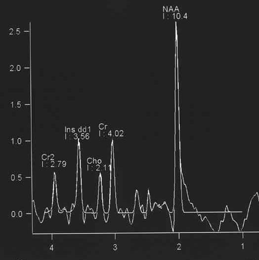
• Later: Decreased intelligence, speech, coordination; early death Later: Decreased intelligence, speech, coordination; early death c/o T. Kennedy, UW-Madison
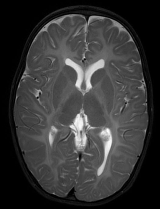
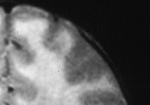
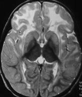
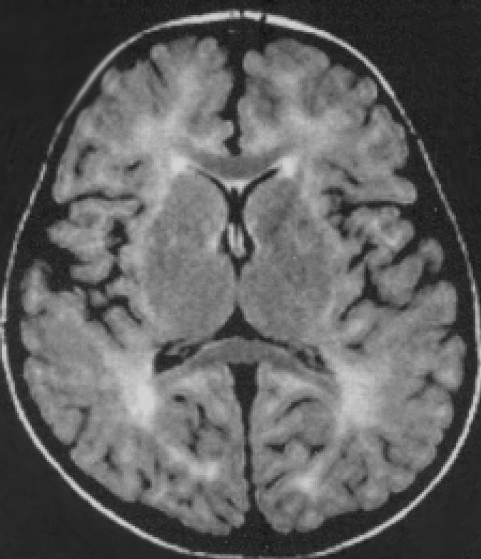
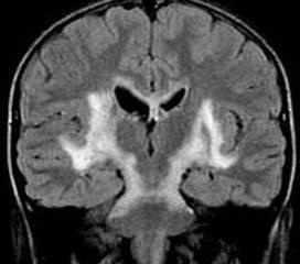
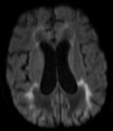
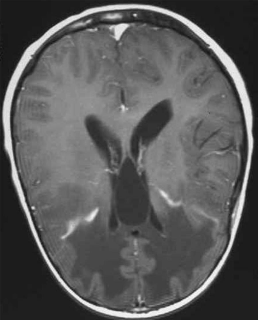
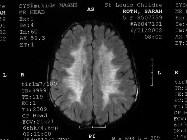
• Symmetric, confluent, deep WM (spares U-fibers) Symmetric, confluent, deep WM (spares U-fibers)
• “ “TigroidTigroid” or “ ” or “leopardleopard” pattern of WM sparing along PV spaces ” pattern of WM sparing along PV spaces
• No enhancement No enhancement
Deep WM Predominant Deep WM Predominant (Subcortical U-Fibers Spared) (Subcortical