TÓRAX. SIGNOS Y PATRONES RADIOLÓGICOS. CONTINENTE TORÁCICO.


VIII. PARTES BLANDAS
Revisado y modificado 20-7-2024


Dr. César Pedrosa Madrid


Revisado y modificado 20-7-2024
Dr. César Pedrosa Madrid
*En el modo “Presentación” se puede acceder a cada signo con un click sobre el nombre *Algunos signos tienen más de una diapositiva *Esquina superior derecha
2. Índice
3. ANATOMÍA GENERAL.
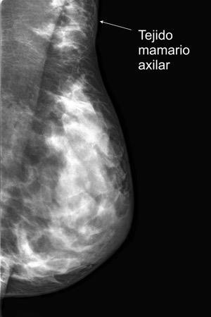
4. AXILA
5. AXILA. LÍNEAS
6. AXILA. MASAS. MAMA SUPERNUMERARIA
7. MASAS. ABSCESO AXILAR
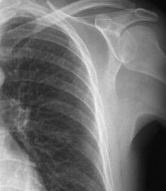
26. HOMBRO
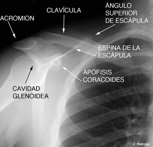
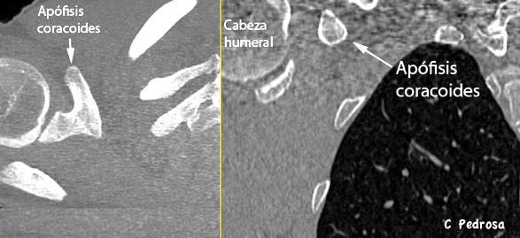
27. HOMBRO. ANATOMÍA
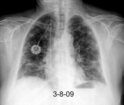
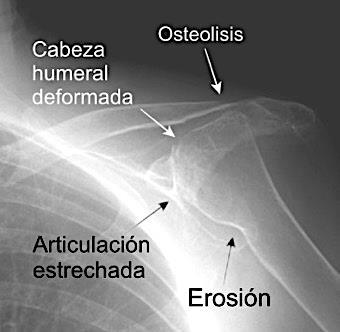
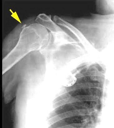
28. HOMBRO. ARTRITIS REUMATOIDE
29. HOMBRO. CALCIFICACIONES
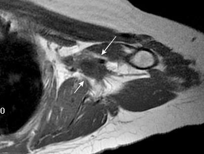
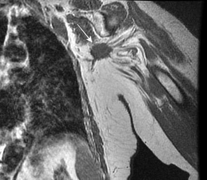
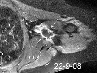
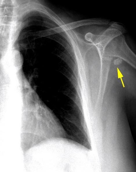
30. HOMBRO. TUMOR DE CÉLULAS
GIGANTES
1.MAMA
32. ANISOMASTIA
33. ASIMETRÍA MAMARIA
”Retorno al índice”
8. MASAS. GANGLIOS. CADENAS
9. MASAS. . HEMANGIOENDOTELIOMA
10. MASAS. LINFOMA DE BURKITT
11. MASAS.LINFOMA DE CÉLULAS B
12. MASAS.LINFOMA DE CÉLULAS B
13. MASAS. . LINFOMA DE HODGKIN
14. MASAS . METÁSTASIS. CA. DE MAMA
16. MASAS. HEMATOMA ESPONTÁNEO
17. CUELLO
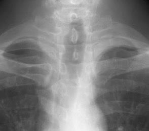
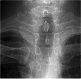
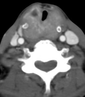
18. CUELLO. ANATOMÍA DE CUELLO Y LARINGE
19. CUELLO. CALCIFICACIONES
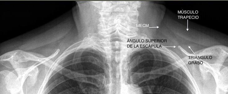
20. FOSA SUPRAESTERNAL
22. MADELUNG. LIPOMATOSIS DE
23. CUELLO. MASAS
24. MASAS. LINFOMA ANAPLÁSICO
25. CUELLO. T UMOR LARÍNGEO
34. MAMA. CALCIFICACIONES
35. GINECOMASTIA
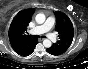
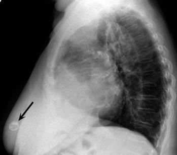
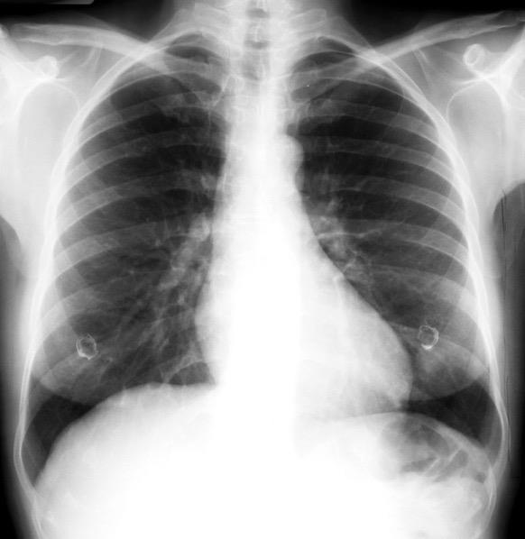
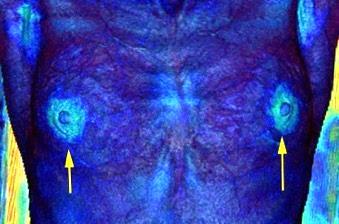
36. PEZONES VERSUS NÓDULOS
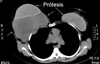
37. MAMA. PRÓTESIS
38. MASAS. TUMOR DESMOIDE
39. PARTES BLANDAS
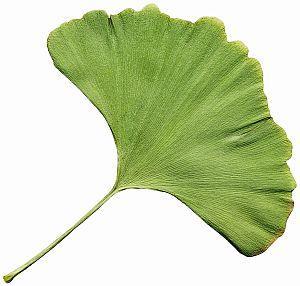
40. GINKGO. SIGNO DE LA HOJA DE
41. PARTES BLANDAS. AUMENTO
42. PARTES BLANDAS. CALCINOSIS
43. PARTES BLANDAS. CISTICERCOSIS
44. CAQUEXIA.
45. EMPIEMA NECESITATIS
46. MASAS. TUMOR DESMOIDE
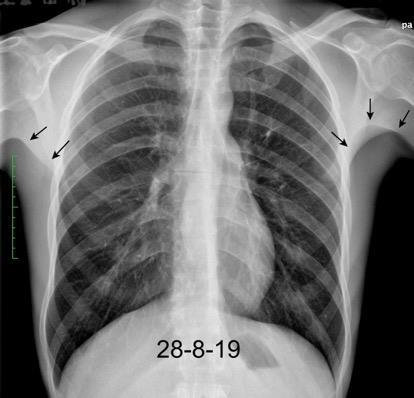
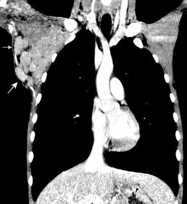
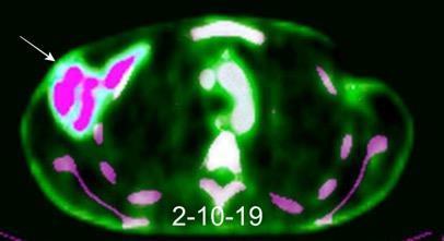
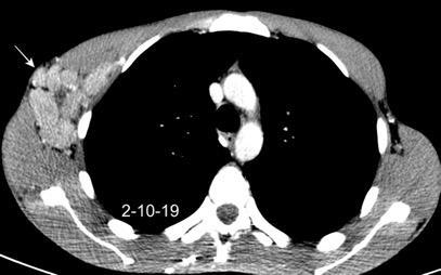
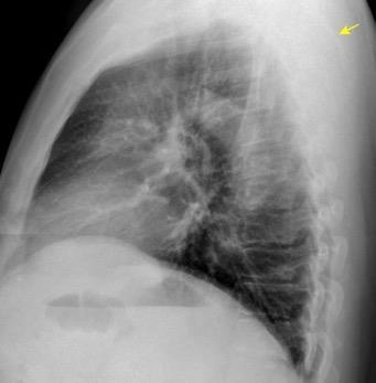
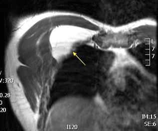
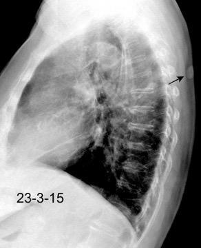
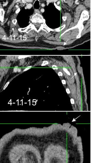
47 MASAS. . ELASTOFIBROMA DORSI
48 MASAS. LIPOMA SUBPECTORAL
50. MASAS. NEUROFIBROMATOSIS 1
51. POLAND. SÍNDROME DE 52.QUISTE HIDATÍDICO DE PARED
53. SEUDOLESIONES PARIETALES. VERRUGA
Piel
Línea del cuello
Fosa supraclavicular
Vías aéreas
Densidad pulmonar
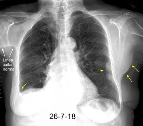
Línea axilar
Sombras mamarias
Diafragmas
Partes blandas de la pared
Pliegue axilar anormal
Clips quirúrgicos
Calcificaciones
Masas costales y de partes blandas
Clips
MAMA. Tejido supernumerario. Fibroadenomas, Cáncer Hamartoma, Necrosis grasa, MASAS DE PARTES BLANDAS. Lipomas, Hemangiomas, Fibromatosis, Quistes epidermoides. Histiocitoma maligno.
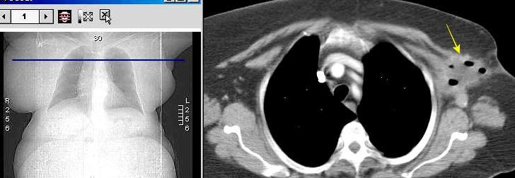
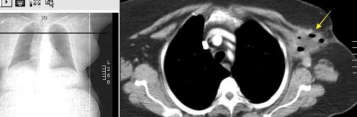
COMPLICACIONES DE DISECCIÓN AXILAR Seromas, Hematomas, Granulomas de sutura, Seudoaneurismas, Linfangiectasia
Kim EY et al. Sonography of axillary masses: what should be considered other than the lymph nodes? .J Ultrasound Med . 2009/ Meghana H, Jane C, Marilyn M. Axillary hematoma secondary to arterialpseudoaneurysm following prophylactic mastectomy. Case Studies in Surgery 201.
Kim EY et al. Sonography of axillary masses: what should be considered other than the lymph nodes? .
J Ultrasound Med . 2009/Meghana H, Jane C, Marilyn M. Axillary hematoma secondary to arterial pseudoaneurysm following prophylactic mastectomy. Case Studies in Surgery 201.
El grupo I tiene 3 subgrupos. Lateral (profundo). Subescapular. (posterolateral) y pectoral (anteromedial)
El II y III, uno cada uno.
Berg JW. The significance of axillary node levels in the study of breast carcinoma. Cancer 1955
Drenaje sigue del Grupo I al II, al III y de ahí al tórax
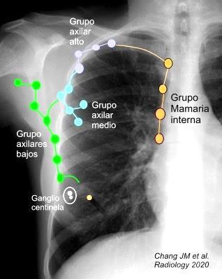
Chang JM et al. Axillary Nodal Evaluation in Breast Cancer: State of the Art. Radiology 2020/ Humprhey KL et alo Do or Not to Do: Axillary Nodal Evaluation after ACOSOG Z011 Tr i a l.. Rdiology /2014/Abe H. Ongoing Demand for Radiologists in Preoperative Axillary Lymph Node Assessment. Radiology 2021/Ecanow JS et al. Axillary Staging of Breast Cancer: What the Radiologist Should Know. Radiographics. 2013/Marino MA et al. Lymph Node Imaging in Patients with Primary Breast Cancer:. Concurrent Diagnostic Tools. The Oncologist 2020
Kim EY et al. Sonography of axillary masses: what should be considered other than the lymph nodes? . J Ultrasound Med . 2009/Meghana H, Jane C, Marilyn M. Axillary hematoma secondary to arterialpseudoaneurysm following prophylactic mastectomy. Case Studies in Surgery 201.
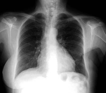
Linfoma muy agresivo. Complica pacientes con HIV+, trasplantes, inmunodeficiencias congénitas.
Linfoma de Burkitt en VIH+. Ganglios en axila izda.
Ferry FA. Burkitt's lymphoma: clinicopathologic features and differential diagnosis. Oncologist . 2006
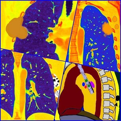
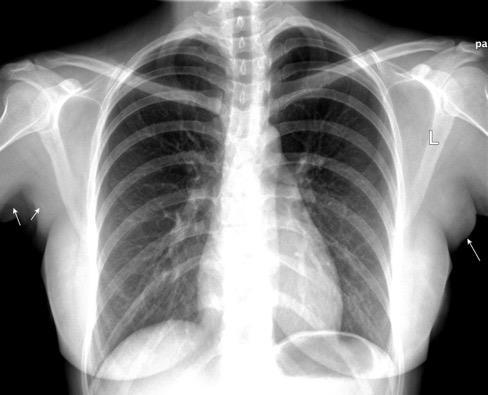
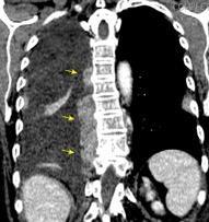
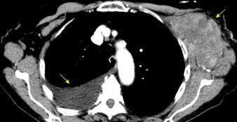
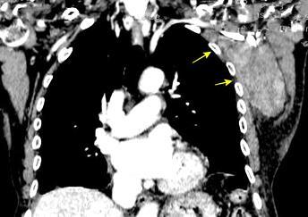
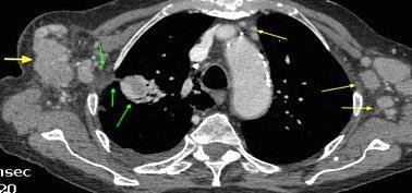
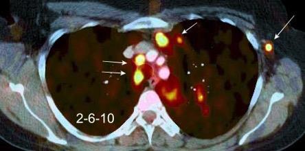
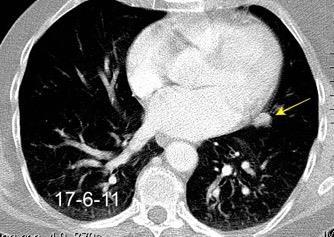
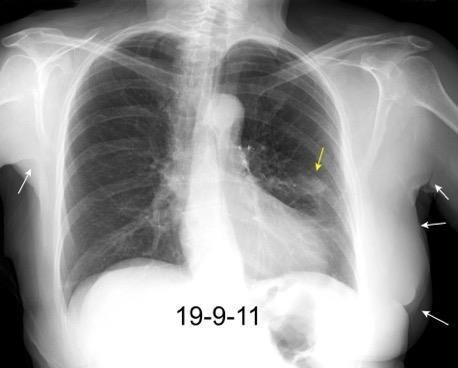
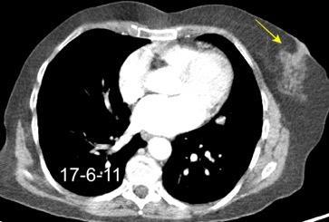
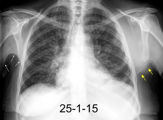
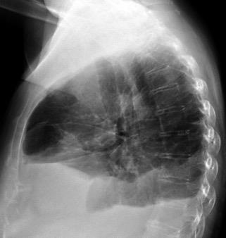
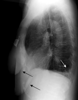
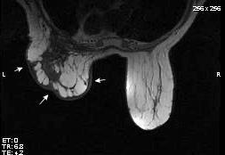
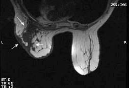
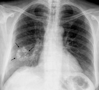
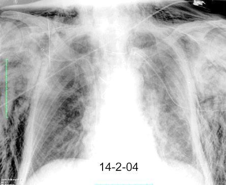
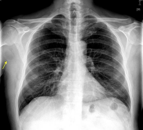
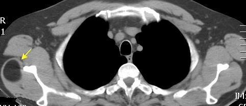
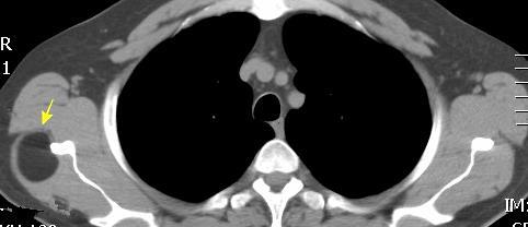
Masa axilar izquierda y derrame pleural derecho. Implantes pleurales, paraespinales . Ganglios en mamaria interna. Linfoma B difuso
Jaffe ES. Diagnosis and Classification of Lymphoma: Impact of Technical Advances.
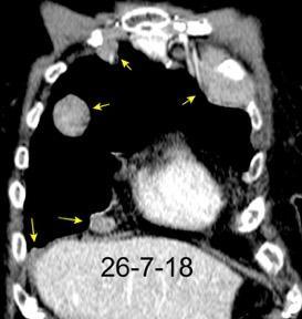
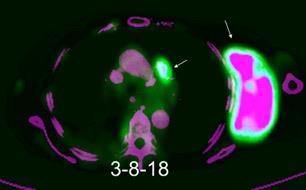
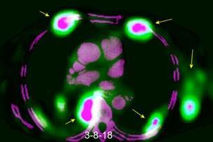
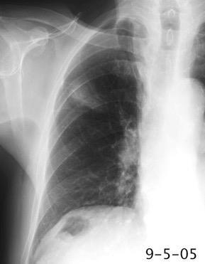
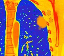
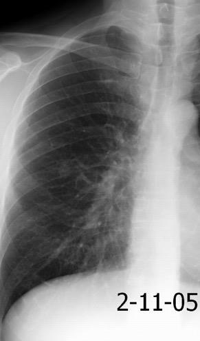
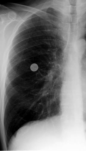
2005. Masa pulmonar.
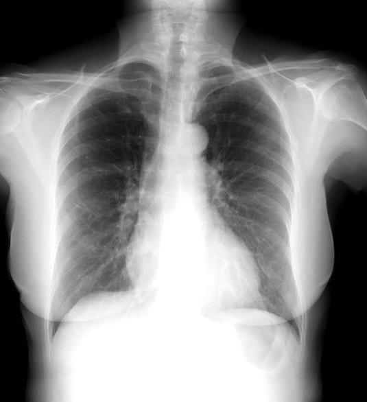
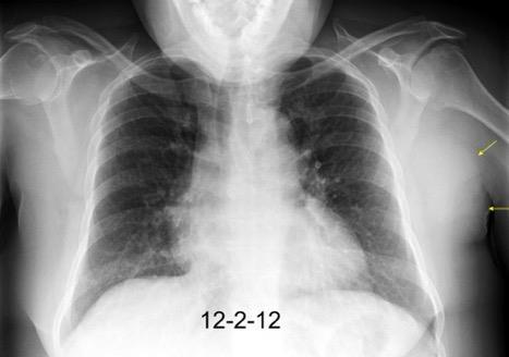
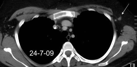
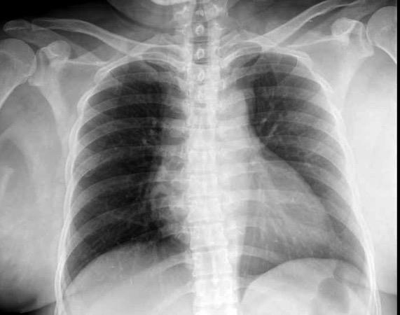
Pliegue axilar Normal
Rehúsa cirugía
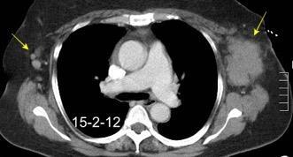
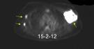
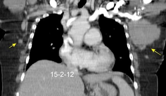
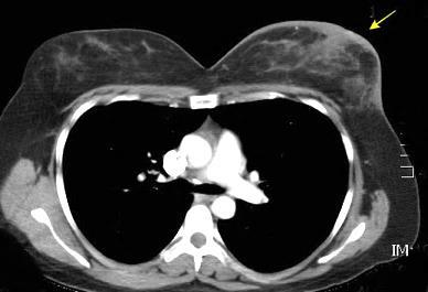
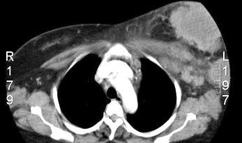
2011. Acude por masa axilar
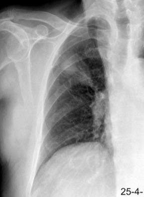
Linfoma NH de cél. B. Invasión Transtorácica.
Afectación axilar
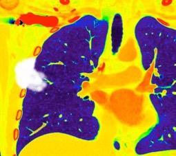
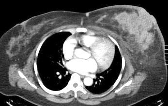
Conglomerado adenopático axila derecha. Linfoma de Hodgkin.
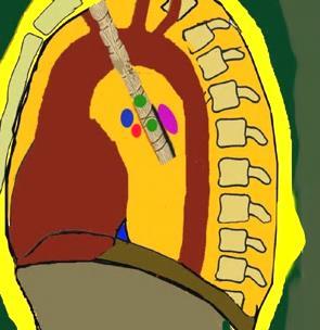
Santamarina MG et al. Multidetector CT for Evaluation of the Extrapleural Space. Radiographics 2017
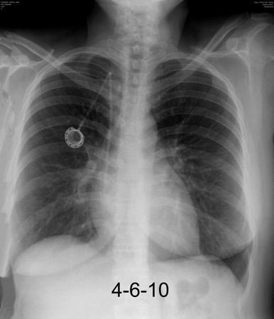
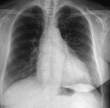
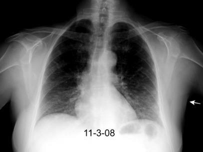
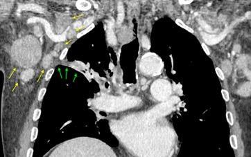
Carcinoma de mama izquierda
American College of Surgeons Oncology Group
Z0011 Trial. Duda sobre la limpieza axilar completa . Humprhey KL et al Do or Not to Do: Axillary Nodal Evaluation after ACOSOG Z011 Tr i a l.. Radiology / Cahoon AR ET AL. Internal Thoracic Lymphadenopathy in Breast Cancer. Radiographics 2017/ Marino MA et al. Lymph Node Imaging in Patients with Primary Breast Cancer:. Concurrent Diagnostic Tools. The Oncologist 2020
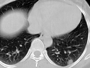
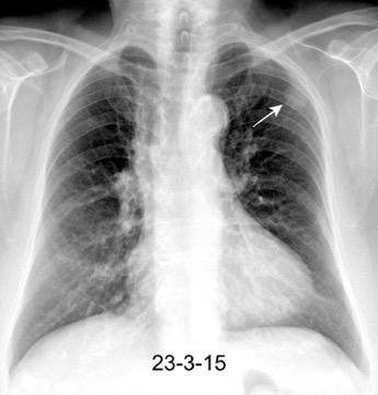
Nódulo en língula.
Cirugía de Ca .de mama hace 23 añosedema de brazo y ganglio en axila. ¡Metástasis!.

Ecanow JS et l. Axillary Staging of Breast Cancer: What the Radiologist Should Know. Radiographics 2013
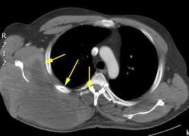
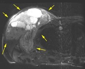
Síndrome mielodisplásico con anemia. Tumoración abrupta en axila por hematoma.
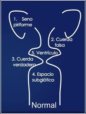
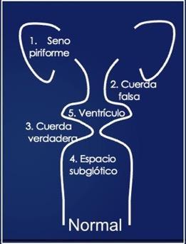
Normal
Becker M et al. Imaging of the larynx and hypopharynx. Eur J Radiol 2008
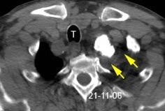
Bocio con nódulo calcificado

Calcificaciones ganglionares en antigua Tb. (“escrófula”). También en abscesos.
Keberle M et al. Physiologic and pathologic calcifications and ossifications in the face and neck. Eur Radiol 2007
Viejo delgado
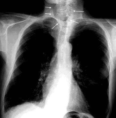
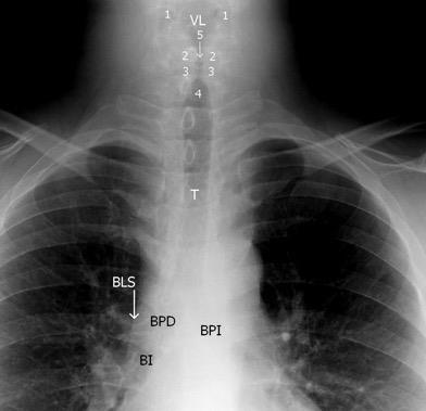
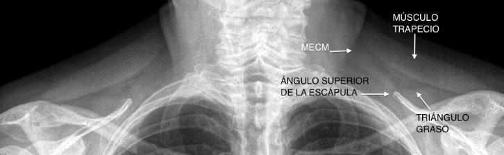
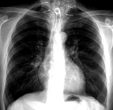
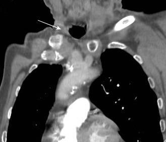
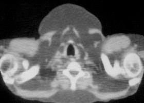
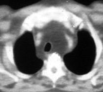
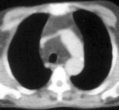
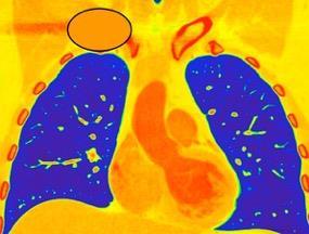
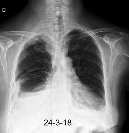
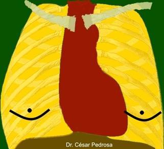
Se forma con las inserciones claviculares de ambos músculos esternocleidomastoideos. Particularmente vista en emaciados, personas muy delgadas y laringuectomizados previamente.
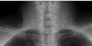
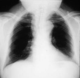
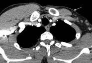
Paciente con cánula de traquestomía y fosa supraclavicular visible
Ominsky S et al. The Suprasternal Fossa. Radiology 1977
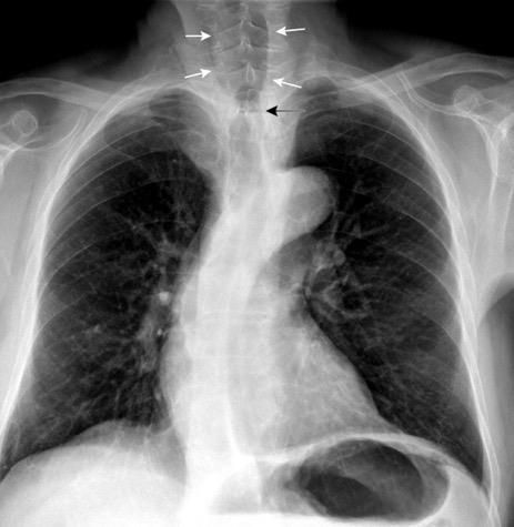
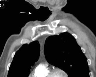
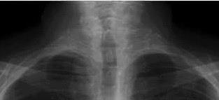
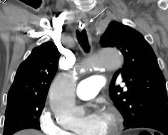
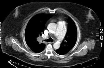
Paciente con traqueostomía previa. Estenosis relativa en el área de la cirugía. Fosa supraesternal visible.
Ominsky S et al. The Suprasternal Fossa. Radiology 1977
1/25000 hombres
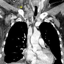
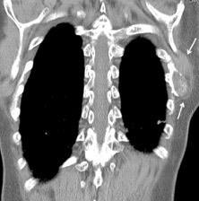
(Lipomatosis simétrica múltiple) Enfermedad rara. Múltiples masas de grasa no encapsulada en cara, cuello, hombros.
Más frecuente en área mediterránea.
90%: Hª de alcoholismo.
.
Distribución.8 pacientes.
Cuello (parte posterior)….8 (parte anterior ) …..7
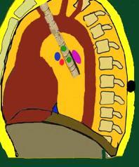
Mediastino superior ……….3
Espacios pre-traqueal y extrapleural
Gao S et al. Madelung disease. A case report. Medicine. 2019.
Ahuja AT et al. Madelung Disease: Distribution of Cervical Fat and Preoperative Findings at Sonography,MR, and CT Am J Neuroradiol. 1998
Linfangioma
Ganglios Linfoma Tb
Sarcoidosis
Metástasis (Laringe, Orofaringe, Pulmón)
van Overhagen H et al. Metastases in SupraclavicularLymph Nodes in Lung Cancer: Assessment with Palpation, US, and CT. Radiology 2004
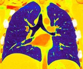
Derrame pleural..33,3% Derrame bilateral.26,7% Ganglios mediastínicos e hiliares..66,7%..
No hiliares
Okada F et al. Chest HRCT findings in acute transformation of adult T-cell lymphoma/leukemia. Eur Radiol 2015
Vidrio deslustrado 60%. Consolidación 33,3% Nódulos…33,3%
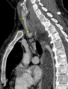
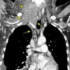
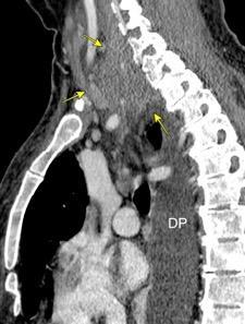
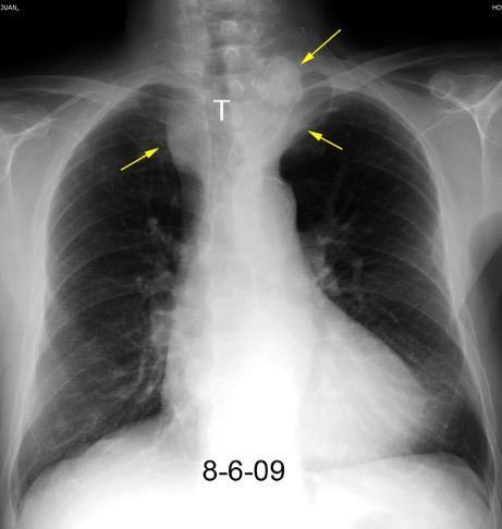
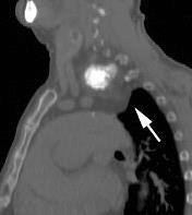
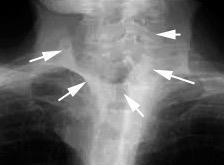
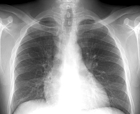
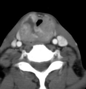
Carcinoma subglótico
Tumor poco frecuente. 1,8% de los tumores laríngeos.
Tendencia a invadir el anillo cricoideo y el esófago.
Ganglios + frecuentes
Mac Neill SD et al. Survival of patients with subglottic squamous cell carcinoma. Curr Oncol. 2018
Hallazgo temprano: Osteopenia general. Erosión marginal del borde inferior de la cabeza humeral.
Osteólisis del extremo externo de la clavícula.
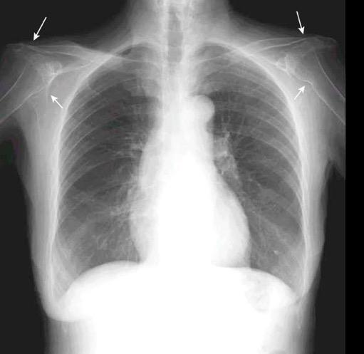
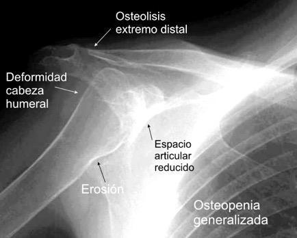
Cuomo F et al. THE RHEUMATOID SHOULDER.
Rheum Dis Clin North Am . 1998
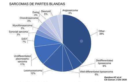
Gamboa AC et al. Soft-Tissue Sarcoma in Adults: An Update on the Current State of
Histiotype-Specific Management in an Era of Personalized Medicine. CA Cancer J Clin 2020
Asimetría mamaria
Puede ser una variante.
Se ha descrito con alteraciones cromosómicas
Trisomía del 18
Sínd. de Turner Disgenesia gonadal
Tipo XY Anomalías genitales Baja estatura
Tejerizo LC, et al..Anisomastia como trastorno de desarrollo en la patología mamaria infanto-juvenil..Rev Senología Patol Mam,. 1992.
Stratakis AC et al. Anisomastia associated with interstitial duplication of chromosome 16, mental retardation, obesity, dysmorphic facies, and digital anomalies: molecular mapping of a new syndrome by fluorescent in situ hybridization and microsatellites to 16q13 (D16S419-D16S503) . J Clin Endocrinol Metab.2000
Hudson SM et al. Left–right breast asymmetry and risk of screen-detected and interval cancers in a large populationbased screening population. Br J Radiol. 2020
Adenoma mamario
calcificado

Desarrollo Pubertad/ Senectud
Hipogonadismo
Tumores
Cirrosis
Fallo renal
Tiroxicosis
Ginecomastia
Ca. de pulmón
Congénitas Klinefelter
Sind. Resistencia a andrógenos
Alcoholismo
Herpes zoster
Trauma pared
Drogas
Sint. de Rohrich. 2003
Raso DS et al. Elemental analysis and clinical implications of calcification deposits associated with silicone breast implants . Ann Plast Surg. 1999
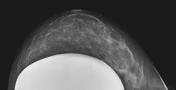
Prótesis
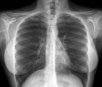
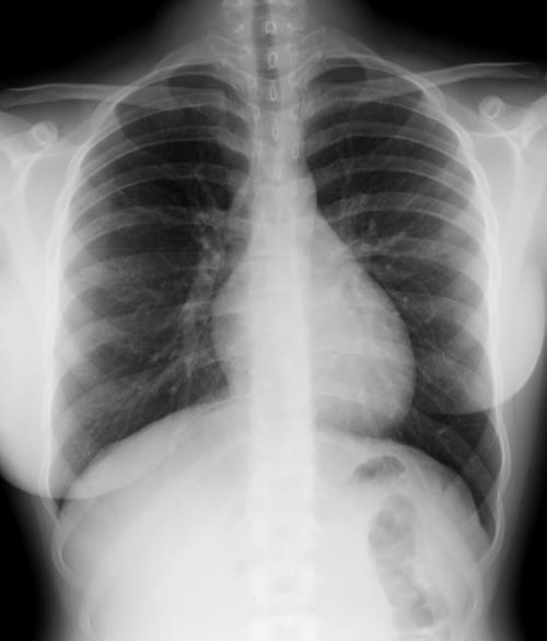
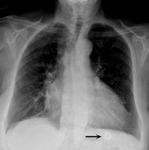
Sombra “simétrica y de densidad “uniforme” de las mamas
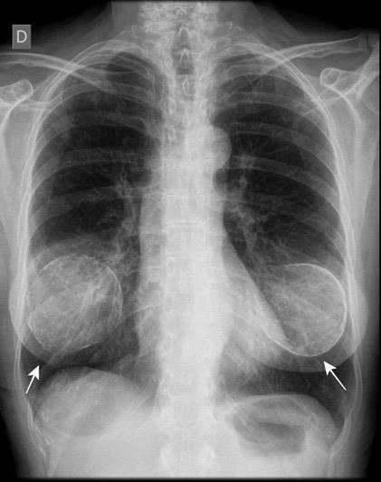
Prótesis de ambas mamas densamente calcificadas
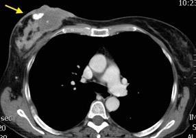
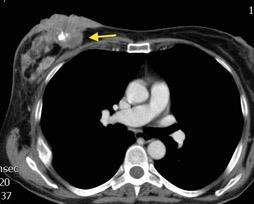
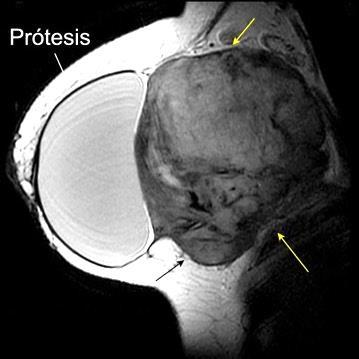
Tumor desmoide derecho. Recidiva.
Tumor maligno. 0,2% de los tumores de mama.
Ocurren en 10% de los implantes de mama.
En los 3 años después del implante.
(Dudas sobre el dato)
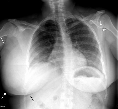
Kilmartin C et al. Desmoid Tumor and Implant-Based Breast Reconstruction. Case Rep Oncol. 2023
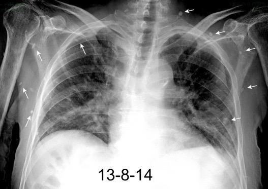
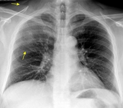
Enfisema subcutáneo Infección
Daño S. respiratorio o GI
Heridas penetrantes
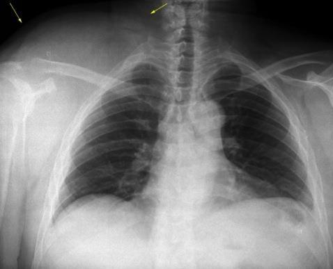
Ho M-L. Chest Radiography in Thoracic Polytrauma. AJR.2009.
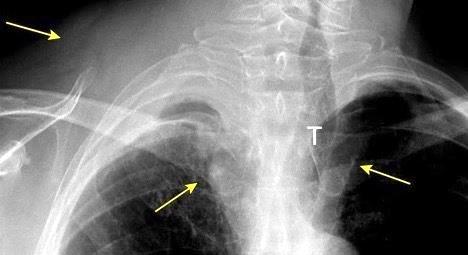
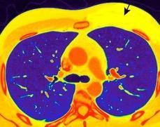
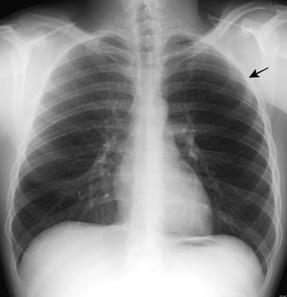
Radiografía AP en politraumatizado con enfisema subcutáneo severo.
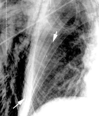
Kumar H M,et al. Ginkgo leaf sign and subcutaneous emphysema.BMJCase Rep .2018
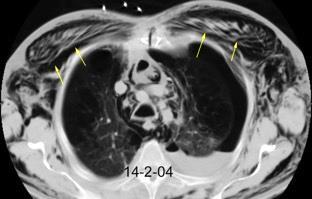
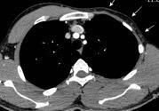
Asimetría de las partes blandas en Ca. de mama
Recidiva local Afectación de la piel
Calcificación metastática
Calcificación distrófica
Calcinosis tumoral
Condrocalcinosis
Dr. César Pedrosa
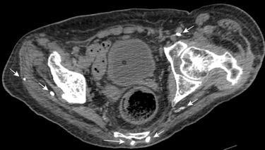
Calcinosis universalis.
Enf. tejido conectivo. Más frecuente:
Dermatomiositis juvenil 40%. Adulto 10%
Síndrome de Sjögren.
Calcinosis de partes blandas
Basado en Carr RB. Soft tissue Calcifications. Stern-Gurney. Expertdd.Chest. Mairsys. 2011/Hwang Z-A et al. Imaging Features of Soft-Tissue Calcifications andRelated Diseases: A Systematic Approach. Korean J Radiol 2018./Wasserman PL et al. MR imaging findings of calcinosis cutis in primary Sjogren syndrome, a rare manifestation. R a d i o l o g y C a s e R e p o r t s. 2 0 2 0.
Calcificación metastática
Calcificación distrófica
Calcinosis tumoral
Condrocalcinosis
Parasitosis
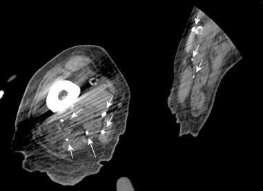
Basado en Carr RB. Soft tissue Calcifications. Stern-Gurney. Expertdd.Chest. Mairsys. 2011/Hwang Z-A et al. Imaging Features of Soft-Tissue Calcifications andRelated Diseases: A Systematic Approach. Korean J Radiol 2018./Wasserman PL et al. MR imaging findings of calcinosis cutis in primary Sjogren syndrome, a rare manifestation. R
Enfermedades. caquequizantes
Anorexia nerviosa
Carcinomatosis
Tuberculosis
Mucoviscidosis
Infec. crónicas
Enf. de priones (cérvidos)
Anorexia nerviosa. Absceso del LSI. Enterobacter
Kinderlehrer DA. Anorexia Nervosa Caused by Polymicrobial Tick-Borne Infections: A Case Study. International Medical Case Reports Journal 2021/Brown RF, Bartrop R, Beumont P, Birmingha CL. Bacterial infections in anorexia nervosa: delayed recognition increases complications. Int J Eat Disord. 2005.
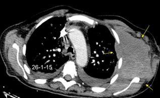
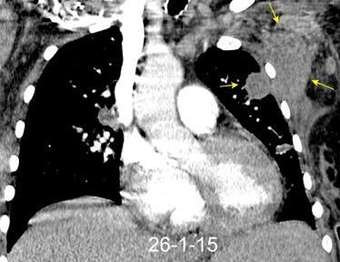
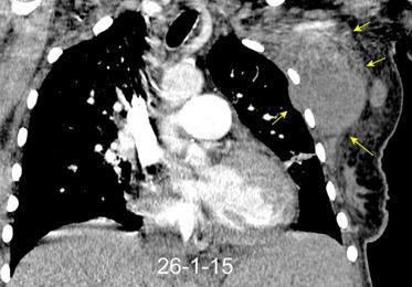
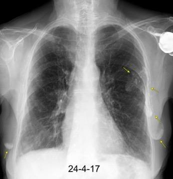
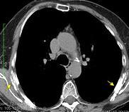
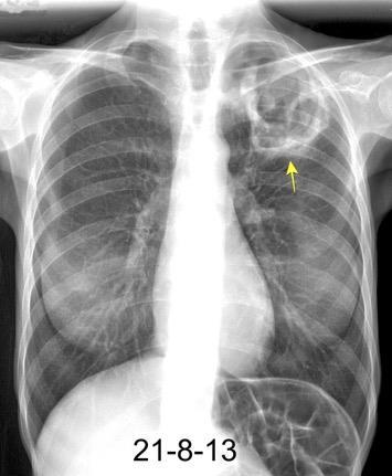
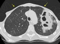
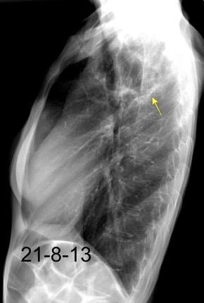
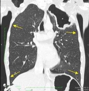
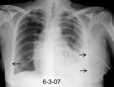
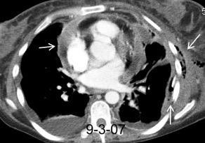
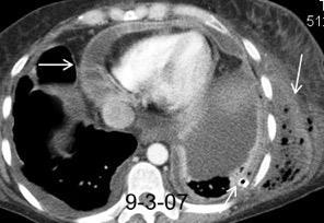
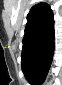
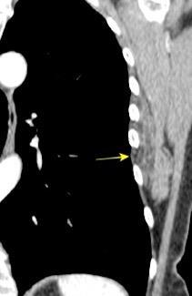
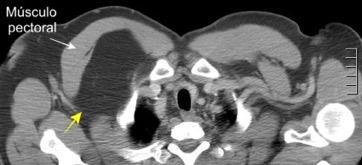
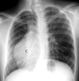
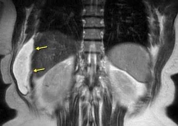
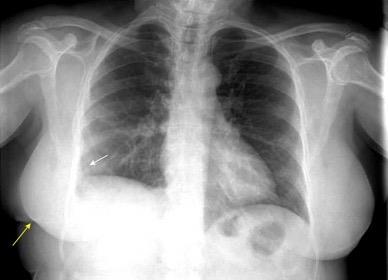
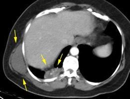
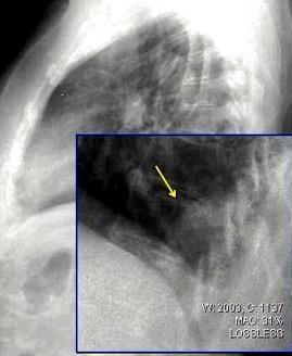
Infección pleural que se disemina a la pared torácica y piel. (Puede drenar espontáneamente)
Organismos: Micobacteria tuberculosa, intracelular. Actinomices
Estreptococo
Estafilococo (raro)
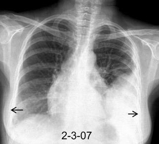
Signo específico: Expansión parietal con la tos
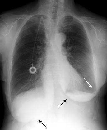
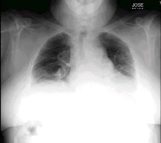
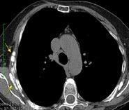
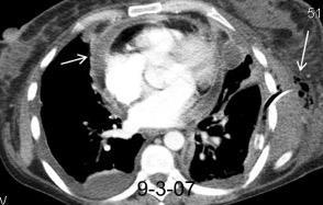
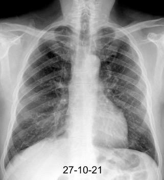
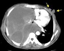
Neumonía por Estrept. neumonía. Derrame pericárdico. Empiema necesitatis (tubo de drenaje)
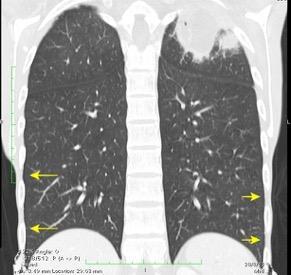
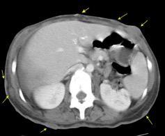
Edema de los tejidos extrapleurales subcostales (60%, 21 of 35).Takasugi et al. The extrapleural fat in empyema: CT appearance. Br J Radiol 1991
Crouch A et al. Spontaneous Rupture of Empyema Necessitans in the Emergency Department. Cureus 2021
Almeida BJ, et al.. Empyema necessitans: After recent thoracostomy in an immunocompromised patient. Respirology Case Reports. 2023;
Considerados como sarcomas de “bajo grado”. Invasión y recurrencia local frecuente.
Kabiri EH et al. Desmoid tumors of the chest wall.
European Journal of Cardiothoracic Surgery . 2001.
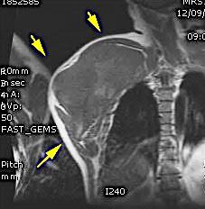
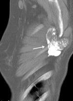
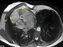
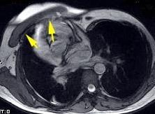
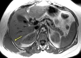
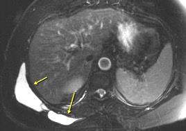
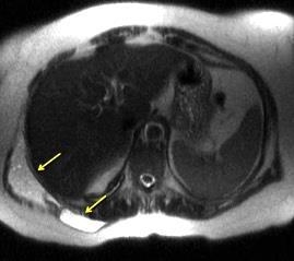
Tumor desmoide (“Fibromatosis agresiva”)
T1: Isointensos con músculos.
T2: Señal intermedia y de alta intensidad.
Presencia frecuente de áreas curvilíneas y lineales dentro de la lesión
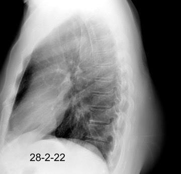
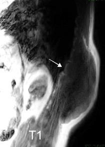
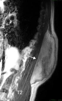
Mansour J et al. Diagnostic and Imaging Approaches to Chest Wall Lesions. Radiographics 2022
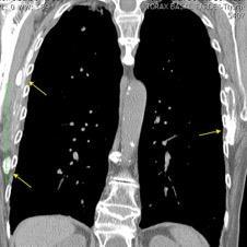
Densidad similar al músculo con estriaciones internas de grasa
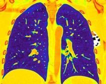
Hallazgo incidental . Región infraescapular 2% de TC del tórax. Bilateral 60%.
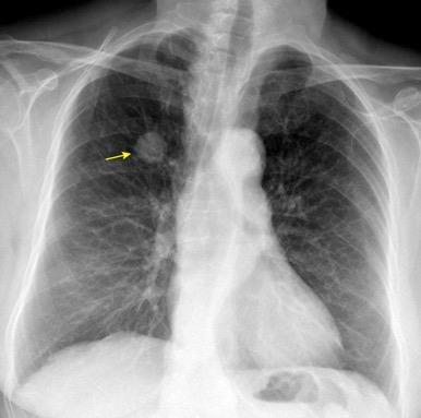
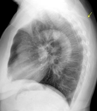
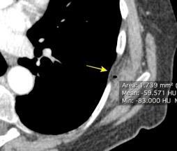
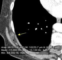
Burt AM et al. Imaging review of lipomatous musculoskeletal lesions. SICOT J2017/ Murphey MD et al. From the archives of the AFIP: benign musculoskeletal lipomatous lesions. Radiographics. 2004
50% de los tumores de partes blandas.
Predilección varones en lipomas profundos
30% “hereditarios”
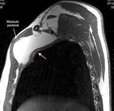
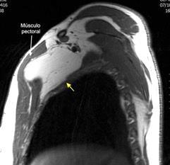
Isointenso con grasa subcutánea
Burt AM et al. Imaging review of lipomatous musculoskeletal lesions. SICOT J2017/ Murphey MD et al. From the archives of the AFIP: benign musculoskeletal lipomatous lesions. Radiographics. 2004
Burt AM et al. Imaging review of lipomatous musculoskeletal lesions. SICOT J2017/ Murphey MD et al. From the archives of the AFIP: benign musculoskeletal lipomatous lesions. Radiographics. 2004
50% sin historia familiar. Manchas en “café con leche” “Pecas”. “Moluscos” cutáneos
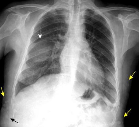
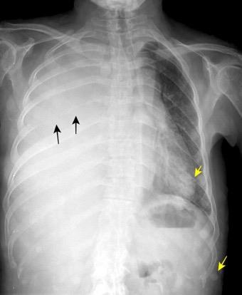
Evolución de neurofibroma a neurofibrosarcoma. Neurofibromas (moluscos) cutáneos múltiples
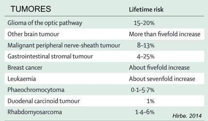
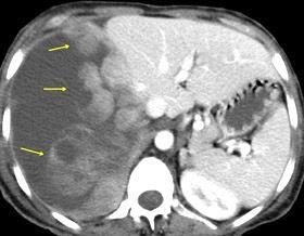
Hirbe AC et al. Neurofibromatosis type 1: a multidisciplinary approach to care. Lancet Neurol 2014;.
1/30.000 nacimientos
Hipoplasia/ausencia de mama o pezón.
Hipoplasia de tejidos subcutáneos.
Ausencia de la porción costoesternal del pectoral mayor.
Ausencia del pectoral menor.
Ausencia de cartílagos costales o costillas, 2,3 y 4.
Variante: Síndrome de Poland con dextrocardia.
Hipoplasia Art. pulmonar dcha. Atelectasia.
Bansal A, et al. Poland syndrome: a case report. BMJ Case Rep 2017
Mutlu H et al. A variant of Poland syndrome associated with dextroposition. J. Thorac. Imaging. 2007
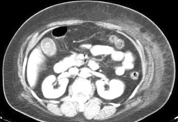
10-20% de pacientes las larvas alcanzan la circulación sistémica y pueden llegar a cualquier órgano También pueden llegar a órganos intrabdominales por migración transmural.
Yagmur Y et al. Unusual Location of Hydatid Cysts: A Case Report and Literature Review. Int Surg. 2012 / Pedrosa I et al. Hydatid disease: radiologic and pathologic features and complications . Radiographics 2000.
Imitadores de lesiones.
Verrugas Tumores dérmicos
Callos de fractura
Verruga en la espalda
Mansour J et al. Diagnostic and Imaging Approaches to Chest Wall Lesions. Radiographics 2022
Mansour J et al. Diagnostic and Imaging Approaches to Chest Wall Lesions. Radiographics 2022