The Member Newsletter of the Society of Breast Imaging
INSIDE THIS ISSUE:
• This issue's theme: Leading Through Adversity, Natural Disasters: Lessons Learned in Academic and Private Practices
• New Year's Resolutions That Help Us Renew and Reconnect
• Meet Our New SBI Fellows
• Highlights From the EUSOBI and RSNA 2024 Annual Meetings
• IDEA (Inclusion Diversity Equity Alliance) Insights on Global Outreach Efforts in India
EDITOR:
Nidhi Sharma
ASSISTANT EDITORS:
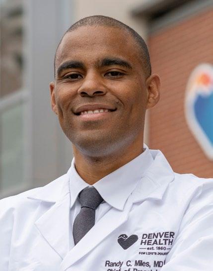
Randy Miles and Shinn-Huey Shirley Chou
TECHNOLOGISTS’ COLUMN:
Robyn Hadley and Sarah Jacobss
WHAT’S NEW IN THE NEWS:
Eleanor DiBiasio and Pamela J. DiPiro
MEMBERS IN TRAINING:
Anita Mehta
WELLNESS COLUMN:
Sarah Jacobs and Claudia Cotes
THE PATIENT'S PERSPECTIVE:
Hannah Perry and Danielle Sharek
LEGISLATIVE UPDATES:
Amy Patel
CANADIAN CORNER:
Supriya Kulkarni
Linda Moy,
MD, FACR, FISMRM, FSBI President, Society of Breast Imaging
OUR SBI MISSION:
For members to be expert and authoritative breast imagers working in supportive practice environments who advance the highest quality of breast care via early detection, diagnosis, and treatment.
OUR SBI VALUES:
Patient-centered and evidence-based care
Excellence in education
Scientific integrity
Collaboration and collegiality
Respect for diversity and inclusiveness
President’s Column
I wish our SBI family a wonderful winter holiday season. Nine months into my presidency, I am impressed with the hard work our SBI committee members perform to support our mission. During Breast Cancer Awareness Month, I saw our tireless efforts to promote screening mammography and to advocate for our patients. Moreover, we are an inclusive society. Our 2024 SBI symposium in Montreal reinforced our bonds with the Canadian Society of Breast Imaging. In this issue, Dr. Kulkarni provides screening updates from Canada. I am so pleased that Dr. Paula Gordon will be awarded the SBI Gold Medal at our 2025 SBI symposium. We also continue to build relationships with colleagues from other societies. The SBI is an organizational supporter of the Society of Interventional Oncology’s Breast Cryoablation Mini Master Class that will take place in Las Vegas in February 2025.
I am also impressed with the resilience of the breast imaging community. We are facing a decrease in the supply of radiologists specializing in breast imaging while the demand for imaging services and maintaining access for our patients is increasing. Although artificial intelligence tools may ease the burden of our ever-increasing workload, they cannot replace the relationships and trust we have formed with our patients.
Finally, I encourage you to attend the 2025 SBI Breast Imaging Symposium, which will be held April 24-27, 2025, at the Broadmoor in Colorado Springs, Colorado. Registration is open.
Linda Moy
Linda Moy, MD, FACR, FISMRM, FSBI President, Society of Breast Imaging
Editor’s Note
By Nidhi Sharma, MD
It’s not only moving that creates new starting points. Sometimes all it takes is a subtle shift in perspective, an opening of the mind, an intentional pause and reset, or a new route to start to see new options and new possibilities.
–Kristin Armstrong1
Each new year ushers in both a sense of possibility and a time for reflection. We’re reminded that true progress is not simply about looking ahead—it’s about understanding the forces shaping our world each moment. January, with its promise of renewal, invites us to think boldly and act with purpose.
Since serving as the newsletter editor for the past few years, I wanted to take a moment to share some observations and ask for your partnership. A question I have been asked often by many connections I’ve made is “What has been the best thing for you in your time in this role and serving on various other committees?” Without hesitation, my response has been an earnest “Our community.”
Whether it’s the many events throughout the year organized by partnerships and new ideas emerging from various committees that showcase our stellar members (summer live webinars, early career series, and Breast Cancer Awareness Month social media campaign, just to name a few) or community events like the talent show, President’s Gala at the symposium, Radiological Society of North America fellows meeting, and more, our community shows up! At the same time, it is also true that members of our community have supported each other through hard times, like the pandemic and all the challenges that came with it.
As I reflect on those things, the strength of our society reminds me of a quote that I once read from the late, great author and activist, Coretta Scott King: “The greatness of a community is most accurately measured by the compassionate actions of its members.”
To me, this perfectly captures one of the very special aspects of our SBI community. On that same note and from a personal view,
I want to express my profound gratitude to everyone I’ve reached out to in the past few years for guest articles, coming forth with excellent informative articles for our membership. Thank you!
Our winter 2025 issue’s theme is “Leading Through Adversity: Natural Disasters.” It is important to be prepared well when faced with an impending extreme weather event. We hear from experts in the field from academic practice in Tampa, Florida, and private practice in Asheville, North Carolina, with tips to prepare and how they dealt with the recent hurricane aftermath. In addition, this issue has multiple engaging articles including wellness New Year resolution tips, cryoablation for breast tumors, screening updates from Canada, and much more.
I am asking for your continued partnership. In addition to the annual symposium, SBI Connect is a vibrant online discussion forum. I invite you to join the conversation in these multifarious SBI initiatives. We encourage microvolunteering, so you don’t necessarily have to commit to a long-term service goal and can still contribute to our SBI News initiatives! If you have any new ideas to share with the community, please reach out to me at nidhisharma31@gmail.com. Thank you for reading this edition of SBI News. I am excited to meet you all in Colorado Springs to celebrate SBI’s 40th anniversary at the symposium, with an amazing lineup of speakers organized by the outstanding program committee, led by Dr. Peter Eby.
Here’s to a 2025 filled with possibility!
References
1. Armstrong K. Mile Markers: The 26.2 Most Important Reasons Why Women Run. Rodale Books; 2011.
Nidhi Sharma, MD
Fellowship Match Committee Update
By Janine Katzen, MD
The Breast Imaging Fellowship Match was established in 2017 with the intent of improving the application and interview process for our future breast imaging radiologists. The Fellowship Match Committee was initially tasked with promoting and supporting the Fellowship Match. For the current 2024-2025 application cycle, 99 programs have signed on to participate in the Match. If all of these programs register with the National Resident Matching Program this will represent record engagement.1 Satisfaction with the Match has been demonstrated among both applicants and program directors, yielding a more equitable process for all.2-4
Due to the tremendous efforts of our predecessors on the committee, the Breast Imaging Fellowship Match is now well established, permitting the members of the committee to pursue additional endeavors focused on education and outreach. Cross-committee engagement has been another priority of the Fellowship Match Committee.
Early Career Curriculum Lecture Series
Created as a joint venture led by Drs. Rend Al-Khalili and Allison Aripoli (from the Early Career Section Committee) and members of the Fellowship Match Committee, the first-ever SBI Early Career Webinar Series launched in September 2023 with an initial lineup of three weekly lectures. Due to the success of the initial series, an expanded four-lecture series was held in the fall of 2024. Topics included identification and pursuit of high-value endeavors; communication skills and working with a multidisciplinary team; maintaining breast community involvement in private practice; and burnout recognition, prevention, and management. We hope we can continue to find new and interesting ways to engage those who are newly entering practice.
Medical Student and Resident Interesting Case Webinar
This initiative was born out of another collaboration between members of the Fellowship Match Committee and the Resident and Fellow Section Committee. Dr. Carol McLaughlin spearheaded an effort to increase engagement and recruitment of future breast imaging radiologists. An open call for submissions from medical student and resident trainees had tremendous response, with 45 case submissions. The top six cases were selected for a live webinar held this past fall with over 160 attendees. A huge thank you to all organizers, judges, and participants!
Standardization of Breast Imaging Fellowship Websites
In another collaboration with the Resident and Fellow Section Committee, led by Dr. Heba Albasha, a suggested standardized breast imaging fellowship web page template has been posted on the SBI website. This template suggests information for breast imaging programs to have on their websites, including application
requirements, available positions, approximate interview dates, curricular information, and call expectations, all of which are highly relevant for applicants attempting to discern which programs fit their educational needs.
Breast Imaging Fellow Evaluation Form
Constructive feedback is a key component of the educational experience. Despite this knowledge, we frequently do not provide our trainees with this feedback as often or consistently as we would like. Members of the committee created a standardized evaluation form and posted it on the SBI website as a valuable resource for all programs to use. The committee hopes this will empower programs to provide their trainees with more consistent feedback.
Inquiry
The Fellowship Match Committee is continually seeking innovative ways to optimize the Fellowship Match and educational process. Previous topics of inquiry have included national trends in call responsibilities, graduated autonomy in the fellowship year, and knowledge of the resources available on the SBI website. Please keep an eye out for future surveys! We look forward to continuing to identify new ways to engage with members of the SBI.
References
1. Results and data: specialties matching service, 2023 appointment year. National Resident Matching Program. April 1, 2023. Accessed January 7, 2025. https://www. nrmp.org/match-data/2023/04/results-and-data-specialties-matching-service2023-appointment-year/
2. Katzen JT, Gotian R, Brem RF. Breast Imaging Fellowship Match: program directors’ perspectives in year two J Breast Imaging. 2019;1(3):244-248. doi:10.1093/jbi/wbz033
3. Lee MV, Katzen JT, Al-Khalili R, Choudhery S, Whitman G, Brem R. Breast Imaging Fellowship Match: applicants’ perspectives of years two and three J Breast Imaging. 2020;2(5):471-477. doi:10.1093/jbi/wbaa065
4. Mullen LA, Nguyen DL, Katzen JT, Brem RF, Ambinder EB. Virtual interviews for breast imaging fellowship during the COVID-19 pandemic: perspectives of program directors and applicants J Breast Imaging. 2022;4(3):309-319. doi:10.1093/jbi/wbac017
Janine Katzen, MD
HIGHLIGHTS OF THE EUSOBI ANNUAL SCIENTIFIC MEETING 2024
By Miguel Braga, MD; EUSOBI Young Club Committee: Anna D’Angelo, MD; Giulia Vatteroni, MD; Ioana Bene, MD; Iva Biondic Spoljar, MD; Marianna Fanizza, MD; Machteld Keupers, MD; Maria Adele Marino, MD; Melis Baykara Ulusan, MD; Simone Schiaffino, MD; Stephanie Meyer, MD; Thiemo van Nijnatten, MD, PhD; Michael Fuchsjäger, MD
The European Society of Breast Imaging (EUSOBI) Annual Scientific Meeting 2024 took place from October 3 to 5 in Lisbon, Portugal. This vibrant city provided the perfect setting for the conference as attendees were able to enjoy Lisbon’s rich history and warm hospitality.
The meeting offered live lectures both in person and online, with sessions available on demand for those unable to attend in person. For the first time ever, these on-demand sessions included subtitles in Arabic, Italian, Portuguese, and Spanish. The program featured a mix of educational and scientific sessions that catered to all levels, from basic to advanced topics.
Building Momentum: Precongress Events
The precongress event was a highlight in itself, featuring the course “Breast Imaging With MRI, Opening to CEM.” In this course, experts discussed the latest techniques in breast magnetic resonance imaging (MRI) and contrast-enhanced mammography (CEM), covering technical aspects, image interpretation, and clinical indications. The course also included small-group workshops in which attendees had the opportunity to discuss clinical cases and build confidence in applying this newly acquired knowledge.
Young radiologists also had the option to attend a symposium organized by the EUSOBI Young Club titled “Top Tips from the Experts.” This session offered valuable insights into effective communication in clinical practice along with guidance on navigating academia and achieving professional growth.
Exploring Innovations: Keynotes and Sessions
The meeting offered a comprehensive overview of current trends and advancements in breast imaging. It opened with a foundational session on topics such as the upcoming BI-RADS update, breast lesion assessment, practical tips for interventional procedures, and the implementation of CEM in clinical practice. Attendees also received an overview of the latest EUSOBI guidelines and recommendations, ensuring the audience stayed up to date with the latest evidence.
The critical role of radiologists in managing benign, high-risk, and malignant lesions was highlighted throughout the conference. Topics such as breaking bad news, staging, and evaluating treatment response, including the challenges of managing residual calcifications after chemotherapy, were addressed. Treatment de-escalation was also a major focus, with discussions on how radiologists play a key role in selecting patients for more conservative approaches. Talks covered watchful waiting for patients with ductal carcinoma in situ, imageguided procedures as alternatives to surgery in low-risk patients, and
To save lives and minimize the impact of breast cancer.
how systemic therapy adjustments can be guided by imaging findings.
Risk assessment was another hot topic, as breast imaging is shifting from a one-sizefits-all approach to more personalized screening strategies. Emerging imaging methods were also explored; these included breast MRI, which is continuing to be developed, and newer modalities like photoacoustic and microwave imaging, which are under research. Artificial intelligence was widely discussed, with a focus not only on recent advancements but also on the challenges of clinical implementation, including the use of large language models.
The meeting concluded with an insightful session on the impact of breast imaging on both radiologists and patients as well as on the environment. Topics included managing burnout, promoting green radiology, and understanding the patient’s perspective.
The congress was also an occasion to strengthen EUSOBI’s connections with breast imaging societies worldwide. A session titled “EUSOBI Meets Singapore” featured talks on imaging after oncoplastic breast surgery and the development of a cross-border distance learning course for radiology residents in Singapore. Additionally, a lecture on precision medicine delivered by Dr. Linda Moy was held in collaboration with the SBI.
Honoring Achievements in Breast Imaging
The EUSOBI Annual Scientific Meeting recognized outstanding contributions to breast imaging. Recipients of the EUSOBI Young Researcher Grant, Young Physician-Scientist Grant, and Carla Boetes Award presented their research. Named in honor of a leading breast radiologist who dedicated her life to advancing breast cancer diagnostics, the Carla Boetes Award supports promising young researchers in this field.
Awards were also given to the most cited article in European Radiology (“Clinical Value of Radiomics and Machine Learning in Breast Ultrasound: A Multicenter Study for Differential Diagnosis of Benign and Malignant Lesions,” by V. Romeo, from Naples, Italy) and the most cited article in Insights Into Imaging (“High-Risk Lesions of the Breast: Concurrent Diagnostic Tools and Management Recommendations,” by D. Avendano, from Monterrey, Mexico). The prestigious EUSOBI Gold Medal was awarded to Anne Tardivon, from Paris, France, for her exceptional career and contributions to breast imaging.
Miguel Braga, MD
Resolutions That Help Us Renew and Reconnect
By Sarah Jacobs, BS, RT(R)(M)(CT)
As the calendar has turned to a new year, we are provided with an opportunity for reflection and renewal and a chance to create a path for fulfillment in the new year. For many of us working in breast imaging, our daily efforts consistently focus on the well-being of our patients, and we often forget to focus on our own well-being. The demanding nature of providing care to our patients in breast imaging can limit time for self-care and result in feelings of burnout. Setting New Year’s resolutions can serve as a tool to renew our professional passion and support healthier work-life integration. Resolutions that focus on strengthening workplace connections, prioritizing self-care, and improving work-life integration can contribute to a higher level of professional and personal fulfillment. Resolutions centered around renewing our sense of purpose and spending more time on activities that promote self-care and collaboration with our team can help us reconnect with one another and with our patients.
Strengthen Workplace Connections
The health care environment thrives on collaboration and teamwork. Building stronger connections with colleagues can transform the workplace into a more supportive and resilient community. The following resolutions can strengthen a team’s resilience and build stronger connections.
• Practice gratitude: A simple yet impactful resolution is to express appreciation for your coworkers. Verbal acknowledgment, thankyou notes, or informal celebrations of team successes can foster a culture of gratitude, strengthen morale, and create resilient teams.
• Provide routine peer support: Make it a priority to check in with your colleagues regularly. Participate in team huddles or rounds opportunities. Consider volunteering to organize or participate in initiatives such as employee engagement or team-building activities.
• Provide in-person communication: In an environment where working remotely or communicating virtually is commonplace, more frequent face-to-face communication is helpful to promote collaboration and teamwork with colleagues and patients.
• Promote inclusion and collaboration: Resolutions that improve teamwork may include inviting less vocal team members to share input during discussions. Providing a safe, supportive environment for team members to share thoughts and actively address conflicts in a constructive manner can promote a more cohesive, productive, and resilient working environment.
• Participate in social events: Workplaces that offer opportunities for social connection such as holiday gatherings and other team-building exercises can promote mutual trust. Attending and organizing such events can strengthen relationships among coworkers.
Prioritizing Self-Care
Members of a busy breast imaging team are often so focused on caring for others that they may neglect their own physical and emotional well-being. A commitment to self-care is not only essential for personal health but also crucial for sustaining the energy and focus needed to care for patients effectively. Resolutions that promote a higher level of self-care may include the following:
• Set boundaries: A key resolution for self-care is learning to establish boundaries that protect your time and energy. Whether it’s declining extra shifts or disconnecting from work communication during days off or at the end of the workday, boundaries can help create a better work-life balance. It’s important to embrace a mindful transition between work and home.
• Adopt a mindfulness practice: Mindfulness has been shown to reduce stress and improve focus. Commit to a daily mindfulness practice such as meditation, yoga, or even three minutes of deep breathing exercises in between tasks, patient examinations, or procedures. There are multiple free applications that you can download in addition to online resources that provide guidance for mindfulness exercises. Employee assistance or employee health programs can also be valuable resources to assist in implementing mindfulness practice in the workplace.
• Prioritize physical health: Another essential resolution is to maintain your physical health through regular exercise, nutritious eating, and adequate sleep. Scheduling time for exercise, meal prepping, and even a specific time each night to disconnect from technology can help improve physical health and energy levels. These activities can be scheduled as you would any other appointment, making them nonnegotiable parts of your daily or weekly routine.
• Indulge in hobbies and passions: Dedicate time to activities outside work that bring joy and relaxation. Moments of leisure such as reading, spending time in nature, devoting time to the arts and music, or spending time with loved ones can recharge your spirit and improve your mental health.
Embrace Work-Life Integration
For many of us, the concept of a balance between work and personal life can be challenging due to the unpredictable nature of our schedules. Instead of striving for balance, embracing work-life integration may be a more practical resolution.
Continued on page 10>
Sarah Jacobs, BS, RT(R)(M)(CT)
Breast Imaging Protocols: Why, How, and When
By Robyn Hadley, RT(R)(M); Sarah Jacobs, BS, RT(R)(M)(CT)
Implementing a policy and procedure manual is standard practice at most imaging facilities and is a requirement for maintaining compliance with regulatory guidelines. However, it is surprising that establishing and adhering to standardized protocols for breast imaging teams is often overlooked by facilities. Many team members expect that necessary imaging protocols are included in the facility’s policy and procedure manual. While this may be true to some extent, a policy and procedure manual generally does not include the comprehensive and inclusive imaging protocols necessary to establish clear expectations for imaging technologists to elevate efficiency and quality practices. This article identifies the value of a standardized imaging protocol manual, the critical elements to include, personnel involved, and recommendations for the development of these protocols.
Standardized imaging protocols provide these benefits:
• Drive efficiency and improve consistency
• Reduce errors
• Help minimize patient anxiety
• Promote positive patient experiences and working environments
• Lower technical callback rates and reduce unnecessary callbacks
• Decrease unnecessary interruptions between the interpreting radiologist and technologist
• Provide an outline for technologist training and competency assessment for new hires and current employees
• Reduce the possibility of litigation
Clear expectations set forth by substantiated imaging protocols limit confusion, define workflow, create uniformity, reduce misunderstandings among team members, and provide every patient with the same high-quality care.1
Imaging departments are extremely busy, which can lead staff members to make small adjustments in an attempt to stay on schedule or catch up. Under those circumstances, there is risk of deviation from established standard processes, potentially causing an increase in errors, neglect of specific aspects of care,
and a decrease in image quality that leads to technical recalls and increased patient anxiety. Revenue is also lost when a technical callback examination takes the place of a revenue-producing new patient screening examination or diagnostic examination for a patient with an acute clinical finding.2 A patient leaving to go to another facility due to an unsatisfactory experience and employee turnover costs related to unsupported practices also carry the risk of additional lost revenue.
Deviation from established protocols can increase the incidence of errors, therefore increasing the risk of litigation. The likelihood of a radiologist being the defendant in at least one lawsuit is 50% by age 60 years; however, the frequency and average number of suits accrued varies widely by state of residence and sex.3 Patients may bring legal charges against radiologists or imaging facilities for a number of reasons, including failure to diagnose breast cancer or negligence due to a fall resulting in injury. Established protocols and appropriate training can help prevent patient falls or injuries during mammography. Although not all falls can be prevented, specific protocols relating to patient falls can help the technologist know exactly what steps to take for prevention and how to appropriately respond and follow up should a fall occur.
When technologists, physicians, or other personnel interrupt an interpreting radiologist to check protocol for an examination or answer questions, the radiologist’s attention may be taken away from the examination at hand, potentially decreasing accuracy and increasing the likelihood of dictation errors, missed diagnoses, and higher recall rates.4 A study by Shah et al found that some radiologists spent as much time on interruptions as they did interpreting studies.5 Standardized protocols that outline clear guidance and imaging expectations can help limit radiologists’ workflow interruptions by decreasing the number
Robyn Hadley, RT(R)(M) Sarah Jacobs, BS, RT(R)(M)(CT)
of questions from imaging staff members and protecting radiologists’ time for examination interpretation.
Establishing Standardized Imaging Protocols
How to establish standardized imaging protocols and who to involve during this process should be strategically considered. Forming a team of representative staff members such as the medical director, interpreting radiologists, radiation officer, physicist, staff technologists, nurses, administrators, and auxiliary personnel such as receptionists should be considered when drafting the imaging protocols. Because technologists and radiologists will be performing and interpreting examinations and finalizing the protocols, their involvement and acceptance is imperative. Protocols must be constructed from evidence-based material, well-recognized references, peer-reviewed literature, and published guidelines.1 This process includes using resources such as the ACR Practice Parameters, Mammography Quality Standards Act regulations, ACR Appropriateness Criteria, and peer-reviewed literature from medical journals and imaging societies. A solid protocol thoroughly and specifically describes what needs to be done. Consistency in protocol performance is essential, especially if a patient receiving diagnostic imaging is having a follow-up examination and accurate, reproducible imaging is critical.
Considerations for protocol development include the following:
• Scheduling guidelines
• Patient preparation and history intake
• Screening and diagnostic imaging guidelines
• Diagnostic callback imaging guidelines
• Breast procedure imaging guidelines
• Use of skin markers for scars, skin lesions, and abnormalities
• Technical callback imaging guidelines: explanation of the technical callback procedure, threshold for technical callback images, and a script to use when calling patients to request a return for additional images due to technical callback
• Image acquisition and quality check:
- A standardized positioning technique, including a standardized sequence of acquiring mammographic views, to be implemented and used by all technologists to ensure consistency and image reproducibility
- A standardized positioning technique to promote proper use of body mechanics to maintain technologists’ physical well-being
- An image quality checklist for reference, along with appropriate and consistent methods of measuring the posterior nipple line on both the craniocaudal and mediolateral views
• Supplemental views:
- Appropriate use of supplemental views, including the exaggerated craniocaudal lateral view, anterior compression view, nipple-in-profile view, and repeat views for motion, artifact, and skin or fat folds
- Well-defined guidelines for imaging skin and fat folds, which are among the most difficult elements to create guidance for within imaging departments
• Special imaging considerations:
- Emergency protocols for patient adverse events, contrast agent reactions or extravasation, and examination or procedure complications
- Protocol for skin tears to include patient education and postexamination care
- Special patient circumstances and patients with physical limitations
- Scripts for documenting patient history and information for radiologists
Imaging protocols should not include adverse or sentinel event procedures but should provide a clear reference to the location of that information in the facility’s policy and procedure manual with the specific policy number or title. Additional detailed facility policies that supplement the protocol should be listed in the protocol manual for quick reference.
All responsible personnel should clearly understand, adhere to, and annually review a comprehensive standardized protocol guidebook. Team members at all of the organizational locations and satellite affiliations must have access to and knowledge of updated protocols. Options for ensuring ease of accessibility may include online authorization to a shared file, a protocol manual binder in each imaging room, and a designated central location at the technologist workstation. Establishing and maintaining standardized protocols is essential for fostering consistency within an imaging team and elevating the quality of patient care.
References
1. Thomas K, Farrell MB. How to write a protocol: part 1 J Nucl Med Technol. 2015;43(1):1-7. doi:10.2967/jnmt.114.147793
2. Smith-Foley S. Elevating the patient experience in breast imaging. AHRA Online Institute webinar. April 30, 2024. Accessed January 8, 2025. https:// onlineinstitute.ahra.org/products/elevating-the-patient-experience-in-breastimaging
3. Baker SR, Whang JS, Luk L, Clarkin KS, Castro A 3rd, Patel R. The demography of medical malpractice suits against radiologists Radiology. 2013;266(2):539-547. doi:10.1148/radiol.12110971
4. Yoon SC, Ballantyne N, Grimm LJ, Baker JA. Impact of interruptions during screening mammography on physician well-being and patient care. J Am Coll Radiol. 2024;21(6):896-904. doi:10.1016/j.jacr.2023.11.024
5. Shah SH, Atweh LA, Thompson CA, Carzoo S, Krishnamurthy R, Zumberge NA. Workflow interruptions and effect on study interpretation efficiency. Curr Probl Diagn Radiol. 2022;51(6):848-851. doi:10.1067/j.cpradiol.2022.06.003
• Blend work with personal goals: Identify opportunities to align work responsibilities with personal values or passions. For example, if fitness is a personal priority, consider forming a workplace walking group during lunch breaks or advocating for more ergonomic workplace improvements that benefit everyone.
• Set flexible routines: Develop routines that adapt to your work schedule. This might involve planning family time or personal activities during your days off, ensuring you have dedicated moments for relaxation and connection despite a demanding workload. Creating a plan to minimize distractions and interruptions during image interpretation and reporting time to maximize your focus, flow, and efficiency is also beneficial.
• Leverage technology for efficiency: Use digital tools and apps to streamline work and personal tasks. Calendar apps, reminders, and task managers can help you stay organized and carve out time for what matters most. Consider adding scheduled moments for gratitude into your calendar or a reminder application on your phone.
• Find meaning in small moments: Instead of waiting for long vacations or extended breaks, focus on finding joy in smaller, everyday moments. Embracing the quick coffee break with a coworker, a heartfelt thank you from a patient, or even a quiet evening with loved ones can contribute to a sense of fulfillment.
(continued from page 7)
Incorporating New Year’s Resolutions Into Daily Life
For resolutions to have lasting impact, it’s important to make them actionable and sustainable. We should start small by dissecting our larger goals into smaller, manageable steps. For example, if the resolution is to prioritize physical health, start by carving out 10 minutes each day for a walk rather than committing to an hour-long workout from the start. Accountability is critical in achieving resolutions. Sharing your goals with a trusted colleague, friend, or family member who offers encouragement and motivation can help keep you accountable. Any progress toward your resolution, even small milestones, should be celebrated! Celebrating progress helps sustain motivation and reinforce positive habits.
Setting New Year’s resolutions that help reconnect you to your purpose, improve workplace relationships, and increase personal well-being are great ways to create a path for fulfillment in the new year. Focusing on strengthening workplace connections, prioritizing self-care, and embracing work-life integration can help breast imaging professionals enhance their personal lives and the lives of their colleagues and patients. As the new year unfolds, let these resolutions serve as a guide to a more fulfilling and balanced journey in breast imaging.
Special thanks to Dr. Jay Parikh for his contributions to this column.
Highlights of the EUSOBI Annual Scientific Meeting 2024 (continued from page 6)
Passing the Torch
The meeting marked a transition in leadership as Prof. Dr. Ruud Pijnappel completed his term as president of the Executive Board, reflecting on the society’s achievements in recent years. He passed the role to Prof. Dr. Michael Fuchsjäger, who will continue to lead EUSOBI in its mission to support research and education within the European breast radiology community and beyond.
Looking Ahead to EUSOBI 2025
The EUSOBI Annual Scientific Meeting 2024 was a remarkable experience of knowledge exchange, bringing together nearly 2000 participants from 78 countries with a diversity of clinical practices and unique insights. We look forward to gathering again at the next meeting, scheduled for September 25 to 27, 2025, in Aberdeen, Scotland. Mark your calendars and join us for another inspiring event! All EUSOBI events are held in the English language!
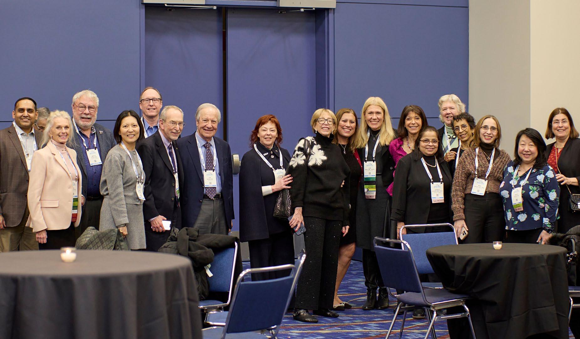
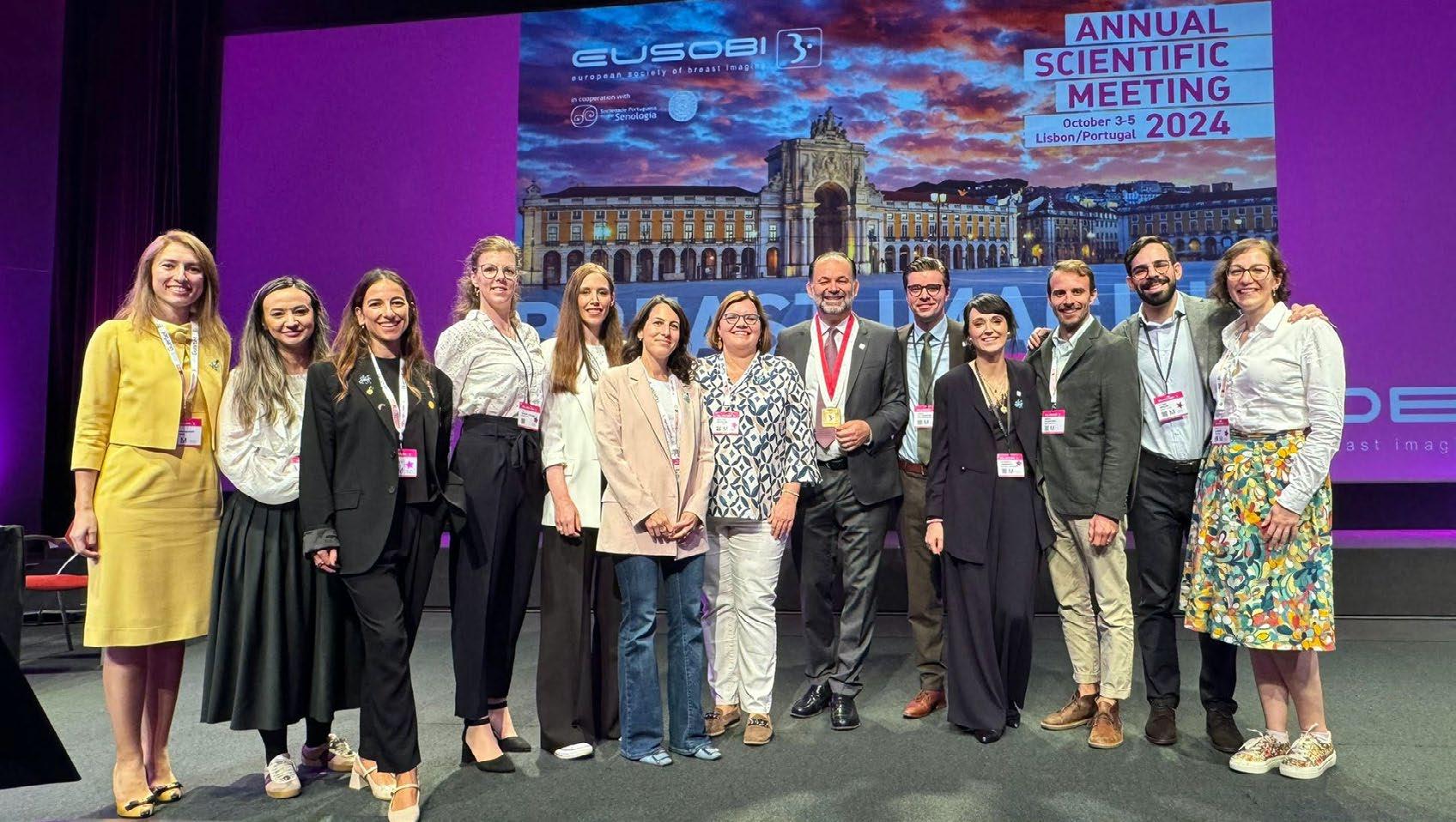
Prof. Dr. Michael Fuchsjäger, EUSOBI president, with the EUSOBI Young Club Committee at the EUSOBI Annual Scientific Meeting 2024 in Lisbon.
EXTERNAL BREAST PROSTHESES: OPTIONS FOR WOMEN WHO DO NOT UNDERGO RECONSTRUCTION AFTER
MASTECTOMY
By Divya Meher Surabhi, MD, MPH; Wynton Bryce Overcast, MD
Each year, more than 100,000 women in the United States undergo a mastectomy for surgical treatment of breast cancer.1 Of these patients, an estimated 25% to 50% opt for breast reconstruction. 2-4 Patients who do not undergo breast reconstruction may opt to wear an external breast prosthesis. Women who use external breast prostheses report that prostheses can increase confidence and enhance body image and self-esteem. 5
The three main types of external breast prostheses are leisure, silicone, and custom prostheses. A leisure breast prosthesis is typically made of foam, fiberfill, polyester fiberfill, or beaded materials encased in a cloth shell. These prostheses are lightweight, which can be helpful for patients when exercising. Silicone prostheses are designed to mimic the appearance of natural breast tissue. Since these prostheses are weighted, they can help improve posture, prevent shoulder drop, and improve balance. However, the additional weight may be uncomfortable, especially in hot weather.
To stabilize leisure and silicone external breast prostheses, women need postmastectomy bras with a spandex stretch pocket. Postmastectomy bras can be purchased from manufacturers or mastectomy boutiques. Some mastectomy boutiques have tailors who create postmastectomy bras, nightgowns, and even swimwear from an individual’s current wardrobe.6 Postmastectomy bras and garment tailoring to accommodate leisure and silicone external breast prostheses are added expenses that women must account for when purchasing these prostheses.
Custom breast prostheses, in contrast, are worn directly against the chest wall, offering a highly personalized fit. Custom breast prostheses are worn with regular bras.7 The benefits of custom breast prostheses include appropriate weight distribution, alleviation of stress and friction against sensitive areas of the chest wall, and a precise match of skin tone, breast shape, and areola size and color. 8 The alleviation of stress and friction against the chest wall is especially significant because women may have scarring and sensitivity secondary to surgery and radiation therapy.
For a patient to qualify for external breast prostheses and postmastectomy bras, the ordering clinician must submit a standard written order documenting medical necessity.9 The patient can then contact their insurance company to obtain a list of in-network external breast prosthesis and postmastectomy bra manufacturers or boutiques. At the boutique, the patient can receive custom measurements and place orders; the boutique typically handles the priorauthorization process.
For Medicare beneficiaries, the number of bras covered is based on medical necessity, as determined by the clinician. However, the coverage depends on the type of prosthesis ordered. For example, Medicare covers one silicone prothesis every two years or one foam prosthesis every six months.10 Medicare and some insurance companies do not cover the cost of custom prostheses. Depending on the type of prosthesis, out-of-pocket costs can range from $400 for a premade silicone prosthesis to $5000 for a custom prosthesis.11
To alleviate the financial, emotional, and time costs of acquiring bras and prostheses, certain organizations have created initiatives. Local organizations may provide for those who are underserved or underinsured. For example, the HERS Breast Cancer Foundation provides bras and external breast prostheses for women in the San Francisco Bay area.12 For those who prefer a lightweight, soft piece, Knitted Knockers provides knitted prostheses that can be worn with a bra; volunteers create the pieces, and women can request them for free.13
Breast prostheses play a vital role in supporting women who have undergone mastectomy, helping them regain confidence and a sense of normalcy after treatment. Beyond their physical function, prostheses contribute significantly to emotional wellbeing and promote a more holistic approach to treatment.
Continued on page 14>
THE PATIENT'S PERSPECTIVE
Lauren McClain
By Danielle Sharek, MD
DS: Please tell me about yourself and your background.
LM: My name is Lauren McClain. I’m 42 years old and have proudly dedicated the past 20 years to teaching elementary school. I live in Zelienople, just north of Pittsburgh, Pennsylvania, with my wonderful husband and our three energetic boys— Connor (12), Jack (11), and Brooks (7). Outside of the classroom, my life is all about balancing family adventures, outdoor activities, and creating lasting memories with my boys.
How were you diagnosed with breast cancer?
A few weeks after celebrating my 40th birthday, I was diagnosed with hormone-positive, HER2-negative invasive ductal breast cancer. It all began when I went for my first mammogram, never expecting anything more than routine results. But soon after, I received a call saying I needed a diagnostic mammogram and ultrasound due to dense breast tissue. After talking to a few people, I was reassured this was a typical occurrence, especially for younger women.
During my ultrasound, I got a terrible feeling almost immediately. Thank goodness my husband was with me because after a couple minutes of taking ultrasound images of my left breast, the ultrasound tech got quiet. I asked if she saw something. I will never forget her saying, “I believe so, but I’m going to get the radiologist to come talk to you. Would you like me to go get your husband?” I said yes and only remember the radiologist saying, “Expect your biopsy to come back as malignant.” While we were there, my husband scheduled my biopsy for me because I couldn’t talk to anyone. I was truly in disbelief. How did I go from walking in this building 45 minutes ago thinking all was just fine, to walking out with a good chance I had cancer?
Later that day, I checked MyChart, such a blessing and a curse, and discovered my tumor was a BI-RADS 4C, which was a 50% to 95% chance it was malignant. For a split second, I had hope that surely it was benign. I took to the internet and compared every word of my results amongst others. Some were benign, but most were malignant.
Nine days later, during my biopsy, I’ll never forget Lynne. Her compassion and presence made all the difference. She was truly meant for her role, and when the doctor confirmed that the tumor looked malignant and had all malignant characteristics, she stood by me, offering a hug as I broke down in tears. My official diagnosis call was a few days later. I knew when they would be calling and took a half day from school because I knew I could not be at school when they called.
How did you feel when you learned of the news?
As strange as it may sound, I felt a sense of relief when I received my official diagnosis. Knowing that there would soon be a plan gave me a sense of calm. I had a few reassurances; the tumor was relatively small, and my type of cancer was very treatable. I’ve always been a stubborn go-getter, and I was determined to tackle this head on and push through treatment as quickly as possible.
What was your treatment process? Did you face any treatment obstacles? How did you overcome them?
I was diagnosed on January 3, 2023, and life instantly felt like a whirlwind—that’s truly the best way to describe the experience of a cancer diagnosis. It seemed like I had a million appointments, blood draws, and scans. On January 24, 2023, I underwent a mastectomy. There was hope for immediate reconstruction, but during surgery, it was discovered that the cancer had spread to one sentinel lymph node. This meant reconstruction had to be postponed, and I was now facing chemotherapy and radiation, a tough pill to swallow after a six-hour surgery.
As I mentioned before, I was eager to get treatment over with, so when I met with my medical oncologist shortly after the mastectomy and learned I would need chemo, I accepted it. I had a feeling this was coming. My immediate question was, “When can I start?” The following week, I had my port placed,
Danielle Sharek, MD
ordered what I needed for cold capping, and started chemo the next Monday—just three weeks after my mastectomy.
Soon after, I went in for my radiation mapping and began radiation at the end of May, completing it on July 5, 2023. Because of the radiation, I had to wait six months to proceed with reconstruction. On the dot, six months later, I had my tissue flap surgery, followed by my first round of fat grafting a few months afterward.
The only obstacle I faced during chemo was having neutropenia. Of course, life does not stop when you have young kids, nor did I want it to. It was hard not being able to go to some of their games because I did not want to get sick and postpone treatment. This also forced me to take a step back from work, something I hadn’t ever really done before. My husband was amazing during chemo and would come up with a family game night every Sunday. I think I looked forward to it more than the kids did.
What motivated you during your diagnosis and treatment process?
I completed chemo during the winter and was fortunate to have a milder season, which allowed me to get outside and walk almost daily. In the first week after treatment, my walks were slow and short, but just moving my body and getting fresh air felt incredible. By the second week, I started feeling more like myself, and by the third week, I was nearly back to normal. I truly believe that staying active and eating healthy made chemo more manageable. I was determined not to let the treatment stop me from keeping up with my kids.
I was also lucky to be selected for a 12-week exercise oncology study through my radiation oncologist. The program connected me with other women going through similar experiences, helped me establish healthy eating habits, and introduced me to the importance of strength training.
What did you learn from your experience?
The biggest lesson I’ve learned through all of this is not to take life for granted. Balancing full-time work and our kids’ busy schedules made my husband and I reassess how we wanted to grow as a family. We realized we’d been caught up in the rat race and needed to prioritize more quality time together. Now, we’re much more intentional about not overcommitting.
Every Monday, I take time to reflect on the positives in my life. With each new week, month, and now, a new year, I’m constantly reminded of just how fortunate I am.
How has this diagnosis impacted your life? How have you used your diagnosis to impact others?
I can’t even begin to express how many blessings have come from my cancer diagnosis. From the start, I made it my mission to help others once I was on the other side of this journey.
During my first appointment with my surgical oncologist, I was assigned a nurse navigator. I asked if there were any books we could read with our kids, as we hadn’t yet told them about my diagnosis. At that point, I had decided to cold cap in an effort to save my hair. It was something I could control, so I embraced the challenge. However, the books offered to us were outdated and focused solely on moms losing their hair and needing a wig. I immediately thought, “But this isn’t going to be me.” So, I politely declined.
Then, one day while walking during chemo, an idea came to me: I could write a children’s book that explains cancer, chemotherapy, and cold capping. I hadn’t seen any books like that, so I did a quick Google search mid-walk to confirm, went home, and told my husband I wanted to write a book. He agreed it was a great idea. I had always dreamed of publishing a book but never had an idea that felt meaningful until this moment.
I started writing right away, found an illustrator (thanks to my tech-savvy fifth grader!), and dove into the process. It was a steep learning curve, but it quickly became therapeutic. Writing my story to help others was more healing than anything else. After about eight months, Mom’s Magical Crown: A Cold Capping Adventure was born. I thought only a few people would buy or read it, but I was wrong. There was such a need for the book, and it quickly gained attention from women across the United States.
Since writing had always been a passion of mine and I now understood the process, I went on to write another book about a mom’s journey through radiation. Brave Rays: A Journey Through Radiation Therapy was published in June 2024. Later that summer, Paxman, a scalp cooling company, reached out to me to revise Mom’s Magical Crown to reflect their system, as I had used the Penguin manual system, and theirs is mechanical. I jumped at the chance, and the new Paxman version was published in October 2024.
Now, I’m almost finished with my fourth book, which will cover mastectomies and surgery. I have one more book I want to write before I feel this series is complete. In addition, I created a website, www.momsmagicalcrown.com, for women to find advice on all things breast cancer. Books may be purchased from my website, on Amazon, or Barnes & Noble online. Through my website, a book may also be donated. Through amazing friends,
Continued on page 14>
The Patient's Perspective: Lauren McClain (continued from page 13)
family, and complete strangers, I have been able to distribute around 300 donated books to cancer centers, foundations, and moms just like me around the country.
Are there any lessons that you think the breast imaging community can learn from your experience?
The breast imaging community plays a pivotal role in a cancer diagnosis as you are the first crucial step in determining a patient’s future. Without your expertise and advocating for women, especially with dense breast tissue, outcomes could be far worse for women.
What advice would you give to other patients who are going through the diagnosis and treatment process for breast cancer?
My greatest piece of advice is to stay as active as possible, prioritize healthy eating, and always advocate for yourself. Never, and I mean NEVER, feel bad about asking your team of doctors questions. You know your body best and will become very in tune with every new ache, pain, bump, bruise, or new ailment. I consider myself still a “fresh” survivor and I always think cancer will live in the back of my head for life, but I can wholeheartedly say that life does get better. Just take it day by day!
Member-in-Training Column: External Breast Prostheses: Options for Women Who Do Not Undergo Reconstruction After Mastectomy (continued from page 11)
References
1. Breast cancer facts & figures. American Cancer Society. Accessed December 19, 2024. https://www.cancer.org/research/cancer-facts-statistics/ breast-cancer-facts-figures.html
2. Greenberg CC, Lipsitz SR, Hughes ME, et al. Institutional variation in the surgical treatment of breast cancer: a study of the NCCN Ann Surg 2011;254(2):339-345. doi:10.1097/SLA.0b013e3182263bb0
3. Kruper L, Holt A, Xu XX, et al. Disparities in reconstruction rates after mastectomy: patterns of care and factors associated with the use of breast reconstruction in Southern California Ann Surg Oncol. 2011;18(8):2158-2165. doi:10.1245/s10434-011-1580-z
4. Morrow M, Li Y, Alderman AK, et al. Access to breast reconstruction after mastectomy and patient perspectives on reconstruction decision making. JAMA Surg. 2014;149(10):1015-1021. doi:10.1001/jamasurg.2014.548
5. Fitch MI, McAndrew A, Harris A, Anderson J, Kubon T, McClennen J. Perspectives of women about external breast prostheses Can Oncol Nurs J. 2012;22(3):162-174. doi:10.5737/1181912x223162167
7. Conner K. Types of breast forms. Breastcancer.org. Updated November 30, 2022. Accessed December 19, 2024. https://www.breastcancer.org/treatment/ surgery/breast-forms/types
8. Breast prostheses and bras for cancer patients. Dana-Farber Cancer Institute. Accessed December 19, 2024. https://www.dana-farber.org/patient-family/ support-services/friends-place-boutique/services/breast-prostheses-bras
9. Ordering external breast prostheses & supplies. Centers for Medicare & Medicaid Services. MLN3754874. November 2024. Accessed December 3, 2024. https://www.cms.gov/files/document/mln3754874-ordering-externalbreast-prostheses-and-supplies.pdf
10. Worstell C. Does Medicare cover mastectomy bras? Medicaresupplement. com. August 22, 2024. Accessed December 3, 2024. https://www. medicaresupplement.com/coverage/does-medicare-cover-mastectomy-bras 11. Donofree D. Custom breast forms 101. AnaOno. Accessed December 3, 2024. https://www.anaono.com/blogs/dressing-room/custom-breastforms-101
12. Charitable programs. HERS Breast Cancer Foundation. Accessed December 3, 2024. https://hersbreastcancerfoundation.org/charitable-programs/ 13. Knitted Knockers. Accessed December 19, 2024. https://www. knittedknockers.org/
Global Outreach, India: Asha Jyoti (Ray of Hope) II
By Harnoor Singh, MD, MBA
The SBI Inclusion Diversity Equity Alliance Committee is fostering change by getting radiologists motivated and aware of various barriers to providing care to our patients and addressing issues of cultural competence. In this endeavor, we extended beyond our national boundaries to encompass international outreach.
I visited a premier institution of training in India, Postgraduate Institute of Medical Education and Research (PGIMER), and became acquainted with its radiology department faculty in December 2022. I wanted to foster a partnership wherein we could share and participate in tumor boards and educate each other. I also wanted to find out if our breast imaging community could help serve the disadvantaged rural population of India. I got in touch with the former radiology chair of PGIMER, Dr. Niranjan Aggarwal. My own radiology department at the University of Texas Health Science Center at Houston and our department chair, Dr. John, are especially supportive of such efforts pertaining to global outreach and collaborative learning. With the blessing of our department, I started exploring the possibility of providing services to the heart of the country of India via mobile efforts.
Rajasthan is a state in India with a substantial rural population. This segment of the population is not aware of recent cancer (especially breast cancer) screening guidelines and recommendations and lacks access to care. It is extremely difficult for these individuals to give up a full day to travel to the tertiary care hospital and get their wellness examinations.
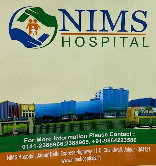
Dr. Aggarwal had contact with a tertiary care hospital, NIMS Hospital, in Jaipur, Rajasthan. We started exploring possible collaboration to provide educational and financial support to launch mobile efforts in cancer screening.
I was delighted to get in touch at the SBI symposium with the RAD-AID team members, who were very supportive of our efforts. RAD-AID had already successfully completed a fiveyear chapter (Asha Jyoti I) in Chandigarh, India, in association with PGIMER, but the project was discontinued during the COVID-19 pandemic. They were eager to begin another chapter.
Dr. Aggarwal and I, in collaboration with RAD-AID and NIMS, have been successful in creating a partnership. I visited NIMS in December 2023 and was pleasantly surprised by the readiness and support from the institution. They are well equipped with all
facets of cancer care, including surgery, medical oncology, and radiation oncology divisions. I was fortunate to meet with the chancellor of the university and the department heads from each of these divisions, all of whom were enthusiastic and supportive of our joint efforts to provide screening care for the marginalized rural population of the state.
RAD-AID also committed to providing certified mammography training program support to the current mammography technologists at NIMS and made a substantial financial commitment to donate a well-equipped Siemens van. The screening van will be equipped to provide breast cancer (with mammography), cervical cancer, and lung cancer screening in addition to screening for tuberculosis, which is direly needed in India given the increased incidence of patients with multidrugresistant tuberculosis. The van will be able to reach residents of rural areas who are unable to travel and gain access to tertiary care.
An awareness campaign is underway wherein local stakeholders, including elected local government officials, participate in programs to educate the population about screening for breast, cervical, and lung cancer. An educational emphasis is placed on the decrease in morbidity and mortality with early cancer detection. NIMS, in association with the state government of Rajasthan, will provide free to minimal-cost care for patients with any abnormal examination findings. Additionally, transportation services will be arranged for travel to and from the institution. The screening examinations will be read in real time by qualified radiologists at NIMS since all examinations will be transmitted to an extensive and robust picture archiving and communication system. The screening van is currently in the production phase, with completion slated for the end of December 2024. All interested parties are planning to launch this extensive screening program in February and March 2025.
We will call this program Asha Jyoti II, or Ray of Hope II, to emphasize our desire to spread hope and welfare to the disadvantaged populations of India. It is heartening to see that
Harnoor Singh, MD, MBA
IDEA (Inclusion Diversity Equity Alliance) Insights: Global Outreach, India: Asha Jyoti (Ray of Hope) II (continued from page 15)
our efforts are near fruition. Our goal is to continue with ongoing educational efforts to improve care. We are eagerly awaiting the
launch of this program and will be publishing our firstphase results in the fall or winter of 2025.
To save lives and minimize the impact of breast cancer. .....
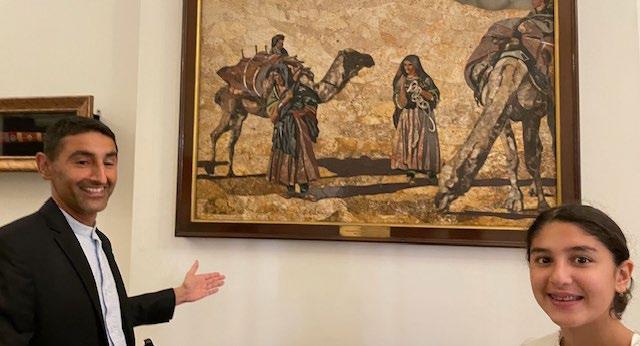
Visitor center to the State of Rajasthan displaying its rural beauty with camels in the desert.
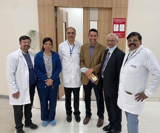
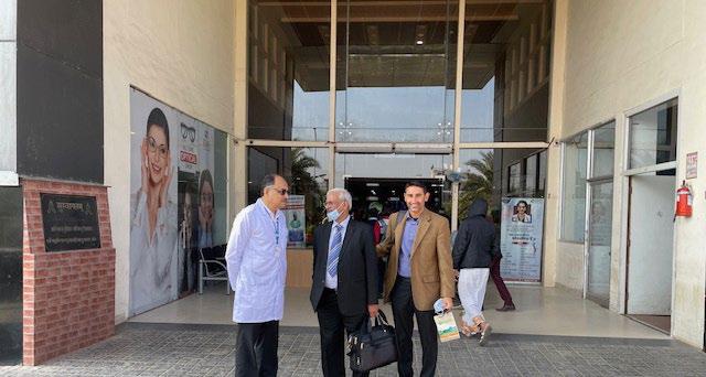
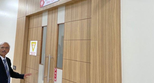
Entrance to NIMS. Left to right: Dr. Sudhir (head of Department of Radiology), Dr. Khandelwal, and Dr. Singh.
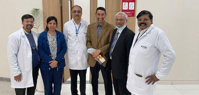
Heads of departments of radiation oncology, medical oncology, surgical oncology, and radiology with Dr. Singh (brown jacket) and Dr. Khandelwal (black jacket).
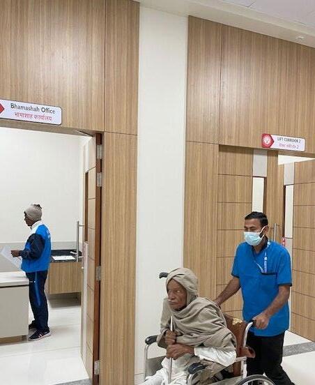
Patient from rural area in a wheelchair in the patient hallway.
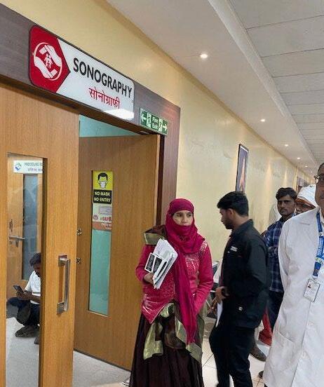
Entrance to sonography suite at NIMS.
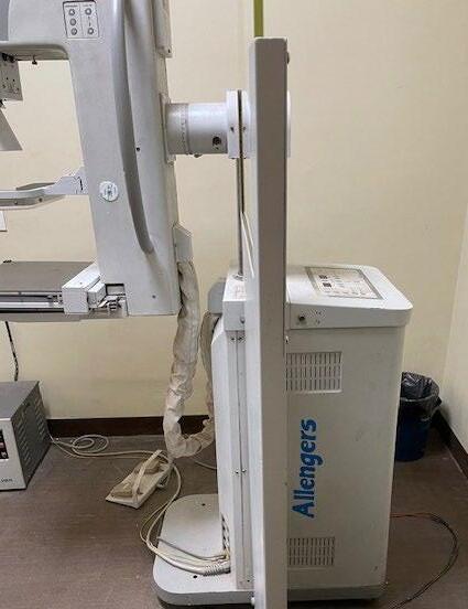
One of the in-house mammography machines.
NIMS Hospital logo.
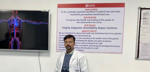
Vision and mission of NIMS.
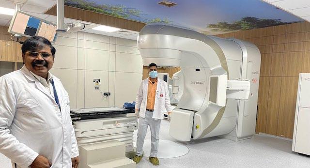
Radiation oncology suite for radiotherapy at NIMS.
Entry door to the radiation oncology area.
Status of Mammographic Screening in Women Aged 40 to 49 Years: The Canadian Story
By Supriya Kulkarni, DMRD, DNB, FRCP(C), FSBI
All Canadian jurisdictions (provinces and territories) offer breast screening. There is variability in how each jurisdiction provides screening services. Most offer fully organized programs, while others screen opportunistically. Screening programs are characterized by essential criteria including screening parameters and eligibility criteria, referral strategies, screening intervals, promotional strategies, quality assurance mechanisms, pathways to diagnostic follow-up, participant recall, and integrated information technology systems. Screening is provided through academic centers, community hospitals, independent health facilities, and mobile vans.
National Guidelines and Challenges
Earlier in 2024 the Canadian Task Force issued draft recommendations that excluded screening of eligible women aged 40 to 49 years. The recommendations conflict with those of other reputable organizations, leading to confusion among health care professionals and patients.
Active advocacy around these recommendations is ongoing, especially given that these recommendations systematically exclude Canada’s evolving ethnoracial population and are based on data that are not fully representative of the population, leading to recommendations that might not be applicable, beneficial, or safe for everyone. Among other recommendations, the Task Force recommended against supplemental screening for women with dense breasts.
Provincial Guidelines
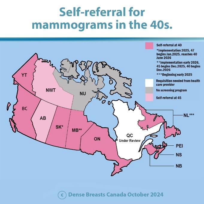
Despite the ongoing challenges, most Canadian provinces have changed their screening programs to allow women aged 40 to 49 years to self-refer into programs for screening mammography. The map in Figure 1 shows the status in the different provinces. This was a welcome change for all of us, including young Canadian women.
Supplementary screening ultrasonography is available in most provinces but requires a requisition from the referring physicians. Capacity remains a huge challenge leading to very limited and patchy availability.
Risk-Based Screening
Many jurisdictions differentiate between average risk, elevated risk, and high risk of breast cancer. These specific levels of risk
have their own definitions and impact screening eligibility. Participants with elevated risk (women with a family history of breast cancer, elevated breast density, or a previous diagnosis of high-risk lesions such as lobular carcinoma in situ, atypical lobular hyperplasia, etc) are recalled for more frequent mammographic screening through the screening programs. Participants with high risk also get enhanced screening with annual mammography and, in some provinces, annual breast magnetic resonance imaging (MRI) starting at a younger age. For example, in Ontario high-risk screening starts at the age of 30 years with annual mammography and annual breast MRI.
Population Engagement
Challenges in accurately identifying equity-denied groups along with lack of awareness, limited access, and limited participation in screening programs among these communities presents a challenge in population engagement apart from the geographical challenges of the Canadian terrain.
Participation Rates
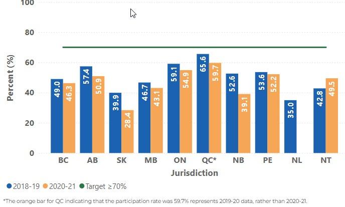
In 2018-2019 and in 2020-2021, screening participation rates in all jurisdictions were below the Canadian target of at least 70%. Participation rates in 2020-2021 were lower than rates in 20182019 in most jurisdictions, given the impact of the COVID-19 pandemic; rates ranged from 28.4% in Saskatchewan to 54.9% in Ontario (Figure 2).
Conclusion
Canadian breast screening programs continue to make important efforts to improve the quality and delivery of breast screening services despite these challenges. The COVID-19 pandemic significantly impacted screening participation rates across jurisdictions, a setback in reaching the 70% national target. Jurisdictions continue to implement population-wide and tailored strategies to restore prepandemic screening volumes and reach underserved populations.
Source: Canadian Partnership Against Cancer (https://www. partnershipagainstcancer.ca/)
Continued on page 19>
Supriya Kulkarni, DMRD, DNB, FRCP(C), FSBI
Artificial Intelligence–Powered Solutions for Breast Cancer Detection in Underserved Communities
By Ameena Elahi; Victoria Mango, MD; Natalie Cain-Wisdom, MD, MPH; Mai Elezaby, MD; Daniel Mollura, MD; Celeste Garcia; Allen Schweitzer; Erica Pollack, MD
RAD-AID International is a nonprofit global health equity organization dedicated to decreasing medical imaging disparities in underresourced communities in the United States and low- and middle-income countries (LMICs).
Access to breast cancer screening for women in underresourced areas is significantly challenging due to geographic barriers, infrastructure limitations, and resource constraints, which lead to diagnostic delays. These inequities are exacerbated in rural areas where diagnostic tools and trained radiologists are even more limited. Through education and the development of reliable imaging informatics infrastructures, RAD-AID is working to increase early detection and consequently increase breast cancer survival rates globally.
Recognizing the need for innovative solutions, RAD-AID’s clinical and administrative leadership team collaborates with the imaging informatics team to use tools such as artificial intelligence (AI) to help close the imaging inequality gap. AI has the potential to strengthen diagnostic accuracy and help bridge workforce gaps experienced in LMICs, thereby reducing delayed diagnosis and promoting early detection.
For example, RAD-AID introduced AI for breast imaging at their partner site in Guyana, Georgetown Public Hospital Corporation, by deploying the intelliMammo platform (Densitas). The AI-driven software is designed to leverage analytics and provide real-time positioning feedback to radiologic technologists, reducing repeat imaging rates and callbacks to optimize quality. By empowering technologists, this tool improves image quality and increases efficiency and departmental effectiveness.
Additionally, RAD-AID implemented Koios Medical’s AI breast ultrasonography tool at two Nigerian hospitals, Obafemi Awolowo University and Lagos State University Teaching Hospital. Koios provides radiologists with decision support in the diagnosis of breast cancer through the analysis of breast ultrasonography findings aligned with BI-RADS categories, with the goal of ultimately improving timely breast cancer diagnosis.
When implementing AI and other information solutions in low-resource settings, RAD-AID collaborates closely with local stakeholders and considers the sustainability of the technology carefully. Beyond addressing infrastructure limitations, such as
save lives and minimize the impact of breast cancer.
an unreliable power supply and internet access, RAD-AID delves deep into the importance of education and the practical use of AI technology.
RAD-AID established a three-pronged AI implementation framework, Teach-Try-Use, to support sustainable and scalable AI technology.
• Teach: RAD-AID expands AI knowledge across the department in this introductory phase. To achieve this, they have developed on-demand courses, including Introduction to Informatics and Introduction to AI online courses, paired with on-site training tailored to administrators, technologists, and radiologists.
• Try: In this phase, RAD-AID works with local partners to access the site’s infrastructure, identifying the hardware and software resources necessary to implement and sustain the AI tools. This evaluation validates the environment and its readiness to deploy the new technology.
• Use: RAD-AID conducts a small pilot study at the partner site during this phase. This real-world deployment is customized to the local needs for optimal performance.
RAD-AID doesn’t implement AI and walk away. After careful deployment, the RAD-AID informatics leadership team maintains an ongoing partnership with the site. Once deployed, they may not always revisit the Teach-Try-Use framework in this exact order; however, they continuously cycle through the phases after deployment to ensure safety, growth, and sustainability.1
Ameena Elahi
Daniel Mollura, MD
Celeste Garcia Allen Schweitzer Erica Pollack, MD
Victoria Mango, MD Natalie Cain-Wisdom, MD, MPH Mai Elezaby, MD
One of the biggest challenges is accessibility to adequate health care in LMICs and many rural areas in high-income countries. Both the World Health Organization and the Centers for Disease Control and Prevention have underlined these disparities.2,3 RADAID’s work revolves around research, awareness, education, and providing accessible breast imaging in underserved communities. These interventions, including non-AI technology, are often tailored to the specific health care needs of the individual community. For example, mobile mammography is designed for women who otherwise may not be able to obtain a mammogram at a hospital or clinic due to various health care barriers, including socioeconomic disparities, culture, location, and lack of awareness.4
The significant resource disparities between LMICs and highincome countries, as well as rural areas within countries, highlight the needs of marginalized and indigenous populations and underscore the need to strengthen communities by improving access to health care.
By addressing inequities in breast imaging, we can significantly reduce the mortality rate and burden of breast cancer. Future RAD-AID goals include expanding initial AI programs beyond the current pilot sites, implementing AI for three-dimensional tomosynthesis, and scaling breast teleradiology solutions. These initiatives represent a stride toward improved health care for all.
RAD-AID International is eager to welcome new volunteers with expertise in any aspect of radiology or breast cancer care who are interested in promoting high-quality care to underserved patients. Attending-level physicians and radiologists in training are welcome to apply, as are physician assistants, technologists, nurses, physicists, and informatics specialists. We invite you to learn more on the RAD-AID website (www.rad-aid.org) and to sign up at https://portal.rad-aid.org/survey/general-volunteersurvey or email breastimaging@rad-aid.org with inquiries. Remember to indicate that you are an SBI member when you sign up!
References
1. Mollura DJ, Culp MP, Pollack E, et al. Artificial intelligence in low- and middle-income countries: innovating global health radiology Radiology 2020;297(3):513-520. doi:10.1148/radiol.2020201434
2. Breast cancer inequities. World Health Organization. Accessed November 30, 2024. https://www.who.int/initiatives/global-breast-cancer-initiative/ breast-cancer-inequities
3. U.S. cancer statistics breast cancer stat bite. Centers for Disease Control and Prevention. June 13, 2024. Accessed November 30, 2024. https://www.cdc. gov/united-states-cancer-statistics/publications/breast-cancer-stat-bite.html
4. Naik S, Varghese AP, Asrar Ul Haq Andrabi S, Tivaskar S, Luharia A, Mishra GV. Addressing global gaps in mammography screening for improved breast cancer detection: a review of the literature. Cureus. 2024;16(8):e66198. doi:10.7759/cureus.66198
Canadian Corner: Status Of Mammographic Screening In Women Aged 40 To 49 Years: The Canadian Story (continued from page 17)
provinces
territories.
Canada.
2. Breast cancer screening participation among individuals aged 50 to 74 years who were screened within a screening mammography program, by jurisdiction in Canada and screening period (2018-2019 and 2020-2021).
Source: Canadian Partnership Against Cancer
Figure 1. Screening practices in Canadian
and
Source: Dense Breasts
Figure
Santo Maimone IV, MD, FSBI
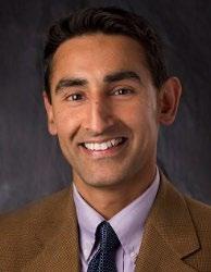
Dr. Maimone is an associate professor of radiology at Mayo Clinic Florida, where he currently serves as the division chair of breast imaging and intervention. He completed his undergraduate and medical school training at Case Western Reserve University in Cleveland, Ohio, and pursued radiology residency training at the Mayo Clinic in Jacksonville, Florida. There, he was appointed as a Mayo Scholar, completing his breast imaging fellowship training at Memorial Sloan Kettering Cancer Center prior to returning to the Mayo Clinic as a staff member. His interests include quality improvement and pragmatic research endeavors that improve clinical practice for both patients and breast imaging radiologists. Dr. Maimone has devoted considerable efforts to legislative advocacy, currently serving on the Board of Officers for the Florida Radiological Society and as the state chair for the Florida Radiological Society Breast Imaging Committee. Outside of breast imaging, Dr. Maimone enjoys traveling with his wife and two sons and sports-related activities; he was recently inducted into both his high school and college athletics halls of fame.
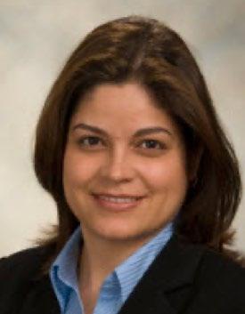
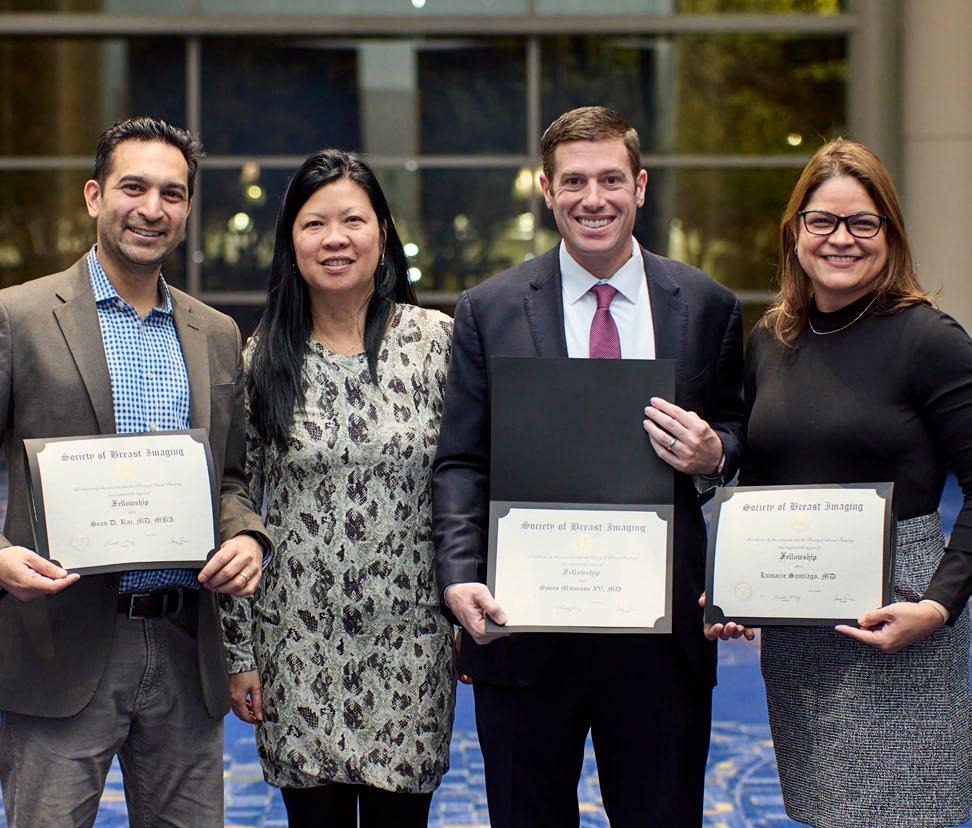
Lumarie Santiago, MD, FSBI
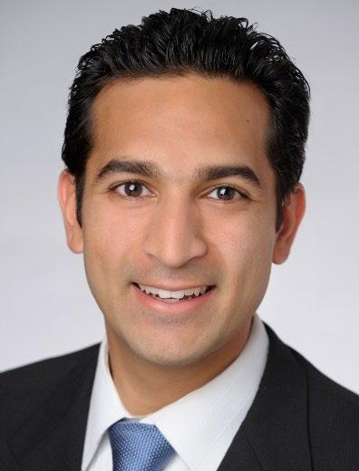
Sean D. Raj, MD, MBA, FSBI
Dr. Sean D. Raj is the chief innovation officer at SimonMed Imaging, where he spearheads initiatives to integrate emerging technologies, particularly artificial intelligence, to enhance health care quality and patient outcomes across one of the nation's largest outpatient imaging networks. A recognized leader in precision health and health care innovation, Dr. Raj has built a career focused on bridging the gap between research and clinical application, using advanced artificial intelligence, radiomics, and genomics to drive personalized, equitable care. He previously served as medical director for Baylor Scott & White’s highrisk breast program, where he developed an award-winning program focused on early cancer diagnosis. Dr. Raj is nationally recognized as an expert in health care innovation and precision medicine. His expertise is sought by industry boards and as a consultant to health-tech startups, private equity, and venture capital groups, reflecting his influence on the future of health care delivery.
Dr. Santiago is a professor of diagnostic radiology in the Department of Breast Imaging at The University of Texas MD Anderson Cancer Center (MDACC) with an interest in novel breast imaging techniques and personalized treatment options, including breast cryoablation and threedimensional (3D) printing. She has led research in neoplastic seeding in breast cancer as well as the impact of 3D-printed breast models in the decisional conflict of patients with breast cancer. Dr. Santiago has helped establish and lead the Medical 3D Printing and Advanced Visualization Laboratory at MDACC and currently serves as the chair of the Radiological Society of North America 3D Printing Special Interest Group.
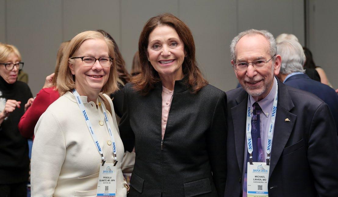
By Rebekah
Novel techniques using artificial intelligence (AI) and radiomics were front and center at the Radiological Society of North America 2024 Scientific Assembly and Annual Meeting (RSNA 2024). Dr. Eric Topol, a cardiologist and executive vice president of the Scripps Research Translational Institute, highlighted the transformative potential of AI in the field of radiology. He asserted that we are on the verge of a seismic shift in which the use of AI will transition from analyzing single data types to integrating multiple data sources, enabling AI to perform a wide range of tasks from making accurate diagnoses to monitoring patients remotely.
Numerous advancements in breast imaging were showcased. Dr. Catharina Oberije1 discussed the transferability of AI across different patient ethnic groups. Human double reading for breast cancer screening must account for the inherent variability of breast cancer among ethnic groups, and AI must be able to perform within this human screening pattern. Her group’s data showed that the positive predictive value (PPV) of double readers was 19.6%, 17.5%, 18.2%, 15.9%, and 13.9% for patients of White, Black, Asian, mixed, and unknown ethnicity, respectively. Double reading with AI demonstrated a comparable trend in PPV, showing absolute improvements of 1.5%, 1.7%, 1.4%, 1.4%, and 1.1%, respectively. A comparison of double reading with and without AI demonstrated no significant difference, indicating the integrity of AI in accounting for ethnicity.
Dr. Manisha Bahl2 discussed the ability of AI to detect falsenegative findings for cancer on screening digital breast tomosynthesis (DBT) and the imaging features of interval cancers detected and not detected by AI. An AI algorithm evaluated false-negative findings on screening DBT at an academic institution from 2013 to 2022. Examination findings considered positive by AI were reviewed by a breast imaging radiologist who determined whether the site corresponded to the following breast cancer. Standard statistical sets were employed to compare the clinical, imaging, and pathologic features of cancers detected and not detected by AI. Results demonstrated that AI correctly identified 27.4% of all false-negative cancer findings and 35.7% of interval cancers. The AI system also identified a higher ratio of interval cancers than asymptomatic
false-negative cancer findings (35.7% vs 14.4%, P < .001). Dense breast tissue, presence of a mammographic finding identified by the radiologist at time of breast cancer diagnosis, and large size on surgical pathologic analysis were all linked to interval cancers detected by AI. The study showed that AI accurately detected one-third of interval cancers when screening DBT examinations were retrospectively examined.
Sarah Verboom, MSc,3 discussed how uncertainty metrics in AI could be used to classify screening mammography readings as certain or uncertain. Quantification of uncertainty was achieved by developing a three-step model: a sensitive region detection algorithm that proposed regions of interest, a region classification model, and generation of an examination-level conclusion. Four metrics were then used to approximate the confidence of the AI malignancy-present decision. When AI predictions were deemed confident, a hybrid reading approach was tested. In the remaining cases, standard radiologist double reading was used. The group retrospectively tested mammographic screening examinations acquired between 2003 and 2018 from a unit of the Dutch national breast cancer screening program (n = 41,469). The AI mammography interpretation model demonstrated an area under the curve of 0.957. The uncertainty metric resulted in a 50% reduced workload, with a recall rate of 27.1 per 1000 (95% CI, 25.6-28.7) and a cancer detection rate of 8.0 per 1000 (95% CI, 7.4-8.7). Using model uncertainty of AI mammography interpretation has the potential to reduce the workload of screening mammography.
In the realm of radiomics, Dr. Xinyi Wang4 sought to develop and validate radiomics models that used ultrasound images to examine benign versus malignant breast lesions with calcifications. The group developed radiomics models based on calcification regions of interest (ROIs) and whole-lesion ROIs. The models based on
Randy Miles, MD, MPH
Rebekah Anders, MD
Anders, MD; Randy Miles, MD, MPH
calcification ROIs demonstrated limited predictive efficacy, but the models using whole-lesion ROIs showed possible prediction in diagnosing breast lesions with calcifications. The study showed that radiomics models have the potential to predict benign and malignant lesions containing calcifications.
Breast imaging posters at RSNA 2024 highlighted radiomics and enhancing the diagnosis of breast cancer by examining data using magnetic resonance imaging (MRI) and ultrasonography. Xiaomei Li and Guijing Jia5 explored the ability of a machine-learning model developed from dynamic contrast-enhanced MRI of breast cancers to predict the level of tumor-infiltrating cells (TILs). Their study divided 132 patients with breast cancer into two groups: those with high TILs and those with low TILs, according to pathologic analysis. They found differences in Ki-67 proliferation index, luminal type (specifically, the proportion of triple-negative breast cancers), and apparent diffusion coefficient (ADC) between the two groups (P < .05). Lower ADC, higher proportion of triple-negative breast cancers, and higher proportion of Ki-67 hyperexpression were associated with high TILs. These three features were then used to construct models with results demonstrating higher predictive efficiency of the combined radiomics-clinical model compared with the clinical model. This study showed the capability of noninvasive evaluation of breast cancer TILs.
Although the presentations largely focused on innovative techniques in breast imaging, operational efficiency in an era of staff shortages and increasing volumes was also emphasized. Telemammography may be one solution to these issues impacting workflow. Dr. Mindy Yang6 discussed the possible incorporation and impact of AI tools in telemammography, Dr. Arlene Sussmann7 detailed the optimal setup for a successful telemammography program, and Dr. Margarita Zuley8 addressed simultaneous telemammography coverage in an academic practice. Telemammography increases radiologists’ flexibility,
which may improve efficiency and address burnout and broadens recruitment opportunities for breast imaging practices.
Ongoing use of AI in breast imaging will create additional opportunities for radiologists to enhance diagnostic accuracy while streamlining workflow. Although current techniques demonstrate promise for detecting breast cancer, the use of AI still depends on breast imaging radiologists to provide guidance and validation. Therefore, workforce shortages, mounting volumes, and radiologist burnout may need to be addressed along with innovative and efficient operational processes for these tools to reach their full potential.
References
1. Oberije C, Ng A, Fox G, et al. Assessing fairness of AI: generalisability evaluation in White, Black, Asian, and mixed ethnicity women in a largescale retrospective clinical study in breast cancer screening. Presented at: Radiological Society of North America 2024 Scientific Assembly and Annual Meeting; December 1, 2024; Chicago, IL.
2. Bahl M, Langarica S, Kniss A, Do S. Use of artificial intelligence to reduce the interval cancer rate of screening digital breast tomosynthesis. Presented at: Radiological Society of North America 2024 Scientific Assembly and Annual Meeting; December 1, 2024; Chicago, IL.
3. Verboom SD, Broeders M, Sechopoulos I. Artificial intelligence should only read a mammogram when it is confident: a hybrid breast cancer screening reading strategy. Presented at: Radiological Society of North America 2024 Scientific Assembly and Annual Meeting; December 1, 2024; Chicago, IL.
4. Wang X. Prediction of breast lesions with calcification using radiomics models from ultrasonic images. Presented at: Radiological Society of North America 2024 Scientific Assembly and Annual Meeting; December 1, 2024; Chicago, IL.
5. Li X, Jia G. MRI-based radiomics analysis to predict the level of tumorinfiltrating lymphocytes (TILS) in breast cancer. Presented at: Radiological Society of North America 2024 Scientific Assembly and Annual Meeting; December 1, 2024; Chicago, IL.
6. Yang ML. Discussion of innovative strategies including possible incorporation/ impact of AI tools in this practice. Presented at: Radiological Society of North America 2024 Scientific Assembly and Annual Meeting; December 1, 2024; Chicago, IL.
7. Sussman A. Optimal set-up for successful telemammography program. Presented at: Radiological Society of North America 2024 Scientific Assembly and Annual Meeting; December 1, 2024; Chicago, IL.
8. Zuley ML. Covering telemammography in the academic setting. Presented at: Radiological Society of North America 2024 Scientific Assembly and Annual Meeting; December 1, 2024; Chicago, IL.
Cryoablation: Tips and Challenges to Implementing a Practice
By Eleanor DiBiasio, MD
Cryoablation continues to receive national attention as an emerging minimally invasive procedure to treat breast cancer in select patients. In September 2024, the five-year results of the ICE3 trial, showing that ipsilateral breast tumor recurrence rates after cryoablation were similar to those after lumpectomy, were published.1 In November, a panel of the US Food and Drug Administration (FDA) Medical Device Advisory Committee convened to make recommendations on the request for clinical use of the IceCure cryoablation system to treat patients with early-stage, low-risk breast cancer. The panel voted in favor,2 and a final decision from the FDA is still pending. We sat down with two breast imaging radiologists who are cryoablation practitioners—Dr. Robert Ward of Brown University Health and Dr. Janice Thai of Massachusetts General Hospital—to discuss how they have established their practices and ask their advice for others interested in performing this procedure. These are some themes that emerged from our discussions.
Perceiving an Unmet Need
At Brown University, enrollment of breast cancer patients for cryoablation began around 2016 as part of the FROST (Freezing Instead of Resection of Small Breast Tumors) trial. However, as breast surgeons and oncologists became more aware of the method, it emerged as an option for other patients as well—particularly those who, although not trial eligible, were poor candidates for traditional surgical therapy. At peak volume before the pandemic, approximately one patient per week was undergoing a breast cryoablation procedure.
The impetus to start a cryoablation program at Massachusetts General Hospital was driven by Dr. Thai’s clinical interest in the procedure, particularly as an option for elderly patients with comorbidities for whom surgery presented challenges. Appreciating that breast imaging radiologists have the essential skills required to perform these procedures, she realized she could offer this option to select patients.
Perfecting the Skill Set
Breast cryoablation can be performed under ultrasound, computed tomography, or magnetic resonance imaging guidance, but it is most frequently performed as an ultrasound-guided procedure with local anesthesia. Breast imaging radiologists are well positioned to perform cryoablation given our extensive training in ultrasound-guided procedures in the breast. Still, cryoablation requires additional learning before it can be safely performed. Both
Dr. Thai and Dr. Ward advised training under more experienced practitioners, either by traveling to an institution that performs breast cryoablation or by taking courses on the subject, before embarking on these procedures independently. Interventional radiology colleagues who perform cryoablation in other parts of the body are unlikely to do so with ultrasound guidance; however, they may provide helpful advice about device storage, transportation, and troubleshooting. It may also be helpful to contact your hospital credentialing office to see what requirements they will perceive necessary before granting hospital privileges to perform the procedure.
Establishing a Network
Both Dr. Thai and Dr. Ward emphasized the importance of consultation with breast surgeons and oncologists for establishing a cryoablation program. Dr. Thai, for example, approached each surgeon in her practice individually to discuss the procedure and gave short presentations at oncology department meetings and tumor boards to provide more detailed information about the scientific evidence, the results of clinical trials, and types of patients who might be candidates. Patient self-referral for cryoablation is rare, so most referrals come from surgeons and oncologists, usually after a multidisciplinary discussion to determine the best treatment plan for the individual patient. It is therefore essential that surgeons and oncologists are familiar with the evidence and willing to offer cryoablation before a program can be established.
Logistical Challenges
The logistical challenges of offering a new therapy can be difficult to navigate. Numerous hospital personnel need to be involved to obtain the necessary equipment, block procedure time in the schedule, book the procedure space and ultrasound machine, and secure technologist availability. Technologist training is offered by equipment vendors. Upfront costs for the ablation devices and probes will need to be negotiated with the vendors, although some costs can be saved by sharing cryoablation units with the interventional radiology department. Space for storing equipment and argon gas tanks will also need to be arranged. Referring clinicians must have a way to order the procedure within the electronic medical record system, which may require
Continued on page 29>
Eleanor DiBiasio, MD
Navigating Radiology Services and Practice Management After a Devastating Hurricane: Lessons Learned in a Private Practice Setting
By Marianne M. Ballisty, MD; Emilie C. Ralston, MD
Our private practice radiology group, ARA Health Specialists in Asheville, North Carolina, provides radiology expertise and services to most of the regional health care systems that serve Asheville and the surrounding 18+ counties. On Friday, September 27, 2024, Hurricane Helene tore through western North Carolina after making landfall as a category 4 hurricane the day before. Helene left in its wake a record $53 billion in damages and recovery needs in western North Carolina. Governor Roy Cooper described the storm as “the deadliest and most damaging storm to ever hit North Carolina.”
In western North Carolina, Helene quickly took out electricity in the early morning hours. At Mission Hospital, the level II trauma center in Asheville, generator power took over immediately, but as the day wore on, city water, cell phone service, and other forms of internet connectivity were soon lost as well. The damage crippled the regional health system’s information technology infrastructure, including the electronic medical records, registration services, and of course the picture archiving and communication system (PACS). In this crippled state, the health system still needed to care for the victims of the historic floods and winds.
Guided by the framework of a catastrophic downtime plan that the operations committee of ARA Health Specialists had developed several months earlier, radiologists at Mission Hospital quickly sprang into action. In the event of a loss of PACS connectivity, plans had been made to station radiologists at the various modalities to provide real-time preliminary reports. The handful of radiologists working that afternoon made their way to the emergency department and managed the computed tomography (CT) consoles, providing paper preliminary reports that were hand delivered by radiology staff to the ordering clinicians. Duplicate copies of the reports were made and were eventually loaded into PACS for documentation purposes. Diagnostic radiology technologists brought their portable machines to the hallways outside the CT control rooms for reads. Radiologists intermittently checked in with the ultrasound and magnetic resonance imaging technologists to batch read examinations. Inpatient imaging was moved to the emergency department, when possible, to improve efficiency.
While this system worked well for Mission Hospital, several smaller surrounding regional hospitals were left with no way of sending their images to PACS for radiologist interpretation. Because of the geological destruction and infrastructure damage, it was not feasible for radiologists to travel to hospitals by normal means. The solution for some of these hospitals was to bring radiologists in via helicopter.
A challenge that had not been anticipated when plans for catastrophe were made was the near-complete loss of communication among physicians and administrative staff when cellular and internet services were completely disrupted. When communication became impossible, our interventional and neurointerventional radiologists transitioned to around-the-clock in-house call. Some of those physicians scheduled to work could not make it to the hospital safely. Ultimately the diagnostic radiologist staffing plan became day to day and hour to hour and was largely based on those who were able to get to the hospital, joined by other radiologists who simply showed up to help.
These were the conditions under which we did our best to provide excellent patient care for over 48 hours. Finally, on the evening of Sunday, September 29, PACS connectivity began to be restored. It was a slow and piecemeal restoration, but we were eventually able to provide final-read dictations for all imaging performed during downtime. Often patient names were misspelled, birth dates were incorrect, and final reports came days later. In the end, every examination was accounted for.
As the emergency department and inpatient systems became stabilized, it became clear that access to outpatient imaging would be our next crisis to manage. The city and surrounding areas were without power for the better part of two weeks and without potable water for nearly two months. The largest outpatient imaging center and largest breast imaging center in the city were completely inoperable for three and a half weeks. This was not only because of the lack of power and potable water but also because the parking lot of
Emilie C. Ralston, MD
Marianne M. Ballisty, MD
this site was serving as an emergency response center that provided water and hot meals to the community. Another breast center in the city was nonoperable for six weeks due to water damage. Once power was restored, outpatient centers resumed operations using a variety of tactics. Bottled water was used for handwashing; all bathrooms were stocked with pallets of water bottles and instructions on their use for handwashing. One center installed portable toilets in the parking lot. Technologists at closed centers were deployed to other departments or other sites to allow for robust and rapid rescheduling of patients with canceled imaging and procedures. Before the hurricane, many mammography technologists were in the process of cross-training in other modalities and rotating at various imaging sites around town. This fortunately provided the flexibility needed to respond to the disaster. Due to the coordinated efforts
to reschedule patients, outpatient imaging volumes rebounded to slightly higher than prehurricane levels within weeks.
While we could have never anticipated the devastation that a tropical event would bring to our beautiful western North Carolina mountains, planning for catastrophe ahead of time allowed us to quickly move to a system that worked in an emergency. In our specialty we are extremely reliant on technology. In radiology departments, we can prepare for the unexpected by analyzing our daily systems and thinking through how we would provide patient care if any or all of these systems became unavailable. It is our responsibility to be prepared, to be ready to pivot, and to preserve excellent patient care as best as we can in the face of crisis.
Radiologist Emilie Ralston, MD, interprets a chest radiograph directly from portable equipment using a paper downtime process.
Radiologist Marianne Ballisty, MD, interpreting an emergency CT scan at the scanner using a paper downtime process.
Radiologist Brianna Vey, MD (right), interpreting a trauma CT scan at the scanner using a paper downtime process while a member of the trauma team (left) listens.
Radiologist Clif Byrd, MD, being transported by helicopter to provide services at Blue Ridge Regional Hospital due to loss of roads and infrastructure.
Hurricane Preparedness and Recovery in a Busy Academic Breast Imaging Practice
By Bethany L. Niell, MD, PhD, FSBI; Kristi Dubois, MHA, RT(R)(CT)(MR); Sonja Ancrile, RT(R)(M)
On September 26, 2024, Hurricane Helene struck Florida as a category 4 hurricane, causing catastrophic flooding throughout the southeastern United States. Less than two weeks later, Hurricane Milton weakened to a category 3 hurricane before making landfall just south of Tampa Bay. Helene and Milton broke the record for the shortest time between two major hurricanes in Florida, leaving behind casualties, massive property damage, and millions without electricity across the southeastern United States. During this time, the Moffitt Cancer Center and Research Institute in Tampa, Florida, closed for multiple business days. When considering how best to manage a busy breast imaging practice before, during, and after these natural disasters, three overarching themes emerge: triage, communication, and caring for your health care team members, patients, and families.
Hurricanes usually have advance warning, so your practice should attempt to perform more breast imaging examinations and procedures several days before the storm by inviting patients to come in earlier for appointments and offering same-day add-ons. More than six million Floridians were issued mandatory evacuation orders for Milton alone. In anticipation of evacuation orders, plan to shift staff members from direct patient care responsibilities to patient rescheduling efforts. To avoid multiple phone calls from different scheduling groups, assign a dedicated scheduling team. If patients can cancel via the portal or a centralized phone number, ensure your organization captures the cancellation requests to avoid additional phone calls. With respect to communication, remind your team members to read every emergency email communication so they can quickly respond to rapidly changing situations. When your organization decides to cease operations, prioritize communication with patients not yet rescheduled. While taking care of patients, remember to also take care of yourself and your family by heeding natural disaster preparation guidelines and abiding by mandatory evacuation orders. You must be flexible with team member scheduling. Be mindful of school and childcare facility closures. Maintain a reasonable working knowledge of your team members’ families (eg, elderly or disabled family members, school-aged or younger children, etc) and living situations (eg, mobile homes, areas more prone to flooding, etc) so you can anticipate how and when critical team members will be impacted.
During a natural disaster, frequent communication is key. If possible despite electricity and internet outages, read regular email communications from your organization and encourage your team members to do the same. Know which department team members are staying in the hospital to provide direct patient care, check in on them, and thank them. Establish group texts among critical team members to increase awareness and timely responses regarding your coworkers’ locations, damage to property, and loss of life. Set aside time and energy to care for yourself and your family. As fatigue, anxiety, and exertion become chronic, physical and mental health quickly degrade.
After the hurricane, leaders should promptly communicate appropriate mechanisms to assess and report damages before resuming operations. Remember that the first step in basic life support is to ensure that the scene is safe, not to immediately resume normal operations. Set realistic expectations. If your mammography facility sustained damage and/or lost records, notify your accreditation body, your state radiological health department, the Mammography Quality Standards Act facility hotline, and your patients.1 Schedule daily or more frequent meetings with your leaders after resuming operations to determine best practices for recovery efforts and make rapid course adjustments.
With respect to triage, attempt to first reschedule BI-RADS category 4c/5 procedures, prioritize breast magnetic resonance imaging examinations for surgical or neoadjuvant treatment planning, and arrange for time-sensitive research examinations or procedures to be completed within any protocol-specified time frames. If you open your imaging facility for additional imaging after hours or on weekends, consider doing so about two weeks after the hurricane rather than immediately. Patients may not be able to return promptly for rescheduled appointments due to lack of electricity, gasoline shortages, childcare facility and school closures, evacuation to more distant locations, and property damage, including flooded homes and cars. Children in the Tampa Bay area missed about two weeks of classes from both hurricanes; schools were also used as temporary shelters. Your patients and team members face similar challenges. Your team members will need scheduling flexibility to return from mandatory evacuation, clean
To save lives and minimize the impact of breast cancer. .....
Bethany L. Niell, MD, PhD
up property damage, meet with contractors and repair personnel, acquire new transportation if their cars were flooded during the storm, and manage emotional or physical exhaustion. Be familiar with your organization’s team member services like emergency childcare, mental health counseling, and other employee wellness and support programs.
Hurricane Helene was the deadliest hurricane to hit the mainland United States since Katrina. Combined with Milton, Hurricane Helene caused more than 250 fatalities, including 100 in North Carolina alone.2 Combined damages from both hurricanes exceed $200 billion.2 Lower-income communities are disproportionately impacted because those communities are more likely to experience financial challenges in preparing and recovering from natural disasters and have limited access to health care.3
Although this article features steps for hurricane preparedness and recovery, many of the principles are broadly applicable to other natural disasters. Our major take-home message is that success hinges upon your ability to be flexible for days to weeks before and months to years after any major natural disaster. Our friends, neighbors, family members, and coworkers are still reeling from the devastating losses they incurred during these two hurricanes. To all the technologists, breast imaging radiologists, and staff members across the country who continue to offer assistance and check in on those of us impacted by Helene and Milton, we sincerely thank you.
References
1. Tips for mammography facilities following natural disasters or severe weather conditions. US Food and Drug Administration. February 5, 2018. Accessed December 13, 2024. https://www.fda.gov/radiation-emitting-products/facilityaccreditation-and-certification/tips-mammography-facilities-following-naturaldisasters-or-severe-weather-conditions
2. Wilcox C, Jacobs P. ‘Daunting’: hurricane-battered researchers assess damage from Helene and Milton. Science.org. October 21, 2024. Accessed December 13, 2024. https://www.science.org/content/article/daunting-hurricane-batteredresearchers-assess-damage-helene-and-milton
3. Ndugga N, Pillai D, Artiga S. Understanding the inequitable impacts of hurricanes and other natural disasters in the wake of hurricanes Helene and Milton. KFF. October 16, 2024. Accessed December 13, 2024. https://www.kff. org/policy-watch/understanding-inequitable-impacts-hurricanes-other-naturaldisasters-the-wake-hurricanes-helene-and-milton/
What’s New in the News: Cryoablation: Tips and Challenges to Implementing a Practice (continued from page 25)
input from the hospital’s information technology department. If your department has a nurse navigator or coordinator, it may be helpful to engage them in scheduling cryoablation consultations, follow-up appointments, and the necessary follow-up imaging tests. Dr. Ward suggested establishing a team to help navigate these challenges and engaging individuals in your hospital who have institutional knowledge to help solve roadblocks as they arise.
Billing Dilemmas
In 2020, the Centers for Medicare & Medicaid Services introduced a category 3 current procedural terminology (CPT) code for breast cancer cryoablation (0581T). Category 3 CPT codes are meant to track use of new and emerging technologies but are not tied to relative value units. Medicare may offer payment of a hospital fee, but no professional fee is given and reimbursement from private insurers is uncertain. Dr. Thai suggested contacting the billing/coding team at your institution before initiating a cryoablation program to ask about any necessary preapprovals and to optimize chances of reimbursement from insurers. Obtaining payment will often require peer-to-peer discussions with private insurers, letters of medical necessity, and letters of appeal if claims are denied. If the FDA grants approval, insurers may be more likely to cover the costs of breast cancer cryoablation in the future.
Excitement Around an Emerging Technology
With continued positive results from clinical trials, interest in and excitement about breast cryoablation has increased over the past several years. It has been a recurring topic in literature in the fields of breast radiology, interventional radiology, breast surgery, and interventional oncology. Both Dr. Ward and Dr. Thai stated how well positioned breast imaging radiologists are to perform this procedure given our extensive skillset in ultrasound-guided procedures and breast imaging. Furthermore, they emphasized how exciting it is as breast imaging radiologists to not only be able to diagnose cancer but now also to have an opportunity to treat it. If you are interested in learning more about breast cryoablation, Dr. Thai and Dr. Ward will be instructors of the breast cryoablation course on Thursday, April 24, 2025, and Saturday, April 26, 2025, during the 2025 SBI symposium in Colorado Springs. In addition, Dr. Thai offers this short animation for those interested in learning more about the procedure: link here. We look forward to the FDA’s final decision on breast cryoablation in the coming weeks to months.
References
1. Fine RE, Gilmore RC, Tomkovich KR, et al. Cryoablation without excision for early-stage breast cancer: ICE3 trial 5-year follow-up on ipsilateral breast tumor recurrence. Ann Surg Oncol. 2024;31(11):7273-7283. doi:10.1245/s10434-02416181-0
2. U.S. Food and Drug Administration Center for Devices and Radiological Health: General & Plastic Surgery Devices meeting: summary of the meeting in relation to the ProSenseTM cryoablation system, November 7, 2024. US Food and Drug Administration. Accessed December 17, 2024. https://www.fda.gov/ media/184077/download
CAREER FAIR
Bartolin Hall The Broadmoor
THURSDAY, APRIL 24, 2025
10:00 AM – 7:00 PM (MT)
FRIDAY, APRIL 25, 2025
10:00 AM – 5:00 PM (MT)
How it Works:
Find new career opportunities from leading employers that are eager to hire.
Connect directly with recruiters-easier and more effective than hitting the ‘Apply Now’ button.
Build your network by continuing your conversations with recruiters weeks (and even months!) after the event is over.
All this and more awaits you at the Career Fair during the 2025 SBI Breast Imaging