Recent History: Likely Reasons Why the US Preventive Services Task Force, Once Again, Supports Screening for Women Aged 40
By Daniel B. Kopans, MD, FACR, FSBI
Sadly, misinformation and lies, similar to what we now see in politics, have existed with regard to breast cancer screening dating back to the 1960s: from the false claim that breast cancer was systemic before it could be found such that early detection would not matter (disproven by randomized controlled trials [RCTs]), to the false claim that it would not be possible to screen large numbers of women efficiently and effectively (disproven by the Breast Cancer Detection Demonstration Project), to the false claim that the radiation from mammograms would cause more cancers than would be cured, to the false claim that screening did not work until the age of 50 years, to the false claim that thousands of breast cancers found by mammography would disappear if left undetected, to the false claim that screening leads to massive overdiagnosis. These false claims and others, over the years, have been repeatedly disproven by science and evidence.
Standing with science (with a weak moment in 1989 supporting biennial instead of annual screening for women aged 40-49 years), the ACR has persistently made the arguments and established the proof that annual screening should include women aged 40 to 74 years—the ages of the women who participated in the RCTs that proved that screening can save the lives of women at these ages.
The power of misinformation has been demonstrated by the oscillation among some of the guidelines panels over the years. The United States Preventive Services Task Force (USPSTF) is an unfortunate example, having supported screening for women aged 40 to 74 years, then dropping support for screening until the age of 50 years, then once again supporting starting screening at the age of 40 years!
In 2021 the USPSTF, still advising women to delay screening until the age of 50 years, began a review of their guidelines. They solicited comments from the public. In addition, the cochair of the USPSTF was on the faculty at Massachusetts General Hospital. Responding to the request for comments, I sent the following to the USPSTF as well as directly to the cochair. These
to 74 Years
facts should have helped to move the USPSTF to, once again, support screening for women aged 40 to 49 years.
At the end of this summary (under the Additional Information on Overdiagnosis heading), I have provided additional material on overdiagnosis that I had not included in my original comments but that represents claims that some have raised and you might encounter.
Comments on the 2021 Plan for the USPSTF Review of Breast Cancer Screening Guidelines
The USPSTF Screening Guidelines Over Time
1. In 2005 the USPSTF supported annual screening for all women aged 40 and over.1
2. In 2009 the USPSTF dropped support for screening women aged 40 to 49 years and urged women aged 50 to 74 years be screened every two years.2
3. In 2016 the USPSTF advised that women aged 50 to 74 years be screened every two years3 despite the fact that the Task Force continued to admit that the most lives are saved by screening beginning at the age of 40 years. They stated, “the USPSTF found adequate evidence that mammography screening reduces breast cancer mortality in women aged 40 to 74 years.”4
4. Apparently the USPSTF is, once again, preparing to review their guidelines for breast cancer screening.
Avoiding the Advice of Experts
The recent pandemic has demonstrated the tragic consequences that result from ignoring science, evidence, and the analysis and advice of experts while being guided by inexpert advice. In a supposed effort to avoid biases from panel members who have a conflict of interest (COI), the USPSTF has prevented anyone with actual expertise in breast cancer screening and the issues involved from serving on the Task Force panel. Consequently, the
Continued on page 24>
Daniel B. Kopans, MD, FACR, FSBI
panel members have been unable to critically sort through the available data and understand the validity or lack of validity of the material they have been asked to review.
This has also given advisers to the panel extraordinary influence to guide an experientially and factually inexpert panel. In the past, the vast majority of advisers to the Task Force Panel reviewing breast cancer screening have been individuals who have expressed their opposition to screening and have clearly had great influence on the panel. Many of the advisers have been viewed as having no COI when, in fact, this is not true. Without an obvious COI it is impossible to gain expertise in a field. Experts such as I, who earn a living related to breast cancer screening, have an obvious and open COI. However, advisers who have received and continue to receive grant support for their research efforts are viewed as free of COI. In fact, theirs is a far less obvious COI. Granting agencies (including the National Cancer Institute [NCI]) and foundations have undeclared biases. When the work of a grantee supports the biases of the grantor, grants are likely to be renewed. Grant or foundation support is a far more insidious COI than those that are out in the open.
The practice of excluding experts should stop. COIs should be detailed, but rather than being excluded, experts are critical for an accurate analysis of the data to provide the most factual and evidence-based advice. Guidelines panels, including the USPSTF, should have leading experts involved in their decisions and the public should be provided with minority reports should there be unresolvable disagreements.
Facts
1. The RCTs of breast cancer screening proved that screening and early detection of breast cancer reduces deaths for women aged 40 to 74 years (the ages of the women who participated in the trials).5 Confusion had been created in 1993 by the inappropriate use of subgroup analysis6 to falsely claim no benefit for women aged 40 to 49 years. The 1993 claims also ignored the fact that an immediate benefit is not expected from a periodic screening program.7 The NCI position was later refuted with longer follow-up8 that showed a clear benefit for screening women aged 40 to 49 years and thus benefit for all women (the RCTs targeted average populations) aged 40 to 74 years. Although it has been suggested that these trials are old, they provide the fundamental proof that early detection reduces deaths.
2. Because of noncompliance and contamination, the RCTs have
underestimated the benefit of early detection. The results of these trials should be viewed as the lower level of the likely benefit.
3. The USPSTF should be aware that the Edinburgh trial is no longer cited with the RCTs because of an apparent imbalance in the socioeconomic factors of participants.
4. The Canadian National Breast Screening Studies (CNBSS) should also have been dropped from guidelines analyses years ago. Not only are their results major outliers among the RCTs but numerous critical analyses over the years have also challenged their validity.9-23 The trials were compromised by poor-quality mammography24,25 and their data compromised by the fact that they violated the fundamental requirements of RCTs by having a nonblinded allocation process.26,27 This resulted in a statistically significant excess of women with advanced cancers being assigned to the screening arm of CNBSS1.28,29 It has been claimed that the CNBSS trials showed a major (22%) rate of overdiagnosis when in fact their own data show that there was only a 4% difference in cancers diagnosed between the two arms.30 The CNBSS results are compromised and unreliable and should not factor into the USPSTF review.31
5. Numerous observational studies have validated the benefit of screening women starting at the age of 40 years in the general population, with reductions in deaths of as much as 40% or more.32-49
6. In a review of the incidence of death among more than 500,000 women in Sweden, there was some benefit from improvements in therapy, but those who participated in mammography screening had a 41% reduction in their risk of dying from breast cancer within 10 years compared to those who had not participated in screening.50
7. There are no data (zero) that show that any of the parameters of screening change abruptly at age 50 years or any other age.51 The RCTs proved mortality reduction for women aged 40 to 74 years. The threshold for initiating screening at the age of 50 years is completely arbitrary with NO scientific support. Grouping of data and averaging has falsely suggested a legitimate threshold when the data show that none exists. The only reason to use the age of 50 years as a threshold is based on individual and scientifically unsupportable biases by analysts. In fact, all major groups, including the USPSTF, agree that the
To save lives and minimize the impact of breast cancer. .....
most lives are saved by annual screening starting at the age of 40 years. There are more years of life lost to breast cancer among women aged 40 to 49 years than among women aged 50 to 59 years.52
8. Radiation risk for the breast from mammography (there is little exposure to any other susceptible organs) drops rapidly with increasing age so that by the age of 40 years it is unmeasurable and may be nonexistent. Even the extrapolated risk is below even the smallest amount of benefit from screening.53,54
9. The NCI/Cancer Intervention and Surveillance Modeling Network (CISNET) models all predict that the most lives are saved by annual screening starting at the age of 40 years.55
10. Despite specious arguments to the contrary, screening has been shown to reduce the rate of advanced cancers,56-67 which has been used as a surrogate for death since these are incurable cancers.
11. In the Harvard Hospitals, 71% of deaths from breast cancer were among the 20% of women who were not participating in screening despite having access to modern therapies.68 Spencer et al had similar results.69
12. The claim that the RCTs did not reduce all-cause mortality is specious.70 All-cause mortality is appropriate in treatment trials where everyone has breast cancer and most of the deaths will be due to breast cancer. You want to be certain that the treatment is not causing an unforeseen risk. In radiation therapy trials, this revealed the unexpected risk that radiation therapy damaged the coronary arteries. In screening trials, however, most deaths will be due to other causes since breast cancer only accounts for 3% of deaths each year from all causes. If you reduce breast cancer deaths by 30% then this will reduce all-cause mortality by 1%. It would take a trial of 2.5 million women to prove that this major decrease in breast cancer deaths significantly reduced all-cause mortality. It would be more appropriate to look at all-cause mortality among women with breast cancer in the RCTs, and this does show that screening reduces the rate of all-cause mortality.71
13. The CISNET models show that the lives of as many as 100,000 women who are now in their 30s and who will die by waiting until the age of 50 years and being screened every two years could be saved by annual screening starting at the age of 40 years.72 Among just the women who are 40 years old today, if they wait until the age of 50 years to be screened every two years, as many as 13,770 will die whose
lives could be saved by annual screening beginning at the age of 40 years.73
The claim of massive overdiagnosis has been manufactured by guessing that the incidence of breast cancer was not steadily increasing as screening was being introduced. Since no one has ever seen a mammographically detected invasive breast cancer disappear on its own (the few “miracles” have all been clinically evident), and Arleo et al showed that none of almost 250 invasive cancers that were untreated regressed or disappeared,74 then waiting until age 50 years and screening every two years will not reduce overdiagnosis if it even exists, because the cancers will still be there.
14. Delaying screening will reduce recalls from screening (inappropriately called false positives) for a few extra pictures or an ultrasound. The recall rate is approximately 10% (approximately the same recall rate as cervical cancer [Pap] testing) and there is a very small chance of having an imaging-guided needle biopsy using local anesthesia with a fairly high yield of cancer. Approximately 2% to 4% of women screened will be advised to have an imaging-guided needle biopsy and 20% to 40% of these lesions will prove to be malignant.
There is no question that recalls make all of us anxious and recalls from screening are no exception, but for most the anxiety is short-lived.75 Given that the major harm (harm is pejorative; it should be called risk) from screening is the anxiety of being recalled, it is beyond paternalistic/ maternalistic to advise women that it is preferable to let them die an avoidable death than to be made anxious by a recall!!?
16. Finally, it has been suggested that only high-risk women aged 40 to 49 years should participate in screening. Although high-risk women are just that—at higher than the average risk—there are no RCT data to prove that screening only high-risk women will save any lives. None of the RCTs stratified patients by risk, so given that RCTs are the only way to prove a benefit, there is no proof that screening only highrisk women will save any lives. In addition, high-risk women account for approximately 25% of all women diagnosed with breast cancer each year, so screening only high-risk women will exclude 75% of the women who develop breast cancer.76,77 At the present time it appears that all women are at risk and should be encouraged to participate in screening. According to the CISNET models (there has been no RCT comparing screening intervals) annual screening is estimated to provide
Continued on page 26>
Recent History: Likely Reasons Why the US Preventive Services Task Force, Once Again, Supports Screening for Women Aged 40 to 74 Years (continued from page 27)
26. Bailar JC, MacMahon B, Randomization in the Canadian National Breast Screening Study: A Review for Evidence of Subversion. Can Med Assoc J 1997;156:193-199.
27. Kopans DB. NBSS: Opportunity to Compromise the Process. Letter to the Editor. Can Med Assoc J 1997;157:247.
28. Kopans DB, Feig SA. The Canadian National Breast Screening Study: A Critical Review. AJR 1993;161:755-760
29. Tarone RE. The Excess of Patients with Advanced Breast Cancers in Young Women Screened with Mammography in the Canadian National Breast Screening Study. Cancer 1995;75:997-1003
30. Miller AB, Wall C, Baines CJ, Sun P, To T, Narod SA. Twenty five year follow-up for breast cancer incidence and mortality of the Canadian National Breast Screening Study: rder ized screening trial. BMJ. 2014 Feb 11;348:g366. Doi: 10.1136/bmj.g366. PubMed PMID: 24519768; PubMed Central PMCID: PMC3921437. See Table 1.
31. Kopans DB. Major failings of trial procedures and quality of screening fatally compromise the results of the Canadian National Breast Screening Studies. J Med Screen. 2021 Jan 17:969141320986186. Doi: 10.1177/0969141320986186. Epub ahead of print. PMID: 33459171.
32. Tabar L, Vitak B, Tony HH, Yen MF, Duffy SW, Smith RA. Beyond randomized controlled trials: organized mammographic screening substantially reduces breast carcinoma mortality. Cancer 2001;91:1724-31
33. Kopans DB. Beyond Randomized, Controlled Trials: Organized Mammographic Screening Substantially Reduces Breast Cancer Mortality. Cancer 2002;94: 580-581
34. Duffy SW, Tabar L, Chen H, Holmqvist M, Yen M, Abdsalah S, Epstein B, Frodis Ewa, Ljungberg E, Hedborg-Melander C, Sundbom A, Tholin M, Wiege M, Akerlund A, Wu H, Tung T, Chiu Y, Chiu Chen, Huang C, Smith RA, Rosen M, Stenbeck M, Holmberg L. The Impact of Organized Mammography Service Screening on Breast Carcinoma Mortality in Seven Swedish Counties. Cancer 2002;95:458-469.
35. Otto SJ , Fracheboud J, Looman CWN, Broeders MJM, Boer R, Hendriks JNHCL, Verbeek ALM, de Koning HJ, and the National Evaluation Team for Breast Cancer Screening* Initiation of population-based mammography screening in Dutch municipalities and effect on breast-cancer mortality: a systematic review Lancet 2003;361:411-417.
36. Swedish Organised Service Screening Evaluation Group. Reduction in breast cancer mortality from organized service screening with mammography: 1. Further confirmation with extended data. Cancer Epidemiol Biomarkers Prev. 2006;15:45-51
37. Coldman A, Phillips N, Warren L, Kan L. Breast cancer mortality afterscreening mammography in British Columbia women. Int J Cancer. 2007 Mar 1;120(5):1076-80.
38. Jonsson H, Bordás P, Wallin H, Nyström L, Lenner P. Service screening withmammography in Northern Sweden: effects on breast cancer mortality –an update. J Med Screen. 2007;14(2):87-93.
39. Paap E, Holland R, den Heeten GJ, et al. A remarkable reduction of breast cancer deaths in screened versus unscreened women: a case-referent study. Cancer Causes Control 2010; 21: 1569-1573.
40. Otto SJ, Fracheboud J, Verbeek ALM, Boer R, Reijerink-Verheij JCIY, Otten JDM,. Broeders MJM, de Koning HJ, and for the National Evaluation Team for Breast Cancer Screening. Mammography Screening and Risk of Breast Cancer Death: A Population-Based Case–Control Study. Cancer Epidemiol Biomarkers Prev. Published OnlineFirst December 6, 2011; doi: 10.1158/1055-9965.EPI-11-0476
41. van Schoor G, Moss SM, Otten JD, Donders R, Paap E, den Heeten GJ, Holland R, Broeders MJ, Verbeek AL. Increasingly strong reduction in breast cancer mortality due to screening. Br J Cancer. 2011 Feb 22. Epub ahead of print42. Mandelblatt JS, Cronin KA, Bailey S, et.al. Effects of mammography screening under different screening schedules: model estimates of potential benefits and harms. Annals of Internal Medicine, 2009; 151: 738-747; see also http://cisnet.cancer.gov, last accessed 16 April 2011.
43. Hellquist BN, Duffy SW, Abdsaleh S, Björneld L, Bordás P, Tabár L, Viták B, Zackrisson S, Nyström L, Jonsson H. Effectiveness of populationbased service screening with mammography for women ages 40 to 49 years: evaluation of the Swedish Mammography Screening in Young Women (SCRY) cohort. Cancer. 2011 Feb 15;117(4):714-22.
44. Broeders M, Moss S, Nyström L, Njor S, Jonsson H, Paap E, Massat N, Duffy S, Lynge E, Paci E; EUROSCREEN Working Group. The impact of mammographic screening on breast cancer mortality in Europe: a review of observational studies. J Med Screen. 2012;19 Suppl 1:14-25. Review
45. Hofvind S, Ursin G, Tretli S, Sebuødegård S, Møller B. Breast cancer mortality in participants of the Norwegian Breast Cancer Screening Program. Cancer. 2013 Sep 1;119(17):3106-12
46. Sigurdsson K, Olafsdóttir EJ. Population-based service mammography screening:the Icelandic experience. Breast Cancer (Dove Med Press). 2013 May 9;5:17-25
47. Coldman A, Phillips N, Wilson C, Decker K, Chiarelli AM, Brisson J, Zhang B, Payne J, Doyle G, Ahmad R. Pan-canadian study of mammography screening and mortality from breast cancer. J Natl Cancer Inst. 2014 Oct 1;106(11).
48. Puliti D, Bucchi L, Mancini S, Paci E, Baracco S, Campari C, Canuti D, Cirilli C, Collina N, Conti GM, Di Felice E, Falcini F, Michiara M, Negri R, Ravaioli A, Sassoli De’ Bianchi P, Serafini M, Zorzi M, Caldarella A, Cataliotti L, Zappa M; IMPACT COHORT Working Group.. Advanced breast cancer rates in the epoch of service screening: The 400,000 women cohort study from Italy. Eur J Cancer. 2017 Feb 18;75:109-116.
49. Morrell S, Taylor R, rder D, Robson B, Gregory M, Craig K. Mammography service screening and breast cancer mortality in New Zealand: a National Cohort Study 1999-2011. Br J Cancer. 2017 Mar 14;116(6):828-839. Doi: 10.1038/ bjc.2017.6. Epub 2017 Feb 9. PMID: 28183141; PMCID: PMC5355933.
50. Duffy SW, Tabár L, Yen AM, Dean PB, Smith RA, Jonsson H, Törnberg S, Chen SL, Chiu SY, Fann JC, Ku MM, Wu WY, Hsu CY, Chen YC, Svane G, Azavedo E, Grundström H, Sundén P, Leifland K, Frodis E, Ramos J, Epstein B, Åkerlund A, Sundbom A, Bordás P, Wallin H, Starck L, Björkgren A, Carlson S, Fredriksson I, Ahlgren J, Öhman D, Holmberg L, Chen TH. Mammography screening reduces rates of advanced and fatal breast cancers: Results in 549,091 women. Cancer. 2020 Jul 1;126(13):2971-2979. Doi: 10.1002/cncr.32859. Epub 2020 May 11. PMID: 32390151; PMCID: PMC7318598.
51. Kopans DB, Moore RH, McCarthy KA, Hall DA, Hulka C, Whitman GJ, Slanetz PJ, Halpern EF. Biasing the Interpretation of Mammography Screening Data By Age Grouping: Nothing Changes Abruptly at Age 50. The Breast Journal 1998;4:139-145.
52. Oeffinger KC, Fontham ET, Etzioni R, Herzig A, Michaelson JS, Shih YC, Walter LC, Church TR, Flowers CR, LaMonte SJ, Wolf AM, DeSantis C, Lortet-Tieulent J, Andrews K, Manassaram-Baptiste D, Saslow D, Smith RA, Brawley OW, Wender R. Breast Cancer Screening for Women at Average Risk: 2015 Guideline Update From the American Cancer Society. JAMA. 2015 Oct 20;314(15):1599-614.
53. Mettler FA, Upton AC, Kelsey CA, Rosenberg RD, Linver MN. Benefits versus Risks from Mammography: A Critical Assessment. Cancer 1996;77:903909.
54. Yaffe MJ, Mainprize JG. Risk of radiation-induced breast cancer from mammographic screening. Radiology. 2011 Jan;258(1):98-105. Doi: 10.1148/radiol.10100655. Epub 2010 Nov 16. Erratum in: Radiology. 2012 Jul;264(1):306.
55. Mandelblatt JS, Cronin KA, Bailey S, Berry DA, de Koning HJ, Draisma G, Huang H, Lee SJ, Munsell M, Plevritis SK, Ravdin P, Schechter CB, Sigal B, Stoto MA, Stout NK, van Ravesteyn NT, Venier J, Zelen M, Feuer EJ; Breast Cancer Working Group of the Cancer Intervention and Surveillance Modeling Network. Effects of mammography screening under different screening schedules: model estimates of potential benefits and harms. Ann Intern Med. 2009 Nov 17;151(10):738-47.
56. Anderson WF, Jatoi I, Devesa SS. Assessing the impact of screening mammography: Breast cancer incidence and mortality rates in Connecticut (1943-2002). Breast Cancer Res Treat. 2006 Oct;99(3):333-40.
57. Tabár L, Yen AM, Wu WY, Chen SL, Chiu SY, Fann JC, Ku MM, Smith RA, Duffy SW, Chen TH. Insights from the breast cancer screening trials: how screening affects the natural history of breast cancer and implications for evaluating service screening programs. Breast J. 2015 Jan-Feb;21(1):13-20
58. Tabár L, Yen AM, Wu WY, Chen SL, Chiu SY, Fann JC, Ku MM, Smith RA, Duffy SW, Chen TH. Insights from the breast cancer screening trials: how screening affects the natural history of breast cancer and implications for evaluating service screening programs. Breast J. 2015 Jan-Feb;21(1):13-20
save lives and minimize the impact of breast cancer. .....
59. Yen AM, Duffy SW, Chen TH, Chen LS, Chiu SY, Fann JC, Wu WY, Su CW, Smith RA, Tabár L. Long-term incidence of breast cancer by trial arm in one county of the Swedish Two-County Trial of mammographic screening. Cancer. 2012 Dec 1;118(23):5728-32. Doi: 10.1002/cncr.27580. Epub 2012 May 17
60. Foca F, Mancini S, Bucchi L, Puliti D, Zappa M, Naldoni C, Falcini F, Gambino ML, Piffer S, Sanoja Gonzalez ME, Stracci F, Zorzi M, Paci E; IMPACT Working Group. Decreasing incidence of late-stage breast cancer after the introduction of organized mammography screening in Italy. Cancer. 2013 Jun 1;119(11):2022-8. Doi: 10.1002/cncr.28014. Epub 2013 Mar 15
61. Tabár L, Faberberg G, Day NE, Holmberg L. What is the optimum interval between mammographic screening examinations? An analysis based on the latest results of the Swedish two-county breast cancer screening trial. Br J Cancer. 1987 May;55(5):547-51
62 Swedish Organised Service Screening Evaluation Group. Effect of mammographic service screening on stage at presentation of breast cancers in Sweden. Cancer. 2007 Jun 1;109(11):2205-12
63. Oberaigner W, Geiger-Gritsch S, Edlinger M, Daniaux M, Knapp R, Hubalek M, Siebert U, Marth C, Buchberger W. Reduction in advanced breast cancer after introduction of a mammography screening program in Tyrol/Austria. Breast. 2017 Apr 15;33:178-182.
64. Puliti D, Bucchi L, Mancini S, Paci E, Baracco S, Campari C, Canuti D, Cirilli C, Collina N, Conti GM, Di Felice E, Falcini F, Michiara M, Negri R, Ravaioli A, Sassoli De’ Bianchi P, Serafini M, Zorzi M, Caldarella A, Cataliotti L, Zappa M; IMPACT COHORT Working Group.. Advanced breast cancer rates in the epoch of service screening: The 400,000 women cohort study from Italy. Eur J Cancer. 2017 Feb 18;75:109-116
65. Malmgren JA, Parikh J, Atwood MK, Kaplan HG. Impact of mammography detection on the course of breast cancer in women aged 40-49 years. Radiology. 2012 Mar;262(3):797-806. Doi: 10.1148/radiol.11111734. PubMed PMID: 22357883
66. Smith RA, Duffy SW, Gabe R, Tabár L, Yen AM, Chen TH. The randomized trials of breast cancer screening: what have we learned? Radiol Clin North Am 2004;42(5):793–806
67. Fracheboud J, Otto SJ, van Dijck JA, Broeders MJ, Verbeek AL, de Koning HJ; National Evaluation Team for Breast cancer screening (NETB). Decreased rates of advanced breast cancer due to mammography screening in The Netherlands. Br J Cancer. 2004 Aug 31;91(5):861-7.
Helvie MA, Chang JT, Hendrick RE, Banerjee M. Reduction in late-stage breast cancer incidence in the mammography era: Implications for overdiagnosis of invasive cancer. Cancer. 2014 Sep 1;120(17):2649-56.
68. Webb ML, Cady B, Michaelson JS, Bush DM, Calvillo KZ, Kopans DB, Smith BL. A failure analysis of invasive breast cancer: most deaths from disease occur in women not regularly screened. Cancer. 2014 Sep 15;120(18):2839-46.
69. Spencer DB, Potter JE, Chung MA, Fulton J, Hebert W, Cady B. Mammographic screening and disease presentation of breast cancer patients who die of disease. Breast J. 2004 Jul-Aug;10(4):298-303.
70. Kopans DB, Halpern E. Re: All-cause mortality in randomized trials of cancer screening. J Natl Cancer Inst. 2002 Jun 5;94(11):863;
71. Tabar L, Duffy SW, Yen MF, Warwick J, Vitak B, Chen HH, Smith RA. Allcause mortality among breast cancer patients in a screening trial: support for breast cancer mortality as an end point. J Med Screen. 2002;9(4):159-62.
72. Hendrick RE, Helvie MA. USPSTF Guidelines on Screening Mammography Recommendations: Science Ignored. Am. J. Roentgenology 2011; 196: W112 –W116.
73. Arleo EK, Hendrick RE, Helvie MA, Sickles EA. Comparison of recommendations for screening mammography using CISNET models. Cancer. 2017 Oct 1;123(19):3673-3680.
74. Arleo EK, Monticciolo DL, Monsees B, McGinty G, Sickles EA. Persistent untreated screening-detected breast cancer: an argument against delaying screening or increasing the interval between screenings. J Am Coll Radiol 2017; 14:863-867.
75. Tosteson AN, Fryback DG, Hammond CS, Hanna LG, Grove MR, Brown M, Wang Q, Lindfors K, Pisano ED. Consequences of false-positive screening mammograms. JAMA Intern Med. 2014 Jun;174(6):954-61.
76. Neal CH, Rahman WT, Joe AI, Noroozian M, Pinsky RW, Helvie MA. Harms of Restrictive Risk-Based Mammographic Breast Cancer Screening. AJR Am J Roentgenol. 2018 Jan;210(1):228-234.
77. Price ER, Keedy AW, Gidwaney R, Sickles EA, Joe BN. The Potential Impact of Risk-Based Screening Mammography in Women 40-49 Years Old. AJR Am J Roentgenol. 2015 Jul 23:1-5.
78. Rosen PP, Groshen S, Saigo PE, Kinne DW, Hellman S. Pathological prognostic factors in stage I (T1N0M0) and stage II (T1N1M0) breast carcinoma: a study of 644 patients with median follow-up of 18 years. J Clin Oncol. 1989 Sep;7(9):1239-51.
79. Chu KC, Connor RJ. Analysis of the temporal patterns of benefits in the Health Insurance Plan of Greater New York trial by stage and age. Am J Epidemiol. 1991;133:1039-49.
80. Elkin EB, Hudis C, Begg CB, Schrag D. The effect of changes in tumor size on breast carcinoma survival in the U.S.: 1975-1999. Cancer. 2005 Sep 15;104(6):1149-57
81. Saadatmand S, Bretveld R, Siesling S, Tilanus-Linthorst MM. Influence of tumour stage at breast cancer detection on survival in modern times: population based study in 173,797 patients. BMJ. 2015 Oct 6;351:h4901. Doi:10.1136/bmj. h4901. PubMed PMID: 26442924
82 Anderson WF, Jatoi I, Devesa SS. Assessing the impact of screening mammography: Breast cancer incidence and mortality rates in Connecticut (1943-2002). Breast Cancer Res Treat. 2006 Oct;99(3):333-40.
83. Kopans DB. Arguments Against Mammography Screening Continue to be Based on Faulty Science. The Oncologist 2014;19:107–112
Added References
84. Puliti D, Duffy SW, Miccinesi G, de Koning H, Lynge E, Zappa M, Paci E; EUROSCREEN Working Group. Overdiagnosis in mammographic screening for breast cancer in Europe: a literature review. J Med Screen. 2012;19 Suppl 1:4256. PubMed PMID: 22972810
85. Zackrisson S, Andersson I, Janzon L, Manjer J, Garne JP. Rate of overdiagnosis of breast cancer 15 years after end of Malmo mammographic screening trial: follow-up study. BMJ. 2006;332:689-92.
86. Bleyer A, Welch HG. Effect of three decades of screening mammography on breast-cancer incidence. N Engl J Med. 2012 Nov 22;367(21):1998-2005
87. Welch HG, Prorok PC, O'Malley AJ, Kramer BS. Breast-Cancer Tumor Size, Overdiagnosis, and Mammography Screening Effectiveness. N Engl J Med. 2016 Oct 13;375(15):1438-1447.
88. Welch, H. G., Gorski, D. H., & Albertsen, P. C. Trends in Metastatic Breast and Prostate Cancer — Lessons in Cancer Dynamics. The New England Journal of Medicine 2015, 373(18), 1685–1687
89. Welch HG, Kramer BS, Black WC. Epidemiologic Signatures in Cancer. N Engl J Med. 2019 Oct 3;381(14):1378-1386.
90. Helvie MA, Chang JT, Hendrick RE, Banerjee M. Reduction in late-stage breast cancer incidence in the mammography era: Implications for overdiagnosis of invasive cancer. Cancer. 2014 Sep 1;120(17):2649-56. doi: 10.1002/ cncr.28784. Epub 2014 May 19. Erratum in: Cancer. 2014 Nov 1;120(21):3426.
Distant Disease: Worrying Trends (continued from page 30)
Our most recent work published in JBI showed that although Black women continue to have declines in breast cancer mortality across all age groups (the only minority cohort to do so), Black women also have the highest breast cancer mortality rates of any subgroup.6 Our results show that this finding is age dependent when compared with rates for White women. For example, breast cancer mortality rates are 39% higher in Black women than in White women overall but 104% higher for Black women aged 20 to 39 years, 51% higher for Black women aged 40 to 74 years, and 13% higher for Black women older than 74 years, compared with their White counterparts.6
This same age-dependent pattern for Black and White women is seen in rates of advanced-stage disease at diagnosis: 55% higher in Black women overall but 97% higher for those aged 20 to 39 years, 58% higher for those aged 40 to 74 years, and 34% higher for those older than 74 years.6 As we reported, “this suggests that advanced stage at diagnosis is an important factor determining breast cancer mortality for Black women overall and under age 40 in particular.”6
Overall, our results show alarming trends that suggest that a review of our current screening and treatment strategies is in order. This is a serious crisis that will affect women of all ages and races/ethnicities; it is especially worrisome for Black women younger than 40 years and Asian women older than 40 years. Our past successes are not being advanced or amplified. In fact, we may be on the verge of losing ground.
We have come a long way since the flat breast cancer death rates of the era before mammography. We must continue to improve breast cancer screening and support breast cancer screening awareness so that our work will continue to be realized in lives saved.
References
1. Coldman A, Phillips N, Wilson C, et al. Pan-Canadian study of mammography screening and mortality from breast cancer J Natl Cancer Inst 2014;106(11):dju261. doi:10.1093/jnci/dju261
2. Tabár L, Yen AM, Wu WY, et al. Insights from the breast cancer screening trials: how screening affects the natural history of breast cancer and implications for evaluating service screening programs Breast J. 2015;21(1):13-20. doi:10.1111/tbj.12354
3. Monticciolo DL, Hendrick RE, Helvie MA. Outcomes of breast cancer screening strategies based on Cancer Intervention and Surveillance Modeling Network estimates Radiology. 2024;310(2):e232658. doi:10.1148/ radiol.232658
4. Monticciolo DL, Malak SF, Friedewald SM, et al. Breast cancer screening recommendations inclusive of all women at average risk: update from the ACR and Society of Breast Imaging J Am Coll Radiol. 2021;18(9):1280-1288. doi:10.1016/j.jacr.2021.04.021
5. SEER cancer statistics review (CSR). National Cancer Institute Surveillance, Epidemiology, and End Results Program. Accessed April 4, 2025. http://seer. cancer.gov/csr/
6. Monticciolo DL, Hendrick RE. Recent trends in breast cancer mortality rates for U.S. women by age and race/ethnicity J Breast Imaging. Published online March 6, 2025. doi:10.1093/jbi/wbaf007
7. Hendrick RE, Helvie MA, Monticciolo DL. Breast cancer mortality rates have stopped declining in U.S. women younger than 40 years Radiology 2021;299(1):143-149. doi:10.1148/radiol.2021203476
8. Hendrick RE, Monticciolo DL. Surveillance, epidemiology, and end results data show increasing rates of distant-stage breast cancer at presentation in U.S. women Radiology. 2024;313(3):e241397. doi:10.1148/radiol.241397
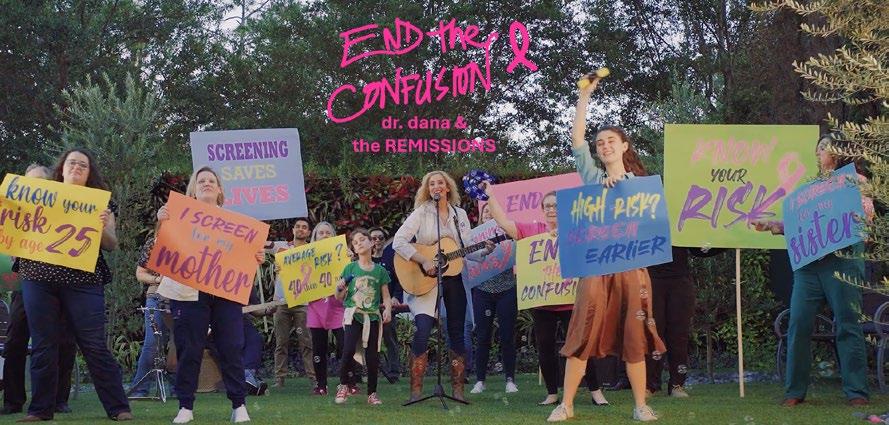
“End the Confusion”: The Journey Behind the Song Calling Women to Screen for Breast Cancer (continued from page 31)
radiologists, mammography technologists, staff members, and volunteers—who show up every day to guide and support women through their breast imaging examinations. To bring this vision to life, I recruited real members of my community, including my breast radiology colleagues, technologists, patients, and support staff, to appear in the video.
Throughout the video, various faculty, staff members, and patients appear holding posters that echo the song’s key messages: “Know your risk by age 25,” “Average risk? 40 then, 40 now,” “High risk? Screen earlier,” and “Screen for you.” These simple yet powerful statements reinforce the importance of understanding personal risk, screening, and early detection.
By the time the video ends, it becomes evident that this song and its message don’t belong to just one person. It’s a collective anthem, owned and shared by every woman and the people that support her.
End the Confusion: The Response
Since we released the “End the Confusion” music video in October, the response has blown me away. Women have messaged me saying, “I booked my mammogram because of your song.” Many women have reached out to share their stories of early detection. One woman wrote, “The song rocks, and now I’m getting screened.” My hope was to use music as a vehicle to empower more women to know their risk and get screened. Hearing that it’s working? That’s everything.
“End the Confusion” is a song for our SBI community and the women we serve. So turn up the volume, share the song, and let’s keep ending the confusion, one mammogram at a time.
You can find the three musical versions of “End The Confusion” (acoustic, full band, dance) by Dr. Dana & The ReMissions on most music platforms.
To save lives and minimize the impact of breast cancer. .....
methods, including their reliance on the Canadian National Breast Screening Studies.8-10 These colleagues include Dr. Shiela Appavoo, Dr. Martin Yaffe, Dr. Dan Kopans, and Dr. Peter Eby.


Dr. Anna Wilkinson, a general practitioner and oncologist, joined forces with Dr. Seely and published several important works with Statistics Canada based on Canadian data. They showed that compared with women in provinces that start screening at age 50 years, those who live in provinces that start screening at age 40 years have significantly lower proportions of advanced-stage breast cancer (stage II and higher)11 and significantly increased 10-year net survival.12 They showed that the peak age of diagnosis for White women was 63 years, compared with ages 52 to 60 years for non-White women, and that Black women were more likely to be diagnosed with advanced breast cancers and 40% more likely to die of their cancers than White women. They found rising incidence of breast cancer in younger women between 1984 and 2019, with the highest annual percentage changes observed among women aged 20 to 29 years and those aged 30 to 39 years.13 Their analysis of the varying costs of breast cancer treatment depending on breast cancer stage and molecular subtype (Figure 1)14 showed for the first time that screening would not only save lives and

improve the quality of life for women with cancer but would also produce net savings of about CAD $400 million each year.15 This research has had a tremendous impact. For over 30 years, only four jurisdictions screened women starting at age 40 years; now seven do. Two more jurisdictions start screening at age 45 years, and two others plan to gradually lower the starting age for screening to 40 years. Soon no jurisdiction will abide by the Task Force guidelines. Now we just need to convince 60,000 health care professionals who still follow the Task Force guidelines to change their practice.
It’s been a bigger challenge reforming the Task Force. Dr. Shiela Appavoo has had a huge impact on this effort. She assembled a coalition of many specialty societies. In addition to members of breast-related specialties, we are joined by specialists in prostate, cervical, and lung cancer; pediatrics; psychiatry; and members of many other specialties in speaking out against the harms of the Task Force methods and guidelines related to their respective fields (Figure 2).
Last month, our federal minister of health ordered a pause on the work of the Task Force, awaiting the outcome of an external expert review of the Task Force to modernize its guideline development. Our group spoke to the panel, and we look forward to seeing their recommendations for reforming the Task Force.
Our current advocacy includes supplemental screening for women with dense breasts. Our recent publication shows the impact of adding supplemental ultrasonography for women with breast density category C or D.16 Unlike women in the United States, most women in Canada undergo screening biennially with digital mammography. Exceptions are women with a first-degree family history and women with category D density, who are offered annual screening in seven jurisdictions. Dr. Seely and colleagues showed that in women with dense breasts, annual screening in these jurisdictions reduced interval cancers by almost 40% compared with biennial screening programs.17 Some provinces also offer magnetic resonance imaging or contrastenhanced mammography for women with category D density and women with greater than 25% lifetime risk. Magnetic resonance imaging is clearly the most sensitive modality for both screening and preoperative planning18 but is by far the most expensive. Limited access in Canada does not permit its use for supplemental screening as advised by the ACR.19
Continued on page 36>
Figure 1. Costs for treating breast cancer in 2023 CAD$ according to stage and molecular subtype of breast cancer. Reprinted with permission of the author.14
Figure 2. Left to right: Dr. Shiela Appavoo (radiologist), Dr. Martin Yaffe (physicist), Dr. Paul Wheatley Price (thoracic oncologist), Dr. Fred Saad (urologist), and Mr. Peter Julian, MP.
Shiela Appavoo, MD, FRCPC
Peter Eby, MD, FSBI
Martin Yaffe, PhD Anna Wilkinson, MD, CFPC
Dan Kopans, MD, FACR, FSBI
Although some authors20 suggest using various risk assessment models to decide which women should be offered supplemental screening, it is important to remember the greater risk of dense breasts in masking cancers on mammography. Our Canadian research16 showed that 84% of the cancers missed on mammography and found on screening ultrasonography were in women with category C density and that 62% of these cancers were in women with no personal or family history of breast cancer. Ideally, all women with category C or D density will have access to supplemental screening.
Another of our goals is to enable self-referral for women aged 74 years or greater, who currently must have an examination requisition to undergo screening. Approximately 15.5% of breast cancer deaths occur in women whose cancers arise between ages 75 and 84 years.21 Most are diagnosed with advanced cancers due to the lack of access to screening.11
By combining rigorous scientific evidence with effective advocacy and collaboration with other specialties in Canada, we’ve improved access to screening for women in their 40s and breast density notification. We will continue to pursue screening for women aged 74 years or greater and supplemental screening for women with dense breasts.
References
1. Gordon PB, Goldenberg SL. Malignant breast masses detected only by ultrasound. A retrospective review. Cancer. 1995;76(4):626630. doi:10.1002/1097-0142(19950815)76:4<626::aidcncr2820760413>3.0.co;2-z
2. Berg WA, Blume JD, Cormack JB, et al; ACRIN 6666 Investigators. Combined screening with ultrasound and mammography vs mammography alone in women at elevated risk of breast cancer. JAMA. 2008;299(18):21512163. doi:10.1001/jama.299.18.2151
3. Geisel J, Raghu M, Hooley R. The role of ultrasound in breast cancer screening: the case for and against ultrasound Semin Ultrasound CT MR 2018;39(1):25-34. doi:10.1053/j.sult.2017.09.006
4. Weigert JM. The Connecticut experiment; the third installment: 4 years of screening women with dense breasts with bilateral ultrasound Breast J. 2017;23(1):34-39. doi:10.1111/tbj.12678
5. Ohuchi N, Suzuki A, Sobue T, et al; J-START investigator groups. Sensitivity and specificity of mammography and adjunctive ultrasonography to screen for breast cancer in the Japan Strategic Anti-cancer Randomized Trial (J-START): a randomised controlled trial Lancet. 2016;387(10016):341-348. doi:10.1016/ S0140-6736(15)00774-6
6. Destounis S, Arieno A, Morgan R. New York state breast density mandate: follow-up data with screening sonography J Ultrasound Med. 2017;36(12):25112517. doi:10.1002/jum.14294
7. Brem RF, Lenihan MJ, Lieberman J, Torrente J. Screening breast ultrasound: past, present, and future AJR Am J Roentgenol. 2015;204(2):234-240. doi:10.2214/AJR.13.12072
8. Seely JM, Eby PR, Gordon PB, Appavoo S, Yaffe MJ. Errors in conduct of the CNBSS trials of breast cancer screening observed by research personnel J Breast Imaging. 2022;4(2):135-143. doi:10.1093/jbi/wbac009
9. Yaffe MJ, Seely JM, Gordon PB, Appavoo S, Kopans DB. The randomized trial of mammography screening that was not-a cautionary tale J Med Screen 2022;29(1):7-11. doi:10.1177/09691413211059461
10. Seely JM, Eby PR, Yaffe MJ. The fundamental flaws of the CNBSS trials: a scientific review J Breast Imaging. 2022;4(2):108-119. doi:10.1093/jbi/wbab099
11. Wilkinson AN, Billette JM, Ellison LF, Killip MA, Islam N, Seely JM. The impact of organised screening programs on breast cancer stage at diagnosis for Canadian women aged 40-49 and 50-59 Curr Oncol. 2022;29(8):56275643. doi:10.3390/curroncol29080444
12. Wilkinson AN, Ellison LF, Billette JM, Seely JM. Impact of breast cancer screening on 10-year net survival in Canadian women age 40-49 years J Clin Oncol. 2023;41(29):4669-4677. doi:10.1200/JCO.23.00348
13. Seely JM, Ellison LF, Billette JM, Zhang SX, Wilkinson AN. Incidence of breast cancer in younger women: a Canadian trend analysis Can Assoc Radiol J 2024;75(4):847-854. doi:10.1177/08465371241246422
14. Wilkinson AN, Seely JM, Rushton M, et al. Capturing the true cost of breast cancer treatment: molecular subtype and stage-specific per-case activity-based costing Curr Oncol. 2023;30(9):7860-7873. doi:10.3390/ curroncol30090571
15. Wilkinson AN, Mainprize JG, Yaffe MJ, et al. Cost-effectiveness of breast cancer screening using digital mammography in Canada JAMA Netw Open 2025;8(1):e2452821. doi:10.1001/jamanetworkopen.2024.52821
16. Gordon PB, Warren LJ, Seely JM. Cancers detected on supplemental breast ultrasound in women with dense breasts: update from a Canadian centre Can Assoc Radiol J. Published online February 21, 2025. doi:10.1177/08465371251318578
17. Seely JM, Peddle SE, Yang H, et al. Breast density and risk of interval cancers: the effect of annual versus biennial screening mammography policies in Canada Can Assoc Radiol J. 2022;73(1):90-100. doi:10.1177/08465371211027958
18. Eisen A, Fletcher GG, Fienberg S, et al. Breast magnetic resonance imaging for preoperative evaluation of breast cancer: a systematic review and meta-analysis Can Assoc Radiol J. 2024;75(1):118-135. doi:10.1177/08465371231184769
19. Monticciolo DL, Newell MS, Moy L, Lee CS, Destounis SV. Breast cancer screening for women at higher-than-average risk: updated recommendations from the ACR J Am Coll Radiol. 2023;20(9):902-914. doi:10.1016/j. jacr.2023.04.002
20. Zaki-Metias KM, Wang H, Tawil TF, et al. Breast cancer screening in the intermediate-risk population: falling through the cracks? Can Assoc Radiol J. 2024;75(3):593-600. doi:10.1177/08465371241234544
21. Oeffinger KC, Fontham ET, Etzioni R, et al; American Cancer Society. Breast cancer screening for women at average risk: 2015 guideline update from the American Cancer Society JAMA. 2015;314(15):1599-1614. doi:10.1001/ jama.2015.12783