The Member Newsletter of the Society of Breast Imaging
INSIDE THIS ISSUE:
• Issue Theme: Thinking Outside the Box: New Ways to Work
• Celebrating History of SBI: Leading Through the Pandemic
• SBI Symposium and ARRS Meeting Highlights
• Sneak Peek Into 2025 Symposium Planning Committee in Action
• New President, Board Members, and SBI Fellows
EDITOR: Nidhi Sharma
ASSISTANT EDITORS: Randy Miles and Shinn-Huey Shirley Chou
TECHNOLOGISTS’ COLUMN: Robyn Hadley and Sarah Jacobss
WHAT’S NEW IN THE NEWS:
Eleanor DiBiasio and Pamela J. DiPiro
MEMBERS IN TRAINING:
Anita Mehta
WELLNESS COLUMN: Sarah Jacobs and Claudia Cotes
THE PATIENT'S PERSPECTIVE: Hannah Perry and Danielle Sharek
LEGISLATIVE UPDATES: Amy Patel
CANADIAN CORNER: Supriya Kulkarni
Linda Moy, MD, FACR, FISMRM, FSBI President, Society of Breast Imaging
OUR SBI MISSION:
For members to be expert and authoritative breast imagers working in supportive practice environments who advance the highest quality of breast care via early detection, diagnosis, and treatment.
OUR SBI VALUES:
Patient-centered and evidence-based care
Excellence in education Scientific integrity
Collaboration and collegiality
Respect for diversity and inclusiveness
President’s Column
I am honored and humbled to begin my term as the president of the SBI. The SBI has been a big part of my professional life. Also, I have made many close friends over the years and have fond memories of prior SBI symposia. Thank you to everyone who attended the SBI 2024 Breast Imaging Symposium in affiliation with the Canadian Society of Breast Imaging. It was our most highly attended symposium, with 1618 attendees and exhibitors from 36 countries. I am particularly pleased that outstanding leaders from the European Society of Breast Imaging spoke at multiple sessions. We inducted 10 new SBI fellows. We also launched our inaugural trainee track. I would like to thank the program planning committee, the faculty, the SBI staff, and most importantly, the attendees for making this meeting a success. The President’s Dinner at the St-James Theatre was a fun and delightful evening. It was wonderful to reconnect, learn, and enjoy each other’s company.
The COVID-19 pandemic has underscored the importance of community engagement and the social impact of science. It has become evident that academic medical centers, community hospitals, and ambulatory practices must enhance their ability to serve underresourced populations. Our collaboration with global colleagues is crucial in supporting screening mammography, addressing issues of disparity in access to breast imaging examinations, and sharing innovative imaging techniques. The significant interest in contrast-enhanced mammography, artificial intelligence, and supplemental screening is a testament to the dynamic nature of the field of breast imaging.
The SBI, a vibrant community with over 3700 members, is committed to fostering inclusivity. In line with Dr. Mimi Newell’s vision, we have ensured that all members interested in serving on a committee have been assigned to one. This has led to a high level of engagement and active participation in our committees, further strengthening our community.
The Journal of Breast Imaging flourishes under the leadership of Jay Baker, MD, our interim editor in chief. The journal is poised to enter its next phase of growth. Web of Science and Scopus index it. Multiple committees continue to produce educational content for our members. The CME and SAM committee and the Young Physician Section will launch several webinars to keep our membership up to date. I am grateful for all the volunteers who support the SBI.
I would like to share two new initiatives we are rolling out in the next year. I am pleased to announce that Bob Nishikawa, PhD, is the chair of the SBI Research and Education Fund Committee. The Research and Education Fund will provide grants for scientifically rigorous pilot and early-stage research broadly related to imaging and the patient experience of screening, diagnosis, or treatment of breast cancer. The Research and Education Fund Committee hopes these grants will translate to improvements in the patient experience and subsequent external funding for investigators at any career stage.
Another initiative is for SBI to engage with local, state, and regional radiology societies, especially those that focus on breast imaging. The goals of this initiative are to support community building, increase awareness of SBI, and advocate for breast imaging professionals. The importance of networking and community for radiologists was raised at the symposium in Montreal. This initiative may allow SBI to showcase its extensive educational content.
Finally, I express my gratitude to John Lewin, MD, who rotated off the SBI Board after his year as past president. John’s remarkable leadership and wit were deeply appreciated as the SBI navigated the post-COVID-19 era. He worked diligently to ensure the success of the SBI 2022 symposium in Savannah.
Best wishes for a great summer.
Linda Moy
Linda Moy, MD, FACR, FISMRM, FSBI President, Society of Breast Imaging
Editor’s Note
By Nidhi Sharma, MD
A popular TED talk by Adam Grant, “What Frogs in Hot Water Can Teach Us About Thinking Again,” explains why humans are slow to react to impending dilemmas and the importance of rethinking. To become original, you must try something new, which means accepting some measure of risk. With continuing staff shortages, discontent, loss of joy at work, burnout galore, and massive quiet quitting, we need to get innovative in bringing joy back to our work. Our theme for this edition is “Thinking Outside the Box: New Ways to Work.”
As the academic year comes to a close and with summer upon us, I take a moment to reflect on the success of our recent outstanding annual symposium in Montreal, with the highest in-person attendance to date. All of us are tired from a full and challenging year. Nevertheless, I am proud of our SBI staff and community for what we have nurtured and created together. During my first year as editor, I have extended my hand to you, and many of you have reached back. Thank you.
While it’s normal to think about endings at this time of year, the end is merely the entrée to a new beginning. New residents and fellows have just begun their programs with beaming enthusiasm. We should be good role models for our next generation of breast imaging radiologists and try to think outside the box to find new ways to achieve better work-life balance. In the winter edition, we delved deep into the nuances of telemammography in both academic and private practices with specific tips on how to make it work for your individual work settings. Adding to this theme of thinking outside the box, this issue’s guest authors highlight part-time work, locums for technologists, and venturing into industry roles.
We have been celebrating the history of the SBI throughout this year for the upcoming 40th anniversary celebrations in 2025. Dr.
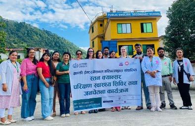
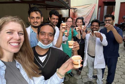
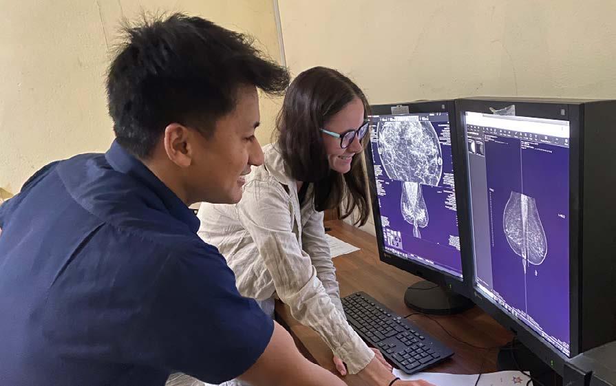
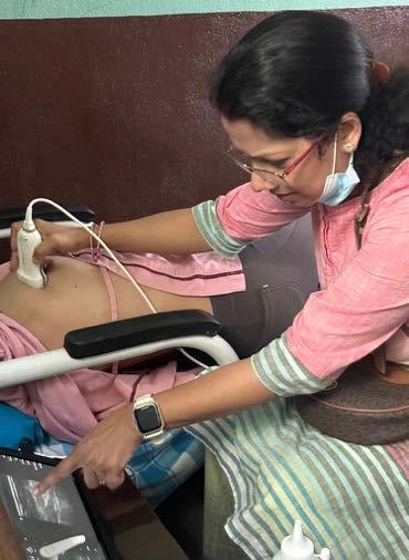
Jessica Leung, past SBI president, shares her experience leading SBI through the pandemic and finding novel ways to work and lead. Sarah Jacobs discusses the connection between mental health and workplace injury for the Wellness Column. Dr. Danielle Sharek, one of our esteemed editorial board members, shares her personal touching story of cancer, encouraging us to be more empathetic to our patients. We also get a glimpse into excellent work being done by RAD-AID in Nepal and highlights from recent SBI and American Roentgen Ray Society meetings. For the first time ever we give our readers a sneak peek behind the scenes with the Symposium Planning Committee, in addition to a multitude of exciting, informative articles.
We have several microvolunteer opportunities at the newsletter. If you or one of your technologists is interested in collaborating with our team for a future article, please reach out to us. As always, I welcome your thoughts and suggestions. Please email me at nidhisharma31@gmail.com. Looking ahead to summer, I urge you to take a moment to relax, connect with your community, and find joy in things you like to do outside work. I hope you all have a fun, sunsational summer!
Nidhi Sharma, MD
ACR Capitol Hill Day and Beyond
By Amy K. Patel, MD
So far, 2024 has been very busy on the advocacy front. Off the heels of the annual SBI symposium, the ACR annual meeting, including Capitol Hill Day, took place in Washington, DC. This year, over 450 radiologists and radiation oncologists of varied career levels met with their members of Congress to discuss key legislative and regulatory issues affecting our profession.
A key issue was the Protecting Access to Medicare Act of 2014 (PAMA). As part of PAMA, Congress established the consultation of physician-developed Appropriate Use Criteria by providers ordering advanced diagnostic imaging examinations. Implementation of the PAMA program is designed to provide the patient with the appropriate examination the first time, curb patient exposure to unnecessary radiation, reduce Medicare spending on low-value imaging, and promote the movement toward value-based imaging care and physician-developed guidelines. A study conducted by the Moran Company modeling the Congressional Budget Office’s scoring process estimated that the draft amendments would provide a Medicare savings of $2 billion over a 10-year period. The Moran Company also estimated that Medicare beneficiaries would save about $1.4 billion over the current budget window via reduced cost sharing. We are urging Congress to amend PAMA this year by including simplification language in its next Medicare-related legislative package.
Given the immense crisis we are facing with a physician workforce shortage, we met with members of Congress to discuss critical pieces of legislation that would address workforce issues. This includes cosponsoring the Resident Physician Shortage Reduction Act (HR 2389/S 1302), which would increase the number of Medicaresupported graduate medical education physicians. While the 1200 positions provided by Congress over the last three years are much appreciated, additional support is needed. This piece of legislation would increase the number of federally supported medical residency positions by 2000 annually over seven years.
We also advocated for cosponsoring the Conrad State 30 and Physician Access Reauthorization Act (HR 4942/S 665). Currently, resident physicians from other countries training in the United States on J1 visas are required to return to their home country for two years after their residency has ended before they can apply for a work visa or green card. The Conrad 30 Program allows 30 qualified residents per state to remain in the United States if they agree to practice in a medically underserved area for three years. This act was introduced in both the House and Senate to reauthorize the program and make minor improvements to its functioning if certain national thresholds are met.
Finally, we advocated for cosponsoring the Healthcare Workforce Resilience Act (HR 6205/S 3211). This legislation would expedite the visa authorization process impacting physicians stuck overseas due to backlogs and also international physicians currently working in the United States on temporary visas with approved immigrant petitions. This would initiate a one-time recapture of up to 40,000 unused employment-based visas: 25,000 for foreign-born nurses and 15,000 for foreign-born physicians.
We continued to advocate for long-term Medicare payment reform and thanked our lawmakers for supporting physicians over the last four years by mitigating scheduled Medicare cuts. Most recently, Congress added an additional 1.68% to the Medicare Physician Fee Schedule (MPFS) conversion factor (CF) beginning March 9 for the remainder of 2024. When combined with the already existing 1.25% CF bump that Congress passed at the end of 2022, the result is a 2.93% increase over what the CF would have been without congressional action. However, this is one of the only fee schedules without a built-in inflationary update. MPFS payment rates struggle to keep pace with the true cost of practice. Therefore, we encouraged our lawmakers to cosponsor, particularly in the House, the Strengthening Medicare for Patients and Providers Act (HR 2474), which adds a Medicare Economic Index–based inflationary update to the MPFS. We also encouraged senators to introduce companion legislation to HR 2474.
On the breast imaging legislation front, many bills were introduced in states aiming to expand insurance coverage for breast imaging services. Many of these bills emphasized requiring health insurance plans to cover supplemental and diagnostic breast imaging examinations without patient cost sharing. Most recently, Vermont and Iowa passed bills into law; the New Hampshire bill is pending action from the governor. In addition to Vermont, Iowa, and New Hampshire, similar bills were also introduced in Alaska, Arizona, Florida, Indiana, Kansas, Massachusetts, Michigan, Mississippi, Nebraska, North Carolina, Pennsylvania, Rhode Island, South Dakota, Virginia, West Virginia, and Wisconsin. The passage of this legislation in multiple states will hopefully provide a path to ultimately passing
Continued on page 12>
Amy K. Patel, MD
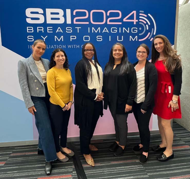
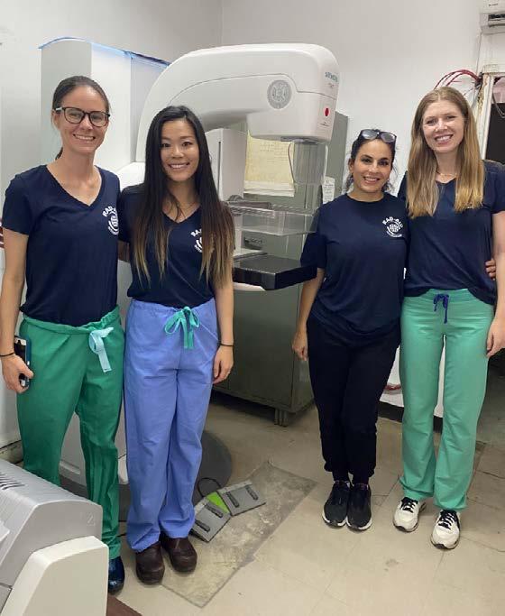
SBI 2024 SYMPOSIUM HIGHLIGHTS
The SBI 2024 Breast Imaging Symposium was held in Montreal, Canada, in affiliation with the Canadian Society of Breast Imaging. With over 1600 breast imaging radiologists from 36 countries, this was the most highly attended symposium to date. The theme of this year’s symposium was “Reimagining Breast Imaging—Seeing Opportunities Together.” The educational and impactful program was curated by the SBI Symposium Program Committee, led by the program chair and incoming SBI president, Dr. Linda Moy. The symposium centered around advancements in breast imaging, with a major focus on screening and surveillance updates, breast reconstruction imaging, and clinical implementations of artificial intelligence (AI) tools. This year brought back the entertaining “Shark Attack” competition, a brand new Trainee Track, and a lovely evening at the welcome reception as attendees watched their colleagues display their talents in the talent show. Attendees also enjoyed a splendid evening at the President’s Dinner as they celebrated outgoing president Dr. Mimi Newell’s service to the society. After a packed week of learning and networking, attendees left the meeting equipped with the latest advances and updates in breast imaging.
Screening and Surveillance
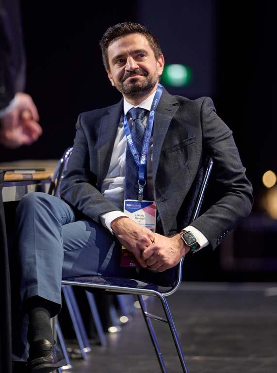
Thursday morning began with a warm welcome by Dr. Linda Moy, followed by an insightful lineup of sessions highlighting updates on screening techniques and modalities. Drs. Supriya Kulkarni and Katy Lowry provided an update on digital mammography and digital breast tomosynthesis, offering insights into recent evidence and ongoing screening trials. Dr. Jessica Leung discussed the benefits and drawbacks of supplemental screening ultrasonography. Drs. Habib Rahbar and John Lewin brought to the stage the debate that many breast imaging radiologists have had within their practices: whether to initiate an abbreviated magnetic resonance imaging (MRI) or contrast-enhanced mammography (CEM) program. Drs. Rahbar and Lewin put these modalities head to head, with compelling evidence for both.
When discussing screening and outcomes, it is critical to consider racial and ethnic disparities. Drs. Tejas Mehta and Stephanie Patterson offered data-supported strategies on how to mitigate these disparities by improving access to care, such as by overcoming barriers and implementing accessible routine screening, as well as by providing education and access to genetic assessment. The Friday afternoon scientific session highlighted research in early detection,
with several presenters addressing screening disparities and health care inequities in diverse patient populations.
As we assess the effectiveness of screening modalities, it is becoming increasingly important to understand how breast cancer outcomes are tied to the initial method of detection.
Dr. Debbie Bennett provided an excellent technically detailed demonstration on how to implement a method-of-detection program with standardized reporting.
Throughout the week, there was a consistent focus on mammography screening and supplemental screening. Expanding on the topic of surveillance, Drs. Janie Lee and Wendie Berg discussed how early detection of second breast cancer in patients with a personal history of breast cancer is essential to improving survival. They discussed data on surveillance outcomes and the recommendation for annual MRI in patients with a personal history of breast cancer before age 50 years and patients with dense breast tissue. Dr. Berg highlighted that MRI might not be accessible or the most suitable choice for certain individuals. She introduced evidence supporting CEM for supplemental screening in patients receiving surveillance. She discussed screening ultrasonography as a viable alternative when neither MRI nor CEM is feasible for certain individuals.
In his keynote lecture, Dr. Jacques Simard shared insights on personalized screening for breast cancer by risk stratification, which differs from the current age-based screening approach. Dr. Simard presented data from the project he leads (Personalized Risk Assessment for Prevention and Early Detection of Breast Cancer: Integration and Implementation), showcasing how a risk-based screening program can be successfully implemented and can even engage patients in making informed choices about their screening.
Breast Reconstruction Awareness
As breast imaging radiologists, we play an integral role on the multidisciplinary team and can assist our surgical and medical oncology colleagues in providing treatment plans. On Thursday afternoon, our colleagues in Montreal, Drs. Maude Labelle (breast imaging), Erica Patocskai (surgical oncology), Kerianne
Boulva (surgical oncology), and Arij El Khatib (plastic surgery), provided attendees with an excellent case-based overview of what surgeons want to know and how breast imaging radiologists can help them. To improve radiologists’ familiarity with posttreatment imaging, Drs. Isabelle Trop, Sujata Ghate, Matthew Seidler, and Michael Fuchsjäger gave a multimodality review of oncoplastic imaging, including normal and abnormal posttreatment imaging findings, “don’t-touch” findings, and complications. The afternoon culminated in an image-rich multidisciplinary tumor board highlighting the nuances of treatment planning and follow-up.
Artificial Intelligence
With the rapid development of AI tools and technologies, breast imaging radiologists are increasingly considering the potential implications for their practices. As AI evolves, the integration of these advanced technologies has the potential to alter clinical practice.
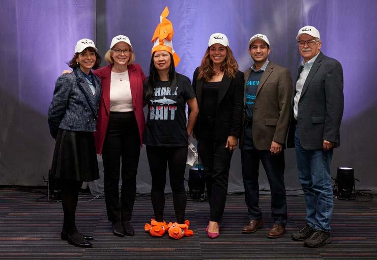
Thursday morning, attendees enjoyed a dynamic session, “Shark Attack,” in which four contestants pitched their AI product to three “sharks.” The contestants shared their vision for future AI applications, including image generation, trainee education, patient education, and mammography positioning. This session offered attendees a preview of how their practice might evolve with the integration of AI tools. The Jaws Award was given to Dr. Maggie Chung for her tool, SimGad, an AI tool for generating simulated contrast MRI.
On Friday, Dr. Julien Cohen-Adad presented the President’s Lecture on AI and bias. She discussed the differences between bias and fairness and reviewed the multitude of ways bias can be introduced into AI algorithms. Dr. Cohen-Adad stressed the importance of detecting and mitigating bias in AI.
Each afternoon of the symposium was dedicated to a series of scientific sessions that showcased the latest AI applications in breast imaging and their implementation. These sessions offered exceptional presentations on how AI can revolutionize breast imaging, particularly in cancer detection.
Drs. Raman Verma, Alana Lewin, and Lisa Mullen provided attendees with essential tips on evaluating an AI product before purchase and implementing it effectively. Highlights included Dr. Verma’s review of AI terminology and the process of AI product regulation and approval. Dr. Lewin discussed key considerations before purchasing an AI product, including learning who and what the AI product is intended to benefit, how the product can be integrated and maintained, and its risks and benefits. Dr. Mullen delved into the evaluation of AI models, equipping attendees with tools to understand and assess the metrics of AI products. Drs. Fernando ColladoMesa, Sally Friedewald, and Connie Lehman continued the discussion with presentations on maximizing the value of AI products, addressing bias mitigation, and effectively monitoring AI products in clinical practice.
A major question on the minds of many breast imaging radiologists is whether we are ready to use AI in clinical practice. Drs. Ritse Mann, Jung Hyun Yoon, and Emily Conant answered with a nuanced yes and no. Dr. Mann presented interesting data showing that standalone AI can now outperform a breast radiologist in reading a screening mammogram and showing how AI can be used as a second reader or used to triage studies. Dr. Yoon emphasized that standalone AI and AI for triage are not quite ready for clinical deployment due to concerns including generalizability, the need to understand the types of cancers AI might miss, the potential for radiologist bias influenced by AI suggestions, and the necessity for quality control measures. Dr. Conant shared how AI could be used to enhance risk estimation and personalized screening recommendations. While AI holds significant promise for these applications, more robust and generalizable data are needed.
Practice Updates and Focus Tracks
On Saturday morning, the conference hall was packed with attendees as Drs. Steven Poplack, Jocelyn Rapelyea, Lilian Wang, and Edward Sickles provided an update on the highly anticipated sixth edition of the BI-RADS manual. This was followed by important updates to Mammography Quality Standards Act regulations.
Attendees enjoyed two inspiring TED talks later that morning. Dr. Caroline Daly shared her heartfelt and eye-opening journey from physician to patient with breast cancer. Dr. Cunningham set attendees’ eyes on the horizon with tips for using credit card points and airline miles to travel smarter.
The first-ever Trainee Track was held on Saturday. The Symposium Program Committee demonstrated SBI’s commitment to engaging trainees by developing this new track and plans to make this a permanent offering at the annual symposium. The goal of this track was to bring trainees together to learn essential leadership, communication, and career development skills while networking with leaders in the field. A combined trainee and Young Physician Section (YPS) session was centered around how to sustain a practice in breast imaging. The afternoon culminated in a combined Resident and Fellow Section and YPS reception hosted by Mammotome, which had an excellent turnout.
Networking and Celebrations
While the days were packed with learning, attendees were able to enjoy an evening of networking and fun at the welcome reception on Thursday evening. Several attendees put their talents on display at the talent show. Additionally, attendees gathered at the sold-out President’s Dinner for a lovely evening celebrating the outgoing president, Dr. Mimi Newell.
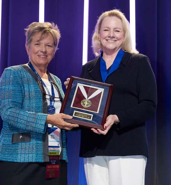
A very special congratulations to Dr. Elizabeth Morris, the recipient of the Gold Medal, and Dr. Tiffany Gowen, the recipient of the Honorary Fellow Award. Furthermore, a heartfelt thank you to everyone involved in making the symposium a success, from organizers to attendees. We look forward to seeing you next year in Colorado Springs!
2025 Symposium Planning Committee: Behind the Scenes
By Dana Ataya, MD
The 2025 SBI symposium at the Broadmoor in Colorado Springs, Colorado, from April 24 through 27, will commemorate the 40th anniversary of the SBI and the commitment of our society to save lives and minimize the impact of breast cancer. What initially began as a small postgraduate course in 1993 has evolved into a three-and-a-half-day conference with over 1600 participants attending the Montreal meeting in person in 2024.
The first SBI meeting was established to bring expert radiologists together to support dialogue, foster research, share best practices, and disseminate information on breast imaging, and this remains true today. Members consistently rank the symposium as the top service provided by the SBI. The Symposium Planning Committee annually commits to creating an educational, cutting-edge, inclusive, and relevant meeting experience that attendees can translate into their clinical practice to improve outcomes for patients and communities. Here’s a peek at what goes on behind the scenes in planning the annual SBI symposium, the preeminent meeting for breast radiology in the world.
The Team
The Symposium Planning Committee comprises 15 breast radiologists and SBI staff members. Required SBI Symposium Planning Committee members include the current SBI president, vice president (education chair), scientific chair, incoming scientific chair, CME-SAM Committee chair or vice chair, a Journal of Breast Imaging representative, and a Resident and Fellow Section/Young Physician Section representative. The remainder of the committee members are selected by the Symposium Planning Committee chair. Frequently, invited committee members are curated to ensure geographical diversity (representation from different parts of the country), practice diversity, diverse levels of professional experience, gender diversity, racial diversity, and insights into a particular area of expertise.
The SBI Symposium Planning Committee also includes (and would not be possible without) the dedicated SBI staff members who are integral to the success of the meeting.
Jennifer Luettinger, BA, CAE, senior director, Education and Meeting, oversees the SBI symposium. She works directly with the program chair to coordinate overall program planning, guide the Symposium Program Committee, manage CME requirements, and oversee exhibits and corporate sponsorship.
In collaboration with the meeting teams, Jennifer ensures a successful and enriching exhibit hall and industry-sponsored learning experience for attendees and industry partners.
Nuria Gramkee, BS, education program manager, manages the education component of the meeting, including over 100 faculty members and guest speakers, the scientific abstract submission and selection process, awards, presentations, audiovisual support, eventScribe, CME/CE accreditation, and faculty biographies. She is the on-site support in the speaker ready room and ensures that every moderator is prepared for the day. She also manages the SBI Symposium Pre-Course, faculty, and program.
Natalie Ward, BA, education coordinator, provides support to the education team, manages the on-site logistics for the meeting, supports the conference app and website, and assists attendees with questions and CME.
Yasmeen Fields, MS, CAE, CEO, joined SBI in 2012 and has been integral to every symposium since her arrival. She has performed all necessary tasks and served various roles in support of the symposium while advancing to become the first CEO of SBI.
Kesha L. Willis, BA, CNP, CAE, senior director, Membership, Marketing, and Communications, provides strategic direction and oversight of the marketing and communications for the SBI symposium. She collaborates with her team to develop an integrated marketing strategy to ensure alignment and integration with the overall organizational brand. Ms. Willis’ team plays a critical role in driving attendance and engagement through strategic marketing campaigns and initiatives.
Rachel Gellman, MS, senior marketing manager, provides hands-on implementation of the marketing strategy. She plays a pivotal role in the execution of marketing campaigns that ensure objectives are met and often exceeded. She collaborates closely with the symposium manager to align and integrate marketing plans with overall SBI symposium plans.
To save lives and minimize the impact of breast cancer. .....
Dana Ataya, MD
The Location
The SBI staff curate a list of possible symposium locations, prioritizing cities that are affordable, are accessible, and possess the required conference and hotel space. Final locations for the SBI symposium are agreed upon by the SBI Board years in advance. This allows SBI staff to prepare budgets and negotiate contracts that are favorable to the SBI and SBI membership.
The Program
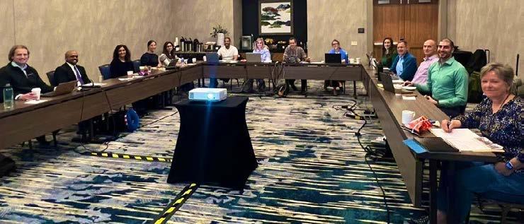
The Symposium Planning Committee is responsible for the content of the program. Each year, the Symposium Planning Committee chair sets the overall vision and theme of the symposium and works with committee members to develop the program structure, lecture topics, and speakers. Each Symposium Committee and chair are unique, resulting in variability over the years in how committee tasks are managed. Despite this variability, the guideposts of every SBI Symposium Planning Committee are the comments and feedback from SBI symposium attendees. Before convening, the Symposium Planning Committee members sift through pages of feedback from SBI members and past symposium attendee surveys, consolidating comments and ratings of prior symposium content. Past symposium attendee feedback is integral in shaping future meeting content, topic selection, symposium structure, and speaker selection.
More than one year before each symposium, the Planning Committee members meet in person to begin establishing the structure, lecture topics, and speaker selection. Symposium content reflects a mix of practice-changing updates, scientific advances, and a balance of “bread-and-butter” and novel breast imaging content. The program can be adjusted every year. On the basis of feedback from members, for example, the SBI Symposium Planning Committee recently developed afternoon tracks, including a 2024 track for early professionals and resident/fellow members. Because of favorable evaluations and suggestions by attendee members, we anticipate expanding these tracks to encompass topics relevant to members working in private practice and academia.
The SBI symposium has historically been an education-focused meeting but continues to evolve and grow as a preeminent scientific forum for breast imaging. The number of abstract submissions has progressively increased since the scientific program was established in 2015. In 2024, there were 244 scientific abstract submissions—a record number—for the 20 oral presentation slots available. Because of the increase in highquality submissions and feedback from members, the committee is expanding available slots to meet the demand of the increased number of submissions for the 2025 meeting. Electronic abstracts are also accepted at the symposium. Each year, the scientific chair invites a dedicated group to serve as abstract reviewers. We receive submissions from over 35 countries and look forward to expanding the program in 2025.
Traditionally, invited speakers at the SBI symposium have been fellows of the SBI. In more recent years, in response to feedback from SBI symposium attendees, this practice has evolved and invited speakers who are not SBI fellows have been integrated into the program. The SBI Symposium Planning Committee remains committed to selecting faculty members who represent the diverse membership of the SBI community for the 2025 symposium and beyond.
The committee strives to produce a meeting program that is fun, evidence based, and professional. “Jeopardy,” “Shark Tank,” and more recently the TED talks and talent show have been various creative approaches the Symposium Planning Committee has integrated into the program to enhance engagement and community building. As integration of the creative arts into society meetings enhances meeting attendee experience,1 we anticipate continued incorporation of the arts and humanities at future SBI symposia.
Continued on page 25>
SBI staff. Left to right: Nuria Gramkee, Rachel Gellman, Kesha L. Willis, Yasmeen Fields, Natalie Ward, and Jennifer Luettinger.
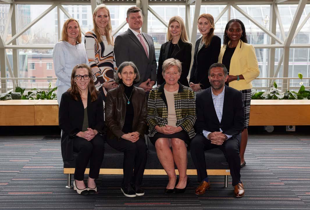
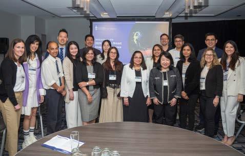
The 2025 SBI Symposium Planning Committee. Left to right: Lars Grimm, Anand Narayan, Tejas Mehta, Elissa Price, Rend AlKhalili, Randy Miles, Dana Ataya, Peter Eby, Nuria Gramkee, Jennifer Luettinger, Gary Whitman, John Scheel, Brian Dontchos, and Mimi Newell. Not shown: Linda Moy, Yasmeen Fields, and Natalie Ward.
blanket federal legislation so that no patient is left behind regardless of insurance plan and geographic location.
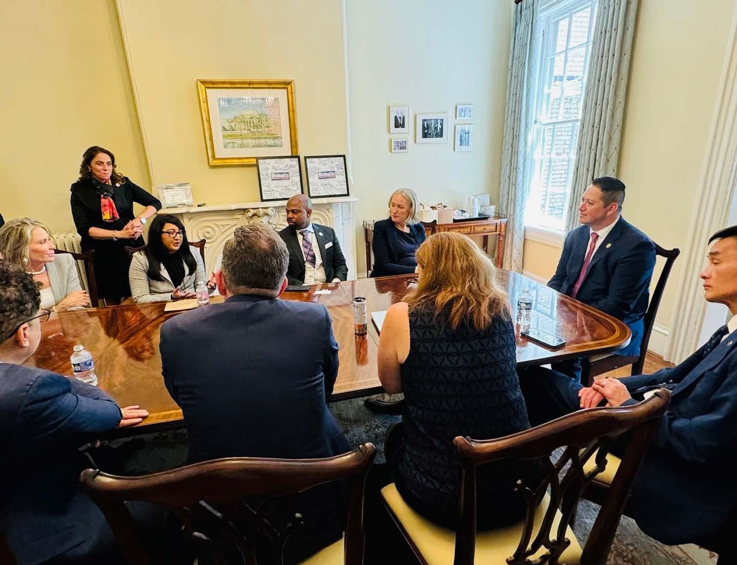
Finally, I was proud to represent the ACR during the House Call on the Mall event in June, in which approximately 100 physicians and dentists representing over 15 organizations met with federal elected officials in the House of Representatives with jurisdiction over health care and also with allies of medical subspecialties, such as radiology. It was a very fruitful event that also highlighted the importance of having direct contact on Capitol Hill with these federal elected officials who are making pivotal health care decisions for our patients. Approximately 94% of members of Congress do not have a health care background, so the more often we are on Capitol Hill advocating for access on behalf of our patients, the more beneficial it is to our profession when Congress votes on critical pieces of radiology legislation. We are increasing our presence on Capitol Hill in addition to the tireless
work our amazing Government Relations team is already carrying out. This new and improved boots-on-the-ground approach is necessary given the challenges we face in a competitive health care environment.
As we continue to advocate for access to patient care and fair reimbursement for the services we provide, coalition building and synergism are imperative. We look forward to your participation in our radiology advocacy efforts throughout the remainder of 2024. As always, thank you for all that you do for radiology and, most importantly, for our patients.
Special thanks to the ACR Government Relations team, particularly Rebecca Spangler, Ashley Walton, and Eugenia Krimer, for assistance with this content. ACR Capitol Hill Day and Beyond (continued from page 5)
Amy Patel, MD, speaking to Tony Gonzales, representative from Texas, at the House Call on the Mall, a medical specialty roundtable discussion of key issues affecting our profession.
New Breast Cancer Screening Recommendations From the USPSTF
By Eleanor DiBiasio, MD
The US Preventive Services Task Force (USPSTF) is a panel of preventive medicine and primary care clinicians that makes evidence-based recommendations about clinical preventive services such as screening.1 The USPSTF assigns each recommendation a letter grade (A, B, C, D, or I) on the basis of the perceived strength of the evidence and the balance of benefits and harms from a preventive service.
In April 2024, the USPSTF issued an updated recommendation on breast cancer screening. The USPSTF concluded with moderate certainty (grade B recommendation) that biennial screening mammography has a moderate net benefit in women aged 40 to 74 years.2 This updated guideline differs from the prior USPSTF recommendations published in 2016, which included biennial screening mammography for women aged 50 to 74 years.3
The USPSTF’s methods included a comparative effectiveness review of mammography-based breast cancer screening strategies4 and collaborative modeling studies.5 The decision to recommend earlier screening came from data showing that the incidence rate of invasive breast cancer in 40- to 49-year-old women increased an average of 2.0% annually between 2015 and 2019.2 In addition, collaborative modeling data estimated that screening beginning at age 40 years would prevent an additional 1.3 breast cancer deaths per 1000 women compared with biennial screening for women aged 50 to 74 years.2,5 This recommendation was also informed by health equity data showing that Black and Hispanic women are more likely than women in other racial and ethnic groups to be diagnosed with invasive breast cancer before age 50 years and to have more advanced stages of cancer and worse outcomes.2,6
The updated USPSTF guideline is now partially in line with screening guidelines from the ACR and SBI, which also recommend screening mammography beginning at age 40 years for women at average risk. However, the ACR and SBI recommend annual rather than biennial screening mammography.7
The USPSTF’s rationale for biennial screening is largely based on a nonrandomized study using data from the Breast Cancer Surveillance Consortium showing no difference in detection of cancers of stage IIB or higher and no difference in detection of cancers with less favorable prognostic features with annual versus biennial screening.2,8 In addition, their comparative modeling studies predicted more life-years gained and breast cancer deaths averted per false-positive result with biennial screening than with annual screening.2,5
However, although cumulative falsepositive results may be higher with annual screening than with biennial screening, the marginal rate of falsepositive results is lower and the overall reductions in breast cancer mortality and years of life saved are greater with annual screening.9,10 Research has also proven that women diagnosed with breast cancer at younger ages and Black women are more likely to have aggressive cancers,6,11 for which annual mammography is critically important to detect.
The USPSTF also issued a statement that evidence is insufficient to determine the balance of benefits and harms of supplemental screening for breast cancer with ultrasound or magnetic resonance imaging (MRI) regardless of breast density (grade I recommendation). In contrast, the ACR recommends annual MRI starting at age 40 years for women with dense breasts who desire supplemental screening, especially if their risk is higher than average.12 The ACR recommendations are supported by research showing higher rates of invasive cancer detection among women with dense breasts undergoing MRI compared with digital breast tomosynthesis.13
Finally, the USPSTF concluded in its 2024 recommendations that the evidence is insufficient to determine the balance of benefits and harms of screening mammography for women 75 years and older (grade I recommendation). The ACR and SBI recommend that women continue screening past age 74 years unless severe comorbidities limit life expectancy7,14 because of the continued high risk of breast cancer morbidity and mortality in this age group.
As breast radiologists, we are often asked by patients and other clinicians about the appropriate use, timing, and frequency of breast cancer screening. The differing recommendations from multiple medical societies can be challenging for patients and referring clinicians to navigate. It is important to familiarize ourselves with the various recommendations so that we may support those of our own society using scientific evidence, data, and sound reasoning. Annual screening mammography starting at age 40 years for women at average risk is ideal, and supplemental screening modalities should be considered for women with dense breasts and those at higher than average risk.
Continued on page 19>
Eleanor DiBiasio, MD
CANADIAN CORNER
News From the Canadian Society of Breast Imaging
By Supriya Kulkarni, DMRD, DNB, FRCP(C), FSBI
As we head into the summer months and the conclusion of another academic year, the Canadian Society of Breast Imaging (CSBI) is also experiencing changing tides. At the 2024 SBI Breast Imaging Symposium in April, Dr. Jean Seely (University of Ottawa) stepped down from the CSBI presidency after the seven steadfast years she spent building up the society. I am humbled to take up the torch and serve as the new president of the CSBI alongside a dynamic and supportive Board of Directors.
Introductions
Supriya Kulkarni, DMRD, DNB, FRCP(C), FSBI, is the CSBI president for 2024-2026. Dr. Kulkarni is the breast division head and associate professor, Department of Medical Imaging, University of Toronto. Her clinical engagement is at University Health Network, Mount Sinai Hospital, Women’s College Hospital, University of Toronto, Canada. Dr. Kulkarni is the regional breast imaging lead for multiple regional cancer programs within the Ontario Breast Screening Program. Her special focus is on teaching and providing mentorship nationally and internationally, and she has received several teaching awards for contributing to cancer education and creating continuing professional development programs and workshops.
Dr. Shushiela Appavoo, MD, FRCPC (University of Alberta), recently joined the CSBI Board of Directors. Dr. Appavoo also chairs the CSBI Patient Engagement Working Group.
We also thank our outgoing trainee board representatives, Dr. Kaitlin Zaki-Metias (Trinity Health Oakland and Wayne State University) and Dr. Sri Sannihita Vatturi (University of Ottawa) for their contributions to the CSBI over the past 18 months. Although no longer on the board, Drs. Zaki-Metias and Vatturi will cochair our new Mentorship Working Group.
SBI/CSBI Joint Annual Meeting
The SBI/CSBI joint annual meeting in Montreal in 2024 was a resounding success, bringing together the best and brightest minds in breast imaging to share knowledge, foster collaboration, and drive innovation. The program showcased multiple talented Canadian speakers from across the country. This collaboration exemplifies the spirit of cooperation and mutual support within the breast imaging community and sets the stage for continued advancements in the field.
save lives and minimize the impact of breast cancer.
Toronto Breast Imaging Conference
The annual Toronto Breast Imaging Conference was held on May 25, 2024, and was hosted by Dr. Kulkarni. This year Dr. Jessica Leung (MD Anderson Cancer Center) was the keynote speaker. The program included a comprehensive session on assessment of the axilla. The conference also hosted multiple vendors and supported interventional workshops showcasing state-ofthe-art interventional technology, including wire-free localization techniques and vacuum-assisted biopsies. This conference is in its ninth year and over the years has hosted various keynote speakers from the SBI, including Drs. Janice Sung, Ed Sickles, Sujata Ghate, Dan Kopans, and Peter Eby.
Canadian Task Force on Preventive Health Care
The end of May brought about a flurry of activity with the release of the draft recommendations on breast cancer screening by the Canadian Task Force on Preventive Health Care (CTFPHC). Unfortunately, these updated guidelines proved disappointing as the CTFPHC recommendations remain in conflict with recommendations from international organizations such as the US Preventive Services Task Force, the European Commission on Cancer Screening, ACR, and SBI and national organizations including the Canadian Cancer Society, Canadian Association of Radiologists, and CSBI. The full CSBI statement regarding the CTFPHC guidelines can be accessed here
CSBI President Dr. Supriya Kulkarni; board members Drs. Jean Seely, Paula Gordon, and Shushiela Appavoo; Advocacy Committee member Dr. Ify McKerlie; and senior scientist Dr. Martin Yaffe recently testified to the Canadian House of Commons Standing Committee on Health against the updated breast cancer screening guidelines. The CTFPHC draft recommendations kept the newly formed CSBI Advocacy Working Group busy and engaged during their initial meeting.
Dense Breasts Canada
Dense Breasts Canada is a nonprofit organization with multiple dedicated volunteers. Dense Breasts Canada raises awareness and advocates for better breast screening for Canadians, provincially and federally. We acknowledge and thank Dense Breasts Canada for their continued advocacy.
Supriya Kulkarni, DMRD, DNB, FRCP(C), FSBI
Dense Breasts Canada has recently released a series of photo essays titled “I Want You to Know” that send emotional messages of action from breast cancer patients. The participants’ messages to those who have not been diagnosed with the disease are to get to know your body, get checked, and be prepared to advocate for yourself.
The “I Want You to Know” series features 31 people aged 26 to 73 years who live in Canada and are of various genders, cultures, lived experiences, abilities, and disease stages. They represent the hundreds of thousands of individuals diagnosed with breast cancer each year in North America. Read more about their moving personal stories here
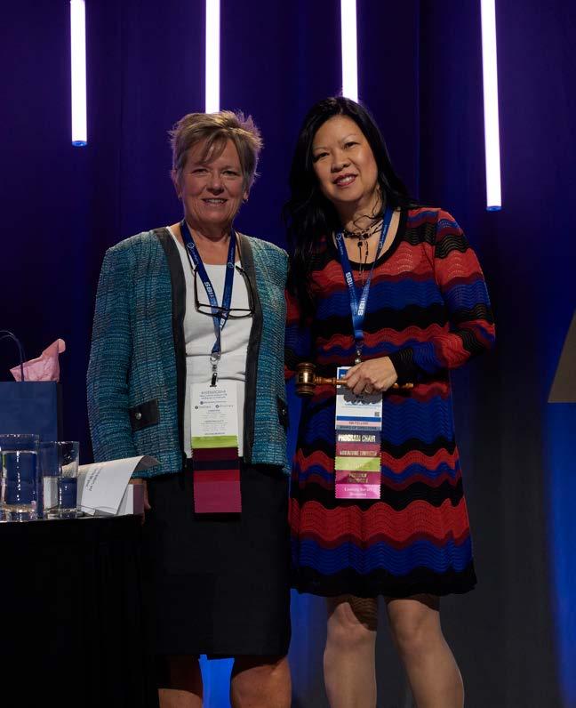
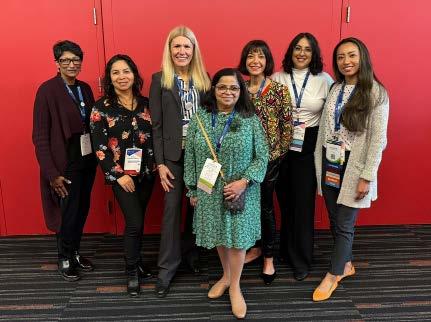
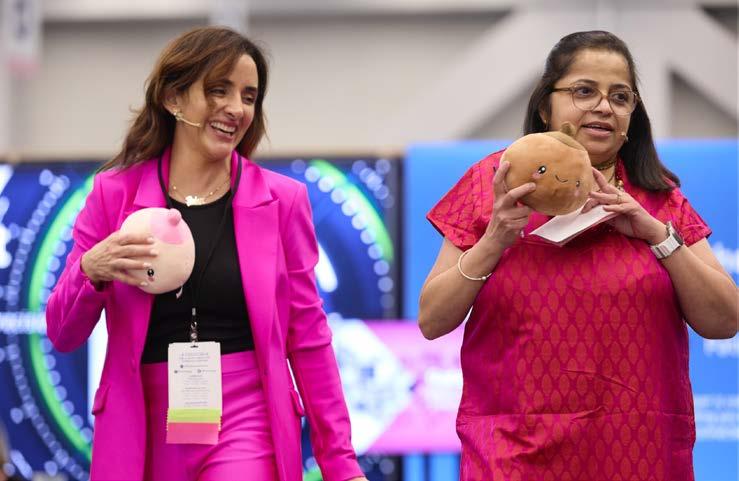
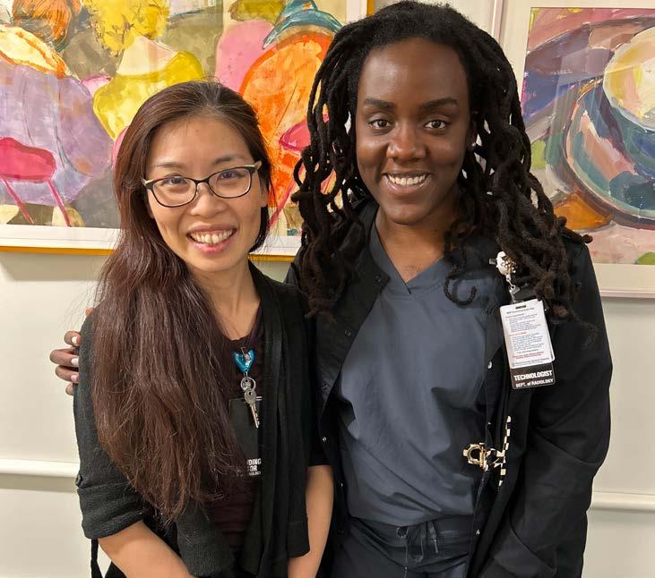
Dr. Supriya Kulkarni (left) and Dr. Jean Seely (right) at the SBI President’s Dinner in Montreal, Canada, on April 12, 2024.
CSBI Board of Directors at the 2024 SBI/CSBI annual meeting. Left to right: Dr. Shushiela Appavoo, Dr. Silma Solorzano, Dr. Jean Seely, Dr. Supriya Kulkarni, Dr. Paula Gordon, Dr. Kaitlin Zaki-Metias, and Dr. Huijuan (June) Wang.
CSBI trainee representatives at the 2024 SBI/CSBI annual meeting. Left to right: Dr. Kaitlin Zaki-Metias, Dr. Sri Sannihita Vatturi, and Dr. Huijuan (June) Wang.
University of Toronto faculty with keynote speaker Dr. Jessica Leung at the Toronto Breast Imaging Conference, May 25, 2024.
CSBI President Dr. Supriya Kulkarni cohosting the inaugural SBI Talent Show on April 11, 2024, with Dr. Robyn Roth (New Jersey).
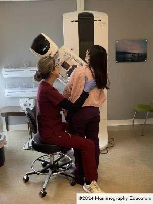
Helping Your Technologists: Tips for Troubleshooting Mammographic Positioning, Part 1
By Robyn Hadley, RT(R)(M); Sarah Jacobs, BS, RT(R)(M)(CT)
Quality imaging is a top priority of each member of the imaging team. Various challenges that are routinely presented throughout a technologist’s workday pose limitations for acquiring quality images. Understanding these challenges and how to troubleshoot them can be difficult for the technologist and the interpreting radiologist. We recommend simplifying the troubleshooting process with your technologists by using the data-driven scientific principles outlined in this article. This article, the first of a twopart series, focuses on tips that radiologists can share with their mammography technologists to correct common positioning problems. The second part will offer ideas for providing image quality feedback to technologists while collaboratively assessing corrective action and amplifying high-level communication skills.
A study by Huppe et al established realistic expectations for specific imaging criteria for the craniocaudal (CC) and mediolateral oblique (MLO) views.1 Because of variations in patients’ physical limitations, body habitus, breast size and shape, mobility, compression tolerance, cognitive abilities, and other unique conditions, achieving all imaging criteria 100% of the time is unrealistic. A study by Salkowski et al found that positioning is the predominant factor in technical recalls. Patient recalls for additional imaging due to technical factors are often related to positioning. Positioning problems account for 47% of full-field digital mammography recalls and 81% of digital breast tomosynthesis recalls.2
The criteria for positioning and image quality assessment have remained unchanged since the publication of the 1999 ACR Mammography Quality Control Manual, which includes a section on clinical image quality. However, this version was developed for use with film-screen mammography and has not been updated to allow for the changes and challenges that technologists encounter with full-field digital mammography and digital breast tomosynthesis, including an increase in the length, width, and thickness of the image receptor (IR). This situation complicates how technologists are taught to position patients for mammograms and can also account for the wide variation in clinical image quality that radiologists encounter, especially from technologist to technologist and from year to year. More importantly, resources for and understanding of how to effectively troubleshoot positioning challenges and work with patient limitations that impact image quality are lacking. Troubleshooting image quality should be based on a solid understanding of correlative anatomy and standardized positioning
techniques. Each step in the positioning process has an effect on a specific image quality criterion. Remember two sources of error when positioning and troubleshooting mammographic images: how the patient is positioned and how the machine is set up or positioned (ie, the angle and height of the IR and the compression paddle size).
Understanding key foundational principles can improve image quality. The following are tips for common positioning challenges with the CC and MLO views.3
Troubleshooting the CC View
Poor Visualization of Posterior Tissue
∙ Ensuring appropriate height of the IR is critical. An IR that is too low will exclude posterior, superior breast tissue. An IR that is too high will exclude posterior, inferior breast tissue. Elevate the breast until the posterior nipple line is perpendicular to the chest wall. Raise the IR to the level of the elevated inframammary fold.
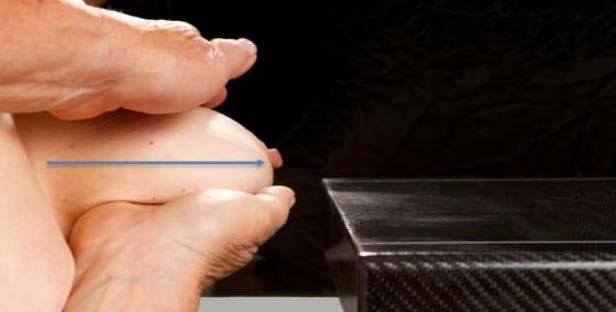
∙ Optimize posterior tissue visualization by pulling the breast onto the IR with both hands (Figure 1).
∙ Anchor the breast with the base of the thumb at the 12-o’clock position and continue to pull the breast forward upon compression.
Robyn Hadley, RT(R)(M) Sarah Jacobs, BS, RT(R)(M)(CT)
Figure 1. Posterior nipple line perpendicular to chest wall and breast pulled onto IR with both hands. Image courtesy of Mammography Educators.
Poor Visualization of Medial Tissue
∙ Once the breast is pulled onto the IR, lift the contralateral breast up and over onto the corner of the IR.
∙ Ensure that the patient’s feet, hips, and shoulders are facing forward. This position will help maximize visualization of deep medial breast tissue.
Troubleshooting the MLO View
Visualization of Inframammary Fold
∙ Do not ask the patient to lean forward while moving the hips and buttocks backward; this position removes the inframammary fold from the field of view.
∙ To ensure the inframammary fold is in front of the IR and visualized on the image, be certain the patient’s feet, hips, and shoulders are facing forward. The patient must sidestep toward the technologist to ensure the bottom corner of the IR is positioned halfway between the patient’s umbilicus and anterior superior iliac spine.
Amount of Pectoralis Muscle
∙ The length of the pectoralis muscle should extend down to the level of the posterior nipple line with a wide margin in the axilla.
∙ The angle of the machine should be parallel to the free margin of the pectoralis muscle with the patient facing forward to achieve adequate length of the muscle. If the angle is too steep or the patient is turned away from the machine and not facing forward, the pectoralis muscle will be shortened.
∙ To obtain a wide margin of muscle in the axilla, place the corner of the IR just anterior to the latissimus dorsi while the patient’s shoulder is directed forward and down and remains relaxed.
Shape of Pectoralis Muscle
∙ The pectoralis muscle should appear convex or straight. This shape indicates that optimal posterior tissue is visualized, with the breast adequately pulled away from the chest wall. A relaxed muscle allows optimal taut compression and separation of breast structures without undue discomfort.
∙ Asking the patient to rest or drape her arm over the side of the IR is often well received. Be mindful not to overuse the word relax. It may also be helpful to ask the patient to soften her shoulders to effectively relax the pectoralis muscle.
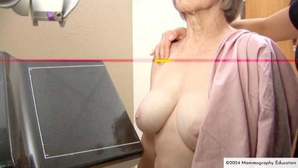
∙ Proper height of the IR is also essential to ensure optimal pectoralis muscle shape. Correct height for the MLO view is achieved when the top corner of the IR is at the level of the sternoclavicular joint, halfway between the top of the shoulder and the axillary crease (Figure 2).
Paddle Size
The same paddle size should be used for both CC views and the same paddle size should be used for both MLO views. Depending on the patient’s body and breast size or shape, using one paddle size for the CC views and a different paddle size for the MLO views may be necessary. Using the appropriate paddle size allows all breast tissue to be imaged within the perimeter and ensures optimal image centering.
∙ Patients with a breast shape that is wide and not long may require the large paddle for the CC view. However, the same patient may require the small paddle for the MLO view to effectively include all superior and inferior tissue and achieve proper image centering without including excess abdominal tissue inferiorly.
∙ Patients with small breasts and a long thorax require the small paddle for the CC view and the large paddle for the MLO view.
∙ Using the half paddle, if available, may be beneficial for male patients, patients with very small breasts, implant-displaced views, and patients with extremely thin breasts. This paddle provides more space for the technologist’s hand, allowing adequate anchoring of the breast to ensure optimal visualization of posterior tissue and compression without having the hand caught between the breast and compression paddle.
Patient Limitations
Technologists face a number of patient limitations that affect the ability to produce images of optimal quality. Producing quality images of all four standard views is often not achievable with patients who have specific limitations or challenges. Including a supplemental view is not only beneficial but may also be necessary to adequately image all breast tissue.4
Continued on page 18>
Figure 2. Proper height of IR for MLO view with top corner of IR at the level of the sternoclavicular joint, halfway between the top of the shoulder and the axillary crease. Image courtesy of Mammography Educators and Volpara Health.
from page 17)
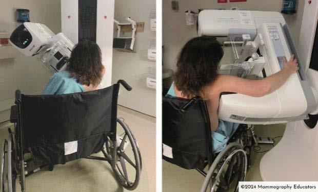
∙ Patients in wheelchairs: For the CC view, place foam blocks or pillows behind the patient to help hold the patient forward. For the MLO view, place the wheelchair at a 45° angle and remove armrests and footrests if possible (Figure 3).
∙ Patients with cognitive compromise: Allow a caregiver or family member to be in the examination room to calm the patient if necessary.
∙ Patients with physical limitations such as kyphosis, scoliosis, pectus carinatum, or pectus excavatum: Patients with kyphosis can have effective imaging for the CC view if they are seated (Figure 4). Angling patients with scoliosis differently for each MLO view may be necessary depending on the degree of curvature of the spine. Patients with pectus carinatum may require two CC views, one to adequately include the lateral aspect of the breast and one to include the medial aspect. Adding a supplemental lateromedial (LM) view can be helpful for visualizing inferior, posterior tissue (Figure 5).
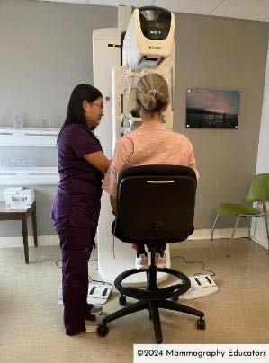
∙ Height difference: When the patient and technologist are of significantly different heights, it may be beneficial for the patient or technologist to be seated during the examination while obtaining the CC views to ensure sound ergonomics for the technologist (Figure 6).
∙ Patients with limited range of motion (shoulder, neck, or back): Exercise care not to force patient movement. Patients should feel comfortable discussing their limitations with the technologist during positioning.
∙ Patients with implanted medical devices: Patients with devices such as pacemakers, defibrillators, ports, shunts, or loop recorders may require two views to accomplish an adequate screening view. For example, the MLO view may require one view with minimal compression to include the posterior tissue (with the device) and an additional anterior compression view with full compression to include anterior breast tissue (Figure 7). Technologists’ Column: Helping Your Technologists: Tips For
To save lives and minimize the impact of breast cancer. .....
Figure 3. Wheelchair with armrests removed, positioned at a 45° angle to the machine for the MLO view. Images courtesy of Mammography Educators.
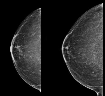
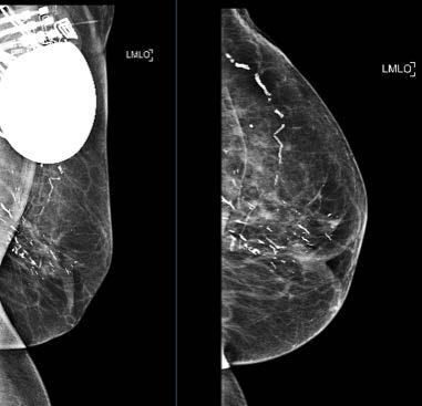
Figure 4. Images obtained from a patient with kyphosis. Left, CC view obtained with patient standing. Right, CC view obtained with patient seated. Images courtesy of Robyn Hadley.
Figure 5. Images obtained from a patient with kyphosis. Left, Standard MLO view. Right, supplemental lateromedial view to include posterior inferior tissue. Images courtesy of Robyn Hadley.
Figure 6. Left, Short technologist performing an examination on a tall patient. Right, Tall technologist performing examination on a short patient. Images courtesy of Mammography Educators.
What’s New in the News: New Breast Cancer Screening Recommendations From the USPSTF
(continued from page 13)
References
1. About the USPSTF. US Preventive Services Task Force. Accessed June 4, 2024. https://www.uspreventiveservicestaskforce.org/uspstf/about-uspstf
2. US Preventive Services Task Force; Nicholson WK, Silverstein M, Wong JB, et al. Screening for breast cancer: US Preventive Services Task Force recommendation statement JAMA. 2024;331(22):1918-1930. doi:10.1001/jama.2024.5534
3. Siu AL; U.S. Preventive Services Task Force. Screening for breast cancer: U.S. Preventive Services Task Force recommendation statement Ann Intern Med 2016;164(4):279-296. doi:10.7326/M15-2886
4. Henderson JT, Webber EM, Weyrich MS, Miller M, Melnikow J. Screening for breast cancer: evidence report and systematic review for the US Preventive Services Task Force JAMA. 2024;331(22):1931-1946. doi:10.1001/jama.2023.25844
5. Trentham-Dietz A, Chapman CH, Jayasekera J, et al. Collaborative modeling to compare different breast cancer screening strategies: a decision analysis for the US Preventive Services Task Force JAMA. 2024;331(22):1947-1960. doi:10.1001/ jama.2023.24766
6. Hendrick RE, Monticciolo DL, Biggs KW, Malak SF. Age distributions of breast cancer diagnosis and mortality by race and ethnicity in US women Cancer 2021;127(23):4384-4392. doi:10.1002/cncr.33846
7. Monticciolo DL, Malak SF, Friedewald SM, et al. Breast cancer screening recommendations inclusive of all women at average risk: update from the ACR and Society of Breast Imaging J Am Coll Radiol. 2021;18(9):1280-1288. doi:10.1016/j. jacr.2021.04.021
∙ Patients with chronic illnesses: Encourage technologists to do their best and enlist the help of other technologists, staff members accompanying the patient, or the patient’s family. Doing so can help achieve quality imaging while keeping the patient as calm and comfortable as possible.
Despite the numerous challenges technologists and radiologists face in their imaging departments, effective troubleshooting during mammographic positioning can help overcome some of the most common positioning challenges. Knowing what actions to take and when to take them can lead to a positive patient experience and improve image quality.
References
1. Huppe AI, Overman KL, Gatewood JB, Hill JD, Miller LC, Inciardi MF. Mammography positioning standards in the digital era: is the status quo acceptable? AJR Am J Roentgenol. 2017;209(6):1419-1425. doi:10.2214/AJR.16.17522
2. Salkowski LR, Elezaby M, Fowler AM, Burnside E, Woods RW, Strigel RM. Comparison of screening full-field digital mammography and digital breast tomosynthesis technical recalls. J Med Imaging (Bellingham). 2019;6(3):031403. doi:10.1117/1.JMI.6.3.031403
3. Miller LC. Mammography Positioning Guidebook: CC, MLO, and Commonly Used Additional Views. 2nd ed. Mammography Educators; 2015.
4. Miller LC. Mammographic Imaging of Challenging Patients. Mammography Educators; 2024.
8. Miglioretti DL, Zhu W, Kerlikowske K, et al; Breast Cancer Surveillance Consortium. Breast tumor prognostic characteristics and biennial vs annual mammography, age, and menopausal status JAMA Oncol. 2015;1(8):1069-1077. doi:10.1001/jamaoncol.2015.3084
9. Monticciolo DL, Hendrick RE, Helvie MA. Outcomes of breast cancer screening strategies based on Cancer Intervention and Surveillance Modeling Network estimates Radiology. 2024;310(2):e232658. doi:10.1148/radiol.232658
10. Berg WA. USPSTF breast cancer screening guidelines do not go far enough JAMA Oncol. Published online April 30, 2024. doi:10.1001/jamaoncol.2024.0905
11. Hung MC, Ekwueme DU, Rim SH, White A. Racial/ethnicity disparities in invasive breast cancer among younger and older women: an analysis using multiple measures of population health Cancer Epidemiol. 2016;45:112-118. doi:10.1016/j. canep.2016.10.013
12. Monticciolo DL, Newell MS, Moy L, Lee CS, Destounis SV. Breast cancer screening for women at higher-than-average risk: updated recommendations from the ACR J Am Coll Radiol. 2023;20(9):902-914. doi:10.1016/j.jacr.2023.04.002
13. Comstock CE, Gastonis C, Newstead GM, et al. Comparison of abbreviated breast MRI vs digital breast tomosynthesis for breast cancer detection among women with dense breasts undergoing screening JAMA. 2020;323(8):746-756. doi:10.1001/jama.2020.0572
14. ACR/SBI statement on new USPSTF breast cancer screening recommendations. American College of Radiology. May 9, 2023. Accessed June 4, 2024. https://www.acr.org/Media-Center/ACR-News-Releases/2023/ACR-SBIStatement-on-New-USPSTF-Breast-Cancer-Screening-Recommendations
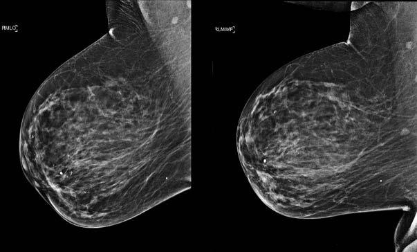
Figure 7. Patient with implanted medical device. Left, MLO view with minimal compression to visualize posterior tissue. Right, Anterior compression view with adequate compression to visualize glandular tissue. Images courtesy of Robyn Hadley.
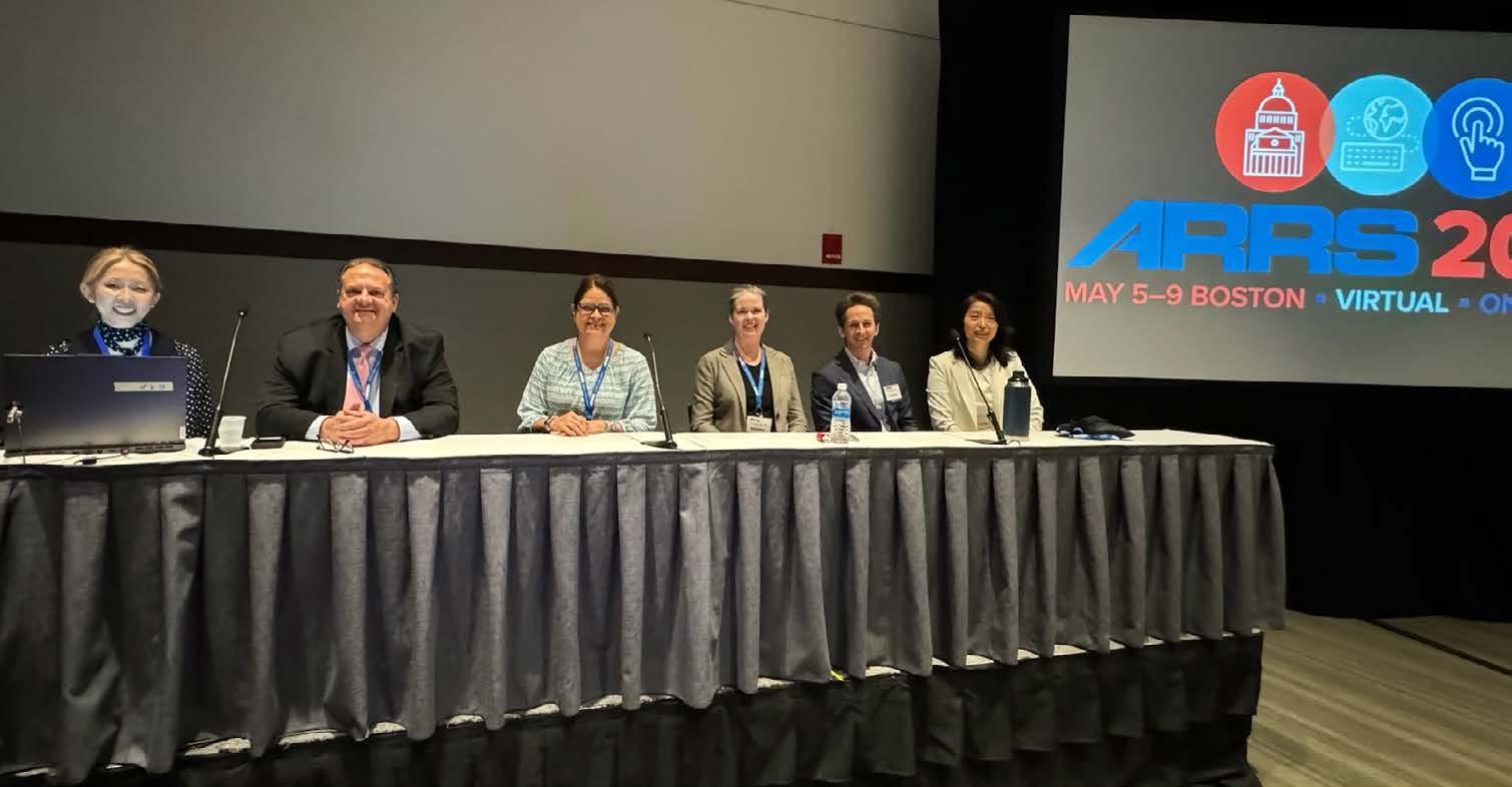
ARRS 2024 Highlights
By Asha Bhatt, MD
The 2024 American Roentgen Ray Society (ARRS) meeting took place May 5 through 9 in the historic city of Boston, Massachusetts. Opening ceremonies began with a multitude of award presentations, followed by the annual passing of the gavel to the new president, Dr. Angelisa Paladin. Dr. Paladin spoke about the science of happiness and quoted Arthur Brooks, reminding the audience that “happiness is not a destination; it’s a direction. We can’t find complete happiness but can be happier.” Finding happiness in the workplace is the key to obtaining and retaining staff members. Showing others that they matter and their work matters will help foster a more resilient, productive, and successful workplace.
As in prior years, the meeting gave attendees the options to see lectures live (in person or virtual) and on demand. This year, the sessions were organized into featured sessions, instructional courses, review tracks, and scientific sessions. Several SBI members and fellows shared their expertise at the meeting. The on-demand option was an excellent feature as a few breast imaging topics occurred concurrently.
In a global partner session between ARRS and the British Institute of Radiology, Dr. Bethany Niell described the lessons learned and future opportunities in breast cancer screening in the United States. The audience was able to compare those options with the national breast cancer screening program in the United Kingdom. Running concurrently on Sunday was a featured course on stereotactic-guided biopsy led by Dr. Tanya Moseley and including Dr. Beatriz Adrada, Dr. Haydee Ojeda-Fournier, and Dr. Mary Guirguis. This was a fantastic opportunity to understand the basics of stereotactic biopsy and review challenging cases with the speakers. The session ended with a demonstration session, which was valuable even when watched on demand.
Sunday afternoon featured a breast cryoablation course with presentations by Dr. Kenneth Tomkovich, Dr. Robert Ward, Dr. Lumarie Santiago, Dr. Deanna Lane, Dr. Monica Huang, and Dr. Lauren Chang Sen. This comprehensive session reviewed the current research on the topic, the appropriate indications for cryoablation, and successful techniques. Dr. Ward discussed the need to understand the culture and structure of your own organization before implementing a new service line. This was a recurrent theme echoed in many of the other instructional and featured courses at the meeting.
In a featured session related to everyday workflow and burnout, Dr. Jay Parikh led a panel discussion on strategies and solutions for driving wellness. This session began with a nice frame of reference for the audience and included real-world solutions for various problems.
Monday morning’s breast imaging session began with awardwinning exhibit presentations moderated by Dr. Jessica Leung. These exceptional exhibits are available online through the portal for attendees to review at their leisure. Overlapping with this session was an emerging breast research session that included various interesting presentations. This session also had three keynote lectures: artificial intelligence and breast imaging (Dr. Shin-Huey Shirley Chou); an update on the initial method of detection (Dr. Peter Eby); and challenging breast magnetic resonance imaging (MRI) cases, specifically nonmass enhancement (Dr. Tanya Moseley).
Monday morning continued with an instructional course on the implementation of new technologies, which included lectures on artificial intelligence (Dr. Manisha Bahl), abbreviated breast MRI (Dr. Holly Marshall), and contrast-enhanced mammography (Dr. Bhavika Patel). The session provided the audience with various pearls and potential challenges in the implementation of each of the specific modalities.
The afternoon included a session on the changing paradigms of breast imaging services and whether we should be adopting programs such as online versus off-line screening interpretations (Dr. Brian Dontchos), same-day biopsies (Dr. Sora Yoon), remote diagnostic imaging (Dr. Vilert Loving), and nonphysician clinicians in the breast imaging center (Dr. Dana Ataya). This session allowed for a fantastic discussion with audience members, who were able to share their experiences and concerns with these various options.
The contrast-enhanced mammography (CEM) session was a fantastic minicourse of this newer modality. The session began with a discussion by Dr. Jordana Phillips on how CEM is being used. Dr. Janice Sung then gave an excellent review of the BIRADS lexicon, which was a 2022 supplement to the current BI-RADS atlas. Dr. Margarita Zuley finished the session with a comprehensive review of CEM-directed biopsies.
To save lives and minimize the impact of breast cancer. .....
Asha Bhatt, MD
An informative session on the proposed updates for the next edition of the BI-RADS atlas included presentations on mammography (Dr. Sally Friedewald), breast ultrasonography (Dr. Jessica Leung), breast MRI (Dr. Roberta Strigel), and auditing/ outcomes (Dr. Peter Eby). The audience was reminded that these updates are not yet official.
The last day included an instructional course on screening women with dense breast tissue. Dr. Randy Miles began the session with a breast density and cancer risk review, which was followed by a discussion on available supplemental screening options by Dr. Lillian Wang. Dr. Amie Lee discussed the impact of breast density legislation on breast cancer screening, a highly relevant topic given that the Mammography Quality Standards Act final rule goes into effect September 10, 2024. The session and final day were concluded by Dr. Rifat Wahab, who rounded out the topic by touching on how radiologists can bridge the gap in breast density education.
Over the course of the meeting, two scientific sessions were dedicated to breast imaging. One of these was on the topic of artificial intelligence and multimodality outcome predictions. The second session was focused on digital breast tomosynthesis and quality improvement. Both sessions were filled with well-rounded abstract presentations along with informative keynote lectures. There was also a breast imaging–focused review session, which was a great way to reinforce concepts seen throughout the meeting.
The 2024 ARRS conference planning committee provided the audience with a wide array of speakers who showcased breast imaging expertise and advances in research. The sessions addressed daily challenges breast radiologists face and provided insight on how to implement and use advancing technologies. Overall, it was a well-rounded conference not to be missed next year!
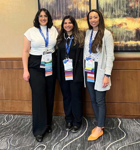
Implementation of new technologies. Left to right: Holly Marshall (University Hospitals), Manisha Bahl (Massachusetts General Hospital), and Bhavika Patel (Mayo Clinic Arizona).
Cryoablation featured course. Left to right: Monica Huang (MD Anderson Cancer Center), Kenneth Tomkovich (Princeton Radiology), Lumarie Santiago (MD Anderson Cancer Center), Deanna Lane (MD Anderson Cancer Center), Robert C. Ward (Brown University), and Lauren Chang Sen (MD Anderson Cancer Center). Image courtesy of Monica Huang and Robert Ward.
ETHICAL CONSIDERATIONS IN THE IMPLEMENTATION OF ARTIFICIAL INTELLIGENCE IN BREAST IMAGING
By Zuby Syed, MD
The integration of artificial intelligence (AI) into breast imaging has the potential to revolutionize the field, offering efficiency and accuracy in the early detection of breast cancer and assessing treatment response. Early detection is the key to improved survival outcomes and de-escalation of treatment. AI can play a pivotal role in our goal to decrease the mortality and morbidity associated with breast cancer. However, the potential scale of impact of this technological advancement brings forth significant ethical considerations that must be carefully addressed to ensure patient safety, privacy, and equitable access to health care. This forum provides an opportunity to explore the ethical concerns associated with AI in breast imaging and propose strategies to mitigate these concerns.
Accuracy and Reliability
While AI algorithms continue to evolve to improve accuracy rates in detecting breast abnormalities, the potential for errors is a concern. Misdiagnosis can lead to unnecessary anxiety for patients or delay treatment, impacting patients’ well-being and ultimately their trust in the health care system. Ensuring high-quality training data with diverse samples representing various demographics and conditions is crucial. Implementing human-AI collaboration, in which radiologists provide feedback and oversight, can enhance diagnostic accuracy. This hybrid approach combines the strengths of AI’s pattern recognition with the nuanced interpretation of human experts. Further continuous monitoring of AI performance in real-world settings allows for prompt adjustments and optimizations.
Bias and Fairness
AI algorithms are trained on large data sets that may contain inherent biases. If these biases are not addressed, they can result in disparities in diagnosis and treatment based on factors such as race, ethnicity, or socioeconomic status. This possibility raises concerns about fairness and equity in health care delivery and the potential for exacerbating existing disparities in breast cancer outcomes. It is essential to employ techniques to detect and mitigate biases in AI algorithms, such as using diverse and representative training data sets, algorithmic audits, and fairness assessments. Involving diverse stakeholders, including patients, clinicians, and ethicists, in the design and development of AI systems for breast imaging can help ensure that AI technology meets the needs and preferences of all users while minimizing the potential for harm or unintended consequences.
Patient Autonomy
Patients undergoing breast imaging may not fully understand the implications of AI technology, including how their data is used and the limitations of AI algorithms. Ensuring transparency and respecting patient autonomy are essential to upholding the principle of beneficence and respect for individual preferences and values. As AI technology evolves, health care professionals should engage in transparent communication with patients regarding the use of AI in breast imaging, explaining its benefits, limitations, and potential risks. This enables patients to make informed decisions about their health care and fosters trust in the health care system.
Professional Responsibility and Oversight
Health care professionals have a responsibility to oversee the implementation and use of AI in breast imaging to ensure its safe and ethical application. This responsibility includes regularly assessing algorithmic accuracy, safety, and effectiveness and soliciting feedback from patients and health care professionals to identify areas for improvement and exercise accountability for any errors or adverse outcomes.
Conclusion
The integration of AI into breast imaging holds immense promise for improving early detection, analyzing tumor biology, and assessing treatment response. However, it also presents significant ethical challenges that must be addressed to ensure patient safety, privacy, and equitable access to health care. By implementing transparent communication, robust data governance, bias detection and mitigation strategies, inclusive design approaches, and continuous monitoring and evaluation, we can mitigate these concerns and harness the full potential of AI to advance breast imaging while upholding ethical principles and promoting patient welfare.
Zuby Syed, MD
THE PATIENT'S PERSPECTIVE
Danielle Sharek
By Danielle Sharek, MD
Please tell me about yourself and your background.
As a committee member for the SBI newsletter, I typically interview patients who have or have had breast cancer. However, I would like to share my own personal story and the impact it has had on my life.
I am a radiologist at Weinstein Imaging Associates in Pittsburgh, Pennsylvania. I completed my fellowship in women’s imaging at Magee-Womens Hospital in Pittsburgh in 2014, and I have been working in the field since.
How were you diagnosed with breast cancer?
At a routine medical appointment in 2019, it was suggested that I have a screening mammogram. I was 36 years old at the time. This recommendation was likely due to my family history, as my maternal grandmother was diagnosed with breast cancer in her forties and my mother had atypia in her fifties.
I was shocked when I was recalled after my screening mammogram for calcifications in my left breast. I subsequently underwent a two-site stereotactic biopsy, which revealed high-grade ductal carcinoma in situ (DCIS) at each site with a suspicion of invasion.
How did you feel when you learned of the news?
I was completely shocked and devastated. Although I was accustomed to seeing breast cancer frequently as a breast imaging radiologist, I never imagined this would happen to me, especially in my thirties. I had always been in good health, exercised regularly, and ate a healthy diet.
What was your treatment process? Did you face any treatment obstacles? How did you overcome them?
After my stereotactic biopsies, I underwent bilateral nipplesparing mastectomies with implant reconstruction. The final pathology [report] showed invasive mucinous carcinoma and extensive DCIS with close margins both anteriorly and
posteriorly. Due to a suggestion of DCIS in the nipple after the mastectomy, I underwent another surgery for nipple removal. I then had four rounds of chemotherapy and six weeks of whole-breast radiation with a radiation boost to the chest wall, given the close margins. I have had two left breast revision surgeries due to painful capsular contracture, which occurred as a side effect of the radiation therapy. I will be on tamoxifen for a total of 10 years.
Looking back on my treatment, I felt a sense of urgency because I wanted to eliminate the cancer cells from my body as quickly as possible. However, in retrospect, I should have slowed down a bit in my decision-making. On multiple occasions, I was given more than one treatment option, and I was in such an overwhelmed state that I didn’t pause and consider all the options. I should have more carefully and thoroughly considered different opinions on my treatment process. I truly was on a mission to rid my body of cancer.
What motivated you during your diagnosis and treatment process?
My children, who were ages 1 and 3 at the time of my diagnosis, were my motivation. I wanted to keep life as normal as possible for them during this time. Thankfully, they were young enough to not be impacted or aware of my diagnosis.
What did you learn from your experience?
Through this experience, I have learned to not take life for granted. I try to appreciate the little things. I am thankful for each day when I wake up healthy. I have also noticed that my anxiety centered around my health has increased. I worry about aches and pains that normally wouldn’t worry a person who had not undergone cancer treatments. Having fought cancer has certainly changed my perspective on my own health.
Continued on page 31>
Danielle Sharek, MD
The Connection Between Mental Health and Workplace Injury
By Sarah Jacobs, BS, RT(R)(M)(CT)
When we hear the term occupational hazard, we tend to think of chemical or drug exposures, needlesticks, workplace violence, and physical injuries. However, in the last several years, workplace stress and anxiety has climbed to the top of the list of occupational hazards.1 This shouldn’t come as a surprise since the surgeon general announced in 2022 that toxic work environments are among the top five health crises in the United States. Atticus released a study last year that found that mental health concerns, including stress and anxiety, are among the most common workplace injuries, contributing to 52% of all reported workplace injury cases.2
In addition to the alarming statistics regarding workplace stress and anxiety, searches for the terms burnout and work-life balance have sharply increased on the Google search engine during the past five years. Searches using these two terms have increased by more than 20% over the last two years, and burnout is searched approximately 823,000 times each month, according to Nigel Frank International, a Tenth Revolution Group company.3
Workplace anxiety and stress can result in depression, burnout, injury, quiet quitting, and eventually employees leaving their jobs. If this stress is overlooked and ignored, the effects are damaging, resulting in distracted employees who are less focused on the job at hand. This can lead to poor performance, other health concerns, and even injury due to interference with safe work practices. Maintaining safe work practices is essential to patient care and employee wellness. Even a minimal distraction in routine work practices can have significant consequences to the employee and the imaging team. Employees with mental health concerns are twice as likely as those with no mental health concerns to have an accident or injury while at work.4 In addition to accidents and injuries, employees with mental health concerns may be more likely to take shortcuts and engage in hazardous behaviors such as being inattentive to standard practices, policies, and procedures that promote safety. For example, inattentive behavior could affect acquisition of a patient’s health history. Technologists and staff members routinely ask for a patient’s updated history during imaging examinations, and if these questions are overlooked or
answered inaccurately, an incorrect history and breast cancer risk assessment may be reported.
Breast imaging professionals have a strong desire to provide high-quality care, which can lead to a perfectionist attitude. Mammographic technologists in particular strive to continuously improve the image quality of the patient’s current examination compared with the patient’s previous examinations. Strict ACR and Mammography Quality Standards Act regulations are crucial to a successful, high-quality imaging center but can also contribute to this desire for perfectionism. Technologists, like many individuals working in service-providing industries such as patient care, are often people pleasers. Technologists aspire to please their interpreting radiologists, leadership teams, and patients while maintaining a kind and caring attitude. In the service industry, many employees are taught that the customer is always right. Working in health care leads to some of the same viewpoints. We aim to serve, please, and care for our patients. We are patient pleasers.
Signs that point to stress, anxiety, and burnout leading to workplace injury include the following:
• Cynicism: employees offering comments that signify a low sense of personal accomplishment
• Increased irritability and emotional exhaustion
• Reduced ability to manage emotions: may be easier to notice in colleagues with whom we have close connections than in ourselves
• Impaired judgment and general distractedness
• Changes in sleep patterns
• Disengagement in activities that are normally engaging, either at home or at work
Sarah Jacobs, BS, RT(R)(M)(CT)
How can you help?
• Encourage a comfortable environment that is open to discussions about mental health.
- Perform monthly check-ins, asking personal questions about work-life harmony.
- Cultivate trust and collaboration.
• Encourage wellness practices.
- Take microbreaks (30- to 60-second breaks every 20 minutes) from repetitive tasks or postures, such as batch reading.
- Go for a walk and stay active during breaks.
- Avoid working through lunch hours and leave your department or work area during lunches and breaks.
Build resilient employees.
- Have compassion for each other, understanding that working in health care is physically and emotionally demanding.
- Encourage communication through peer support or professional channels.
- Plan off-the-clock activities outside your work environment, focusing on activities that bring relaxation, fulfillment, and joy.
- Know your strengths. Consider taking a personality or strengths assessment every few years and build upon these strengths by taking a deep dive into the results.
- Respect boundaries between working and nonworking hours for all employees.
Having open and honest conversations about mental health challenges can be difficult, but these conversations are crucial to maintain an inclusive environment where everyone feels safe discussing concerns. Being attentive to signs of stress and anxiety in our colleagues and in ourselves can lead to discussions and practices that promote wellness before injury happens.
References
1. Robinson B. Mental health issues soar to number 1 workplace injury. Forbes. October 9, 2023. Accessed May 20, 2024. https://www.forbes.com/sites/ bryanrobinson/2023/10/09/mental-health-issues-soar-to-number-1workplace-injury
2. Muñoz V. Exploring the most (and least dangerous states for workers. Atticus. August 29, 2023. Accessed May 20, 2024. https://www.atticus. com/advice/workers-compensation/exploring-the-most-and-leastdangerous-states-for-workers
3. Russell B. Battling burnout: insights from Google searches demand action. IFA Magazine. June 27, 2023. Accessed May 18, 2024. https://ifamagazine.com/ battling-burnout-insights-from-google-searches-demand-action/
4. Cowan G. The link between accidents and mental health in the workplace. LinkedIn. March 28, 2023. Accessed May 21, 2024. https://www.linkedin.com/ pulse/link-between-mental-health-accidents-workplace-graham/
2025 Symposium Planning Committee: Behind the Scenes (continued from page 11)
Industry Partners and the In-Person Meeting
The SBI symposium brings together radiologists and industry partners (vendors), providing unique opportunities for highly efficient and concentrated individual interactions at the largest gathering of breast radiologists in North America. Industry partners receive a prospectus detailing exhibit hall opportunities, sponsored learning sessions, signage options, and branded giveaways. Long-standing industry partners have first right of refusal in rebooking booths and additional sponsored opportunities for the upcoming annual symposium.
The SBI relies on revenue from industry partners to cover a significant percentage of the symposium meeting cost. In 2024, revenue from the exhibit hall covered a substantial portion of the symposium cost. Not surprisingly, industry partners prefer inperson over virtual exhibits and pay more to exhibit at a meeting with more in-person attendees. This is one of the reasons why the SBI symposium remains an in-person-only meeting. Dr. Lewin’s President’s Column2 covered additional details on the dilemmas of virtual and hybrid meetings.
Call to Action: Your Voice
The slogan for the 2025 symposium will be “40 Then. 40 Now” to commemorate the SBI’s 40th anniversary and long-standing commitment to screening and early detection beginning at age 40 years. Although the 2025 symposium curriculum will be finalized 10 months before the symposium, we invite you to submit comments, feedback, and suggestions for the 2026 symposium content and speakers to info@sbi-online.org with “Attn: 2026 SBI Symposium” in the subject line.
Your voice matters. If you’ve filled out a past symposium attendee survey or submitted comments, thank you. Your voice has been and continues to be instrumental in shaping the SBI symposium.
References
1. Belfi LM, Slanetz PJ, Cooke EA, et al. Creativity, connection, and community: reimagining the radiology society meeting Acad Radiol. 2024;31(2):351-359. doi:10.1016/j.acra.2023.10.033
2. Lewin J. SBI News Winter 2023 President’s Column. Society of Breast Imaging. January 31, 2023. Accessed June 27, 2024. https://www.sbi-online. org/news/sbi-news-winter-2023-presidents-column
Ann L. Brown, MD, FSBI
Dr. Brown is an associate professor and associate section chief of breast imaging in the Department of Radiology at the University of Cincinnati. Dr. Brown earned her medical degree at the Medical University of South Carolina (MUSC) and was elected to the Alpha Omega Alpha and Gold Humanism honor societies. She completed her radiology residency and breast imaging fellowship at Harvard’s Beth Israel Deaconess Medical Center, where she served as an executive member of the Massachusetts Radiological Society. Dr. Brown has stayed actively involved in legislative advocacy at the state and national levels. She coauthored Ohio House Bill 371, which was passed in 2022 to improve breast cancer screening in her state. She is also passionate about quality improvement, health equity, and evidence-based practice as a current chair of the ACR Appropriateness Criteria.
Christine E. Edmonds, MD, FSBI
Hospital and subsequently returned to Massachusetts General Hospital, where she is assistant professor and serves as codirector of the Breast Imaging Research Center. Dr. Lamb is a prior recipient of the University of Ottawa Gold Medal Award and Dean’s Award of Excellence and the American Roentgen Ray Society President’s Award. She is currently a Radiological Society of North America Research Scholar Grant recipient whose research interests include the application of artificial intelligence to improve outcomes of women at high risk and breast cancer risk assessment.
Rebecca J. Leddy, MD, FSBI
Dr. Edmonds is an assistant professor of radiology, Division of Breast Imaging, Hospital of the University of Pennsylvania, Penn Medicine. She completed a combined diagnostic and research radiology residency at the University of Pennsylvania, followed by a breast imaging fellowship at Memorial Sloan Kettering Cancer Center. Her areas of interest and research include health equity and cancer screening disparities, optimization of breast magnetic resonance imaging (MRI) (including abbreviated MRI) for screening and diagnosis, and molecular oncologic imaging. She leads a breast cancer education and screening outreach program in west Philadelphia and is investigating novel applications of positron emission tomography (PET) for improved staging and treatment planning in patients with estrogen receptor–positive breast cancer.
Leslie R. Lamb, MD, FSBI
Dr. Lamb is originally from Ottawa, Ontario, Canada, and completed her undergraduate degree, master’s degree, medical doctorate, and residency at the University of Ottawa. She then completed her breast imaging fellowship at Massachusetts General Hospital. She returned to Canada to work at the Ottawa
Dr. Leddy received her bachelor of science in interdisciplinary studies and her medical doctorate from the University of Florida. Dr. Leddy then completed a diagnostic radiology residency at Emory University in Atlanta, Georgia. Dr. Leddy completed a fellowship in breast imaging radiology at MUSC and joined the MUSC faculty in 2010. Dr. Leddy is currently professor in radiology at MUSC. She is the medical director, division chief, and fellowship director of breast imaging at MUSC. Dr. Leddy has presented talks nationally on her breast imaging research and is involved in trainee education and mentorship. Her interests are breast education, breast care disparities, and quality improvement.
Eniola T. Oluyemi, MD, MPH, FSBI
Dr. Oluyemi is a breast imaging faculty member at the Johns Hopkins Department of Radiology. She graduated from Northwestern University School of Medicine and completed her radiology residency at the University of Wisconsin, Madison. After completing her residency, she did a breast imaging fellowship at the University of Washington. She subsequently completed the master’s in public health degree program at the Johns Hopkins
School of Public Health. Her main research areas of interest are racial and socioeconomic disparities in the timely diagnosis of breast cancer and breast imaging access and also evaluating advanced breast imaging modalities.
Toma S. Omofoye, MD, FSBI
Dr. Omofoye is an associate professor of radiology in the Department of Breast Imaging at University of Texas MD Anderson Cancer Center. She completed her medical degree, diagnostic radiology training, and chief residency at Duke University Medical Center. She completed her breast imaging fellowship training at MD Anderson Cancer Center, after which she joined the faculty. Dr. Omofoye’s research interests include global radiology and education and assessing capacity building in low- to middle-income countries. As the inaugural director of breast imaging global oncology, she develops and implements MD Anderson’s breast imaging–related programs addressing breast cancer disparities in Africa and the Caribbean. In this role, she works closely with the International Atomic Energy Agency and the World Health Organization to achieve the Global Breast Cancer Initiative’s goal of decreasing global breast cancer mortality by 2.5% yearly.
Katja Pinker, MD, PhD, FSBI
Dr. Pinker is member at Memorial Sloan Kettering Cancer Center, attending radiologist at Memorial Hospital for Cancer and Allied Diseases, and professor of radiology at Weill Medical College of Cornell University in New York. She is the director of research and director of breast MRI for Memorial Sloan Kettering Cancer Center. She is affiliated faculty at the Department of Scientific Computing at Florida State University, Tallahassee, and research associate at the Department of Radiology at the Medical University of Vienna in Austria. She is an expert in translational and clinical breast and oncologic gender imaging. Her research interests focus on advanced breast imaging with highresolution MRI, hybrid imaging (PET/MRI), and the application of artificial intelligence in oncologic imaging. She has published more than 200 peer-reviewed articles on breast and oncologic imaging.
Akshat C. Pujara, MD, FSBI
Dr. Pujara completed his residency training in diagnostic radiology at New York University School of Medicine and a breast imaging fellowship at Northwestern University School of Medicine. In addition to his clinical duties as assistant professor, Dr. Pujara
enjoys teaching residents and fellows and participating in research studies to advance the field of breast imaging. His current research focus is improving patient care and radiologist satisfaction by streamlining workflows, developing novel image acquisition and reconstruction techniques, and using artificial intelligence. Dr. Pujara also enjoys breast imaging leadership and administrative opportunities.
R. Jared Weinfurtner, MD, FSBI
Dr. Weinfurtner was born and raised in Athens, Ohio, and received his bachelor’s degree at Princeton University. He completed medical school at Case Western Reserve University in Cleveland, Ohio. During medical school, he completed a Doris Duke Clinical Research Fellowship at Washington University in St. Louis. Upon graduation, Dr. Weinfurtner began pursuit of a neurology residency at the University of Pennsylvania in Philadelphia before switching to radiology and returning to Case Medical Center in Cleveland, where he served as chief resident. He then completed his breast imaging fellowship at Moffitt Cancer Center, where he is now an associate member of radiology with a joint associate professorship at the University of South Florida. His research interests currently focus on immunotherapy response monitoring with MRI, and he recently presented abstracts at the European Society of Breast Imaging meeting and the European Congress of Radiology. He is the breast fellowship director at Moffitt Cancer Center.
Nicole S. Winkler, MD, FSBI
Dr. Winkler is a fellowship-trained breast radiologist at Huntsman Cancer Institute at the University of Utah. She completed medical and residency training at the University of Utah, followed by a clinical breast fellowship at Brigham and Women’s Hospital. She has over 10 years of clinical and teaching experience in breast radiology at Huntsman Cancer Hospital and previously served as the breast imaging fellowship director. Currently she is the diagnostic radiology program director for the University of Utah residency program. Her main areas of focus are radiology education and MRI-guided focused ultrasonography to treat breast cancer.
Resident and Fellow Section Update
By Heba Albasha, MD; Zuby Syed, MD
The SBI Resident and Fellow Section (RFS) brings together a group of enthusiastic volunteers who work hard to curate and share society and breast imaging content relevant to members in training. The focus of the committee in the past year has been twofold: to expand the educational and informational resources available for members in training and to recruit and retain members in training as they transition to early career. Through the efforts of our committee members and in partnership with other SBI committees, the RFS has successfully worked toward these missions.
The fellowship application season can be exciting! Residents are sifting through all the excellent programs to find the right fit. When reflecting on this process, the RFS received feedback that fellowship websites often lack the essential information an applicant is looking for. This makes it difficult to evaluate a program’s fit before applying and interviewing. To address this issue, the RFS created a standardized fellowship website template for programs to use when advertising their fellowship online. This template provides a list of information that residents find useful on a fellowship program’s website. In partnership with the SBI Fellowship Match Committee, the RFS is preparing this template for distribution to fellowship programs.
The RFS has also been working in close partnership with the SBI Fellowship Match Committee in reimagining the Case of the Month. The Case of the Month was previously published online with no oral presentation. Together we are redesigning the submission and presentation format for the SBI Summer Series. We plan to solicit case submissions soon, with select cases chosen for virtual presentation during the SBI Summer Series. Through this project, we hope to increase trainee participation in the SBI while encouraging trainees to share their excellent work.
As with any subspecialty, we hope to capture the interest of trainees early to allow them to explore our subspecialty and learn what it has to offer. To better engage and educate medical students in breast imaging, our team has created a “Medical Student Guide to Breast Imaging” that touches on the breadth of work we do as breast radiologists and offers a glimpse into our daily lives. This guide is nearly complete and will be shared soon. On the basis of additional feedback, we hope to expand our efforts in catering to medical students with additional resources and events.
In past years, we created and shared Fellowship Match spotlights on social media to highlight the accomplishments and Match results of our peers. This year we celebrated the many residents who have chosen to train in breast imaging!
We have many more exciting projects in the works that we hope to share with fellow members in training soon. Our committee works closely together to accomplish our goals and meets virtually on a quarterly basis to discuss progress and new ideas. We are looking forward to another successful year and welcome anyone interested in serving on this committee!
Heba Albasha and Zuby Syed are cochairs of the RFS.
Heba Albasha, MD Zuby Syed, MD
Advancements in Breast Imaging: Mitigating Health Care Disparities for All
By Derek L. Nguyen, MD
Early detection of breast cancer reduces patient mortality. However, despite treatment advancements, health care disparities exist in breast imaging, particularly among underserved populations such as racial and ethnic minority groups and individuals of low socioeconomic status. Concerted efforts have been made to improve access to breast imaging services and reduce these disparities. This article reviews strategies that have been recently implemented to address health care disparities in breast imaging.
Targeted Interventions: Screening
Increasing Awareness and Adherence
Patient navigators have effectively increased awareness and adherence to annual screening mammography recommendations among racial and ethnic minority patients, nearly tripling the odds of adherence compared with no intervention.1 Other strategies, such as multilingual educational materials, transportation assistance, direct communication through in-person or telephone encounters, personalized reminders, and free vouchers for screening mammograms, have successfully addressed specific socioeconomic barriers common to minority populations, thereby enhancing adherence to annual screening mammography.2 Implementation of walk-in screening programs can improve access and engagement of racial and ethnic minority individuals and Medicaid-insured patients for screening mammography.3
Mobile Mammography Units
Mobile mammography units offer a geographically flexible alternative to breast imaging facilities by providing screening mammography services directly to patients.4 Mobile mammography units effectively address racial, ethnic, and socioeconomic barriers by eliminating cost to patients and providing location flexibility, thus reaching rural and other underserved populations and encouraging regular annual screenings.4,5
Inclusive Guidelines for Transgender and Gender-Diverse Patients
The ACR, with support from the National Comprehensive Cancer Network, has provided consensus-based guidelines for transgender and gender-diverse patients, recognizing the increased risk of breast cancer associated with gender-affirming estrogen treatments.6,7 These guidelines ensure that transgender and gender-diverse patients receive timely and appropriate breast care.
Targeted Interventions: Diagnostic Imaging
Reducing Number of Patients Lost to Follow-Up
Multiple targeted communication interventions have been proposed to reduce the number of patients lost to follow-up after
abnormal screening mammography results. Improved readability of the mandated Mammography Quality Standards Act recall letter significantly increased adherence rates among racial and ethnic minority patients and patients of low socioeconomic status.8 Multiple telephone reminders in conjunction with recall letters further improved adherence to diagnostic mammography follow-ups among patients of all racial and ethnic minority groups.9 Patient navigation programs, particularly those offering bilingual services, have also effectively reduced delays and improved followup rates for racial and ethnic minority patients after abnormal screening mammography results.2,10
Same-Day Services
Offering same-day screening programs along with same-day diagnostic mammography appointments after abnormal screening mammography results has significantly reduced access disparities among racial and ethnic minority groups.11,12 Delays in scheduling breast biopsies are common due to limited appointment availability and resources. Offering same-day biopsy services has eliminated racial, ethnic, and insurance-related disparities associated with these delays and has provided timely diagnostic evaluations for all patients, including those with serious mental illness.13-15
Targeted Interventions: Access and Inclusivity
Gender-Inclusive
Practices
Transgender and gender-diverse individuals often face significant challenges in accessing health care due to negative interactions within the health care system and the adherence of many clinical practices to a gender-binary model. Practices can improve care for transgender and gender-diverse patients by adopting genderinclusive practices, such as providing gender-neutral restrooms, displaying signage inclusive of lesbian, gay, bisexual, transgender, and queer (LGBTQ+) individuals, training staff to use patients’ preferred names and pronouns, and displaying pronouns on staff members’ name badges.16
Continued on page 30>
Derek L. Nguyen, MD
Accessibility for Patients With Disabilities
Patients with disabilities face unique challenges in accessing breast imaging services, often encountering suboptimal facilities and staff members inexperienced with this specific patient population.17 Enhancing accessibility for these patients involves multifactorial approaches including improving the scheduling process, optimizing clinic workflows, employing effective communication strategies, and tailoring imaging options to fit individual needs.
Remote Diagnostics
The COVID-19 pandemic accelerated the adoption of remote work in radiology, presenting both challenges and opportunities.18,19 Remote diagnostics can increase geographic access to breast imaging specialists for patients in underserved or rural areas, improving overall access to care. As a result, patients who previously faced barriers to accessing breast imaging care can now receive timely and expert evaluations. Additionally, the availability of remote diagnostics presents the opportunity for more flexible scheduling and reduced wait times for diagnostic appointments.
Contrast-Enhanced Mammography
Contrast-enhanced mammography (CEM) offers a viable alternative to contrast-enhanced breast magnetic resonance imaging (MRI), with similar sensitivity and specificity. CEM increases access to contrast-enhanced imaging while reducing costs and accommodating patients who cannot tolerate MRI, such as those with disabilities or contrast agent allergies, and patients in rural areas without MRI availability.20-22 The recent availability of CEM biopsy capability may offer a cost-effective alternative to MRI-guided biopsies.23
Moving Forward
While significant strides have been made in addressing health care disparities in breast imaging, there is still much work to be done. It is important to continue innovating and improving the interventions that are currently in place. The following strategies can help further reduce disparities and ensure equitable access to breast cancer care:
• Promote workforce diversity: Increase diversity in the health care workforce by recruiting more health care professionals who identify as LGBTQ or are members of racial and ethnic minority groups and can understand and address the needs of our patient populations.
• Expand health care coverage: Advocate for policies that ensure comprehensive insurance coverage for breast cancer screening, diagnosis, and treatment for all individuals, regardless of socioeconomic status or identity.
• Financial support: Create institutional or foundational programs to help reduce out-of-pocket diagnostic and interventional costs for uninsured populations.
Increase contrast-enhanced examination availability: Expand access to breast MRI and/or CEM appointments to improve availability for rural and uninsured populations.
Addressing disparities in breast imaging requires a comprehensive approach that includes structural and logistical improvements as well as cultural and interpersonal strategies that may need to be tailored to specific communities. By advocating for policy changes, tackling social determinants of health, and promoting cultural sensitivity within the health care system, we can make considerable progress in closing the disparities gap. As we continue to innovate and improve our ability to detect breast cancer, prioritizing equitable access to quality breast cancer care for all individuals, regardless of their background or identity, remains essential.
References
1. Gong AJ, Lee EE, Visvanathan K, Oluyemi ET. Impact of patient navigation on reducing breast imaging disparities and applications in the COVID-19 era J Breast Imaging. 2023;5(3):346-350. doi:10.1093/jbi/wbac093
2. Nguyen DL, Ambinder EB, Myers KS, Oluyemi E. Addressing disparities related to access of multimodality breast imaging services before and during the COVID-19 pandemic Acad Radiol. 2022;29(12):1852-1860. doi:10.1016/j.acra.2022.03.017
3. Wang GX, Pizzi BT, Miles RC, et al. Implementation and utilization of a “pink card” walk-in screening mammography program integrated with physician visits J Am Coll Radiol. 2020;17(12):1602-1608. doi:10.1016/j.jacr.2020.07.007
4. Funaro K, Niell B. Screening mammography utilization in the United States J Breast Imaging. 2023;5(4):384-392. doi:10.1093/jbi/wbad042
5. Tsapatsaris A, Reichman M. Project ScanVan: mobile mammography services to decrease socioeconomic barriers and racial disparities among medically underserved women in NYC Clin Imaging. 2021;78:60-63. doi:10.1016/j.clinimag.2021.02.040
6. Patel H, Arruarana V, Yao L, Cui X, Ray E. Effects of hormones and hormone therapy on breast tissue in transgender patients: a concise review Endocrine. 2020;68(1):6-15. doi:10.1007/s12020-020-02197-5
7. Chowdhury A, Saad Eddin A, Kim Hsieh S, Policeni FC. Review of breast imaging in transgender and gender-diverse patients: gender-affirming care, histopathologic findings, breast cancer risk, and screening recommendations J Breast Imaging 2024;6(3):238-245. doi:10.1093/jbi/wbae016
8. Nguyen DL, Harvey SC, Oluyemi ET, Myers KS, Mullen LA, Ambinder EB. Impact of improved screening mammography recall lay letter readability on patient follow-up J Am Coll Radiol. 2020;17(11):1429-1436. doi:10.1016/j. jacr.2020.07.006
9. Nguyen DL, Oluyemi E, Myers KS, Harvey SC, Mullen LA, Ambinder EB. Impact of telephone communication on patient adherence with follow-up recommendations after an abnormal screening mammogram J Am Coll Radiol. 2020;17(9):1139-1148. doi:10.1016/j.jacr.2020.03.030
10. Percac-Lima S, Ashburner JM, McCarthy AM, Piawah S, Atlas SJ. Patient navigation to improve follow-up of abnormal mammograms among disadvantaged women J Womens Health (Larchmt). 2015;24(2):138-143. doi:10.1089/ jwh.2014.4954
11. Dontchos BN, Achibiri J, Mercaldo SF, et al. Disparities in same-day diagnostic imaging in breast cancer screening: impact of an immediate-read screening mammography program implemented during the COVID-19 pandemic AJR Am J Roentgenol. 2022;218(2):270-278. doi:10.2214/ajr.21.26597
12. Oluyemi E. Editorial comment: offering immediate screening mammography interpretation may be an effective way to reduce the racial and ethnic disparity gap in the time to diagnostic follow-up AJR Am J Roentgenol. 2022;218(2):278. doi:10.2214/ajr.21.26798
13. Yoon SC, Taylor-Cho MW, Charles MG, Grimm L. Racial disparities in breast imaging wait times before and after the implementation of a same-day biopsy program J Breast Imaging. 2023;5(2):159-166. doi:10.1093/jbi/wbad003
14. Dontchos BN, Narayan AK, Seidler M, et al. Impact of a same-day breast biopsy program on disparities in time to biopsy J Am Coll Radiol. 2019;16(11):1554-1560. doi:10.1016/j.jacr.2019.05.011
15. Wang GX, Hwong AR, Mercaldo SF, Lehman CD, Dontchos BN. Impact of a same-day breast biopsy program on disparities in time to biopsy for patients with serious mental illness J Am Coll Radiol. 2022;19(1 Pt B):146-154. doi:10.1016/j. jacr.2021.09.009
16. Ly D, Hoyt AC, Weimer A, et al. Breast cancer among transgender and nonbinary patients: paradigms for improving data collection and inclusion in breast imaging settings J Breast Imaging. 2023;5(1):73-79. doi:10.1093/jbi/wbac067
17. Net JM, Feliciano YZ, Podsiadlo V, Dialani V, Grimm LJ. Optimizing the patient experience for women with disabilities in the breast imaging clinic J Breast Imaging 2024;6(2):183-191. doi:10.1093/jbi/wbad106
18. Neitzel E, vanSonnenberg E, Markovich D, et al. The new normal or a return to normal: nationwide remote radiology reading practices after 2 years of the COVID-19 pandemic J Am Coll Radiol. 2023;20(8):804-808. doi:10.1016/j. jacr.2023.04.014
19. Zhang Z. Opinions on remote and in-person breast imaging work: survey results from the Society of Breast Imaging J Breast Imaging. 2024;6(1):23-32. doi:10.1093/jbi/wbad082
20. Cozzi A, Magni V, Zanardo M, Schiaffino S, Sardanelli F. Contrast-enhanced mammography: a systematic review and meta-analysis of diagnostic performance Radiology. 2022;302(3):568-581. doi:10.1148/radiol.211412
21. Jochelson MS, Lobbes MBI. Contrast-enhanced mammography: state of the art Radiology. 2021;299(1):36-48. doi:10.1148/radiol.2021201948
22. Lee CI, Bogart A, Germino JC, et al. Availability of advanced breast imaging at screening facilities serving vulnerable populations J Med Screen. 2016;23(1):24-30. doi:10.1177/0969141315591616
23. Weaver OO, Yang WT, Scoggins ME, et al. Challenging contrastenhanced mammography-guided biopsies: practical approach using real-time multimodality imaging and a proposed procedural algorithm AJR Am J Roentgenol 2023;220(4):512-523. doi:10.2214/ajr.22.28572
How has this diagnosis impacted your life? How have you used your diagnosis to impact others?
I have become more aware of the importance of screening exams. If I hadn’t had a screening exam in my thirties and had waited until age 40, I am not certain my life would be the same. I have an increased appreciation for early detection and screening, as they can save lives.
Additionally, I feel that I can better relate to those diagnosed with breast cancer and can be more empathetic. I know exactly how it feels to be diagnosed and all the emotions that go along with it. This has allowed me to become a more compassionate provider.
I also have an increased appreciation for the entire medical community. During my diagnosis, many medical professionals were involved in the process, including radiologists, pathologists, radiation and medical oncologists, and multiple breast surgeons. It is incredible that this team approach has saved my life. I am so grateful to everyone who was involved with my care.
Are there any lessons that you think the breast imaging community can learn from your experience?
Although as radiologists we typically do not spend a tremendous amount of time with our patients, our words and actions matter. It has been almost five years since my diagnosis, but I can still vividly remember the doctors, nurses, and staff who were kind and compassionate. Spending a few extra minutes with your patients to explain a procedure or to talk them through things can go a long way and is very meaningful.
Leading Through the Pandemic
By Jessica W. T. Leung, MD, FACR, FSBI
On March 11, 2020, the World Health Organization declared the COVID-19 outbreak a pandemic, affecting the entire world. Until that time, the SBI was planning to hold one of the largest symposia to date in Denver, just a little more than a month away. When it became increasingly clear that this outbreak was like no other in recent history, we quickly learned to adapt to move forward as a society and serve our members. As the incoming president of SBI at that time, I initially had a vision of how the year would proceed. I, too, had to quickly let go of all previous ideas and adapt. I learned the meaning of force majeure. Like all of us, I practiced resilience and relied on teamwork.
Making timely decisions based on the currently available information was very important. Reactive adenopathy secondary to vaccination against COVID-19 was being reported worldwide, and confusion and anxiety about its management were widespread. One of the most impactful SBI achievements during this time was the policy statement from our Patient Care and Delivery Committee regarding adenopathy after COVID-19 vaccination, titled “SBI Recommendations for the Management of Axillary Adenopathy in Patients With Recent COVID-19 Vaccination.” This was one of the first such recommendations from any group. It served as the foundation for subsequent guidelines from other organizations and played an important role in patient care.
The ability to pivot and build was essential. We brushed off the disappointment associated with the cancellation of the 2020 SBI symposium and channeled our energy into the first ever all-virtual SBI symposium in 2021, which was an immense success! We were able to transpose much of the original 2020 program, and the 2021 symposium attracted a very large, diverse, and international audience. The symposium had 3409 participants from 109 countries. Just for reference, there are 195 countries in the world, which means that 56% of all the countries in the world were represented at the 2021 SBI symposium! Even though this was our first such venture, it was well executed, joy filled, and so very inclusive and meaningful.
In addition to crafting a system that worked for the time being, it was important to keep in mind the future. We made the most of the virtual platform and turned it into a lasting format that has
expanded the reach of SBI. This is well exemplified in our online educational materials, societyhosted webinars, and continuing partnerships with overseas radiology and breast imaging organizations. Despite the absence of travel during the pandemic years, our educational efforts reached various parts of the globe. Through the International Education Outreach Committee, we taught at virtual conferences to audiences in diverse places including Pakistan, South Korea, and Egypt. We partnered with RAD-AID and Radiology Across Borders for impactful and effective educational endeavors.
Recognizing and ameliorating the downstream negative effects from the pandemic were also crucial. In many states, governors issued public health emergency declarations in response to the pandemic, limiting “nonessential” medical procedures, including screening mammography. The triaging of limited medical resources and the pause in nonemergency medical care were certainly understandable and indicated at the time. However, doing so resulted in delayed diagnoses of breast cancer, as discussed in recent publications in Journal of Breast Imaging and elsewhere. Our job now is to ensure the timely, appropriate, and comprehensive return to breast cancer screening everywhere through education and access.
As we look back on the pandemic years, we realize more than ever that SBI is strong and resilient. Being humble and willing to pivot, appreciating the positive aspects of a less-than-positive situation, working together as a team for the good of the community, and deriving satisfaction from being of service—these are key components for emerging from the pandemic into brighter times. As Charles Darwin said, the species that are most adaptable to change are the ones that survive. More than simply surviving, SBI led the response to the COVID-19 pandemic in many ways, and it is thriving.
Nepal is a landlocked country in Southeast Asia situated along the Himalayan mountain range. The population of Nepal is approximately 30.5 million and the average life expectancy is 68 years.1 In 2014, RAD-AID established a partnership in Nepal. RAD-AID International is a global nonprofit organization that was established in 2008 with the goal of increasing access to medical imaging in low-resource areas of the world. When a devastating earthquake struck Nepal in 2015, the RAD-AID Nepal program contributed to disaster relief efforts.
The RAD-AID Nepal program has continued to grow under the direction of Dr. Carlin Ridpath, director of the RAD-AID Nepal country program, and through collaboration with the Tribhuvan University Teaching Hospital (TUTH) in Kathmandu and the Hospital & Rehabilitation Centre for Disabled Children, a renowned orthopedic pediatric hospital in Banepa, Nepal. In 2020, the newly formed University of Colorado (CU) RAD-AID chapter established a partnership with RAD-AID Nepal. Members of the CU RAD-AID chapter regularly join RAD-AID teams deployed to Nepal. Between in-person visits, members of the CU RAD-AID chapter and the TUTH radiology department meet every two weeks over Zoom for case-based teaching conferences.
TUTH and RAD-AID performed a thorough assessment that identified gaps in breast cancer early detection outreach and organized breast cancer screening. The reported total of 2255 new cases of breast cancer in Nepal in 2022 corresponded to a prevalence of 0.007% among the total population and 0.014% among all women.2 However, these numbers are thought to underestimate the actual disease prevalence; a significant number of women with breast cancer likely do not present for diagnosis and treatment, resulting in inaccurate official recordkeeping. Mortality among patients diagnosed with breast cancer in 2022 was 1149 persons, or 51%.2
Currently, Nepal has no national breast cancer screening program. Although a few hospitals offer screening mammography, screening examinations remain infrequent, and most breast imaging examinations are diagnostic evaluations of a clinical symptom. At TUTH, the diagnostic breast imaging workflow varies, often beginning with ultrasonography and occasional biopsy in a surgical clinic before workup with mammography. Dr. Anamika Jha leads the breast imaging program, serves on other general radiology clinical and teaching services, and also has clinical responsibilities as an
attending radiologist at a private hospital in Kathmandu. Dr. Jha and the RAD-AID breast imaging team identified the following breast imaging goals: optimizing mammography technique, developing an efficient diagnostic workflow, creating a radiology fellowship in breast imaging, and eventually implementing a breast cancer screening program.
In 2023, a RAD-AID team composed of radiology residents from CU collaborated with faculty members and residents from TUTH to coordinate the first breast cancer screening health fair, which took place in a semirural region north of Kathmandu. The event was attended by 85 patients, all of whom received a clinical breast examination. Ultrasonography was performed when indicated with handheld devices donated by RAD-AID, and arrangements were made for follow-up image-guided biopsy as needed. TUTH hopes to expand patient breast health education and outreach via regular breast cancer screening day camps offering participants free clinical histories, breast examinations, and targeted diagnostic ultrasonography using portable ultrasonography devices. The camps will target patients in medically underserved villages around Kathmandu, with significantly discounted or free follow-up appointments scheduled at TUTH if needed. RAD-AID will continue to support this endeavor through donation of Lumify ultrasound systems and by providing resources for patient outreach and education.
Incorporating a picture archiving and communication system (PACS) into the radiology department at TUTH is a potential way to increase capacity throughout the radiology service line, including breast imaging. RAD-AID has donated a PACS, and the RADAID informatics team is working with TUTH on implementation. Once a functioning PACS is integrated into the radiology workflow, Continued on page 34>
Corey Thompson, MD
Carlin Ridpath, MD
Ivy Ewald, MD
Erica Pollack, MD
Anamika Jha, MD
breast imaging will become more efficient and have higher yield. Comparison with the stored prior images will allow the radiologists to appreciate small but significant differences over time.
A recent RAD-AID team in Nepal included Asmut Abdallah, a mammography technologist and navigator from CU. Asmut led hands-on positioning workshops and delivered didactic lectures on mammography quality control. The audience included the mammography technologists at TUTH and technologists from other hospitals in Kathmandu, who also attended the workshops to take advantage of Asmut’s expertise. As a breast imaging navigator, Asmut also helped the team consider potential mechanisms to implement a breast cancer navigation system and medical audit system for breast imaging. The lack of electronic medical records and complicated workflow limit TUTH’s ability to track patients receiving breast imaging. Navigation and auditing remain works in progress for continued collaboration.
The next RAD-AID Nepal team from CU will visit Nepal in September 2024 and will focus on supporting PACS integration,
breast imaging workflow and navigation, breast cancer patient outreach and education, and development of a breast imaging fellowship program at TUTH.
The RAD-AID breast imaging team is eager to welcome new volunteers with expertise in any aspect of breast cancer care who are interested in promoting high-quality care to underserved patients. Attending-level physicians and radiologists in training are welcome to apply, as are physician assistants, technologists, nurses, physicists, and informatics specialists. We invite you to learn more on the RAD-AID website (https://rad-aid.org/) and to sign up at https:// portal.rad-aid.org/form/Pvn9P/general-volunteer-survey or email breastimaging@rad-aid.org with inquiries. Remember to indicate that you are an SBI member when you sign up!
References
1.Nepal. World Bank Group. Accessed June 26, 2024. https://data.worldbank.org/ country/nepal
2.Estimated number of new cases from 2022 to 2025, incidence, both sexes, age [0-85+], Nepal. Cancer Tomorrow. Accessed June 26, 2024. https://gco.iarc. fr/tomorrow/en/dataviz/tables?years=2025&populations=524&cancers=20&mode=cancer&group_populations=1&multiple_populations=0
To save lives and minimize the impact of breast cancer. .....
RAD-AID Nepal Director Dr. Carlin Ridpath (center) with TUTH faculty members and RAD-AID volunteers in Kathmandu.
Drs. Anamika Jha and Erica Pollack discussing breast imaging findings at TUTH.
Asmut Abdallah (right), the lead RAD-AID mammography technologist at TUTH.
CU residents Drs. Emily Sterbis, Maili Lim, and Ivy Ewald with CU mammography technologist and navigator Asmut Abdallah in the mammography suite at TUTH.
TUTH breast radiologist Dr. Anamika Jha and CU residents Drs. Emily Sterbis, Maili Lim, and Ivy Ewald with staff at a breast cancer screening camp outside Kathmandu, Nepal.
TUTH radiology residents and CU radiology residents enjoying a cup of tea after work in Kathmandu.
CU resident and RAD-AID volunteer Emily Sterbis reviews mammography on PACS with a radiology resident at TUTH.
Dr. Anamika Jha, the primary breast radiologist at TUTH, using a portable ultrasonography system to evaluate a patient during the breast cancer screening camp in Jhor, Nepal.
Perspectives From a Traveling Mammography Technologist: Raquelle Jadotte, RT(R)(M)(CT)
By Shinn-Huey Shirley Chou, MD, MPH, FSBI
The following interview has been edited.
SC: How long have you been a mammography technologist? Have you always been a mammography technologist?
RJ: I’ve been a mammography technologist for about seven years. I have always wanted to be a mammography technologist. When I was going through x-ray school, I knew I wanted to do another modality. So when they asked us to choose what we were most interested in, I chose mammography and computed tomography (CT). I ended up doing both, but I like mammography more.
Why did you choose to become a mammography technologist?
I chose mammography because the environment is calmer, more peaceful, more sensitive, and more similar to my personality. It is not too rushed or rough.
Have you always been traveling?
No, I have been traveling for four years. I started traveling just before the COVID-19 pandemic was declared. I was here in Boston actually.
Oh, interesting! So you started traveling before the pandemic. I had thought traveling technologists came about because of the pandemic, but it is not something new.
Yes, but more people are aware now since the pandemic, because when staff get sick, and the practices don’t have anyone to help cover, that is when they figure out there are agencies to provide temporary support.
So were you based at a single institution before you started traveling in 2020?
Yes, I was a full-time dual technologist performing bone density x-rays and mammography. Then I left to pursue CT schooling, followed by training at a trauma hospital. Afterward, I did CT for a couple of months a year and mammography per diem. Ultimately, I decided to pump the brakes because CT became a lot, plus it was overnight and very stressful. That’s when I left CT and started traveling and doing only mammography.
FSBI
How did you find out about the traveling technologist positions?
My sister is a nurse. She had a conversation one time with the x-ray technologists that came up to her floor and found out that they were travelers. She knows I like to travel and so she asked me, “Did you know that they have this opportunity for radiologic technologists?” I said “No!” I immediately started doing some investigations and found out more about these traveling positions. Since I’ve picked up this job, I love it!
Besides traveling, are there other aspects of your job that you find most enjoyable and most challenging? Is it difficult to work with new people and learn new protocols?
It doesn’t bother me. I feel that it is only going to make me stronger and better. Protocols are generally consistent and quite similar. I find that a lot of hospitals and facilities use Hologic, so I have not encountered too many issues. Some doctors might want something different from what you are used to, but as long as you know how to perform the specific views, you are good to go.
Do you feel that you’ve learned new things at every place every time?
Not too much, but I’ve definitely picked up different tactics and different skills based on the patient population sometimes. For example, when I worked in Tucson, Arizona, there were many elderly [patients] there, so I picked up different ways of how to move my body mechanics to get them positioned properly for quality imaging. Other places have different demographics.
What about the challenges?
In terms of working with new people, everyone seems nice. I haven’t experienced any struggles with that, which is why I like mammography. I did a contract for CT and x-ray, and you do feel a bit more struggle when traveling as a CT or x-ray technologist because of the pay differences.
Shinn-Huey Shirley Chou, MD, MPH,
Who would you recommend your job to among your colleagues or aspiring peers?
I think this job is suitable for those who are not tied down or don’t have young children. Even if you are married, you can still do this job. I see people travel with their spouses.
What have you learned about this job?
I learned about the importance of efficiency. You have to be willing to learn quickly and be adaptable for sure. You must have a good work ethic; you can’t be lazy. You have to be willing to pick up the cases because nine times out of ten, you are going into a place that needs people, so you are not there to chill; you are going to be there to pretty much hustle.
How can the breast imaging community and practices improve our work environment to incorporate, promote, and help technologists in your position?
I think breast imaging practices can provide more frequent and better group communications, hands-on training for procedural skills, and opportunities to work alongside a radiologist to improve any essential skills for one to function as a well-rounded technologist to better serve our patients. It would also be helpful to give recognition for a strong work ethic. For the most part, for mammography typically, the practices are pretty good.
Who provides you with evaluations or feedback so you can know how you are doing?
I’ve never had that. Or it has never been brought to my attention. That is something that can be beneficial. The managers can always send evaluations to the agency if they want to. The agency has given me positive feedback verbally in the past, but I am not aware of any formal evaluation system.
Is there a community among traveling technologists?
Yes. When I go to a practice where there are other travelers, we would connect and exchange our numbers and experiences: “Have you been to this place? What is it like?” But Facebook mainly. We have a page with a whole bunch of travelers where we talk amongst ourselves. The agency itself does not connect or introduce us to each other.
What are the agencies doing to help with your career development? Do you feel that there are career advancement opportunities?
There really aren’t any career advancement options as a traveler. There are some perks. For example, some agencies provide continuing education unit reimbursements. Some offer tuition reimbursements, but I’m not sure how to do that unless you take all
online courses. A lot of technologists choose to travel because they need, number one, the change, and number two, money is a factor as well, so they pretty much know if they are either doing this for the long run or just temporarily. Some people do it and then back out to pursue career advancement or to change their careers to do something else, mainly due to burnout.
Talking about burnout, how do you think being a traveling technologist affects the risk of burning out? Do you think it helps prevent burnout?
The burnout will still exist, but the circumstances are different as your term on the contract is not forever. You can still be burned out if you go somewhere that is super busy and needs you. The thing to keep in mind is that it is only temporary. It is only three months, and you have wiggle room with that. You can either stay there, go somewhere else, or just take a break. You are not committed, so that is what makes things better. A minimum of three months at a place is generally the standard.
What is the process of finding the next practice? How do you find out what position is available? It seems stressful.
It is both stressful and exciting at the same time. If a desirable place pops up among the postings on the agency’s website, you get very excited and you want to go there. It is very stressful to figure out where to rent and live and if it is safe. The housing cost is on our own, so it can be pricey. The moment you see a location that you are interested in, you have to jump on it. But many people are also intrigued by that location, so it depends on your resume, too. If the manager thinks you have more to offer, then they will go with you or whoever is the best candidate, so it is not 100% [sure] that you will get the position. There is a formal interview over the phone, which is not as intense as an in-person interview.
Perspectives From a CrossTrained Radiologic Technologist: Ashley O’Brien,
By Shinn-Huey Shirley Chou, MD, MPH, FSBI
BS, RT(R)(M)
The following interview has been edited.
SC: How long have you been a mammography technologist? Have you always been a mammography technologist?
AO: I have been a mammographer for just about a year now. I began my radiology career as an emergency room radiologic technologist. After a few years, I transferred into a clinical education position where I trained radiologic technologist students. I was a radiologic technologist for five years before deciding to specialize in mammography.
Why did you choose to become a mammography technologist?
I realized that many of my female patients I was caring for did not receive a lot of education on women’s health, especially breast health. I wanted to help spread the knowledge of how annual screening mammography and clinical breast exams are important for early detection.
Why did you choose your current job/arrangement?
I chose to train in breast imaging to specialize in an imaging modality that I was passionate about and to expand my clinical skills.
What do you find most enjoyable and most challenging about your current job?
The most enjoyable part of being a mammographer is meeting people from all parts of the country, even from other parts of the world. Many patients travel to Boston to receive medical care, and it’s great to learn about their experiences and about their families during their exams. The most challenging aspect of being a mammographer is how different each patient’s body habitus, surgical history, and breast tissue is. Positioning that works for one patient may not necessarily work for the next patient. Critical thinking through positioning to obtain the best possible images is challenging.
Who would you recommend your job to among your colleagues or aspiring peers?
My coworkers who prioritize patient care would be perfect candidates for becoming a mammographer.
What have you learned from this job that you did not know before?
I learned about the various risk factors for developing breast cancer and the many pathologies involved in breast imaging. There is a large range in types of breast cancers, severities, and treatments.
How can the breast imaging community and practices improve our work environment to incorporate, promote, and help technologists in your position? How can we improve job satisfaction?
I believe that more opportunities for educational events may be a fun way to incorporate, promote, and support technologists in the breast imaging departments. It would be beneficial to learn about healthy body ergonomics as we work in a physically demanding profession when performing mammography exams and procedures. It would also be helpful to learn more about keeping our minds and bodies healthy when balancing our busy work and home lifestyles.
Shinn-Huey Shirley Chou, MD, MPH, FSBI
So You Want to Go Part Time?
Decision-Making, Process, Experience, and Practical Considerations for Going Part Time
By Jane Conlin, MD
Jane Conlin, MD
Upon fellowship graduation, eight weeks after having a cesarean delivery, I dove headfirst into a 100% breast imaging private practice near family. I set a save-the-world pace and was living the dream. Later pregnant with number two and having bought our first house, my boss lost his contract, and therefore so did I. Death of a dream. I moved on as a locum, then an employee, and now as a shareholder in a multispecialty private practice. A new dream.
All came to a grinding halt during COVID-19 shutdowns. For the first time, I had time to reflect on life’s ephemeral nature and fragility, time to play with my kids, learn the guitar, and cultivate wild sourdough bread; time to rest. Most importantly, I had time to acknowledge intermittent burnout I had experienced over the years and weigh what really matters to me in this one life, one body in an uncertain number of turns around the sun. I had thought it was money and professional success. But it was actually giving myself permission to explore what is possible.
My current practice allows part-time partnership (0.5-0.9 full-time equivalent [FTE]), something I would do “someday.” Last fall, there was a department leadership transition and someday became now. Ten years post fellowship, I went from “take no prisoners” to a terrifying and exciting 0.5 FTE. I now work two clinical days doing screening, diagnostics, procedures, and tumor board and one half-day reading remotely from home. That first job? We kept in touch and now I help read screens remotely. In my off days, I became a certified life coach, joined a local dance company, and started performing on stage. I cohost a book club for women physicians. I took my daughter to see Taylor Swift in Paris and my son to see Stonehenge. Medicine has transformed for me, too. Last week after a biopsy, a patient thanked me so earnestly for my gentle hands that tears streamed down both our faces, reigniting my awe and love of medicine.
Physicians are daring to question whether the dogma of sacrificing all at the altar of health care, established in the early 1900s, meets our current professional needs and personal dreams or serves patients as well as they deserve. Physicians are humans too. Flexibility, creativity, agency, work from home, and part-time partnerships are available to radiologists now more than ever—yes, even to breast radiologists.
Practical Considerations
Who: Why not you?
Why: What is your why? According to a 2024 study, 78.4% of breast radiologists reported being highly burned out.1 Factors contributing to radiologist burnout have been well reported, and less exposure may immediately alleviate these stresses. Other considerations include pursuing passion projects, spending time with family, and optimizing mental and physical health and overall well-being.
What: Options include locums, part-time partnerships, amount of part-time work (0.5 to 0.9 FTE), telemammography, and remote reading.
When: Are you required to give notice? What is the process in your group? Is there a natural ebb in your group’s cycle that would make this transition easier for your partners (for example, after new hires arrive or after the busy holiday season)? Are you going for public service loan forgiveness and do you need to maintain 30 hours/week to remain eligible?
Where: Does your own practice offer this, especially after the COVID-19 pandemic? Find out what arrangements other local practices are offering their radiologists. Expand your search as needed. Check the SBI and ACR job boards and join social media groups.
How: Be valuable. Ask for what you want in a reasonable way. Negotiate and offer solutions. Let them know you’re motivated to remain useful and make this work. What are your tax implications? Can you keep accrued paid time off and CME time (and funds)? Do you need to renew your private disability insurance before or after reducing your FTE hours? Talk to others who have done it.
Reference
1.Parikh JR, Sun J, Mainiero MB. Prevalence of burnout in breast imaging radiologists J Breast Imaging. 2020;2(2):112-118. doi:10.1093/jbi/wbz091
Perspectives From an Industry Clinical Application Specialist Technologist
By Grace Ashworth, BS, RT(R)(M)
Women’s health, specifically breast imaging, has been close to my heart since my mother and maternal aunt were both diagnosed with breast cancer within years of one another. I have been a mammography technologist for seven years. Currently, I am working for MOLLI Surgical, a medical device company, as a clinical application specialist. This position has allowed me to see a unique side of breast imaging as I travel all over the world to educate technologists, nurses, radiologists, and surgeons about how our breast localization technology can help improve breast imaging and surgical workflow. I have been in this role for a little over two years while still having the privilege of working per diem as a mammography technologist at Massachusetts General Hospital.
My favorite part of my current role is being able to help a facility transition to a technology that works better for their practice. I get to be a guide throughout every step of the breast localization
process, from breast imaging, surgery, and pathology to sterile processing. This access has given me a new perspective of the totality of the industry and patient care.
The most challenging aspect of the job is constantly working with new people. Everyone has their own personalities, methods, and preferences. Traveling to different hospitals gives me the opportunity to become comfortable in new environments and learn best methods to withstand the pressures of unfamiliar territory.
I would recommend my role to anyone who loves a challenge and is looking for a job that can push them. Traveling to different hospitals every week can take a toll, but it is very rewarding to know you helped make a difference.
Providing the best possible care for patients in an extremely vulnerable time is and will always be the number one priority!
Perspectives From a Traveling Mammography Technologist: Raquelle Jadotte, RT(R)(M)(CT) (continued from page 37)
Oh…so you might not get to go wherever you would like to go. Are there a lot of traveling mammography technologist positions everywhere these days?
Recently, the job availability has been increasing, more than it was in the past. But there are also more mammography technologists becoming traveling technologists, so it has become more competitive and relatively fewer opportunities specifically for desirable locations.
How many places or practices have you worked at since you started traveling?
About 10 or more: in San Francisco, California; Dallas, Texas; Portland, Oregon; Tucson, Arizona; Chicago, Illinois; Staten Island, New York; Livingston, New Jersey; Boston, Massachusetts, for three times, and then I came here (Boston).
Do you have a favorite facility? Here? (Laugh) Just kidding, you don’t have to say that!
I wouldn’t say that I have any favorite facility, but I did enjoy some
places more than others. The doctors here are nice; the staff have been great. That is why I’ve been here for this long (one year). Also, I am trying to build myself and my future; I knew I had to stay at a place, and this was a good place to stay at. Before this place, I typically stayed at each place for three months, just pick it up and go; that is how I was able to see so much.
How do you choose which places to go?
I don’t choose the practice per se. I do get to choose my contract as the need for traveling mammography technologists arises for a particular location. When a position is posted on the agency’s website, I would decide whether its location is where I would like to be based on climate, etc.
Do you get to explore these places?
For sure!
How fun!
It seems like you want to travel now! Do you want to travel? (Laugh)
Grace Ashworth, BS, RT(R)(M)
SBI Live Summer Webinar Series
July 30, 8:00 PM EDT
Pediatric and Adolescent Breast Disease: Imaging & Management
August 27, 8:00 PM EDT Remote Breast Imaging: Tips to Take Home
Please visit the SBI Calendar of Events at www.sbi-online.org for a complete