The Member Newsletter of the Society of Breast Imaging
INSIDE THIS ISSUE:
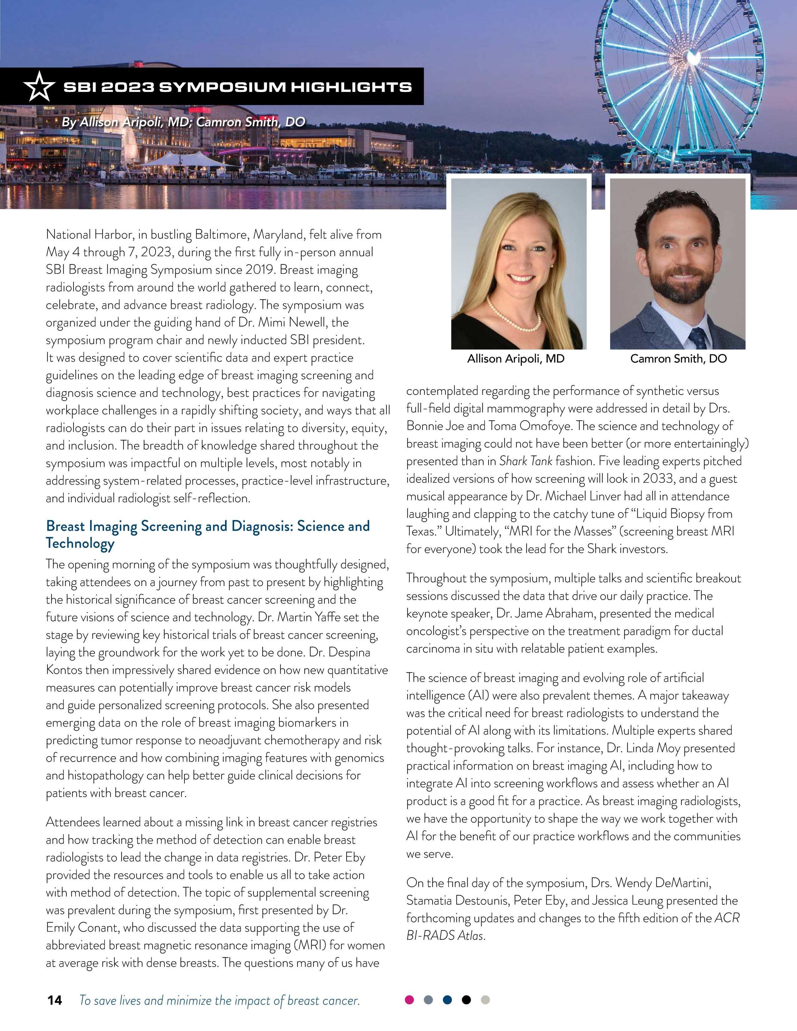
• SBI 2023 Symposium Highlights
• Contrast Enhanced Mammography Imaging Screening Trial (CMIST) Is Now Open!
• Highlights From the 2023 ARRS Meeting
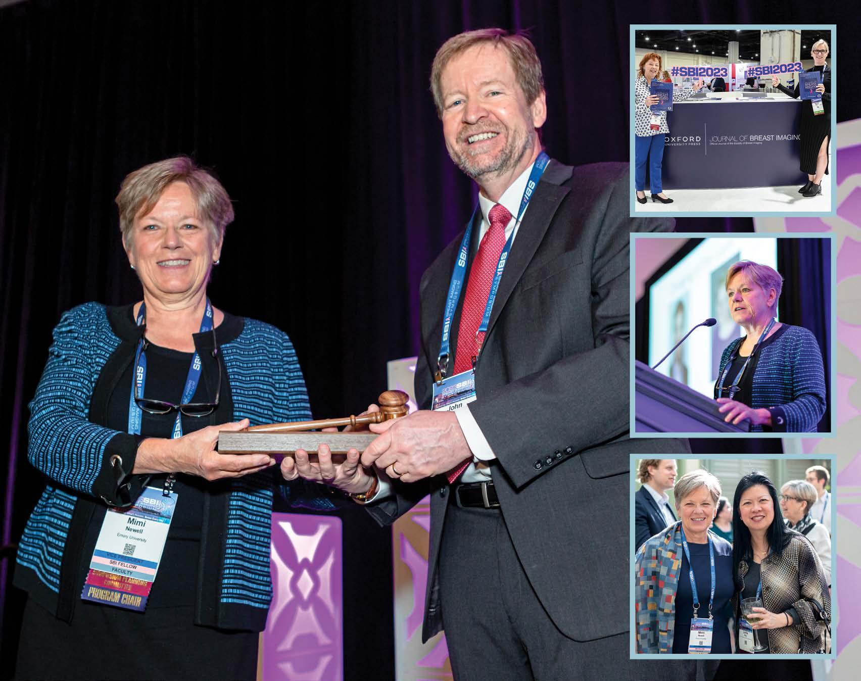
Welcome new SBI president and JBI editor!
Pictured: Mimi Newell, MD, FACR, FSBI; John Lewin, MD, FACR, FSBI; Jennifer Harvey, MD, FACR, FSBI; Wendy DeMartini, MD, FSBI; Linda Moy, MD, FACR, FSBI, FISMRM.
EDITOR: Nidhi
ASSISTANT EDITORS: Randy Miles and Shinn-Huey Shirley Chou
SBI COMMITTEE UPDATES: Yasmeen Fields
TECHNOLOGISTS’ COLUMN: Robyn Hadley and Sarah Jacobss
WHAT’S
IN THE NEWS: Anita Mehta
MEMBERS IN TRAINING: Wenhui Zhou
WELLNESS COLUMN: Claudia Cotes and Sarah Jacobs
THE PATIENT'S PERSPECTIVE: Hannah Perry and Danielle Sharek
LEGISLATIVE UPDATES: Amy Patel
OTHER MEMBERS: Jean Seely
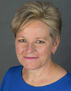
Dr. Mimi Newell, MD, FACR, FSBI President, Society of Breast Imaging
OUR SBI MISSION:
To save lives and minimize the impact of breast cancer
OUR SBI VALUES:
Patient-centered and evidence-based care
Excellence in education Scientific integrity Collaboration and collegiality
Respect for diversity and inclusiveness
On Belonging
The sense of belonging is a basic human need. We all want to fit in, be heard, and feel included: in essence, to matter.
While the SBI, of course, does not represent anyone’s ground zero for affirmation of self, our members have told us that a sense of belonging and inclusion within our organization is very important to them. Prior survey results outline that while a large majority of members feel respected, only 52% agree with the statement “I feel that I am seen, heard, and valued as a member of SBI.” We want that to change.
Our diversity, equity, and inclusion strategic plan will be unveiled in the near future and will contain many initiatives that will be implemented over time with the help of our Inclusion Diversity Equity Alliance, our members, and the Board. One interesting concept is “pop-up” work groups. In addition to our standing committees (which are relatively fixed in number and size for logistical reasons), the SBI can address issues that arise or the need for rapid input with ad hoc work groups and microvolunteering opportunities. We are thinking about other ways to increase involvement for members who desire it, in the dose that works best for their situation.
But belonging is not actualized only through volunteerism. It can also occur when you see people that look like you among attendees on a webinar or at the annual meeting, when you hear talks that address your clinical or workplace questions, and when you see decisions being made by colleagues who reflect your specific situation. Our work in these areas is ongoing but intentional, and we hope that you will continue to hold us accountable for our progress.
Please send ideas and suggestions for improvement to me via info@sbi-online.org (attention Mimi Newell).
Mary S. (Mimi) Newell, MD, FACR, FSBI President, Society of Breast Imaging
Editor’s Note
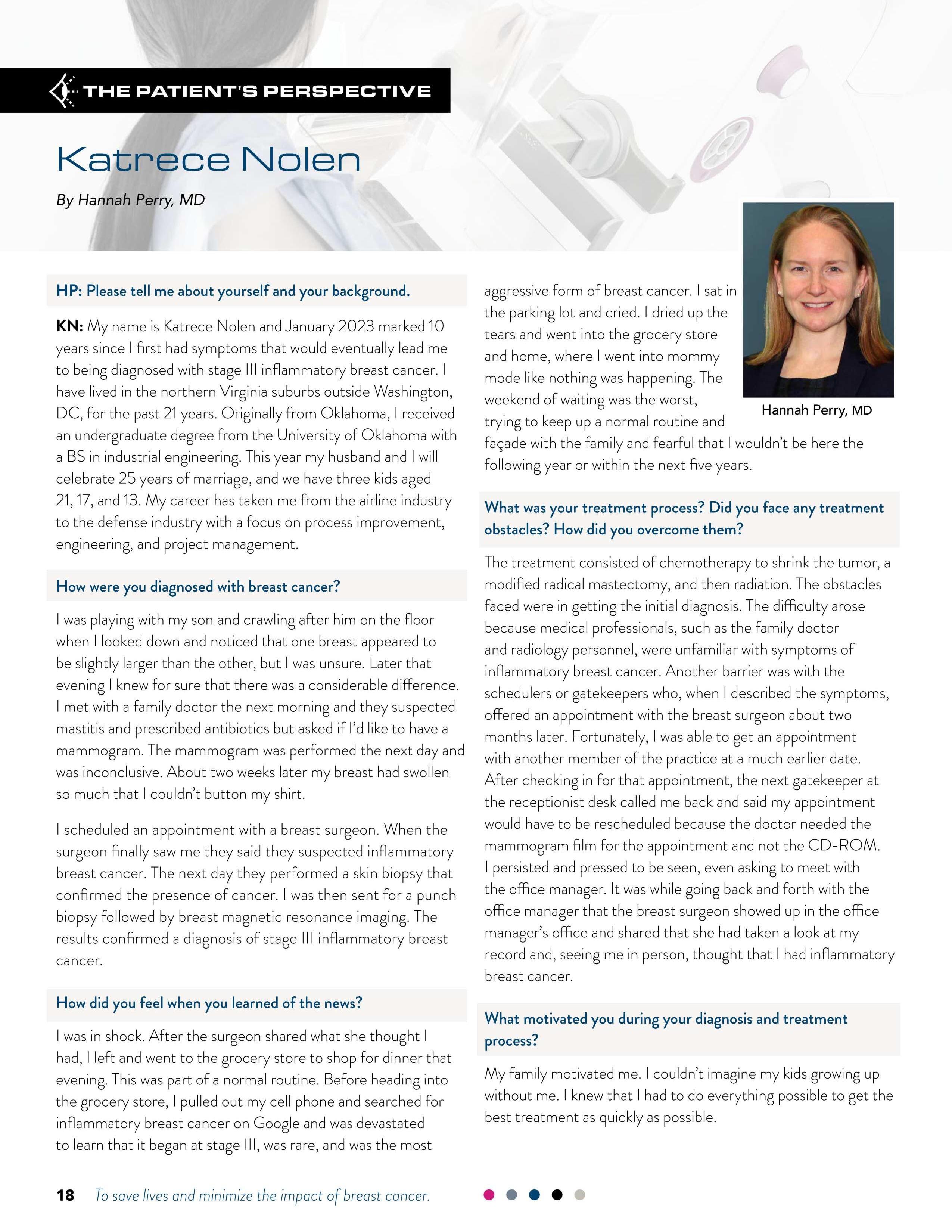
By Nidhi Sharma, MD
Do more than belong: participate. Do more than care: help. Do more than believe: practice. Do more than be fair: be kind. Do more than dream: work.
“Do More” by William Arthur Ward
The sense of belongingness refers to a human emotional need to affiliate with and be accepted by members of a group. The need to belong is an intrinsic motivation to affiliate with others and be socially accepted.1
This need plays a role in self-presentation, social comparison, and seeking stability and happiness.2 Over time one realizes that happiness is not finding success by a certain time but finding something to do that one is so passionate about that time does not count. True belonging only happens when we present our authentic, imperfect selves to the world,3 something that often comes with practice and nurturing a growth mindset.
The recent SBI symposium was invigorating, pedagogical, and delightful, with a strong effort on community building and belonging. When entire organizations like ours embrace a growth mindset, the members feel far more empowered and committed and make greater strides in collaboration and innovation. Peter Eby, MD, FACR, FSBI, one of my SBI newsletter editor predecessors and current director at large for the SBI Board of Directors, established the mission of the SBI newsletter: “To empower and expand the diverse global breast imaging community through honest journalistic coverage of significant events, personal perspectives, scientific discoveries, and inspirational stories from all facets of our field for our members.”
All our editors’ tireless, diligent efforts have laid a strong foundation for the newsletter, enabling it to serve as a reliable source of information, knowledge, and inspiration for our members. As I embark on this exciting new chapter of my career, I am filled with profound gratitude and a deep sense of purpose. With great honor and humility, I pledge to produce exceptional content for our esteemed members and the wider breast imaging community. Importantly, this newsletter is a year-round team effort. Joining me are the associate editors, Randy Miles, MD, and Shinn-Huey Shirley Chou, MD. The stellar newsletter team will continue to deliver interesting and practical stories that keep you apprised of myriad topics impacting the worldwide breast imaging community. In this SBI News edition, we cover such topics as various meeting highlights, latest nuclear medicine breast imaging innovations, placing a focus on physical wellness, and CMIST (Contrast Enhanced Mammography Imaging Screening Trial) updates.
Keeping in theme with our president’s note on belonging, the SBI has developed many resources in addition to the newsletter to engage the breast imaging community, and I encourage you to make the best use of solutions that might meet your personal needs. In addition to the annual symposium, SBI Connect has blossomed into a vibrant online discussion forum. In an era of ever-evolving medical breakthroughs, effective communication becomes paramount. Our newsletter makes a profound impact by disseminating critical information, facilitating professional networking, and inspiring continuous learning.
My personal journey of volunteering at SBI over the past decade has been hands down the best time investment for immense learning and growth. One of my core beliefs is that greatness is achieved through collaboration. I am eager to foster a culture of inclusivity and openness, encouraging diverse perspectives and embracing the wealth of knowledge within our society. I will actively seek multifarious contributions to create a platform that reflects the breadth and depth of our collective expertise. Embracing innovation and newer approaches, we can enhance the reach and impact of our newsletter. Let us explore ways to engage our readership, including interactive content, multimedia elements, and personalized experiences. Together we can create a dynamic and evolving publication that can positively impact all.
If you have any stories, questions, ideas, or breast imaging–related personal passion projects, I invite you to write to me: nidhisharma31@gmail.com. Thank you for reading this summer edition of SBI News. I hope you are having a great summer, filled with happy times and good sunshine!
References
1. Schneider ML, Kwan BM. Psychological need satisfaction, intrinsic motivation and affective response to exercise in adolescents Psychol Sport Exerc. 2013;14(5):776-785. doi:10.1016/j.psychsport.2013.04.005
2. Pillow DR, Malone GP, Hale WJ. The need to belong and its association with fully satisfying relationships: a tale of two measures Pers Individ Dif. 2015;74:259-264. doi:10.1016/j.paid.2014.10.031
3. Brown B. Finding our way to true belonging. Ideas.ted.com. September 11, 2017. Accessed June 27, 2023. https://ideas.ted.com/finding-our-way-totrue-belonging/
To save lives and minimize the impact of breast cancer. .....
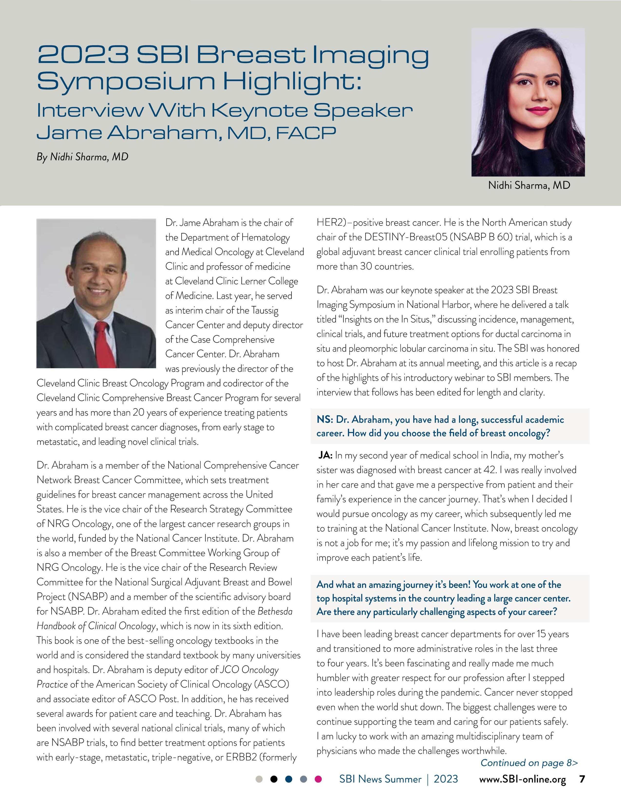
Nidhi Sharma, MD
Patient Care and Delivery Committee Update
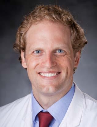
By Lars Grimm, MD, FSBI
The Patient Care and Delivery (PCD) Committee continues to be incredibly active producing clinically relevant content for the larger SBI community. As chair, I am fortunate to have such a wonderful group of collaborators with such great ideas. The PCD Committee completed several projects this past year and have many more in the works.
Current and Upcoming Projects
In collaboration with the Lobular Breast Cancer Alliance and led by Dr. Kristen Coffey, we recently surveyed the SBI membership about invasive lobular breast cancer. Analysis is currently in progress, but we are looking at modality-specific confidence for making the diagnosis of invasive lobular cancer, especially in women with dense breasts. We also want to understand when radiologists recommend supplemental imaging.
There are advantages and disadvantages to same-day study interpretations. Led by Dr. Brian Dontchos, we recently released a survey to understand how often members are performing sameday imaging and procedure services. The results will help practices benchmark their workflow to other breast imaging groups.
In the near future, SBI members should see projects related to wellness, imaging for patients with disabilities, synthesized mammography, and virtual mammography services.
Recent Journal of Breast Imaging Publications
Three manuscripts were published in the Journal of Breast Imaging this past year. Led by Drs. Sally Goudreau and Katia Dodelzon, the PCD Committee developed an evidence-based review of bleeding complications after breast core-needle biopsy.1 The risks of breast biopsy are different from risks of biopsy in other organ systems, but most published guidelines are for general radiology or interventional radiology procedures. These guidelines review breast-specific bleeding complications and provide practical approaches to treat patients.
Outside study interpretations are an increasingly large component of many radiology practices. Dr. Dontchos
spearheaded a survey to understand how practices approach outside study interpretation.2 Respondents reported that they are commonly asked to give second opinions on biopsy recommendations but that physician time constraints are a major barrier.
Abbreviated breast magnetic resonance imaging (MRI) is a means to expand access to breast MRI for women who may not be covered by insurance. Led by Dr. Lars Grimm, the PCD Committee surveyed members on current and future plans to adopt abbreviated breast MRI programs.3 Adoption of abbreviated breast MRI is growing rapidly, but patient eligibility, cost, protocols, and follow-up intervals vary.
The PCD Committee aims to produce content of value to the SBI membership. The quality of our survey work is highly dependent on the participation of SBI members, and we would like to give a special thanks to all survey respondents. We are always looking for new topics of interest to SBI members. Please do not hesitate to reach out with new ideas or collaborations (lars.grimm@duke.edu).
References
1. Goudreau S, Grimm LJ, Srinivasan A, et al. Bleeding complications after breast core-needle biopsy—an approach to managing patients on antithrombotic therapy J Breast Imaging. 2022;4(3):241-252. doi:10.1093/jbi/wbac020
2. Dontchos B, Dodelzon K, Dogan BE, et al. Variations and challenges to performing outside study interpretations in breast imaging: a national survey of the Society of Breast Imaging membership J Breast Imaging. 2022;4(2):153-160. doi:10.1093/jbi/wbab101
3. Grimm LJ, Conant EF, Dialani VM, et al. Abbreviated breast MRI utilization: a survey of the Society of Breast Imaging J Breast Imaging. 2022;4(5):506-512. doi:10.1093/jbi/wbac048
Lars Grimm, MD, FSBI
Contrast Enhanced Mammography Imaging Screening Trial (CMIST) Is Now Open!
After almost five years of planning, development, and delays from the COVID-19 pandemic, the ACR, the Breast Cancer Research Foundation, and GE HealthCare are happy to announce the enrollment of the first patients in the Contrast-Enhanced Mammography Imaging Screening Trial (CMIST).
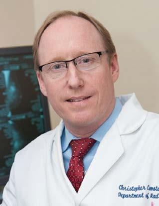
Breast cancer is one of the most commonly diagnosed cancers among American women, with approximately one in eight women facing this diagnosis in her lifetime. Although mammography is considered the standard breast cancer screening tool, its effectiveness is reduced in women with dense breast tissue. “Despite incremental improvements in mammographic technique over the past 50 years, screening mammography may still not detect many lethal breast cancers, especially in women with dense breasts,” said Christopher Comstock, MD, FACR, FSBI, CMIST study chair.1 Dense breast tissue is also a lesser-known risk factor that can double one’s risk for breast cancer. Effective September 10, 2024, there will be a national requirement for patient notification regarding breast density under a single dense breast reporting standard as a result of recent updates to the Mammography Quality Standards Act (MQSA) announced by the US Food and Drug Administration.
Contrast-enhanced mammography (CEM) combines mammography and iodinated contrast media in a simple and quick procedure to highlight areas of unusual blood flow patterns that may indicate malignancy. “This trial will help us to determine whether CEM can find many cancers that would otherwise be missed on screening mammography and help to usher in a new screening paradigm,” said Dr. Comstock.
The CMIST study seeks to determine if CEM provides more accurate cancer detection than digital breast tomosynthesis (DBT), or 3-dimensional (3D) mammography, in women with dense breasts. As part of CMIST, 2032 women with dense breasts will be enrolled to compare the CEM technique with 3D mammography.
Carolina Breast Imaging Specialists in Greenville, North Carolina, led by Dr. Bruce Schroeder, medical director and chief executive
officer, is the first of 15 planned sites to start recruiting patients. Additional sites in the United States and Canada will commence recruitment in the coming months. The trial is set to deliver the first results in 2025. The trial is registered on ClinicalTrials.gov under NCT05625659.
Title: ACR 4707: Comparison of Breast Cancer Screening With Dual-Energy Contrast-Enhanced Spectral Mammography to Digital Breast Tomosynthesis in Women With Dense Breasts (Contrast Mammography Imaging Screening Trial)
Study chair: Christopher Comstock, MD, FACR, FSBI
Primary aim: To determine if dual-energy contrast-enhanced spectral mammography (CESM) can detect more cancers with fewer false positives than DBT in women with dense breasts.
Eligibility (see protocol for complete list):
• Women aged 45 to 74 years scheduled for routine screening DBT are eligible.
• Participants must have mammographically dense breasts (ACR BI-RADS density category C or D) on their most recent screening mammogram.
• Participants must be asymptomatic.
• Participants must be able to undergo intravenous administration of iodinated contrast material.
• Participants must not have had breast magnetic resonance imaging or CESM in the 36 months before enrollment.
• Participants must not have undergone screening breast ultrasonography within 12 months before enrollment.
Study design (N = 2032 patients):
• Paired design: Each patient undergoes both DBT and CESM on the same day at year 0 and year 1.
• DBT and CESM images are read by two radiologists, each of whom reads images from only one test and is blinded to the results of the other test.
Continued on page 11>
To save lives and minimize the impact of breast cancer. .....
Christopher Comstock, MD, FACR, FSBI
You are the chair of the North American NSABP-60 trial, which is a global adjuvant breast cancer clinical trial for trastuzumab, enrolling patients from 30 countries. You lead many such important roles and wear so many hats. What keeps you motivated to continue to be involved in breast oncology research?
It goes back to my story about my initial journey. Cancer is real and we want the best therapies available for our patients. One of the most important ways we can make an impact in cancer care is with research and clinical trials. Throughout my journey, I have been fortunate to be a part of large clinical trials and landmark studies like NSABP, which helped shape cancer treatment globally. What drives me is my continued belief that we can make things better for tomorrow for our patients.
In 2010, you received an award from the president of India for your outstanding contributions to the field of breast cancer care. Will you please share some high points in your career like this award that made it all feel worthwhile?
Thank you. I was fortunate to receive this eminent award in the presence of globally renowned Indian artists, sportsmen, and personalities. I am lucky and trying to make my impact in a small way.
What can early-career and midcareer physicians do to get more involved with their national societies?
I always say finding a good mentor is really important. I have had some amazing sponsors and mentors throughout my career who really lifted me up. Your mentor doesn’t have to be necessarily from your home institution or practice. National societies like the SBI provide a platform to network and make it easier to find a few mentors whose careers and personalities align with your goals and then try to work closely with them on a few projects. It’s important to develop the right connection with your mentors. Work hard, volunteer in the societies, take on assignments, put your name out there, and go the extra mile. Everyone is looking for people willing to work hard and contribute. Likewise, we are always looking for the next leaders to carry the mantle.
Of everything you’ve achieved, your outstanding patient care stands out as an exceptional accomplishment. In your recent TEDx talk, you discussed building trust. Can you please share some important pointers that have helped you build these trustworthy, strong connections with your patients and peers?
We are so privileged to walk into someone’s life in the most vulnerable phase, in their worst moments with a new cancer diagnosis. It’s a huge responsibility; always try to treat an individual, not another cancer diagnosis. For me, it’s really important to get to know my patients by listening to their life stories and making that connection. Try to learn what makes your patient worried or happy;
save lives and minimize the impact of breast cancer.
only then can we build trust as the foundation of the physicianpatient relationship.
What are your tips for preparing oneself to become a successful future leader and creating a healthy, rewarding work environment and career?
Everything we do as clinicians in our respective fields, we make a huge impact in the health care space. Find the niche or passion that inspires you every day and try to get better at it. It sounds cliché to say this, but I truly don’t feel like I am working long hours every day; instead I feel I’m living my dream and passions. As a leader, your strength lies in how to bring people together and get them excited about something that will make tomorrow better for your field. Leadership is not about you; it’s about the team. Listen to your team and really care about the people you’re working with.
What advice would you give to your early-career self?
That’s a tough one! I don’t think I’m good at work-life balance. I came to this country as an immigrant like you and we are driven to prove ourselves. Finding the right balance is something I continue to work on.
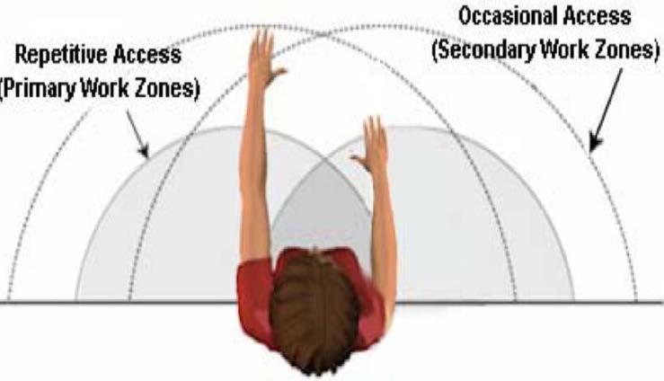
Repetitive Access (Primary Work Zones)
Seldom Access (Tertiary Work Zone)
Occasional Access (Secondary Work Zones)
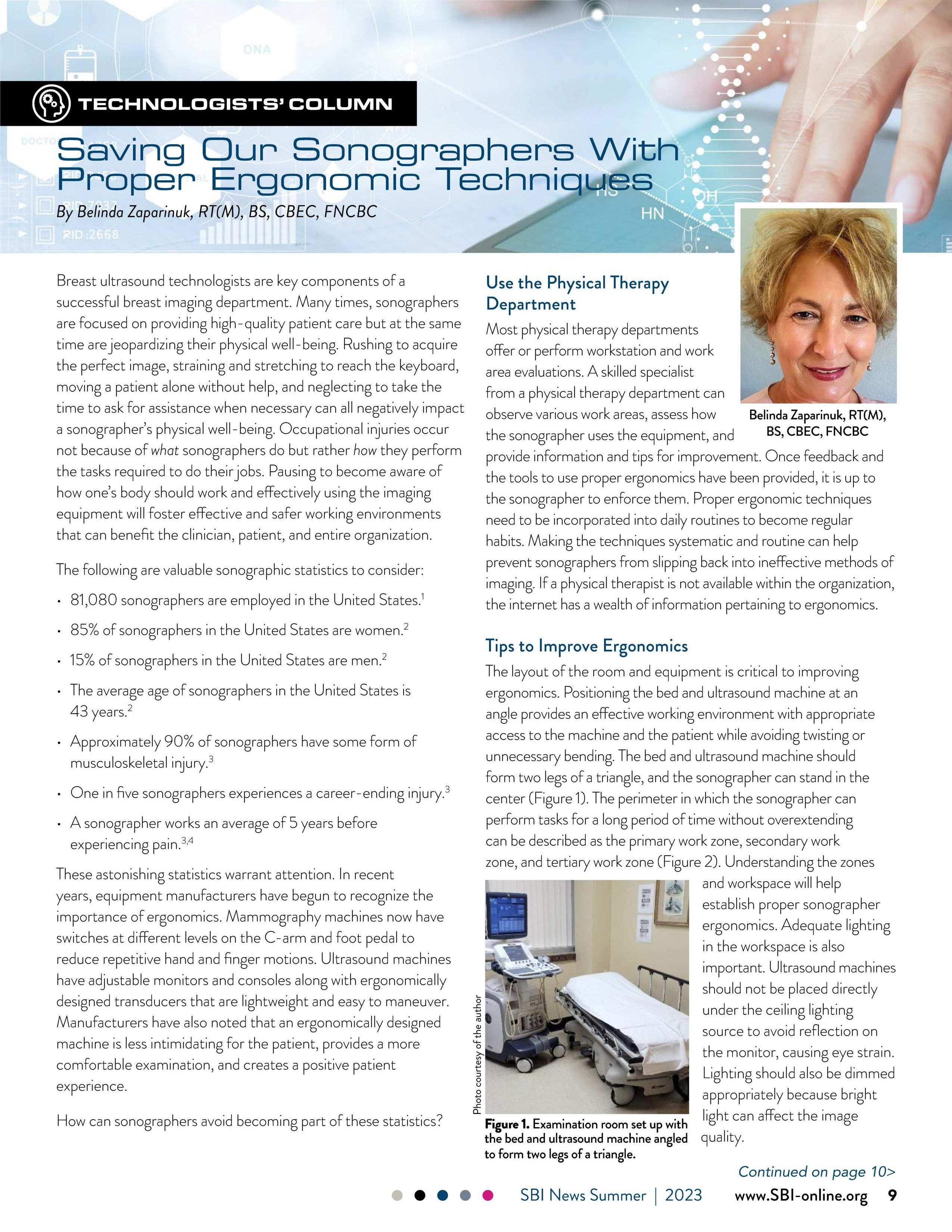
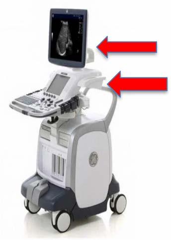
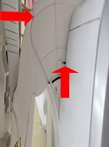
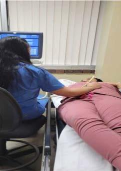
Once the layout has been addressed, the monitor should be adjusted. The console should be pivoted toward the technologist with presets built into imaging protocols. This adjustment reduces the number of keystrokes and overreaching during the examination. The height of the monitor must be adjusted to the sonographer’s eye level, with a minor tilt downward. For staff members who share equipment, it is useful to save monitor adjustments by placing a mark on the side of the console and on the side of the monitor where it raises and lowers. Each staff member may choose an individual color specific to their settings and place marks on the unit (Figure 3). These marks provide a means to quickly and accurately adjust the unit for the technologist performing the examination.
Another critical aspect of organizing the examination room is the location of the patient’s bed. The bed should be raised or lowered to the appropriate height for the sonographer. This adjustment will reduce any possible extension of the sonographer’s body while avoiding leaning or twisting, which may cause injury. Some examination beds include electronic, stationary, and manual setting options that can be modified for the sonographer performing the examination. Motorized beds are the most ideal option for optimal ergonomics. The height of these beds can be adjusted to accommodate patient transfer and the sonographer. Stationary
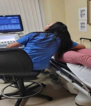
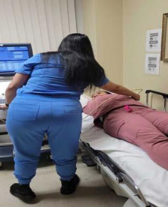
gurneys pose more of a challenge, but the lack of bed movement can be compensated for. If the examination room does not allow you to place the bed at an angle and the bed is against a wall, try turning the patient to avoid overextension by the sonographer. Having the patient lie supine with the patient’s head in the routine examination position may be adequate to obtain imaging of the right breast. Having the patient sit up and reverse their position on the gurney to obtain imaging of the left breast may be more ergonomically friendly. When using a stationary bed, sonographers are usually scanning while seated. It is essential to be mindful not to lean into the bed (Figure 4A) to avoid a shoulder, neck, or twisting injury. Standing (Figure 4B) would not be suggested with a stationary bed.
Manual beds that require pumping a foot pedal to raise and lower the bed require the use of the sonographer’s legs and feet. Using an alternating-leg technique to raise and lower manual gurneys will help reduce repetitive motion injuries.
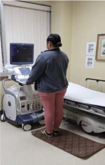
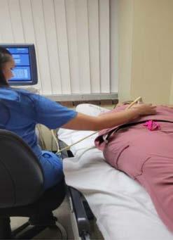
Sonographers may sit or stand when performing imaging. Those who prefer to stand may choose to use a fatigue mat to absorb stress on the legs and feet over time and help support a standing position (Figure 5A). The mat can be stored under the bed when not in use. When sonographers choose to sit in a chair to perform imaging, the chair should be close to the machine and patient gurney to ensure proper technologist posture and minimal arm extension. For adequate arm extension, a 30° abduction is recommended. Imaging staff should be mindful of placing their feet flat against the floor and using a chair that provides lumbar support (Figures 5B and 5C).
To save lives and minimize the impact of breast cancer. .....
Figure 2. Work zones. Working in the triangular area is optimal for ergonomics.
Figure 3. A, Arrows indicate locations where the monitor and console are raised and lowered. B, Arrows indicate locations where marks can be placed on the unit to represent preferred settings for individual users.
3B
3A
Photo courtesy of Google
Photo courtesy of the author
Photo courtesy of the author
4A
Figure 4. A, Incorrect sitting technique. B, Incorrect standing technique.
4B
Photo courtesy of the author
Figure 5. A, Correct use of fatigue mat. B, Proper sitting technique with lumbar support and feet flat on the floor or supported. C, Scanning position with the patient as close as possible to reduce extension of the arm.
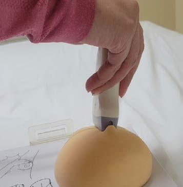
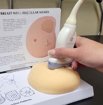
The final part of ergonomic technique is the transducer and the sonographer’s grip. It is very important to use transducers that foster the palmar grip (Figure 6). The palmar grip is an ergonomic, wrist-neutral grip that reduces torque and tension in the hand, fingers, and wrist.
The following are tips for achieving optimally sound ergonomic technique while performing sonographic examinations:
• Position the machine within proper reach.
• Adjust the console adequately for seated and standing scanning positions.
• The base of the console should not interfere with the ability of the sonographer to reach the patient and the console.
• There should be adequate clearance for the sonographer’s legs and feet when scanning in a seated position.
• Adjust the height of the monitor to minimize excessive neck rotation, flexion, and extension.
• The monitor should be at eye level.
• The touch screen and keyboard should not restrict neutral posture or require excessive reaching beyond the primary work zone.
• Use customized preferences to reduce keystrokes.
• Transducers should be lightweight, balanced, and designed with the palmar grip (neutral wrist position) to reduce torque in the wrist.
• Examination beds should be raised to allow arm abduction of less than 30°.
• Sonographer examination chairs should swivel and should have lumbar and thigh support and a footrest.
• Chair height should be adjusted appropriately for the sonographer.
• The sonographer’s feet should remain flat on the floor when seated or should be supported with a fatigue mat when standing.
• The patient should be positioned close to the sonographer.
• Adjust bed height, if possible.
• Reverse the patient’s position to facilitate scanning of specific body parts.
This information should serve as a reminder that sonographers need to protect their bodies so they can continue to provide lifesaving examinations and procedures for their patients in a healthy manner. Their bodies, patients, and organizations will reap the benefits of the best in patient care.
References
1. Occupational employment and wage statistics: diagnostic medical sonographers. US Bureau of Labor Statistics. May 2022. Updated April 25, 2023. Accessed June 29, 2023. https://www.bls.gov/oes/current/oes292032.htm
2. Sonographer demographics and statistics in the US. Zippia. Accessed June 29, 2023. https://www.zippia.com/sonographer-jobs/demographics/ 3. Ultrasound ergonomics: an overview. Institute for Advanced Medical Education. Accessed June 29, 2023. https://www.iame.com/ultrasound_ergonomics_CME
4. Industry standards for the prevention of work related musculoskeletal disorders in sonography. Society of Diagnostic Medical Sonography. 2021. Accessed June 29, 2023. https://www.sdms.org/docs/default-source/Resources/industry-standardsfor-the-prevention-of-work-related-musculoskeletal-disorders-in-sonography.pdf
Contrast Enhanced Mammography Imaging Screening Trial (CMIST) Is Now Open! (continued from page 6)
• DBT and CESM are performed with the GE Pristina SenoBright HD.
• The contrast agent is iohexol, 350 mg iodine per milliliter.
Study costs: The costs of the year 0 and year 1 CESM and the costs for start-up, data entry, and image submission will be covered by the trial.
Site qualifications (approximately 15 sites expected):
• Interpreting physician/reader requirements are MQSA qualification, a minimum of 5 years’ experience reviewing digital mammography, and interpretation of a minimum of 20 prior GE Pristina CESM examinations.
• Sites must have completed a minimum of 50 clinical GE Pristina CESM examinations.
• The technologist performing study CESM examinations must have performed a minimum of 10 clinical GE Pristina CESM procedures.
If you are interested in participating in the study or would like more information, please contact:
• Christopher Comstock, MD, FACR, FSBI (comstocc@mskcc.org)
• Leslie Sears, ACR project manager, Diagnostic Imaging Clinical Trials, 1818 Market St, Suite 1600, Philadelphia, PA 19103 (lsears@acr.org)
Reference
1. First patients enrolled in Contrast Enhanced Mammography Imaging Screening Trial (CMIST). News release. American College of Radiology. May 4, 2023. Accessed June 30, 2023. https://www.acr.org/Media-Center/ACR-NewsReleases/2023/First-Patients-Enrolled-in-Contrast-Enhanced-MammographyImaging-Screening-Trial
Figure 6. A, Incorrect grip. B, Correct grip. 6B
6A Photo courtesy of the author
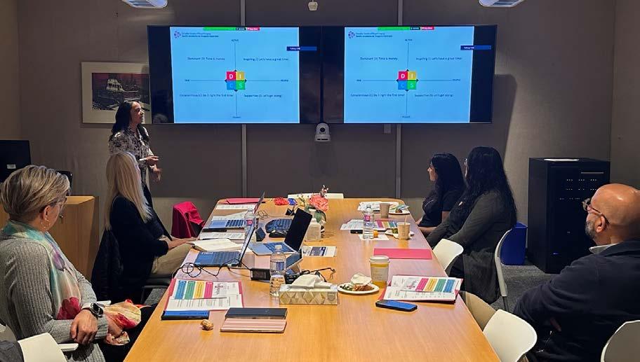
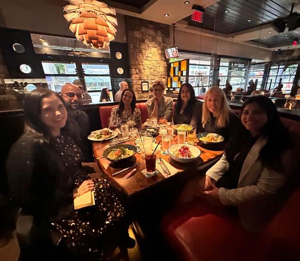
Image from the strategic planning meeting: Amy Smith, Executive Director of CSBI; Dr. Carolyn Flegg; Dr. Jean Seely; Dr. Sri Sannihita Vatturi; Dr. Kaitlin Zaka-Metias; and Dr. Raman Verma.
breast cancer among women in their 40s by approximately 8% over the past 20 years (0.39% annualized percent change), with an even steeper increase since 2015 (1.6% annualized percent change). Increased breast cancer treatment costs in the past 10 years may establish the cost-effectiveness of early detection from breast cancer screening, with the costs of early diagnosis exceeded by the costs of treating advanced-stage breast cancer at the time of diagnosis.
Resources the health care system needs to treat advanced-stage breast cancer and the physical and mental penalties patients and their family members bear are large. Radiologists and technologists who diagnose breast cancer and health care professionals who care for breast cancer patients have incentive to reduce the emotional toll and negative impact that advanced-stage breast cancer can inflict upon the workforce. The CSBI is strongly motivated to implement changes to improve early detection of breast cancer more equitably.
References
1. Heaney RM, Zaki-Metias KM, McKee H, et al. Correlation between breast arterial calcifications and higher cardiovascular risk: awareness and attitudes amongst Canadian radiologists who report mammography Can Assoc Radiol J 2022:8465371221140347. doi:10.1177/08465371221140347
2. Wilkinson AN, Billette JM, Ellison LF, Killip MA, Islam N, Seely JM. The impact of organised screening programs on breast cancer stage at diagnosis for Canadian women aged 40-49 and 50-59 Curr Oncol. 2022;29(8):56275643. doi:10.3390/curroncol29080444
Image from the strategic planning meeting: Amy Smith, Dr. Carolyn Flegg, Dr. Jean Seely, Dr. Sri Sannihita Vatturi, Dr. Kaitlin Zaka-Metias, Dr. Raman Verma, and Dr. Charlotte Yong-Hing.
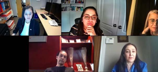
Screenshot from the CSBI annual general meeting: Dr. Sri Sannihita Vatturi, Dr. Silma Solarzano, and Dr. Kaitlin Zaki-Metias.
Screenshot from the CSBI annual general meeting. Clockwise from top left: Dr. Jean Seely, Cheryl White, Dr. Anat Kornecki, and Sandra MacFarlane, MRT.
Screenshot from the CSBI annual general meeting. Clockwise from top left: Dr. Charlotte Yong-Hing, Dr. Supriya Kulkarni, Dr. Mona El-Khoury, Dr. Rola Shaheen, and Dr. Derek Muradali.
Best Practices for Navigating Workplace Challenges
Did you know that a recent SBI member survey showed that 79% of respondents would prefer a hybrid model, believing it would allow them to be more efficient and improve work-life balance without negatively impacting their work or career? Drs. Jennifer Harvey and Margarita Zuley discussed data and realworld experiences regarding hybrid work models in breast imaging. Dr. Catherine Giess navigated the pertinent topic of conflict in a practice and how to build effective practice teams, with great examples from her practice of implementing proven leadership principles to bring about positive change. This talk in particular elicited self-reflection in all of us.
Louise Miller, Dr. Bethany Niell, and Dr. Jay Parikh also shared their experiences with maximizing efficiency in busy, modern practices. The focus on applicable knowledge-sharing for both private and academic practices is one of the great strengths of the annual SBI symposium, as shown by these talks as well as the other excellent presentations!
Diversity, Equity, and Inclusion
Each full day’s plenary session had a talk devoted to understanding and addressing issues with diversity, equity, and inclusion and narrowing disparities in breast cancer screening, diagnosis, and treatment in disadvantaged communities we serve. Drs. Tanya Moseley, Sughra Raza, and Donna Plecha taught us about equality versus equity of care, the biases we all must combat as members of the human race, how to be more inclusive in our workplaces, the disparities between populations we serve that are based on socioeconomic factors, and ways we can narrow those disparities. Allostatic load (look it up!) leads to worse outcomes for our patients, and Black women with breast cancer have a 40% higher mortality rate than their White counterparts. Drs. Cindy Lee, Victoria Mango, and Rachel Brem led an educational session sharing their real-world experiences with minimizing barriers to care. It is encouraging to see attention given to these topics, and speakers provided tools and hope for positive change.
The Value of Personal Interaction
As the world returns to a state of face-to-face interaction, the symposium added valuable opportunities for in-person connections. Additional roundtable discussions were offered with Breakfast Briefs, small-group mentoring and training sessions on various topics, including screening boot camp, early-career advice, leadership, use of social media, thriving as a breast imaging fellow, and others. Before, between, and after main symposium lectures, meeting areas bustled with conversation, introductions, and socialization. It was wonderful to see networking at its peak.
For SBI members active on the virtual SBI forum, Dr. Jay Baker led a discussion of topics of interest from the forum in “Dear Jay Baker” format, creating memorable evidence-based responses
to common questions. Many programs and consortiums also held individual gatherings allowing colleagues from far and wide to reconnect or perhaps meet for the first time! Breast imaging radiologists with similar goals and experiences had so many opportunities for meaningful personal interaction.
Celebrations
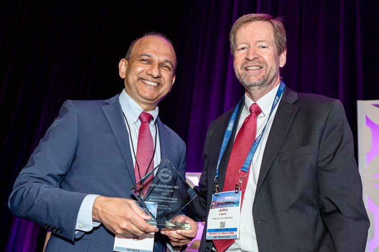
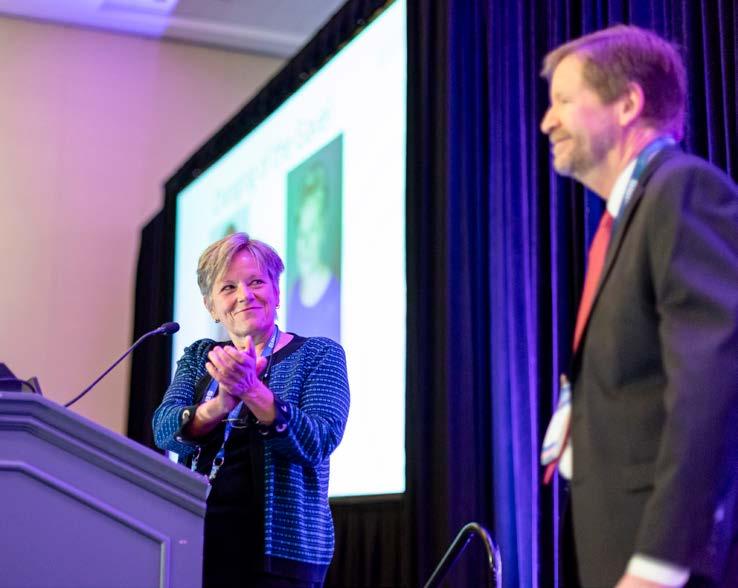
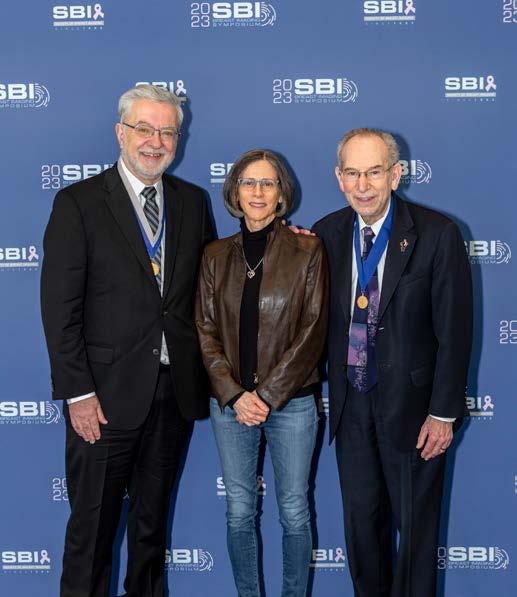
There was plenty to celebrate with the induction of the new president, Dr. Mimi Newell, by the past president, Dr. John Lewin. The 2023 Gold Medal recipient was Dr. Murray Rebner. It is humbling and inspiring to see how the work of our leaders and members has shaped our practice and the society.
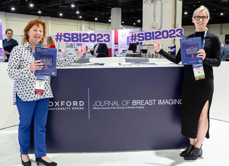
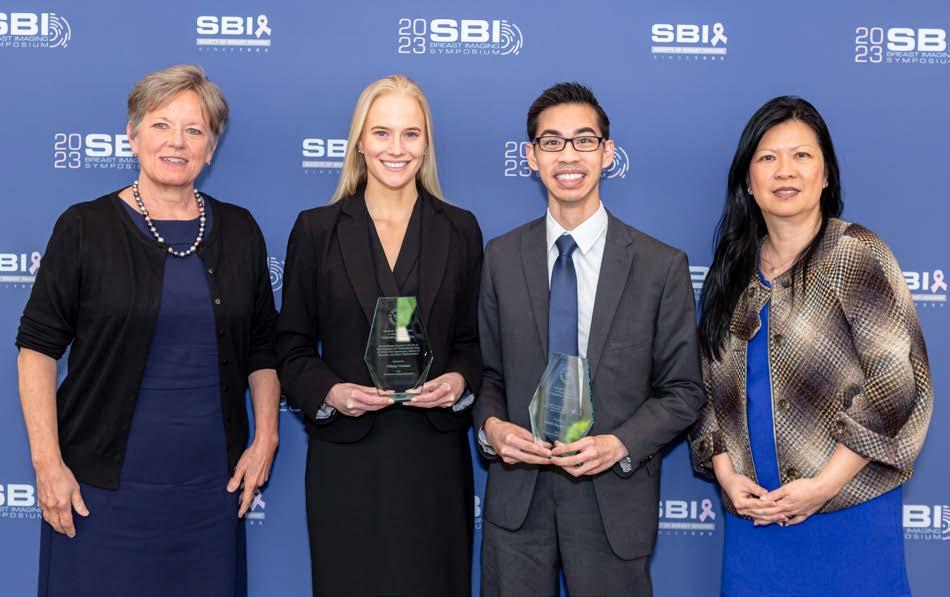
The monumental work of Dr. Jennifer Harvey as editor in chief of the Journal of Breast Imaging was honored. She passes the torch to Dr. Wendy DeMartini. The future of the society and research within it were celebrated as the SBI Research & Education Fund award winners, Drs. Tiffany Clausen and Derek Nguyen, presented their abstracts. Many also celebrated aboard the Odyssey DC on a beautiful spring evening with dinner and dancing on a river cruise up the historic Potomac for the President’s Gala.
Lots to Look Forward To
Clearly, there are great things happening and great things coming for breast imaging! If you were unable to attend and are feeling mounting regret or piqued curiosity (“What can I do to narrow disparities in my community?” “What might the future of screening look like?” “How could a hybrid model work for me?”), despair not! Lectures and lecture slides from the symposium will be posted for asynchronous content review. And next year’s symposium is now less than a year away, to be held April 11 through 14 in Montreal! Upcoming SBI webinars and multiple other educational opportunities are also available through the SBI website. See you next year in Montreal!
Continued on page 16>
Dr. Mimi Newell and Dr. John Lewin opening the symposium.
save lives and minimize the impact of breast cancer.
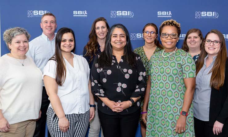
SBI administrative team worked hard to make the symposium a great success.
Outgoing and incoming JBI editors Dr. Jennifer Harvey and Dr. Wendy DeMartini.
Dr. Tiffany Clausen, 2023 Gerald D. Dodd Jr. Research Award winner, and Dr. Derek Nguyen, 2023 Wendell Scott Award winner, with Dr. Mimi Newell and Dr. Linda Moy.
SBI Gold Medal recipient Dr. Murray Rebner with past recipients Dr. Debra Monticciolo and Dr. Michael Linver.
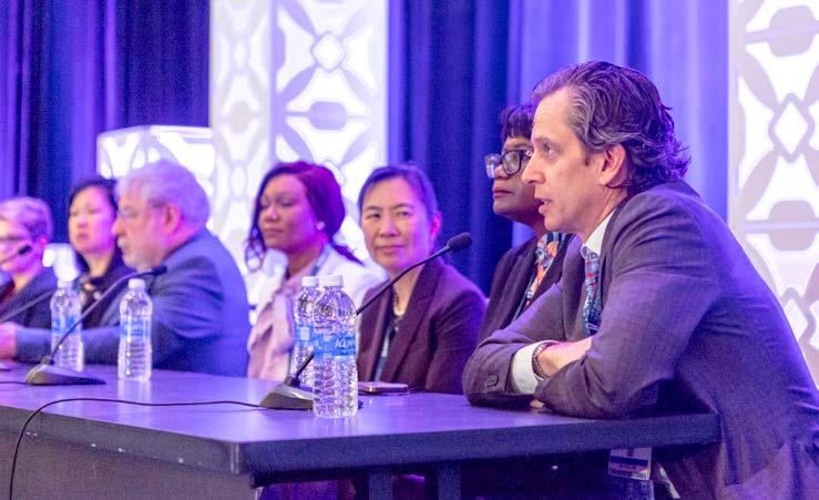
Morning plenary session in progress.
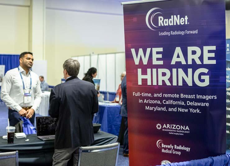
SBI career fair was a huge success.
What
did you learn from your experience?
I learned that the statistics for life expectancy following cancer are just that, statistics. There are several factors that drive life expectancy and one of those is timely access to treatment and excellent care. I made sure that I had access to both, but I learned that is not always the case for everyone going through a similar situation. I was sure that if given timely access to treatment and excellent care, the life expectancy for others following such a cancer diagnosis would also be improved.
How has this diagnosis impacted your life?
It has completely altered my life and changed my life’s purpose. Given the obstacles I faced and my ability to get access to timely treatment and excellent care, I decided to write a book about my experience and that of others and how others can get access to cancer support that can meet their unique needs. The book is called I’ve Been Diagnosed, Now What? Courageously Fighting Cancer in the Face of Fear, Uncertainty and Doubt. I have also consulted with cancer nonprofit organizations and advised cancer centers on support throughout the cancer experience. Lastly, I launched a website at www.FindCancerHelp.com as a marketplace to connect cancer patients, survivors, and caregivers to cancer nonprofit and for-profit organizations that can meet the unique needs of a cancer patient’s journey.
Are there any lessons that you think the breast imaging community can learn from your experience?
Interestingly enough, I was pretty upset with the imaging center that missed the original cancer diagnosis. They wrongly assumed it wasn’t cancer from a mammogram and ultrasound, when it turns out that those two diagnostic tests can be limited in their ability to detect inflammatory breast cancer. The symptoms are classic for inflammatory breast cancer and additional diagnostic testing was not recommended until my visit with a breast surgeon. I had a conversation with my hematology-oncology doctor about the missed diagnosis and she indicated that she did meet with a representative of the radiology center to better educate them on the need for additional testing in such a case.
I think the breast imaging community could work with their general imaging colleagues to make sure others are knowledgeable about the different imaging tools that should be offered to patients to help get to the bottom of the symptoms.
What advice would you give to other patients who are going through the diagnosis and treatment process for breast cancer?
The advice I would give to other patients is that they have no choice but to be an advocate for themselves and surround themselves with a cancer support system that can fill this important need. Take advantage of every resource that is offered to you and ask for additional resources that can help you. It’s a devastating time, but it’s also not the time to mentally separate yourself from others. Support groups will help, as well as connecting with those who have faced the same type of cancer. It’s a steep learning curve, and a quick one at that, to learn how to navigate this journey. The quickest way to get up to speed is to have an effective support team in place, listen to the experts, and ask questions when you don’t clearly understand the plan.
Learn more about Katrece Nolen at www.KatreceNolen.com. Follow her on Twitter, Instagram, or Facebook @KatreceNolen.
Katrece Nolen
These 5 tips can help ensure that physical health remains high on your facility’s priority list:
1. Provide wellness program incentives.
It’s no secret that employees are motivated by incentives. Offer subsidized gym memberships or discounted fitness classes. Encourage goal setting and make an effort to help employees reach their personal goals.
2. Make wellness fun.
Bring out the inner child in your employees and watch their faces light up when an event or activity gets them away from their desk while still getting paid. Organize a competition or group walk or bring in a fitness instructor for a group activity midmorning on a random Tuesday. Consider partnering with local gyms or companies that sponsor recreational activities.
3. Make healthy dietary options available.
If employees are going to place effort on their physical fitness, encourage them to fuel their bodies with healthy dietary options and snacks at work. Nothing sabotages a nice midday walk like a soda or candy bar from the vending machine.
4. Create consistent physical habits.
Bring in an educator or wellness coach to help identify employees’ personal goals, then assist employees and leadership teams in reaching those goals and overcoming obstacles that they may face while at work.
5. Encourage movement.
In a health care setting, taking a break or going out for a walk can be challenging because of schedules. Making a focused effort to step away from your environment and breathe fresh air for a few minutes can make a huge impact. Consider using standing desks and encourage breaks and physical activity. Even a few minutes of movement can decrease mental fog and feelings of burnout.
Ultimately, leaders must set the tone. Staff members will care only as much as leaders require them to care. If we want to see the benefits of physical wellness in the workplace, we need more than just a few people to get on board. Making wellness a priority requires inviting others to participate. The benefits of physical wellness are contagious and the rewards are countless. Seventy-three percent of employees with senior managers who show support through involvement and commitment to well-being initiatives said their organizations helped employees develop a healthy lifestyle.4 When making mental and physical health a priority, leaders should understand that their words and actions have consequences that flow down the organization chart.4 Staff members are more likely to engage if the leadership team leads with excitement and provides strong support for wellness initiatives.
Enthusiastic leaders and individuals must be vigilant when attempting to build momentum and excitement focused on engaging others to participate in wellness initiatives. Building a team of employees who prioritize their own physical health will not happen overnight. Remember to exercise patience as you slowly witness a culture shift that even your patients will recognize.
References
1. Garzaro G, Clari M, Ciocan C, et al. Physical health and work ability among healthcare workers. A cross-sectional study Nurs Rep. 2022;12(2):259-269. doi:10.3390/nursrep12020026
2. Ilmarinen J. From work ability research to implementation Int J Environ Res Public Health. 2019;16(16):2882. doi:10.3390/ijerph16162882
3. What is physical fitness? MIT Medical. Accessed June 28, 2023. https://medical. mit.edu/sites/default/files/Physical_Fitness_101.pdf
4. White M. Five ways to drive excitement for your health and wellness benefits. Access Perks. December 19, 2020. Accessed June 28, 2023. https://blog. accessperks.com/five-ways-to-drive-excitement-for-your-health-and-wellnessbenefits
Highlights From the 2023 ARRS Meeting
By Harnoor Singh, MD
The 2023 American Roentgen Ray Society (ARRS) Annual Meeting was hosted in the stunning locale of Honolulu, Hawaii, from April 16 through 20, 2023, with the overall message of “love and peace.” The conference commenced with a warm message of positivity, sharing, and giving. This in-person meeting was a perfect setting to network and reconnect with radiology colleagues from all over the country and world in a picturesque locale with delicious cuisine.
The annual meeting kicked off on Sunday with a great opening ceremony in which the ARRS demonstrated its long-standing commitment to global partnerships by presenting Dr. Jeong Min Lee, president of the Korean Society of Radiology, with honorary membership in the ARRS. The society also acknowledged the outstanding contributions of Dr. Jon Jacobson as the 2023 ARRS Distinguished Educator and awarded Dr. Bernard King the prestigious 2023 Gold Medal, recognizing their exemplary achievements in the field of radiology.
The main focal points of this annual meeting were the concepts of teamwork and wellness. The conference emphasized teamwork based upon the time-honored values of truth, open communication, caring, pride, and respect. Wellness was extensively discussed as a way to reduce burnout, which is currently rampant in the radiology community (reportedly affecting up to 46% of practicing radiologists). As part of wellness, remote radiology was discussed as a potential means to enhance efficiency, promote work-life balance, and ameliorate staffing shortages. Fairness, transparency, and rotating tasks were also discussed.
In addition, proactive approaches to achieving well-being and an optimal state of health to promote work-life satisfaction, including ways to generate a sense of purpose and strategies to manage stress, were presented. Challenges to achieving wellness, including ineffective leadership, lack of diversity, and barriers to professional fulfillment, were also discussed. On an individual level, techniques of self-awareness and mindfulness (to pause, recognize, and reflect) were highlighted.
It is important to build connected communities and embody the philosophy of ubuntu (“I am because we are”). The vital importance of diverse teams and equity was discussed as part of our growth as global citizens and radiologists. The conference also laid importance on targeted efforts to bridge health disparities, which need to be understood and resolved.
The conference hosted numerous breast imaging sessions organized into instructional courses, scientific sessions, and categorical courses. Several SBI members and fellows shared their expertise by presenting sessions. Breast imaging sessions included a session dedicated to the technology of contrastenhanced mammography (CEM), which involves obtaining a high-energy image and a low-energy image. The low-energy image is considered diagnostic, with no statistically significant differences from a full-field digital mammogram. A recombined image is generated via a dual-energy subtraction technique by using the K-edge of iodine after intravenous administration of iodinated contrast material. Maximum contrast is achieved in the breast tissue 2 to 8 minutes after contrast material administration. Advantages of CEM over breast magnetic resonance imaging (MRI) include lower cost, shorter examination time, and less likelihood of claustrophobia. A team approach was again discussed as a way to achieve wider adoption of this technology by aligning the interests of all stakeholders, including patients, referring clinicians, technologists, and radiologists. Training in the billing and coding of CEM was provided, and the most recently updated BI-RADS CEM lexicon was reviewed.
Sessions on breast MRI cited mixed research findings on disease-free survival with or without the use of breast MRI.
The importance of having baseline MRI for comparison with posttreatment MRI to evaluate for treatment response was reported. Breast MRI benefits were reported for dense and nondense breasts after a discussion on breast density as a risk factor for breast cancer. Topics in other emerging breast imaging technologies, such as artificial intelligence (AI), prostate-specific membrane antigen correlation in basal-like breast cancer, and cryoablation, were also presented. AI is rapidly evolving, and the discussion highlighted its potential role in helping improve radiologists’ efficiency and accuracy by decreasing interval cancer rate and recall rate. The conference shed light on the various current barriers and challenges to AI.
To save lives and minimize the impact of breast cancer. .....
Harnoor Singh, MD
Updates on screening mammography recommendations based on age were presented. The importance of performing risk assessments starting at the age of 25 years and continuing screening mammography past the age of 75 years for women in good health was emphasized. The sessions reviewed the revised National Comprehensive Cancer Network guidelines for genetic testing, various risk assessment models for breast cancer, and guidelines for breast screening and assessment for transgender patients.
An insightful session discussed improving breast imaging practices by promoting awareness (including global awareness), proactive acceptance, and action. Patient-centered care was emphasized via a demonstration of empathy and positive intention (sitting down at eye level with patients and practicing active listening). It was interesting to hear how patient education is moving toward online resources and how practices can tailor their communication accordingly.
Sessions on screening mammography featured several intriguing topics, including the importance of uninterrupted reading of screening mammograms by offline batch reading or uninterrupted online reading. Results of various studies continue to reinforce the importance of annual mammography screening, and digital breast tomosynthesis consistently shows increased cancer detection and decreased recall rates. As an initiative to demonstrate the continued relevance of screening mammography and as part of efforts by the ACR Commission on Breast Imaging, a national mammography database is being generated. In this database, the initial method of detection will be systematically recorded to help determine how screening mammography impacts patient outcomes. Presentations at this year’s meeting indicated poorer outcomes among patients with symptomatic breast cancer and favorable tumor characteristics in MRI-detected cancer. The molecular subtypes of breast cancer and differences in their imaging manifestations, recurrence, and prognosis were presented. A comprehensive discussion of the types and management of high-risk breast lesions was a highlight of the meeting.
A session on positron emission tomography/computed tomography covered its role in diagnosing breast cancer, including inflammatory breast cancer. The significance of neoadjuvant systemic therapy was underscored, and informative presentations reviewed treatment response classifications, including stability, complete response, partial response, and
disease progression. According to the presentations, MRI is accurate for monitoring treatment response, and tumor volume is a better indicator than the largest dimension of the known malignancy. Synthetic two-dimensional mammograms generated from digital breast tomosynthesis were presented as a tool to reduce radiation dose to the breast without loss of cancer detection capability. The session concluded that compared with standard digital mammography, synthetic mammography is noninferior for cancer detection rate; is not different for breast density assessment, recall rates, and detection of masses; and shows slightly decreased conspicuity of asymmetries.
A session relevant to practicing breast radiologists was an overview of the anticipated ACR BI-RADS Atlas update. Additions or changes to terminology (eg, using the term lobular for masses, changing milk of calcium to layering calcifications, and using the term echogenic rind on ultrasonography) and new definitions of multifocality and multicentricity were mentioned.
The 2023 ARRS Annual Meeting was a phenomenal showcase of breast imaging expertise and advances in research by a wide array of accomplished breast imaging faculty members. It provided a platform for breast radiologists to come together in the spirit of love, peace, and sharing, and organizing such a fantastic program is a commendable accomplishment by the ARRS planning committee. Key themes of this meeting were teamwork, wellness, and disparities, and the meeting addressed a multitude of day-to-day challenges we face in our varied practice settings. Overall, it was a well-rounded conference and I hope many of us can attend in 2024!
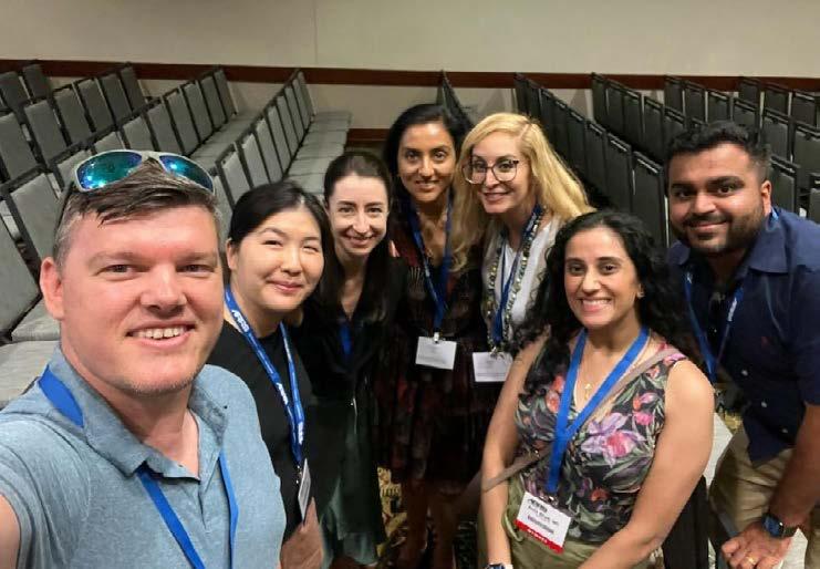
Image from the ARRS meeting. Left to right: Dr. Jared Weinfurtner, Dr. Amie Lee, Dr. Noemi Schmidt, Dr. Uzma Waheed, Dr. Dana Ataya, Dr. Asha Bhatt, and Dr. Maulik Patel. (Photo courtesy of Dr. Dana Ataya.)
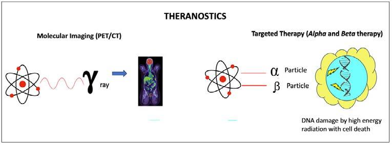
Figure. Theranostics.
Results of the VISION trial published in 2021 found that radioligand therapy with lutetium Lu 177 PSMA-617 prolonged imaging-based progression-free survival and overall survival when added to standard care in patients with advanced PSMApositive, metastatic, castration-resistant prostate cancer.13,14 The success of PSMA-based diagnostic imaging and therapy has set the stage for a similar model in breast cancer management.
Fluoroestradiol F 18
For breast cancer, two radiotracers, fluoroestradiol F 18 (18FES) and fibroblast activation protein inhibitor (FAPI), have shown the most promising results in imaging and treatment. 18FES, approved by the Food and Drug Administration in 2021, is a synthetic estrogen combined with fluorine 18, a positron-emitting isotope. It closely mimics the binding affinities of estradiol for estrogen receptors (ERs) and can noninvasively depict ER expression in the whole body. In breast cancer, in which more than 70% of tumors are positive for hormone receptors, targeted therapies like tamoxifen and aromatase inhibitors have high efficacy and fewer adverse effects than traditional chemotherapy. Likewise, 18FES has emerged as a targeted radiotracer with superior sensitivity for hormone receptor–positive breast cancer and can be used in conjunction with 18FDG PET/CT to assess the heterogeneity of breast metastases and overall tumor metabolism.15,16
Data demonstrate that 18FES PET/CT will have the most clinical impact in patients with advanced breast cancer, specifically those with stage III and IV disease and some with stage IIB disease. Clinical indications for 18FES PET/CT include assessing metastatic lesions that are difficult to biopsy, guiding therapy for metastatic disease, and detecting ER-expressing sites when other imaging results are equivocal. Additional clinical indications are being explored and include assessing ER-expressing lesions in lieu of biopsy and staging invasive lobular carcinoma and low-grade, ER-expressing invasive ductal carcinoma.17 A recent metaanalysis of seven studies assessing 18FES PET/CT found a pooled sensitivity of 86% and a specificity of 85%,18 and other studies have shown a diagnostic specificity superior to 18FDG PET/CT for ER-positive breast cancer.15 18FES PET/CT also has superior detection for metastatic disease in the brain and skull because of the low background uptake of 18FES in the brain19 and has greater detection of bone metastases compared with 18FDG PET/CT for patients with invasive lobular carcinoma.20 Perhaps one of the most promising applications of 18FES PET/CT is its ability to differentiate
ER-positive from ER-negative metastatic sites, given the heterogeneity of metastases despite the hormone receptor status of the primary tumor. Thus, 18FES PET/CT can be used to predetermine which patients will benefit from endocrine therapy before treatment failure occurs.16
Fibroblast Activation Protein Inhibitor
FAPI is a new and promising targeted radioligand that works by binding to cells in the surrounding microenvironment of tumor cells. The tumor microenvironment is a complex and dynamic collection of cellular and noncellular components in and around a tumor.21 It is pivotal in tumor development, progression, and response to therapies. In addition to cancer cells, the tumor microenvironment contains noncancer cells, including cancerassociated fibroblasts, which promote tumor development and progression and influence therapeutic response.22 FAP is a cell surface protein overexpressed by cancer-associated fibroblasts, making it a potential target for molecular imaging and theranostics.
FAPI PET/CT uses radioactive tracers, such as gallium Ga 68 FAPI-06, that bind to FAP, allowing for visualization and quantification of FAP-expressing cancer-associated fibroblasts in various solid tumors.23 Early data have shown that breast cancer has high FAPI uptake and that FAPI PET/CT has superior detection compared with 18FDG PET/CT due to the high maximum standardized uptake value of FAPI. FAPI not only provides insight into the tumor microenvironment but also can be used as a therapeutic radioligand. This innovative approach involves combining FAPI with radioactive isotopes such as lutetium 177 and actinium 225, which emit ionizing radiation. Ongoing research and clinical trials are further exploring the efficacy, safety, and optimal use of FAPI radioligand targeted therapy in various cancer types.24,25
Conclusion
With the development of new cancer-specific PET radiotracers, the role of molecular and metabolic imaging in cancer management is progressing. The clinical successes of PSMA PET in diagnosis and treatment of prostate cancer have paved the way for potential applications in breast cancer patients. Thus far, 18FES, Ga 68 FAPI-06, and FAPI have shown the most promise, with data supporting their value in detecting disease, clarifying equivocal findings, and assessing response to treatment. It is essential that breast imaging radiologists stay up to date with advances in nuclear medicine. Continued development and large clinical trials are crucial to establish the clinical role for these newer agents. The new radiotracers have raised the possibility for targeted breast cancer imaging and theranostics and will likely be integral in breast cancer management in the near future.
Continued on page 27>
Legislative and Regulatory Update: An Eventful 2023 Thus Far!
By Amy K. Patel, MD
The year 2023 has proven to be incredibly eventful on the radiology advocacy (radvocacy) front! The Find It Early Act was reintroduced on May 8, 2023, by Representatives DeLauro (D-CT) and Fitzpatrick (R-PA).
This act includes sweeping federal legislation requiring private payer, Medicare, Medicaid, and TRICARE coverage of screening and diagnostic imaging without copay or deductible. The following is an excerpt from the bill:
(A) [W]ith respect to an individual who is at increased risk of breast cancer (as determined in accordance with the most recent applicable American College of Radiology Appropriateness Criteria or the most recent applicable guidelines of the National Comprehensive Cancer Network) or with heterogeneously or extremely dense breast tissue (as defined by the Breast Imaging Reporting and Data System established by the American College of Radiology), screening and diagnostic imaging (with no limitation applied on frequency) for the detection of breast cancer, including 2D or 3D mammograms, breast ultrasounds, breast magnetic resonance imaging, or other technologies (as determined in accordance with such applicable criteria or guidelines); and
(B) [W]ith respect to an individual who is not described in subparagraph (A) and who is determined by a health care provider (in accordance with such most recent applicable criteria or guidelines) to require screening or diagnostic breast imaging by reason of factors, including age, race, ethnicity, or personal or family medical history, screening and diagnostic imaging (with no limitation applied on frequency) for the detection of breast cancer, including 2D or 3D mammograms, breast ultrasounds, breast magnetic resonance imaging, or other technologies (as determined in accordance with such applicable criteria or guidelines).1
Nearly contemporaneously, on June 6 the Access to Breast Cancer Diagnosis Act of 2023 was introduced by Representatives Allred (D-TX), Dingell (D-MI), Fitzpatrick (R-PA), and Wasserman Schultz (D-FL). This act requires the same coverage but only of private insurance companies. Some advocacy groups, such as Susan G. Komen, are supporting passage of both bills
but are placing an emphasis on this bill because the organization feels it has a better chance of passage given its narrower scope of coverage requirements.
The ACR Radiology Advocacy Network recently organized a call to action to radiologists and radiation oncologists asking them to contact their elected federal officials to support Medicare reform/stabilization through HR 2474, with bipartisan cosponsors Representatives Ruiz (D-CA), Bucshon (R-IN), Bera (D-CA), and Miller-Meeks (R-IA), all of whom are doctors of medicine. This introduced legislation applies an automatic inflationary update to Medicare physician payments. Approximately 1556 members took action in response to this call to action. If you were one of them, we thank you!
Very recently, the ACR Radiology Advocacy Network organized another call to action, once again asking radiologists and radiation oncologists to contact their elected federal officials to support HR 3674, the Providing Relief and Stability for Medicare Patients Act of 2023, introduced with bipartisan cosponsors Representatives Bilirakis (R-FL), Cárdenas (D-CA), Murphy (R-NC), and Davis (D-IL), to help mitigate practice expense cuts. We appreciate your efforts! It takes only a few seconds to respond to these calls to action, which are so imperative to help change policy for our patients and profession.
We also just concluded a successful 2023 ACR annual meeting, which included advocating on Capitol Hill Day to address Medicare physician payment reform, issues with implementation of the No Surprises Act, and a legislative solution for implementation of the proposed amendment to the Protecting Access to Medicare Act. Capitol Hill Day drew over 400 radiologists and radiation oncologists. Thank you all for your time and efforts!
I am pleased to announce that RADPAC, the ACR’s nonpartisan political action committee, nearly doubled its fundraising at the ACR annual meeting this year compared with last year. Just as
To save lives and minimize the impact of breast cancer. .....
Amy Patel, MD
important as our lobbying efforts on Capitol Hill Day, this effort is crucial for supporting elected federal officials who vote in favor of proradiology legislation. We also solicited 100% state councilor participation from multiple states. RADPAC currently ranks fourth in contributions among US medical political action committees (following political action committees for anesthesiology, orthopedic surgery, and dentistry), and our hope is to move up to first or second place, as we have been in years past. We will need your help to achieve this goal in the years to come. To learn more, please visit the Radiology Advocacy Network website.
As many of you are aware, the US Preventive Services Task Force (USPSTF) issued draft recommendations for screening mammography that lowered the starting age to 40 years but maintained the recommendation for biennial screening. The ACR and SBI have re-
leased a statement and submitted a joint letter urging the USPSTF to recommend screening mammography annually beginning at age 40 years for women with average risk. The USPSTF final recommendations are now in progress.
As we continue to advocate for access to patient care and fair reimbursement for the services we provide, coalition building and synergy are imperative. We look forward to your participation in our radvocacy efforts for the remainder of 2023. As always, thank you for all that you do for the house of radiology and, most importantly, for our patients.
Reference
1. Find It Early Act, HR 3086, 118th Cong (2023). Accessed June 29, 2023. https://www.congress.gov/bill/118th-congress/house-bill/3086
What’s New in the News: New Breast-Specific Radiotracers in Positron Emission Tomography: The Future of Molecular Imaging in Breast Cancer Diagnosis and Therapy (continued from page 25)
References:
1. NCCN guidelines. National Comprehensive Cancer Network. Accessed June 29, 2023. https://www.nccn.org/guidelines/category_1
2. Kumar R, Rani N, Patel C, Basu S, Alavi A. False-negative and false-positive results in FDG-PET and PET/CT in breast cancer PET Clin. 2009;4(3):289-298. doi:10.1016/j.cpet.2009.09.002
3. Chakraborty D, Basu S, Ulaner GA, Alavi A, Kumar R. Diagnostic role of fluorodeoxyglucose PET in breast cancer: a history to current application PET Clin 2018;13(3):355-361.
4. Ulaner GA. PET/CT for patients with breast cancer: where is the clinical impact? AJR Am J Roentgenol. 2019;213(2):254-265. doi:10.2214/AJR.19.21177
5. Vogsen M, Naghavi-Behzad M, Harbo FG, et al. 2-[18F]FDG-PET/CT is a better predictor of survival than conventional CT: a prospective study of response monitoring in metastatic breast cancer Sci Rep. 2023;13(1):5552.
6. Koolen BB, Vegt E, Rutgers EJ, et al. FDG-avid sclerotic bone metastases in breast cancer patients: a PET/CT case series Ann Nucl Med. 2012;26(1):86-91.
7. Evangelista L, Panunzio A, Polverosi R, et al. Early bone marrow metastasis detection: the additional value of FDG-PET/CT vs. CT imaging Biomed Pharmacother. 2012;66(6):448-453.
8. Groheux D, Hindié E, Giacchetti S, et al. Triple-negative breast cancer: early assessment with 18F-FDG PET/CT during neoadjuvant chemotherapy identifies patients who are unlikely to achieve a pathologic complete response and are at a high risk of early relapse J Nucl Med. 2012;53(2):249-254.
9. Rousseau C, Devillers A, Sagan C, et al. Monitoring of early response to neoadjuvant chemotherapy in stage II and III breast cancer by [18F] fluorodeoxyglucose positron emission tomography J Clin Oncol 2006;24(34):5366-5372.
10. Wang CL, MacDonald LR, Rogers JV, Aravkin A, Haseley DR, Beatty JD. Positron emission mammography: correlation of estrogen receptor, progesterone receptor, and human epidermal growth factor receptor 2 status and 18F-FDG AJR Am J Roentgenol. 2011;197(2):W247-W255.
11. Hofman MS, Lawrentschuk N, Francis RJ, et al; proPSMA Study Group Collaborators. Prostate-specific membrane antigen PET-CT in patients with highrisk prostate cancer before curative-intent surgery or radiotherapy (proPSMA): a prospective, randomised, multicentre study Lancet. 2020;395(10231):1208-1216.
12. Hofman MS. ProPSMA: a callout to the nuclear medicine community to change practices with prospective, high-quality data J Nucl Med. 2020;61(5):676-677.
13. Sartor O, de Bono J, Chi KN, et al; VISION Investigators. Lutetium-177PSMA-617 for metastatic castration-resistant prostate cancer N Engl J Med 2021;385(12):1091-1103.
14. Hofman MS. Bringing VISION to nuclear medicine: accelerating evidence and changing paradigms with theranostics J Nucl Med. 2021;62(12):1710-1711. doi:10.2967/jnumed.121.262890
15. Liu C, Gong C, Liu S, et al. 18F-FES PET/CT influences the staging and management of patients with newly diagnosed estrogen receptor-positive breast cancer: a retrospective comparative study with 18F-FDG PET/CT Oncologist 2019;24(12):e1277-e1285.
16. Kurland BF, Peterson LM, Linden HM, Mankoff DA. FDG PET and FES PET predict PFS on endocrine therapy-response Clin Cancer Res. 2018;24(1):249-250.
17. Chae SY, Ahn SH, Kim SB, et al. Diagnostic accuracy and safety of 16α-[18F] fluoro-17β-oestradiol PET-CT for the assessment of oestrogen receptor status in recurrent or metastatic lesions in patients with breast cancer: a prospective cohort study Lancet Oncol. 2019;20(4):546-555.
18. Mo JA. Safety and effectiveness of F-18 fluoroestradiol positron emission tomography/computed tomography: a systematic review and meta-analysis J Korean Med Sci. 2021;36(42):e271.
19. Appropriate use criteria for estrogen receptor-targeted PET imaging with 16a-18F-fluoro-17β-fluoroestradiol. Society of Nuclear Medicine & Molecular Imaging. Accessed June 29, 2023. https://www.snmmi.org/ClinicalPractice/content. aspx?ItemNumber=42023
20. Ulaner GA, Jhaveri K, Chandarlapaty S, et al. Head-to-head evaluation of 18F-FES and 18F-FDG PET/CT in metastatic invasive lobular breast cancer J Nucl Med. 2021;62(3):326-331.
21. Marsh T, Pietras K, McAllister SS. Fibroblasts as architects of cancer pathogenesis Biochim Biophys Acta. 2013;1832(7):1070-1078.
22. Dumont N, Liu B, Defilippis RA, et al. Breast fibroblasts modulate early dissemination, tumorigenesis, and metastasis through alteration of extracellular matrix characteristics Neoplasia. 2013;15(3):249-262.
23. Kratochwil C, Flechsig P, Lindner T, et al. 68Ga-FAPI PET/CT: tracer uptake in 28 different kinds of cancer J Nucl Med. 2019;60(6):801-805.
24. Lindner T, Loktev A, Giesel F, Kratochwil C, Altmann A, Haberkorn U. Targeting of activated fibroblasts for imaging and therapy EJNMMI Radiopharm Chem 2019;4(1):16.
25. Kelly JM, Jeitner TM, Ponnala S, et al. A trifunctional theranostic ligand targeting fibroblast activation protein-α (FAPα) Mol Imaging Biol. 2021;23(5):686-696.
MARK YOUR CALENDAR
for the 2023 SBI Summer Live Webinars
Come learn from breast imaging experts from the comfort of your own home! Live Q&A sessions with every webinar ensure you will have the opportunity to ask those burning questions relevant to your clinical practice. Our webinars also include interactive polls to help assess practice patterns across different centers—particularly useful for controversial and challenging scenarios. This summer's lineup includes:
August 15
Navigating Imaging AI Bias
August 22
Imaging Algorithms and Pathologies of the Breast During Pregnancy & Lactation
August 29
Missed Cancers: Pearls & Pitfalls
Please visit the SBI Calendar of Events at www.sbi-online.org for a complete listing of events.