The Member Newsletter of the Society of Breast Imaging
INSIDE THIS ISSUE:
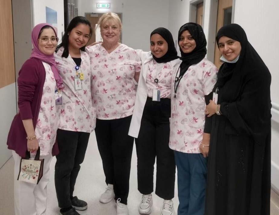
• Breast Cancer Awareness at Ground Root Level
• SBI’s Upcoming 40th Anniversary: History of Screening Mammography Regulations
• Celebrating the Life of Dr. G. W. Eklund
• Lessons to Learn From Wellness Initiatives in Aviation Industry
EDITOR:
Nidhi Sharma
ASSISTANT EDITORS:
Randy Miles and Shinn-Huey Shirley Chou
TECHNOLOGISTS’ COLUMN:
Robyn Hadley and Sarah Jacobss
WHAT’S
Eleanor DiBiasio and Pamela J. DiPiro
MEMBERS IN TRAINING:
Anita
WELLNESS COLUMN:
Sarah
THE
and Claudia Cotes
PERSPECTIVE:
Hannah Perry and Danielle Sharek
LEGISLATIVE UPDATES:
Amy Patel
CANADIAN CORNER:
Supriya Kulkarni
Linda
Moy,
MD, FACR, FISMRM, FSBI President, Society of Breast Imaging
OUR SBI MISSION:
For members to be expert and authoritative breast imagers working in supportive practice environments who advance the highest quality of breast care via early detection, diagnosis, and treatment.
OUR SBI VALUES:
Patient-centered and evidence-based care
Excellence in education Scientific integrity
Collaboration and collegiality
Respect for diversity and inclusiveness
President’s Column
October is a wonderful time to reflect on the energy and mission of the SBI. It is Breast Cancer Awareness Month. Radiologists, medical physicists, researchers, and technologists in our community participate in many activities to promote breast imaging. Many practices and hospitals have outreach programs to advocate for screening mammography and to engage in fund-raising events for breast cancer research and innovative programs to improve access to screening mammography.
We are fortunate to have many breast imaging radiologists who participate in television and radio shows, newspapers, blogs, and social media programs to discuss the importance of screening mammography. There is irrefutable evidence that mammography saves lives and minimizes the impact of breast cancer. More importantly, our members are compassionate and patient centric and practice the highest-quality breast care.
There is also an undeniable energy at the SBI. Our committees are composed of dedicated individuals who develop the best educational content for our members. The CME & SAM Committee organized topical and informative webinars over the summer. Our Patient Care and Delivery Committee has several initiatives focusing on workflow issues in our practices and methods to improve practice efficiency and address burnout. Dr. Michael Cohen is also diligently working as the new editor of the Journal of Breast Imaging. Dr. Peter Eby and his program committee have developed an exciting program for the 2025 SBI symposium in Colorado Springs.
As we navigate through October, it’s a perfect time to consider new ways to engage with the SBI. Your voice, your perspective, and your expertise are vital to us. I encourage you to volunteer to serve on our committees. The SBI is an inclusive and welcoming community, and we value your contribution.
Linda Moy
Linda Moy, MD, FACR, FISMRM, FSBI President, Society of Breast Imaging
Editor’s Note
By Nidhi Sharma, MD
Imagine that Dorothy, upon landing in Oz, was actually told all the experiences and personalities that she would encounter. Imagine that she was given the option to take a direct route to the Wizard’s home rather than follow an unknown path and have to make many decisions along the way. Imagine that Luke Skywalker was told as a preadolescent that Darth Vader was his father and how the rest of his life was going to play out
I often played video games as a young kid. As a very intense competitor, I always wanted to win, whether that was knocking out Mike Tyson, finding the key to the castle, beating the A’s in R.B.I. Baseball, or destroying the alien heart on the final stage of Contra. Yet when I was offered a cheat code of some kind from a friend, as tempting as it was, it wasn’t nearly as fun or rewarding as doing it on my own—grinding it out, playing it 1000 times, learning the algorithm to beat Soda Popinski, getting a hit off Eckersley, and the like.
In this era of instant gratification, artificial intelligence, exposure, and access to tools that simplify work or pursuits like academic publishing, it is ever important to remind ourselves that success comes from multiple factors coming together: talent, tenacity, humility and desire.
If we acknowledge that we have talent in our community, we quickly arrive at one-fourth of the key ingredients for success. The second variable, tenacity, is a growth mindset grounded in the concept of resilience. Resilience is learned and built through doing and failing in real time, not simply a theoretical or philosophical concept. Now let’s factor in humility, arguably a human quality that is exceptionally difficult to teach. In my experience, most of the time it is learned through failure, yet again. Personally, I have learned the most through my fair share of failures, a list much longer than my successes, and I’m glad each one gave me an opportunity to pivot and grow.
And then there is desire. On its face, the word desire appears to be similar to tenacity, but it’s specific to the kind of desire that’s needed to be successful. The desire to make the world better involves correctly focused motivation, selfless spirit to service, and a big
heart for others who may be very different from you. In other words, it’s the desire to make a positive dent in the universe. If we approach our next generation of breast imagers with these few factors in mind, we’re bound to succeed as a community!
Our fall issue theme is “Breast Cancer Awareness at Ground Root Level.” October is the perfect time of year to focus on this topic as we all work in our individual practice settings to create clinical awareness of the importance of screening mammography for our patients. We have so many amazing, inspiring organizations working toward this core cause. With the latest Food and Drug Administration regulations on breast density notification, we have leaders from two organizations with focused efforts in this arena, DenseBreast Info and My Density Matters, sharing their strong work. A pertinent part of awareness is the education of our staff and trainees, and our 2017 SBI honorary fellow, Louise Miller, has left no stone unturned in concerted efforts to spread the knowledge worldwide. We also highlight the work being done at Mammography Educators and learn from their shared experiences, setting a great example for the rest of us. We continue to celebrate the upcoming 40th SBI anniversary in our newsletter issues this year, with Dr. Monticciolo sharing the historic legislative changes in breast imaging. We have many more exciting articles, and I hope you enjoy reading them as much as our newsletter team did in compiling this issue.
I would love to hear if you have any new ideas to share with the community. Please reach out to me at nidhisharma31@gmail.com
Thank you for taking time out to read this edition of SBI News. I hope you all get some time to relax and spend with loved ones this upcoming holiday season. We all have so much to be grateful for!
Nidhi Sharma, MD
How to Advocate at Your State Legislature
By Amy K. Patel, MD
Do research to find state legislators who may be interested in championing breast imaging legislation. These might include junior legislators who are looking to make their mark in the legislature and established legislators who are known to be pro medicine and pro radiology. If you are not sure who these people are, contact your state radiological society’s lobbyist or, if you do not have one, your state medical association’s lobbyist.
You could also contact both lobbyists to get their individual perspectives. They can provide invaluable insight on how to approach these legislators and how your state legislature passes legislation, which varies significantly from state to state. It is beneficial to identify both a representative and a senator who can lead efforts in their respective chambers.
Once you have identified legislators, contact them either via email or by calling their office. If you are able to obtain their cell phone number and this is their preferred method of communication, you can contact them via text or phone call. Keep your correspondence brief while conveying to them who you are, why you are reaching out, and that you would appreciate their time to discuss the issue by phone, by video chat, or in person. Then follow through with the next step (eg, call them if they prefer to speak over the phone, send a virtual meeting invitation, or go to a place of their choosing to meet in person).
When you meet, give them a fact sheet or packet that they can take back to their office for legislative assistants who did not attend the meeting. If you spoke over the phone, email them the fact sheet or packet afterward. The fact sheet or packet should include your reasons for wanting to embark on the legislative process, all the states that have similar legislation, and the language in existing legislation.
During your in-person or video meeting, look engaged, smile, be enthusiastic, make eye contact, and do not slouch. When speaking on the phone, make sure you use an enthusiastic and friendly tone.
Follow up on correspondence in a timely fashion. This follow-up might include sending proposed language that could be introduced in the state legislature or a modification of existing legislation.
If the legislators become sponsors of the proposed legislation, contact your state radiological society’s lobbyist and/or your state medical association’s lobbyist to let them know and to help garner support. Support letters that the sponsoring elected officials can submit during hearings on new proposed legislation can be extremely helpful, especially if the letters are from the medical association in your state and other medical specialty societies. Be
sure you also have the support of your state radiological society; there is immense strength in numbers. Ask the societies’ lobbyists to help you track your proposed legislation. Pieces of legislation are often added to different bills that move to different hearings and can therefore move at an incredibly fast pace, making them hard to track without a contact person who is at your state capitol regularly, knows what is happening, and is able to effectively track bills. The same advice applies if you are partnering with lobbyists of other organizations, such as Susan G. Komen.
Offer your time to testify in both House and Senate committee hearings if the legislation is going to be discussed at a hearing that has significant jurisdiction over health care and is the next step for the bill to advance. If you cannot attend in person, submit a written testimony. In your testimony, include who you are, why you are present, the legislation you are advocating for (whether it is a House or Senate bill and the number), breast cancer statistics, and breast cancer patient stories that are heartfelt and relatable to committee members. Just providing statistics is often not enough and can result in losing the interest of committee members. Stories that tug on the heartstrings are a must.
From this point, make yourself available to the collaborating lobbyists and legislators in case the language needs to be modified, they need additional information, and so forth. The sponsors of these bills will be working behind the scenes to make sure the bills advance.
If your legislation does not pass on the first or even second try, do not give up! Reaching out to others who have been successful in passing legislation can help tremendously, as can gauging the climate of that legislative session year. Passing legislation will be inherently easier in some years than in others, depending on whether certain upcoming issues may overshadow what you are trying to accomplish. The bottom line is to not give up! Regroup for the next legislative session, if need be, either with the same legislators or different ones who want to support and champion the legislation.
Good luck!
Amy K. Patel, MD
DenseBreast-info.org
By JoAnn Pushkin
DenseBreast-info.org (DBI) was launched in 2015 to educate patients and clinicians about the screening and risk implications of dense breast tissue and to inform discussions about supplemental screening. The site’s Medical Advisory Board, led by Dr. Wendie Berg, is composed of internationally recognized, published experts in breast imaging and obstetrics/gynecology. The website supports advocacy, education, legislation, and policy conversations. It is now the world’s leading website on the topic and is on track to host over 1 million website visits in 2024 alone.
In 2010, as the number of patient advocates fighting for density “inform” laws began to rise, I wrote to the US Food and Drug Administration (FDA) to request consideration of a national dense breast reporting standard in the lay letter women receive after their mammograms. In 2011, the topic was added to the National Mammography Quality Assurance Advisory Committee meeting agenda. I testified in support, and the committee agreed that women should indeed be provided this information. The requirement finally went into effect on September 10, 2024, and all mammography facilities now must provide a patient with either a “not dense” or “dense” standardized notification from the FDA.
Now that all women in the United States will be informed definitively if they have dense breasts, there will be new patient-clinician conversations on what that means in terms of breast screening. The next educational frontier for DBI is helping patients and clinicians understand risk and considerations for supplemental screening.
Patient Education
Patients will now receive notification that their breasts are either not dense or dense, and many women will be learning they have dense breasts for the first time. Two new DBI web pages have been launched to help them understand the notice and to prepare them for what it means. Is My Mammogram Enough? details breast cancer risk factors, and Screening Tests After a Mammogram (currently the website’s most popular page) features a table showing the average number of cancers detected by the various types of breast screening test.
To reach patients, the DBI team speaks on the topic, participates in webinars, attends community events, chats with and provides resources to the media, writes blog posts, and has a robust social media calendar to highlight educational tools. Website content within the For Patients tab is extremely popular and available in more than 25 languages. To assure medical accuracy, all translations are by breast radiologists who are native language speakers. Because Spanish is the second most common language in the United States, a dedicated suite of content, including videos, is available in Spanish at SenosDensos.org
Clinician Education
To help clinicians prepare for patients’ questions, the website provides, among other educational tools, an educational series (six short videos) on topics such as the implications of dense tissue, supplemental screening, risk assessment, screening guidelines, and insurance coverage. Additionally, we have collaborated with the American Society of Radiologic Technologists on a continuing education credit opportunity titled “Dense Breasts, Supplemental Screening and New MQSA Reporting Requirements.” Videos and courses are available on this web page Because one-size-fits-all screening protocols are being transitioned to those that include personal risk assessment, available tools also include a risk model tutorial, a screening decision support flowchart titled “Who Needs More Screening?” and a new table specifically for clinicians, “Cancer Detection by Screening Method in Dense Breasts.”
Is Supplemental Screening Covered?
Currently 33 states have enacted laws requiring some level of expanded insurance coverage for breast imaging beyond mammography (see the DBI insurance map). As was true for the previous density inform laws, the individual state insurance laws are inconsistent. They vary in what is covered, the qualifications for coverage, and whether copays/deductibles apply. Additionally, many types of plans (eg, self-funded plans, out-of-state plans, and federal plans like Medicare) are exempt from state insurance laws. Unfortunately, insurance coverage by individual state laws is usually required “when medically necessary,” a determination that an individual insurance carrier may not agree with or approve.
Federal legislation was needed to address these inherent loopholes in state insurance laws. To ensure that all health insurance plans cover screening and diagnostic breast imaging with no out-of-pocket costs for women with dense breasts or at higher risk for breast cancer, DBI worked with the office of Representative Rosa DeLauro on the draft and introduction of the federal Find It Early Act. DBI has been active on Capitol Hill garnering support for this important legislation as we gear up for its reintroduction in the next Congress. To learn how to contact your representative to ask for their cosponsorship of the bill, see our Find It Early Act web page
To save lives and minimize the impact of breast cancer. .....
JoAnn Pushkin
World Dense Breast Day
The implications of dense breast tissue are not limited by US borders. The European Society of Breast Imaging now recommends that women be informed about their breast density and that supplemental screening be offered to women aged 50 to 70 years who have extremely dense breasts. To expand awareness, DBI has gone global in its educational outreach. Sponsored and launched by DBI and officially listed on the National Day Calendar, World Dense Breast Day is recognized annually on the last Wednesday of September. It is a one-day social media blitz to raise awareness about dense breasts and share medically sourced educational tools. To
educate people in every time zone in every country, on September 25th, DBI launched over 300 posts in a single 24-hour period. Education on the topic is clearly welcome. We had participants from 104 countries, and over 16.8 million people saw, read, and shared orld Dense Breast Day posts!
Please visit DenseBreast-info.org for many more educational resources and videos.
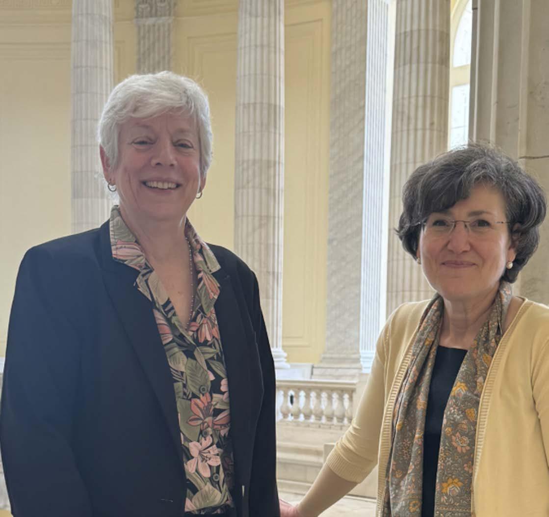
Lobbying for the Find It Early Act: DBI Board of Directors members Laurie Scofield and JoAnn Pushkin.
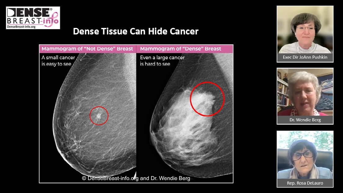
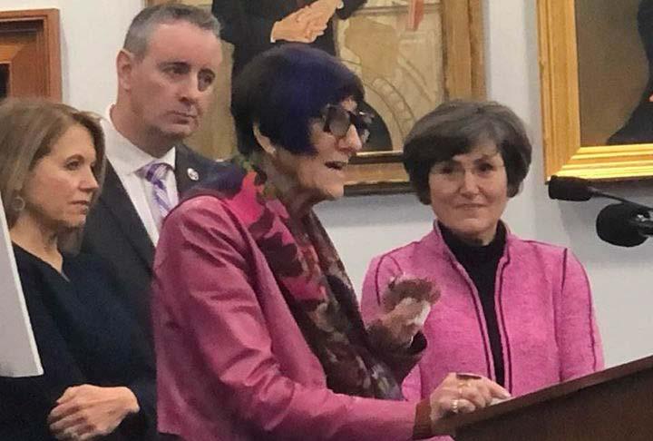
Screenshot from a national press briefing on the FDA dense breast reporting rule with DBI’s JoAnn Pushkin, Dr. Wendie Berg, and Rep. Rosa DeLauro.
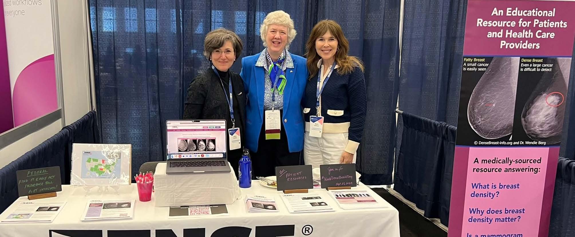
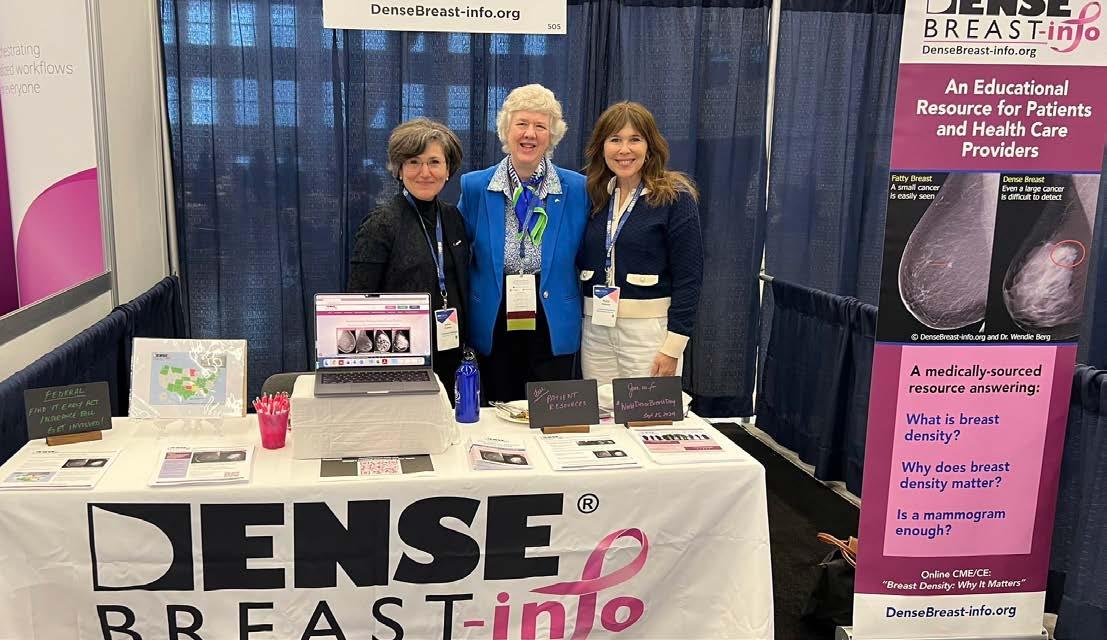
DBI booth and team at the SBI annual meeting. Left to right: JoAnn Pushkin, Dr. Wendie Berg, and Dr. Robin Seitzman.
Introduction of the Find It Early Act on Capitol Hill. Left to right: Katie Couric, cosponsor Rep. Brian Fitzpatrick, cosponsor Rep. Rosa DeLauro, and JoAnn Pushkin.
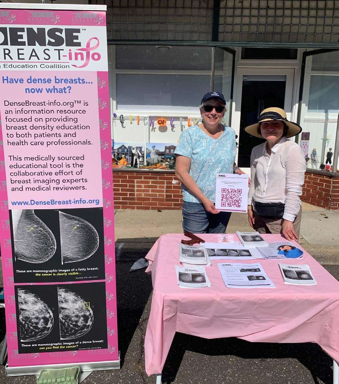
Health fair community outreach with DBI Board of Directors members JoAnn Pushkin and Laurie Scofield.
Stepping Off Autopilot May Hold the Key for Physician Wellness
By Claudia C. Cotes, MD
After summer travels and visits, an idea came to mind—one that feels particularly close to home. My brother, a commercial pilot, visited us and as usual, we dived into deep conversations about our respective professions. Although our fields are different, they are in many ways similar: difficult hours, shifts, and a high degree of responsibility. While discussing wellness and work-life balance, he mentioned something that has stuck with me: “Regulations in aviation are written with blood.” It’s a harsh reality, and it highlights a serious question: how many lives must be lost before meaningful change is implemented?
Fatigue and burnout are significant issues that compromise safety in both aviation and medicine. In aviation, a single mistake by a fatigued pilot can have catastrophic consequences, leading to the loss of hundreds of lives in an instant. This potential for disaster has driven the aviation industry to adopt stringent regulations aimed at mitigating the risks associated with pilot fatigue. The medical field deals with its own version of burnout crisis. Studies have shown that physician fatigue significantly increases the likelihood of medical errors, with burnout doubling the risk of serious mistakes.1 How are we dealing with this crisis in comparison to our aviation counterparts?
Medical errors may account for approximately 250,000 deaths annually in the United States, making them the third leading cause of death.1 In contrast, air travel is one of the safest modes of transportation, with only approximately 1300 deaths per year.2 Burnout unfortunately affects both pilots and physicians, with data showing that up to 40% of pilots experience burnout, while more than 50% of in-training and practicing physicians in the United States report symptoms of this condition.3,4
The aviation industry has set a high standard for addressing the dangers of fatigue through proactive regulation. The tragic crash of Colgan Air Flight 3407 in 2009, partially attributed to pilot fatigue, led the Federal Aviation Administration (FAA) to implement new regulations under 14 CFR Part 117.5 These rules mandate rest periods, limit flight duty hours, and establish cumulative rest requirements to ensure pilots are well rested and alert. Specifically, pilots must have at least 10 hours of rest between duty periods, including a minimum of 8 hours of uninterrupted sleep. Additionally, they are limited to 60 flight duty
hours in any 7-day period, 190 hours in any 28-day period, and a maximum of 9 to 14 hours of flight duty within a 24-hour period.
Unfortunately, the medical field lags in this regard. Although the Accreditation Council for Graduate Medical Education (ACGME) introduced regulations in 2003 to limit physician-in-training work hours, these rules are not as stringent as those in aviation. Resident physicians are capped at 80 hours of work per week and are allowed a maximum of 24 continuous hours of patient care, with an additional 4 hours for care transition. After a 24-hour shift, residents must have 14 hours off before their next shift.6 Unlike the FAA, the ACGME does not impose cumulative duty hour limits but does require one day off each week, averaged over four weeks. While these regulations are a positive step, they fall short of the protections established for pilots.
Many justify the situation by saying, “At least we’ve made some changes.” And it is this “at least” that troubles me. Why do we accept “at least” as enough? Why are pilots federally regulated, but practicing physicians are not? Why does the responsibility for managing fatigue and burnout frequently fall on individual physicians and the institutions that employ them rather than being addressed through comprehensive federal regulation, if we know the consequences of burnout and fatigue and their potential relationship to medical errors? The FAA was created in response to a clear need for centralized regulation to protect public safety by those who recognized that without strict oversight, the risks were too great. Why are we, as physicians, not doing the same?
We need to step off autopilot and demand the same level of regulatory oversight that has made aviation one of the safest industries in the world. This is not just about protecting our patients; it’s also about safeguarding ourselves. We must advocate for stronger regulations that prioritize physician well-being, recognizing that our health is inseparable from the safety and care of our patients. As we’ve all heard, “Put your own oxygen mask on first.”
Continued on page 11>
Claudia C. Cotes, MD
Helping Your Technologists, Part 2: Elevating Feedback With Value and Impact
By Sarah Jacobs, BS, RT(R)(M)(CT); Robyn Hadley, RT(R)(M)
Part 1 of this series, published in the summer 2024 issue of SBI News, provided tips for troubleshooting mammographic positioning. Part 2 explores essential strategies for delivering effective image quality assessments, ensuring that feedback is meaningful and well received.
Recently, Hadley and Jacobs conducted an independent unpublished survey of mammography technologists in the United States. The survey revealed that 89% of technologists received routine feedback pertaining to image quality. Of the technologists who received routine image quality feedback, 39% received the feedback both for images of excellent quality and for those of suboptimal quality. An analysis of positive and negative feedback in the workplace by Goller and Späth showed that receiving positive feedback had a favorable impact on subsequent performance, whereas negative feedback did not have an effect.1 The recent survey of technologists also revealed that technologists prefer to receive feedback via email and/or verbally in a confidential setting, rather than receiving feedback in nonconfidential settings or among their peers.
Although the skills for acquiring quality images are mainly entrusted to modality-specific technologists, the radiologist is ultimately responsible for image quality.2 The ACR technical standard states, “The physician performing the official interpretation must be responsible for the quality of the images being reviewed.”3 According to the US Food and Drug Administration, “[t]he LIP [lead interpreting physician] is viewed as the individual most responsible for ensuring image quality is continuously maintained by the facility.” 4 Although interpretation and final image quality are the interpreting radiologist’s responsibility, the imaging technologist “acts as an agent through observation and communication to obtain pertinent information for the physician to aid in the diagnosis and treatment of the patient.” 5 Elevating skills to continually optimize image quality requires a team effort and is essential for providing a final diagnostic study that is acceptable for interpretation. This article presents potential tools for delivering feedback effectively and aims to minimize friction, enhance receptivity, foster strong team unity, and promote image quality excellence.
Importance of Standardized Positioning Techniques and Protocols
Creating and establishing written protocols for imaging acquisition and quality are essential and can help ensure that breast imaging teams have the same goal in mind. Establishing and implementing
standardized positioning techniques are crucial when setting expectations for image quality and can help technologists effectively troubleshoot suboptimal images. Using standardized positioning techniques can also increase image reproducibility, contributing to earlier cancer detection. Image quality criteria benchmarks can be met more frequently with the implementation of standardized positioning techniques.6 By placing a copy of the protocols in each examination room and at technologists’ workstations, radiologists can ensure that protocols are readily accessible during examinations and when preparing for mammographic procedures.
Delivering Image Quality Feedback
Asking a patient to return for additional images when the initial examination was suboptimal can be a considerable inconvenience. Complacency and friction fears are common reasons to offer routine feedback both for images of excellent quality and for images that need improvement. Dr. Kuehn-Hajder (University of Minnesota Physicians) said, “Offering balanced feedback is important in maintaining quality. Too frequent/too negative can backfire. Too infrequent results in complacency.”
Establishing clear expectations and engaging in regular discussions about image quality are crucial. Limiting feedback to only a few times per year can often be perceived as punitive or inconsequential by technologists. Providing balanced feedback on a regular basis helps foster a team-oriented atmosphere and upholds high quality standards.
Ensuring that feedback is offered to technologists in a way that reduces friction fears and increases receptivity is key. Feedback that technologists see as valuable and driven by leaders who are genuinely invested in their growth can lead to significant positive outcomes.
• Ask technologists about their preferred methods for receiving both positive feedback and suggestions for improvement.
Continued on page 10>
Robyn Hadley, RT(R)(M) Sarah Jacobs, BS, RT(R)(M)(CT)
Technologists’ Column: Helping Your Technologists, Part 2: Elevating Feedback With Value and Impact
(continued from page 9)
• Start with positive acknowledgement, highlighting areas where the technologist excels. This can include areas such as communication skills and patient care in addition to image criteria metrics.
• Be specific with examples of how the change could enhance the image.
• Emphasize the benefit and empower the technologist to see the value in making the adjustments.
• Leave room for dialogue. Framing the feedback as a shared effort to improve the study encourages collaboration. Dr. Daly (Bronson Healthcare, Michigan) suggested, “Start by asking about a specific case: tell me about this patient and what challenges you encountered. Ask them what they think about the study and how they would assess the images; then offer suggestions for improvement.” 7
• Encourage technologists to ask questions or share suggestions when facing challenging patient scenarios, and remind them that their input is welcome.
Create a Lead Technologist Alliance
Time and resources within imaging departments must be strategically allocated, given the continuously growing workloads and often understaffed departments. Lead mammography technologists can play a vital role in maintaining a clinical image quality review program that is educational and beneficial. Clinical image quality review programs maintained and managed by the lead mammography technologist can be an effective way to use resources. Key areas of focus for such programs include the following:
• Clear, written expectations and protocols for image quality criteria
• Adequate education and training of the designated lead technologist:
- Solid understanding of standardized positioning techniques and correlative anatomy to effectively troubleshoot imaging
- Knowledge and understanding of image criteria and the radiologist’s expectations
- Core understanding of electronic reporting and artificial intelligence (AI) software system reporting, if applicable
- Ability to provide valuable feedback and train/educate others
• A means for the lead technologist and lead interpreting physician to clearly communicate and share information in a simple, effective way on a routine basis
• A process for routine image review and time for the lead technologist to complete the task
• Goals or desirable benchmarks set for individuals and for teams to promote quality in an engaging way
• Time set aside each week, month, or quarter (per facilityestablished guidelines) for the lead technologist and radiologist to discuss image quality reports, trends in quality, and opportunities for improvement
Using Electronic Platforms for Feedback and Image Analysis
AI is a valuable option for objective image review and reporting of quality. AI removes the subjectivity from the image review process, potentially reducing friction fears and providing an optimized review of overall image quality over a period of time. This option offers a stronger understanding of the team’s overall image quality by reviewing every image rather than a select number of random cases from a specific time period. AI can also provide real-time analysis and troubleshooting assistance. Ensuring that a technologist logs in to the AI software system regularly is essential for the tool to be optimally useful. In addition to direct radiologist feedback, AI can deliver feedback through email or a shared system integrated with the electronic medical record platform. Using the reporting features within an AI platform allows leaders to effectively review and track trends in image quality. This data-driven approach provides insights into areas where image quality might be declining or improving, enabling a more focused and proactive strategy to address trends. The reporting function can help streamline troubleshooting efforts by pinpointing common challenges, thus enhancing the overall performance of the AI system and the teams using it. Using AI to boost team engagement can be highly effective. By leveraging AI-driven insights, imaging teams can focus on one specific quality metric and work collaboratively to improve in that area. Turning this into a gamified challenge in which teams compete to achieve the best results can further drive engagement, motivation, and collaboration. Offering incentives or recognition on a quarterly basis encourages continuous improvement and teamwork. This approach can help foster a positive, goal-oriented work environment.
Using electronic systems already in place, such as electronic medical records or reporting systems, can be a simple yet highly effective method for providing image quality feedback. These platforms often have built-in features that allow for seamless communication, enabling radiologists or leaders to offer real-time comments, recognition, and suggestions regarding image quality directly to technologists. Collaborating with system specialists often allows these tools to be customized for optimal efficiency. This is a simple way to foster a culture of continuous improvement through real-time, actionable feedback while using existing resources and minimizing the need for additional software.
Although image quality is ultimately the responsibility of the interpreting radiologist, achieving high-quality images begins with the initial image acquired by the technologist. A team approach incorporating regular feedback for image quality is essential for maintaining a high-performing breast imaging team.
Special thanks to the following individuals for their valuable comments and research contributions to this article: Dr. Caroline Daly, Bronson Healthcare, Michigan; Dr. Bryan Donald, Midwest Radiology, Minnesota; Dr. Jessica Kuehn-Hajder, University of Minnesota Physicians; Dr. Laurie R. Margolies, Icahn School of Medicine at Mount Sinai, New York; Dr. Anusuya Mokashi, Petaluma Valley Hospital, California; Dr. Georgia Spear, Endeavor Health/NorthShore University Health System, Illinois; and Suzanne Watring, BS, (RT)(R)(M), Endeavor Health/NorthShore University Health System.
References
1. Goller D, Späth M. “Good job!” The impact of positive and negative feedback on performance arXiv. Preprint posted online January 27, 2023. doi:10.48550/ arXiv.2301.11776
2. Parsee AA, Ahmed A. Role of medical simulation in radiology. In: StatPearls [Internet]. StatPearls Publishing; 2023. Accessed September 3, 2024. https:// www.ncbi.nlm.nih.gov/books/NBK560893/
3. ACR-AAPM-SIIM technical standard for electronic practice of medical imaging. American College of Radiology. Updated 2022. Accessed September 3, 2024. https://www.acr.org/-/media/ACR/Files/Practice-Parameters/elec-practicemedimag.pdf
4. Mammography Quality Standards Act (MQSA) Enhancing Quality Using the Inspection Program (EQUIP) frequently asked questions—facilities. US Food and Drug Administration. Updated June 2018. Accessed September 3, 2024. https://www.fda.gov/media/101293/download
5. ARRT standards of ethics. American Registry of Radiologic Technologists. Updated September 1, 2023. Accessed September 3, 2024. https://assetsus-01.kc-usercontent.com/406ac8c6-58e8-00b3-e3c1-0c312965deb2/ bbb73119-fa02-429c-be17-1f6896047106/2023%20ARRT%20 Standards%20of%20Ethics.pdf
6. Huppe AI, Overman KL, Gatewood JB, Hill JD, Miller LC, Inciardi MF. Mammography positioning standards in the digital era: is the status quo acceptable? AJR Am J Roentgenol. 2017;209(6):1419-1425. doi:10.2214/ AJR.16.17522
7. Daly C. Technologist feedback on images. SBI Connect forum. August 7, 2024. Accessed September 3, 2024. https://connect.sbi-online.org/discussion/ technologist-feedback-on-images
Wellness Column: Stepping Off Autopilot May Hold The Key For Physician Wellness (continued from page 8)
References
1. Tawfik DS, Profit J, Morgenthaler TI, et al. Physician burnout, well-being, and work unit safety grades in relationship to reported medical errors Mayo Clin Proc. 2018;93(11):1571-1580. doi:10.1016/j.mayocp.2018.05.014
2. Airplane crashes. National Safety Council Injury Facts. Accessed October 2, 2024. https://injuryfacts.nsc.org/home-and-community/safety-topics/ airplane-crashes/
3. Demerouti E, Veldhuis W, Coombes C, Hunter R. Burnout among pilots: psychosocial factors related to happiness and performance at simulator training Ergonomics. 2019;62(2):233-245. doi:10.1080/00140139.2018.1464667
4. West CP, Dyrbye LN, Shanafelt TD. Physician burnout: contributors, consequences and solutions J Intern Med. 2018;283(6):516-529. doi:10.1111/ joim.12752
5. Flight and Duty Limitations and Rest Requirements: Flightcrew Members. 14 CFR Part 117 (2012). Accessed October 2, 2024. https://www.ecfr.gov/current/title-14/chapter-I/subchapter-G/part-117?toc=1
6. Accreditation Council for Graduate Medical Education. ACGME Common Program Requirements: Section VI With Background and Intent. 2017. Accessed October 2, 2024. https://www.acgme.org/globalassets/PFAssets/ProgramRequirements/CPRs_Section-VI_with-Background-and-Intent_2017-01.pdf
NAVIGATING THE JOB SEARCH: TIPS FOR RESIDENTS AND FELLOWS
By Heba Albasha, MD
Congratulations! Many of you have recently taken the Core exam and are realizing your training is soon coming to an end. If you need to start the job search but are not sure where to start, keep reading!
How to Search for Jobs
Job boards are an excellent way to begin your search. Thankfully, SBI has a job board on their website that makes searching for breast imaging jobs across the nation a breeze: https://rad.sbi.associationcareernetwork.com/
You should also make use of your network. Ask your attending clinicians if they have heard of any openings or if they have any contacts within any of the groups or institutions you are interested in. Furthermore, it does not hurt to send a job inquiry email to a group even if they do not have an advertised opening—you might be surprised!
When to Search for Jobs
When to start your job search can depend on several factors: the current job market, whether you plan to do a fellowship, whether you have geographic limitations, and other personal factors. Generally, most residents who are pursuing fellowship will start their job search in late R4 year or early in fellowship. In the current landscape, it is possible to sign for a job during your R4 year if you plan to do a fellowship or R3 year if you are not planning for fellowship. However, when the job market is tighter, groups may not be willing to wait for your future start date. Additionally, if you know that you are tied to a specific geographic location, it may be helpful to reach out to and connect with groups in the area early in residency. When you start applying to jobs, you will have already formed a relationship with these groups.
Deciding What You Want From a Job
As you start your job search, it is important to evaluate what kind of job you want and also to search beyond these parameters. Many residents first consider whether they are interested in academics or private practice. However, practice settings have more nuanced differences that may influence your decision, including practice mix, procedure requirements, call requirements, location and travel, flexibility in work setting, partnership tracks, and full-time versus part-time options, among others.
Deciding between academics and private practice may be a simple choice for some according to their short- and long-term career goals. For others, the choice may not be so easy. If you are unsure,
it is helpful to interview in both practice settings to gain a better understanding of what each has to offer and what might fit your preferences. Traditionally, private practices offer higher salaries and more vacation but also come with higher volumes and call requirements and less subspecialty focus. However, these distinctions are becoming less apparent and the practice differences between academics and private practice are blurring. If your interests lie in academic pursuits, your job search should be focused on academic jobs. However, some private practices offer opportunities to engage in research, product development, and trainee education to varying degrees. Similarly, if you are not interested in academic pursuits and find yourself preferring to apply to private practices, keep in mind that there are academic programs that allow for a greater clinical focus and even academic-affiliated groups that staff the nontraining sites in their health care system. Such an affiliated group, while tied to a large academic center, essentially functions as a private practice in its day-to-day work. Understanding the practice model of each group is important, and interviewing at practices with different operational models has advantages.
Beyond the question of academic versus private practice, it is important to determine whether you would like to take a 100% breast imaging position or continue to read general radiology studies. This is a highly personal preference, and both have advantages and disadvantages. If you are doing general work, does this also include nonbreast procedures or general call? How will your subspecialty focus affect your eligibility for becoming a partner, if offered at the practice?
Location is another big determinant during the job search and geographic preferences help you narrow your search. After determining your geographic preferences, you should consider the practice locations and commute expectations in each group. It is crucial to know how many sites you will be expected to staff, what the commute is like depending on where you plan to live, and if there is any flexibility in working from home for part of the week. Some of these details will become clearer during the interview process and may influence your decision.
If you have been on the job boards, you may have noticed that hybrid and fully remote positions have increased in number and
Continued on page 14>
Heba Albasha, MD
THE PATIENT'S PERSPECTIVE
Shannon Ausfahl
By Hannah Perry, MD
HP: Please tell me about yourself and your background.
SA: I’m a Colorado native. I began my career in radiology in 2012; however, I didn’t specialize in breast imaging until 2016. My first career was in sales in several different arenas. I’m a single mom of one adult daughter. I’m fortunate that my immediate family all lives here in the greater Denver area. I’m a fur mom to a very cute pair of dogs, half Shar Pei and [half] French bulldog, known as Tater and Tot.
How were you diagnosed with breast cancer?
I was diagnosed with my routine screening mammogram in the fall of 2022.
How did you feel when you learned the news?
I wasn’t surprised to be called back. I have found myself on a B3 pathway on several occasions; however, it had always been for asymmetries. This callback was for calcifications, which was new. If I was being completely honest, I was slightly annoyed at the inconvenience of having to go to biopsy. Knowing that most biopsy [results] are negative I thought mine would be too. After my stereo biopsy came back as atypical ductal hyperplasia and hearing I needed a surgical biopsy, my annoyance just increased. I wasn’t concerned, just mildly irritated at the inconvenience. When I got the news that my diagnosis was being upstaged to ductal carcinoma in situ I was stunned. I hadn’t believed it possible for myself to have a positive diagnosis. I’m an advocate, after all, one of the warriors who fights and supports those on their journey, not one of the numbers. My irritation at being inconvenienced quickly changed to dismay. I felt angry and lost my gratitude for quite a while.
What was your treatment process? Did you face any treatment obstacles? How did you overcome them?
When I met with the breast surgeon for the first time, I was prepared to go and discuss surgical planning for a lumpectomy. She broke the news to me that the calcifications were so diffuse a lumpectomy would be ill advised. A right breast mastectomy would be necessary. Of course I had looked at my images; however, some of the calcifications were so faint I had not seen them. I also met with the plastic surgeon and viewed some postmastectomy reconstruction images. At this point things got very difficult for me.
I didn’t see images of women I could identify with and there were so many decisions I had to make. I knew I didn’t want implants (long-term maintenance didn’t appeal to me). The real tough question [was] would it be bilateral or unilateral mastectomy? It was an agonizing process and a weeks-long effort of weighing the pros and cons of this decision, not to mention the lifelong ramifications of my choice. Initially I thought I would choose a unilateral mastectomy to preserve the nipple symmetry and sensitivity. After weeks of deliberation, I came to accept that my breasts wouldn’t be symmetrical, but my nipples would be the same. I returned to the breast surgeon to discuss my decision and proceed with surgical planning. I was hit with another curveball. She told me I wasn’t a candidate for a nipple-sparing mastectomy as some of the calcifications were subareolar and too close to the skin. I felt as though every time I settled into a decision the benchmark moved farther away. I went back and did a great deal of soul searching. It took weeks to find my peace with my decision. I would have a bilateral mastectomy and nipple reconstruction. It was so hard to say those words. It felt so final to say my choice was to sacrifice all sensation for symmetry. I had a bilateral mastectomy and reconstruction on March 30, 2023. It was almost twelve hours of surgery, but I felt fortunate to have had both procedures at the same time.
What motivated you during your diagnosis and treatment process?
Once I had decided my surgical path I chose to focus on the silver lining. I would have a new and improved body. There would no longer be a need for routine mammography. I had spent my fair share of time on a B3 pathway, so this felt like a gift. For myself, focusing on the notion that I would have a body that would be symmetrical and mature at the same pace and in the same manner meant everything to me. I reminded myself the early catch potential spared me from chemo and radiation. I really didn’t want to take tamoxifen and that was now off the table.
Continued on page 14>
Hannah Perry, MD
The Patient's Perspective: Shannon Ausfahl (continued from page 13)
What did you learn from your experience?
The value of life and how quickly it can change. I now feel an abundance of gratitude. I feel very blessed to have had an early diagnosis. I’m thankful for the care I received and the skill of my surgeons. Never take your health for granted; health is wealth.
How has this diagnosis impacted your life?
I now feel an abundance of gratitude. I feel very blessed to have had an early diagnosis. I’m thankful for the care I received and the skill of my surgeons. After everything I love the skin I’m in and you can’t put a price on that. I appreciate my relationships and my family more than ever.
Are there any lessons that you think the breast imaging community can learn from your experience?
When I was consulting with the breast surgeon it would have helped me to know nipple-sparing mastectomy wasn’t an option in the beginning. That really hit me hard. I also think having many images of postmastectomy reconstruction would be beneficial. My surgeon didn’t have any models in his portfolio that looked like me, so I had a hard time relating.
What advice would you give to other patients who are going through the diagnosis and treatment process for breast cancer?
I tell my patients, “This is a crappy chapter but there are many more chapters in your book.” On this journey, some days may seem nearly impossible, but no matter how hard your day is, continue to put one foot in front of another. Before you know it you will have turned the page. Never underestimate the importance of annual screening. I think of myself as the poster child of why we perform annual surveillance.
Member-in-Training Column: Navigating the Job Search: Tips for Residents and Fellows (continued from page 12)
availability in recent years, including in breast imaging! Choosing a hybrid or fully remote position may fit one’s personal needs better than a fully on-site position. Of course, all three models have advantages and disadvantages. A trade-off with a fully remote position is the inability to perform procedures.
During the interview process, you are interviewing the groups as much as, if not more than, they are interviewing you! Create a list of questions you want answered from each group. Do not overlook the benefits package. Benefits packages, including health insurance, retirement and health savings account contributions, CME stipends, and others, can add significant indirect monetary value to your overall compensation.
Making Your Decision
We have all heard that most radiologists do not stay at their first job after one to two years. However, the goal is still to make the best decision possible, and you may find yourself sticking with your first job long term. As you evaluate all the information you have gathered, seek advice from peers, mentors, and family. Additionally, connect with current and even former radiologists at the practice to gain more honest insight into the practice culture.
Before signing an offer letter or employee agreement, make sure you are comfortable with all the terms. It can be incredibly valuable to have a lawyer review these documents with you. They can help interpret the technical terminology for you and also compare your terms to the norm so that you have a sense of how your terms may differ. And please negotiate! Your attorney can assist you in knowing what and how to negotiate. Most of the terms might be rigid because they are standard across the group. While there can be flexibility in the salary, you may find more flexibility in the signon and relocation bonuses, CME allowance, board and licensing expenses, and other added-value benefits.
Most importantly, make the decision that is best for you at this time. Best of luck! This is an exciting time—enjoy the process!
Rad IDEAS for Successful Outreach
By Anne Darrow, MD, MA
Are you passionate about Rad IDEAS (radiology inclusion, diversity, equity, advocacy, and sustainability) like I am? Do you have a Rad IDEA to share with the world? Do you have an interest in contributing to your community in a meaningful way? Have you noticed inequities and wished you could do something to improve the health of underserved groups? Do you have ideas about improving access to care or health literacy for a marginalized group but feel unsure of how to start? I have experienced these feelings too. Here are some of my reflections on projects I have managed and some advice that might help you do the same.
As I approached the age of 40, I mentioned to my spouse that I needed to make an appointment for a screening mammogram. My spouse, who is transmasculine, turned to me and asked, “Do I need to get a screening mammogram too?” I was stumped. I did not know off the top of my head the guidelines for transgender breast cancer screening. When I searched online, it seemed that there were not yet any universal guidelines for this population (this was before the 2021 release of the ACR Appropriateness Criteria for transgender breast cancer screening,1 which I am very grateful now exists). I was motivated to learn more about this topic and to share what I learned about transgender breast and general imaging with others so that we can all be better prepared to serve this group.
My spouse and I also had the good fortune of experiencing pregnancy together—both at the same time. As a cisgender woman, I always felt that I was receiving excellent, competent care. I noticed that my transgender spouse, on the other hand, often received less competent care, even from the most well-intentioned and affirming clinicians. I think this is largely because medical training at every level does not adequately prepare health care workers to serve transgender patients. Most of us are not taught about types of medical and surgical procedures that transgender patients may pursue or even the basics of inclusive language to use during our training. With the younger generations reporting greater diversity in identity related to ethnicity, race, gender, and sexual orientation than prior generations, including 28% of Gen Z individuals reporting that they identify as LGBTQ+,2 it is ever more important for health care professionals to be comfortable working with diverse populations. I was inspired to do something to help fill the knowledge gap for radiologists.
I started with presentations at my home program, for my department, then for other departments, and later at nearby institutions in town and beyond. As I learned more about the topic and gained partnerships and support from others, our work in this area grew like a snowball, gaining momentum and scale over time. I spearheaded a statewide program called MORE (Men-
toring, Outreach, and Resources for Equity). The program has 3 components: staff training, community outreach, and paired mentorship, with an emphasis on LGBTQ+ health equity and transgender imaging. Our initial goal was to host three in-person presentations and one virtual presentation for radiology staff members on the topic of transgender imaging. We surpassed the goal during the grant period with five in-person and eight online presentations. Our goal for the staff presentations was to achieve a 5% increase in self-reported comfort with LGBTQ+ terminology. For the mentoring program, our goal was for at least 50% of participants to report a positive impact of the program on their lives. These goals were also surpassed, with results in both categories near 100%.
This pilot program focused on LGBTQ+ health equity was a success, and my own commitment to this work continues. If you want to create a community outreach program, yours can be a success too! Radiology residents and attending clinicians particularly enjoyed the review of normal and pathological imaging findings for transgender patients. Our presentation includes case reviews of normal postoperative and post–hormone therapy breast imaging in transgender patients, cases of breast cancer in transwomen, normal and complicated postoperative imaging following masculinizing and feminizing surgical procedures of the head and neck, phalloplasty, vaginoplasty, and more.
It is important for breast imaging radiologists to know that any woman with breast tissue, whether cisgender or transgender (and receiving feminizing hormones for ≥5 years), should be having a conversation with her doctor about recommendations for breast cancer screening. We are thinking of ways to share the information more broadly through collaborative efforts with other organizations. We are continuing our program beyond the initial grant period with presentations at multiple sites throughout the United States. We are happy to share our resources, train others to give the presentations, and continue to promote collaborative efforts for a more equitable future. Perhaps you have a similar passion
Continued on page 16>
Anne Darrow, MD, MA
for sharing information and promoting health equity. If you are also passionate about LGBTQ+ health equity and transgender imaging, I sincerely hope we can connect and collaborate! If your passion is something else, that is fantastic. Either way, here are some tips that I hope all find helpful.
• Identify your passion. Why do you want to do this work? What is your passion; what drives you and motivates you? Is it a vision for decreasing barriers to health care? A desire to improve health outcomes for a particular group, such as members of racial or ethnic minority groups, immigrants, or people with mobility issues? A hope for a more diverse future workforce that better reflects the community?
• Create a plan. Know your goal; write down the group you want to serve, how, and why. Include small and large specific, measurable, achievable, relevant, time-bound goals3 and a timeline. Keep this in mind as you move forward with your work; it will keep you motivated and on track.
• Collaborate. Find partners, allies, and supporters. I found support from my residency and fellowship program directors; mentors from other programs, my home institution, and others throughout the area; my local radiological society; and community organizations. Think about the people and programs in your community that may like to collaborate or support your outreach.
• Secure funding. Not all programs need funding, but it can certainly help. Our pilot program secured a grant from the ACR as well as in-kind and small contributions from local programs and departments. Many institutions have a budget for community outreach or diversity, equity, and inclusion work. You may find funding at your home institution or within your community from partner programs.
• Start small. Design a pilot program that can be implemented on a small scale with room for growth and expansion. Start with your home institution, your own department, or within your neighborhood, for example.
• Collect data. It is essential to know if the work we do is having the desired impact, and collecting data will help continuously evaluate aspects of the program that should be updated, improved, or removed to stay relevant. Design a short before-and-after survey for any presentations. Surveys can be on paper, online with easy QR code access, or even in person with hand raising or verbal feedback. Whatever method of data collection you choose, make sure to be consistent and track your data. It’s better to collect data and not need them than to not collect data and find later that you need them.
• Be open to change. If your original project isn’t taking off as planned, keep an open mind to updating or changing your program. Maybe your program will be more successful with new partners to spread the word, maybe a new outreach method will be more effective, or perhaps your content can be updated to be more relevant. Be open to continuous feedback and improvement. Following a plan-do-study-act cycle4 keeps us open to ongoing growth.
• Get social! Social media can be a great way to share information and advice from your outreach and can increase the impact of your work. I created new social media profiles to share information about my programs (@DrRadIDEA on Instagram and Twitter/X). Maybe you can do the same! I found a social media mentor to help me get my social media up and running. I was super lucky to connect with social media legend @theBoobieDocs, who I met at the SBI annual meeting in 2022. Seek advice from others with more experience who are willing to give you tips along the way. Maybe they can even promote or repost your posts to amplify your impact!
References
1. ACR Appropriateness Criteria: transgender breast cancer screening. American College of Radiology. 2021. Accessed October 3, 2024. https://acsearch.acr.org/ docs/3155692/Narrative/
2. Pappy A. ICYMI: new data shows that nearly 30% of Gen Z adults identify as LGBTQ+. Human Rights Campaign. January 24, 2024. Accessed October 3, 2024. https://www.hrc.org/press-releases/icymi-new-data-shows-that-nearly30-of-gen-z-adults-identify-as-lgbtq
3. Martins J. What are SMART goals? Examples and templates. Asana. November 1, 2023. https://asana.com/resources/smart-goals
4. Plan-do-study-act worksheet, directions, and examples. Agency for Healthcare Research and Quality. 2024. Accessed October 3, 2024. https://www.ahrq.gov/ health-literacy/improve/precautions/tool2b.html
Historic Legislative Changes in Breast Imaging
By Debra L. Monticciolo, MD, FACR, FSBI
One of our main goals as a society is to detect breast cancer at the earliest possible stage to allow for better treatment options and the least risk of mortality for our patients. A key component of early detection is high-quality imaging; another is access for all eligible women. This article focuses on select legislative efforts over the past several decades to ensure both.
The ACR has a political action arm and can lobby the US government, unlike the SBI, which as a nonprofit educational organization cannot. The SBI has been a strong patient advocate but lobbying for legislation is outside of our purview. Therefore, the ACR has often taken the lead to influence governmental policy.
In the 1980s, dedicated mammography units became widespread, replacing standard x-ray equipment that had been in use to image the breast.1 In spite of technical advances in screen-film systems, imaging quality varied widely. In 1985 the Food and Drug Administration (FDA) conducted the Nationwide Evaluation of X-ray Trends (NEXT) study, showing quality and radiation doses to be worryingly inconsistent, even from dedicated units.2,3 In response, in 1987 the ACR created a voluntary accreditation program, the first subspecialty program of its kind: the ACR Mammography Accreditation Program (MAP).
The ACR MAP introduced quality assurance measures, personnel and facility qualifications, and image quality (clinical and phantom) review for sites performing mammography. The program was well received. Between initiation of the program in the fall of 1987 and early 1991, more than half of the 10,000 mammography units in the United States had been submitted for accreditation by the ACR.2 The program was voluntary and those applying generally believed that they would meet or exceed the standards, yet the failure rate in the early years of the program was consistently around 30%.2 On second attempts, the failure rate fell to about 12%, demonstrating improvements to the sites as a result of the program.
The ACR MAP showed the need for and benefit of consistent national standards. This drew national legislative attention in 1990, when Congress passed a law providing Medicare coverage for screening mammography—but only for sites performing extensive quality control measures that mirrored the ACR MAP.1,2 Recognizing the need for national standards and that a mandate for such might be imminent, the ACR worked with the FDA to establish the appropriate program. The ACR worked behind the scenes advising the Agency for Healthcare Research and Quality and participating in national reports on quality standards from the National Cancer Policy Board, the Institute of Medicine, and
the National Research Council. In 1992 Congress passed the Mammography Quality Standards Act (MQSA), largely based on the ACR MAP. It went into final effect in October 1994.
The ACR’s influence and guidance in establishing the MQSA is remarkable. If the ACR had not formulated its voluntary MAP and shown its usefulness, the government might have come up with a plan independent of users in the field. Instead, radiologists, technologists, and physicists who cared deeply about highquality imaging at the lowest possible dose were able to help craft a solid and thorough program. The ACR continued to take ownership, becoming the first nationwide accrediting body for the FDA MQSA. Improvements to the program were made in the early years, with the final rules put into effect in 1999. Currently there are 25,971 accredited mammography units at 8894 certified facilities in the United States4 and the initial pass rate for accreditation is above 95%.
The MQSA addressed screening quality but not access. The Patient Protection and Affordable Care Act was passed in 2010 and included provisions to eliminate patient cost sharing for cancer screenings, which can be a barrier to access. The Affordable Care Act required insurance plans to provide certain cancer screenings according to the recommendations of a variety of agencies and advisory bodies, including the US Preventive Services Task Force (USPSTF), without out-of-pocket costs for participants. Unfortunately, the USPSTF stopped supporting screening for women aged 40 to 49 years in 2009 and reiterated this position in 2016, giving breast cancer screening for women in this age range a C grade in both reports. This represented a change from their 2002 recommendations and is in contradiction to the recommendations of many organizations, including the ACR, SBI, and the National Comprehensive Cancer Network, among others. The USPSTF stance would directly prevent women aged 40 to 49 years from participating in breast cancer screening without copays.
To prevent this barrier to screening from taking effect, the Protecting Access to Lifesaving Screenings (PALS) Act was introduced, supported by the ACR and multiple patient advocacy
Continued on page 23>
Debra L. Monticciolo, MD, FACR, FSBI
Sharing and Receiving the Gifts of Education and Friendship Around the World
By Louise C. Miller, RT(R)(M)(ARRT), CRT(M), FSBI, FNCBC
Breast Cancer Awareness Month is here! We put on our special T-shirts, decorate our waiting rooms, stuff the goodie bags (if we have the budget for them these days!), and prepare for a packed schedule. The energy and enthusiasm we had the first week has faded by the third week: tired feet, tired brains, tired techs, tired rads, and crabby patients if we ran out of goodie bags.
But when it’s over we can all feel a sense of accomplishment for celebrating the work we do and the patients we care for, which we often forget about during these busy periods. Then we are on to holiday season. We decorate the department again and look forward to celebrations with friends, families, and colleagues. During this time of year we often focus our attention on finding the perfect gifts to give to our friends, family members, and even pets! We think about the gift itself and how we will wrap it, anticipating if the recipient will be delighted. If we run out of time, we pop the gift into a holiday gift bag that (thank goodness!) we saved from last year. (Hot tip: you can iron wrinkled tissue paper on very low heat if you are desperate!)
When I was much younger, Santa would bring us books, chemistry sets, and yes, of course, the Tiny Tears dolls (and in later years, Barbie dolls). But I couldn’t figure out why he would not bring just the good stuff: toys! Another gift that I did not understand and frankly was annoyed by was one I got from my Nana. She was my elderly (in my eyes, ancient) great-grandmother who would give our family a subscription to National Geographic every year. Ugh! An educational magazine? Even sending a dollar in an envelope would be better! But as I have gotten older I have realized that she was giving us the gift of education and exposure to worlds that were far beyond my childhood home in suburban Chicago.
It was in one of those magazines that I first saw breasts of women from different tribal nations whose chests were not covered and whose uncovering was considered natural and beautiful. Little did I know that although this was my first exposure to the world of breasts, over the next 60 years I would see (and position) thousands of breasts, some that looked like those I saw in the magazine and others that looked very different. This was where my education about breasts began. I am sure my Nana had no idea where her gift would lead me!
So, education as a gift? My childhood self cringes. But perhaps it is worth a look from the perspective of personal and professional growth. Those of us who have been lucky enough to have the experience of positive mentorship understand how important it is in the educational process and that it will firmly set the basis for our
place in the world. My mentors were Cathy Coleman, RN, PhD, an amazing oncology nurse, and Drs. Kopans, Tabár, Linver, and Sickles, among many, many others. They taught me, encouraged me, and supported me through over 30 years as a mammography tech. They gave me educational experiences that shaped my career and life. They also opened my eyes to the possibility of providing mammography education throughout the world, especially in countries where mammography education was relatively unknown. I started to teach in countries where I never dreamed I would go and worked with breast imaging professionals who were eager and excited to learn.
My work in many developing countries has helped give me some perspective on this kind of gift. Education, especially for women, is viewed as a privilege. Often there is nothing beyond a basic radiology course for technologists, so they are eager to learn and treasure every little piece of information that is shared. There are no mammography classes, required or not. Perhaps the scarcity of resources makes them seem more valuable? We, living in a country that is so rich in educational resources, often do not stop to think about the value of education.
We also must consider that there are very few reliable educational resources for breast imaging technologists even within our own country. In 2013 I cofounded Mammography Educators with Amy Chatten, MPH. Although Mammography Educators is a professional business (not a nonprofit organization), one of our primary goals is to provide high-quality, practical, and applicable education. We have hundreds of free resources on our website. Almost every month we offer free webinars featuring worldclass experts and relevant topics that are informative and, more importantly, applicable to each breast imaging radiologist and breast imaging practice. Countless lectures, videos, and educational programs are available with a click of a button to help technologists grow as valuable members of the breast imaging team. With the use of virtual formats we have had the opportunity to educate over 100,000 technologists worldwide! But hands-on positioning training is still extremely important as it is very hard to teach without positioning actual patients and evaluating the resulting images.
To save lives and minimize the impact of breast cancer. .....
Louise C. Miller, RT(R)(M) (ARRT), CRT(M), FSBI, FNCBC
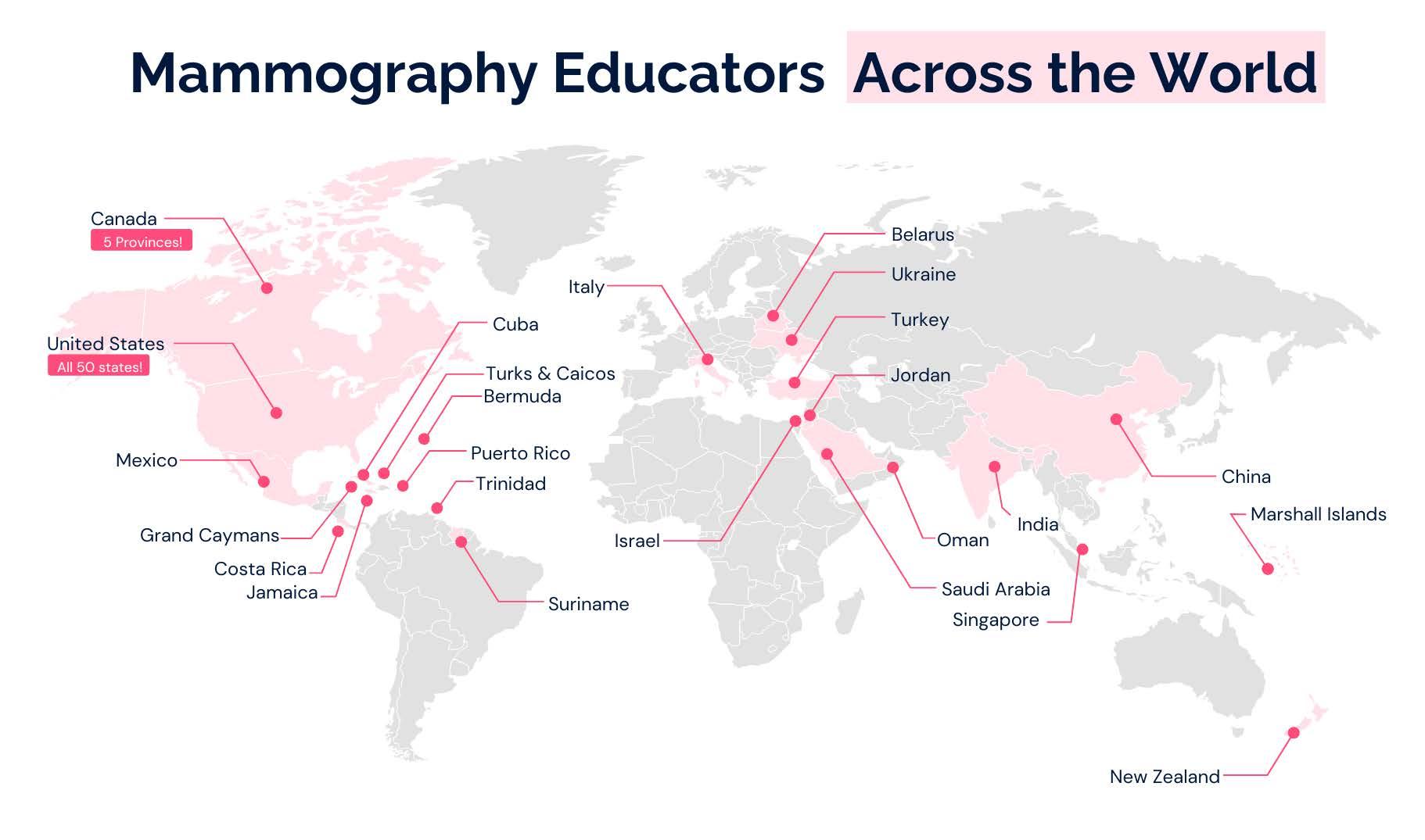
During this huge growth process, we seized the opportunity to mentor 12 new consultants to be part of our team to help us meet our goals. In the process, those outstanding mammographers were also given the opportunity to participate in mammography technologist educational programs and trainings throughout the United States (sometimes in very remote areas!) and in many other countries. Most international work is done on a volunteer basis with only basic travel expenses covered. They are truly giving the gift of education to technologists who may not otherwise have access to such high-quality teaching standards. Mammography Educators donates all educational resources (lectures, etc) to assist in this endeavor and offers scholarships to foreign students with limited resources. We are proud to say we have participated in mammography education programs in over 25 countries, including China, Ukraine, Cuba, Jordan, Oman, and Ghana.
We also recognized the need for better resources and connection on the national and international levels. The ACR manual was last updated (with positioning standards) in 1999,1 so mammographers are looking for a more current, reliable resource that applies to full-field digital mammography and digital breast tomosynthesis. The teaching techniques we use for positioning and image evaluation are data based (published in the American Journal of Roentgenology in 2017),2 which is unique in itself. It was clear that technologists, specifically, needed a forum to ask questions of their colleagues and seek advice concerning the myriad of issues related to their work. Following the concept of the SBI forum, Mammography Educators started a Facebook forum (Quality Breast Imagers) to provide positive and professional support and education for mammographers
throughout the world. Since its inception in 2021 we now have over 10,000 members worldwide, and we keep growing. The need is obviously there!
We are proud to be a group of medical professionals who realize the gift of education. However, in speaking of gifts, I would be remiss if I did not mention the gift of friendship and camaraderie that comes with being part of the special and unique sisterhood (and brotherhood) of breast imaging professionals. We learn so much from each other in so many ways. This, like education, is a true gift in life. As we move through Breast Cancer Awareness Month and into the holiday season, I hope we will all be able to see the special gifts around us and hold them for the treasures that they are. Sometimes we need to change our focus, but they are there, shining so brightly, even amid the daily stress and negativity that can be so prevalent in today’s world.
• Quality Breast Imagers Facebook group: https://www.facebook.com/groups/ qualitybreastimagers
• Breast Cancer Awareness Month website: https://www.nationalbreastcancer.org/ breast-cancer-awareness-month
References
1. American College of Radiology. Mammography Quality Control Manual: Radiologist’s Section, Clinical Image Section, Radiologic Technologist’s Section, Medical Physicist’s Section. American College of Radiology; 1999. Accessed October 5, 2024. https://www.acr.org/-/media/ACRAccreditation/Documents/ Mammography/1999_Mammo_QCManual_Book_final.pdf
2. Huppe AI, Overman KL, Gatewood JB, Hill JD, Miller LC, Inciardi MF. Mammography positioning standards in the digital era: is the status quo acceptable? AJR Am J Roentgenol. 2017c;209(6):1419-1425. doi:10.2214/AJR.16.17522
Countries where Mammography Educators has provided educational services as of September 2024.
Sharing and Receiving the Gifts of Education and Friendship Around the World (continued from page 19)
To save lives and minimize the impact of breast cancer. .....
Mammography Educators Director of Education Louise Miller in Ghana at the 2010 Breast Cancer Project, sponsored by the International Atomic Energy Agency.
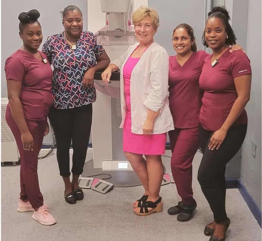
Mammography Educators consultant Belinda Zaparinuk at the 2023 mammography positioning training in Jamaica.
Mammography Educators consultant Pamela Fulmer at the 2023 mammography positioning training in Bermuda.
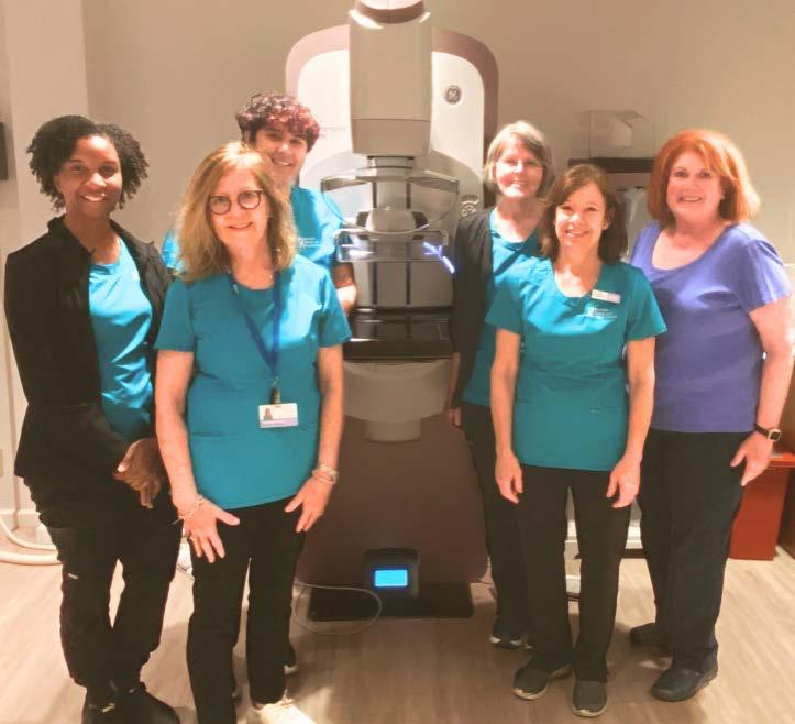
Mammography Educators consultants Sarah Jacobs and Robyn Hadley at the 2023 Jordan Breast Cancer Program.
Mammography Educators Director of Education Louise Miller at the 2022 mammography conference and positioning training in Oman.
Mammography Educators consultant Dawn Derenburger at the 2020 mammography positioning training in Turks and Caicos.
In Memoriam: G. W. (Bill) Eklund, MD, FACR, FSBI (1932-2024)
By Michael Linver, MD, FACR, FSBI
It is with great sadness that we mourn the passing of Dr. Bill Eklund on July 19. An innovative mammography pioneer and consummate teacher, Bill led the SBI as its president in 1998 and 1999.
Bill brought so much of his talents and his energy to breast imaging from a unique and varied background. Born to a medical missionary and an American dentist in Wai, India, in 1932, Bill spent his first 10 years there. His father served as dentist to many of the crowned heads of India, including the Maharaja of Jaipur, and Bill often accompanied his father, befriending and playing with young “Bubbles,” the future Maharaja, who became an international polo star. During World War II, Bill moved to the United States with his father aboard a troop transport, dodging German U-boats along the way. He attended high school in San Antonio and undergraduate and medical school at the University of Texas. After an internship in Denver, he moved to Lamar, Colorado, where he practiced general medicine for seven years. In 1968, he saw the light and entered a radiology residency program in Denver. During his residency, he met and married Elizabeth (Betsy) Pattee, who became his life partner for the next 55 years.
In 1971, Bill joined a general radiology practice in Portland, Oregon, Betsy’s hometown, and developed a strong interest in mammography, setting up the Portland Mammography Center, one of the first of its kind in the Northwest. While directing the Portland Mammography Center, Bill also served as director of breast imaging at the Oregon Health and Science University School of Medicine. He became one of the first breast imaging radiologists to take on a strong clinical role, speaking to every patient and taking the extra time to treat them as people instead of numbers at the top of the report.
In 1988, Bill was recruited to head the inaugural Komen Memorial Breast Center in Peoria, Illinois. It was there over the next seven years that Bill established an international reputation for developing the Eklund imaging technique for the augmented breast. He also published, with Dr. Gilda Cardenosa, landmark articles with definitive guidelines for mammography image quality through the use of the posterior nipple line and other techniques. Consequently, he became a sought-after speaker with a teaching style that brought artistry and a presentation presence enhanced by his unique background as a general practitioner and his remarkable ability to make mundane material come to life at the podium.
For all these accomplishments, Bill was invited to become one of the initial fellows of the SBI in 1990. He continued to be an active and vital force within SBI during the 1990s, culminating with his tenure as SBI president in 1998 and 1999.
Bill developed a strong friendship with Dr. László Tabár, who served as a mentor as well as a colleague to Bill and with whom Bill brought the concept of extended processing to American breast imaging radiologists in the early 1990s. During this period, Bill in turn became one of my mentors as well. In 2003, he flew to my home in Albuquerque and spent a week with me, moving me out of slide trays and into PowerPoint. His generosity as a friend and as a teacher was legendary, and his popularity as a speaker led to invitations to present over 1000 lectures throughout his storied career.
His breast imaging expertise and exploits continued to be acknowledged by the breast imaging community. He was appointed to the ACR Breast Imaging Task Force from 1992 to 1997 and served as a board examiner in breast imaging for the American Board of Radiology. Bill was chosen as one of the initial fellows of the SBI in 1990 and was elected a fellow of the ACR in 1996. In 2005, he received the prestigious IMPACT award from the National Consortium of Breast Centers.
Bill’s artistry as a teacher and lecturer extended into his love for creative photography. He excelled at capturing nature through the camera’s lens in spectacular fashion, with many of his photos garnering prizes throughout the Northwest.
Bill’s compassion for others extended far beyond his role as a physician. He was always available to young people interested in photography or other artistic expressions. He served as president of his religious congregation, a position he filled with the same mix of humility and commitment he demonstrated in his professional career.
As much as Bill was passionate about quality breast imaging, teaching, photography, and community, the true love of his life was always his family. He and Betsy presided over a large and loving family of a son, three daughters, eight grandchildren, and three great-grandchildren, with each holding a special place in their hearts. They truly made beautiful music together, as Betsy is a talented and well-known opera singer and Bill was always her most appreciative audience.
As a breast imaging radiologist, Bill was universally loved by his patients and colleagues alike. He represented all that is good within our specialty: excellence as a radiologist, a patient advocate, a clinician, a teacher and educator, and a researcher, all delivered with kindness, compassion, competence, and caring. He set standards to which we can all aspire. Indeed, Bill, you will be dearly missed.
Continued on page 22>
Michael Linver, MD, FACR, FSBI
In Memoriam: G. W. (Bill) Eklund, MD, FACR, FSBI (1932-2024) (continued from page 21)
save lives and minimize the impact of breast cancer.
Bill and Betsy with Mike and Mina Jo Linver at Santa Fe Opera, 2008.
Bill the photographer by the Willamette River, Oregon.
Bill with his three daughters and their families. Front row, left to right: Karen Eklund Gilmore (holding Harley Gilmore), Hillary Eklund, Bill Eklund, Katherine Eklund Steen, and Michael Steen. Back row, left to right: Greg Larsen (Hillary’s husband), David Pattee (Betsy’s brother), Ella Steen, and Andrew Steen (Katherine’s husband).
Bill with Dr. Debra Monticciolo at the SBI fellows meeting at the Radiological Society of North America annual meeting, 1999.
One of Bill’s photos: Brilliant Morning In Portland.
Bill with fellow faculty members at Santa Fe Mammography Conference, 1998. Front row: Gilda Cardenosa. Back row, left to right: Mike Linver, Bill, Gillian Newstead, Richard Bird, and Robert Schmidt.
groups. The 2010 Affordable Care Act included a five-year moratorium on the 2009 USPSTF guidelines; the 2015 PALS Act picked up where the moratorium left off (personal communication, ACR Government Relations Committee). The PALS Act requires breast cancer screening coverage without patient copay for annual mammography starting at age 40 years. The ACR has advocated for and achieved multiple extensions of the PALS Act to keep the law in place. It currently remains in place until January 1, 2026, regardless of the recent USPSTF guideline updates.
Together, the MQSA and PALS Act have ensured access to high-quality mammography screening in the United States. These are by no means the only legislative efforts but they have been important to help achieve these goals. Additional legislation (for example, legislation to address breast density, including notification of patients) is intended to improve outcomes. Current legislation is focused on the expansion of coverage beyond screening so that supplemental and diagnostic examinations will be available to women without cost sharing. There is much work to be done to help women
who have breast cancer. These continuing legislative efforts have helped usher in advances in quality and in breast imaging care that benefit all of us.
My personal thanks to Penny Butler, Gloria Romanelli, and Eugenia Brandt for their assistance in formulating this article and for their tireless work on behalf of the women we serve.
2. McLelland R, Hendrick RE, Zinninger MD, Wilcox PA. The American College of Radiology Mammography Accreditation Program AJR Am J Roentgenol 1991;157(3):473-479. doi:10.2214/ajr.157.3.1872231
3. Reuter FG. Preliminary report-NEXT-85. In: National Conference on Radiation Control. Proceedings of the 18th Annual Conference of Radiation Control Program Directors. Conference of Radiation Control Program Directors (CRCPD) publication 86-2. 1986:111-120.
4. MQSA national statistics. US Food and Drug Administration. Accessed October 3, 2024. https://www.fda.gov/radiation-emitting-products/mammography-information-patients/mqsa-national-statistics
Bill and colleagues at second SBI conference, Orlando, 1995. Left to right: Dr. Orah Moskovitz, Tamar Moskowitz, Dr. Carl D’Orsi, Dr. Myron (Mike) Moskowitz, Dr. Lester Kalisher, Bill, and Dr. Robert Schmidt.
Bill receiving Impact Award at National Consortium of Breast Centers conference, 2005. Also pictured: Dr. Cary Kaufman.
Historic Legislative Changes in Breast Imaging (continued from page 17)
Beyond Breast Density Notification: How My Density Matters is Changing Breast Health Conversations
By Leslie Ferris Yerger, MBA
As of September 10, 2024, every woman getting a mammogram is informed about her breast density. While this is a crucial step forward, it also raises new questions. What does this mean for the individual? What additional actions should be taken? And how can women make informed decisions about their health?
This is where My Density Matters comes in, making our grassroots educational efforts more relevant than ever. We are dedicated to empowering women to not only know their breast density but also fully understand its implications and take charge of their breast cancer screening journey.
At My Density Matters, our vision is clear: we want a world where breast density no longer hinders the early diagnosis of breast cancer. Through our work, we educate, empower, and prepare women to advocate for their health. Our efforts are driven by our commitment to ensuring that every woman knows what to do with the information she receives about her breast density.
Educate
Our journey begins with education. We focus on teaching women about breast density and its potential impact on mammography effectiveness. Across local events in the Chicagoland area and nationwide through virtual presentations, we meet women where they are—at community health fairs, corporate wellness programs, and other gatherings—to ensure that they understand the significance of breast density in breast cancer screening.
Our approach is straightforward: we provide easy-to-understand materials in English and Spanish, share real-world stories, and answer questions. Women often tell us they’ve never heard about breast density before speaking with us. This revelation emphasizes the critical gap in public awareness, and we work diligently to close it. We are passionate about engaging the community and spreading the word.
For medical professionals, this effort can be particularly helpful. By supporting and amplifying grassroots educational efforts like ours, physicians can help ensure that their patients come to them equipped with basic knowledge about their breast density and prepared to ask the right questions about their additional screening options.
Empower
Education is only the first step. The second crucial aspect of our work is empowerment. Knowing about breast density is
important, but women need to feel confident in navigating the health care system and advocating for themselves.
At events and through virtual sessions, we provide information and share tools that women can use to take action. We explain additional screening options for women with dense breasts and we emphasize the importance of individualized care. By giving women the knowledge to understand their breast density and the power to ask for appropriate follow-up screenings, we are helping them take control of their own health.
It’s about more than just facts. It’s about making sure every woman feels heard, supported, and empowered to take the next step. When women leave our sessions, they often express a new sense of agency and confidence, ready to pursue the right path for their health.
Prepare for Discussion
The final element of our grassroots work is preparation. We aim to ensure that women know about breast density and are ready to have informed conversations with health care professionals. We prepare women to discuss their breast density with their doctors and ask the right questions to collaboratively make informed decisions about additional screening, if necessary.
Many women struggle to bring up these topics during appointments. We equip them with resources so they feel more comfortable advocating for themselves. In doing so, we help bridge the gap between patients and physicians. Breast imaging professionals play a crucial role in these conversations, and by engaging with our efforts, you can ensure that your patients are informed and prepared for meaningful dialogue during their appointments.
Check Your Chart: A Mobile Platform for Nationwide Impact
In addition to our in-person and virtual educational efforts, My Density Matters developed Check Your Chart, a scalable mobile platform that empowers women to access their mammography reports, learn about their breast density, and prepare for conversations with their health care team.
To save lives and minimize the impact of breast cancer. .....
Leslie Ferris Yerger, MBA
Patients can simply text “check your chart” to 211-411 to access educational resources that guide them through understanding their breast density and the next steps to take. This platform extends our mission, making education accessible nationwide and ensuring that no woman’s breast density stands in the way of early cancer detection.
Check Your Chart supports health care professionals by improving patient education, facilitating informed discussions, and enhancing patient engagement. For more information about a semicustomized version for your breast center, contact susan@mydensitymatters.org
Conclusion
As breast imaging professionals, you recognize the critical role of breast density in breast cancer screening. By partnering with My Density Matters, you can help amplify our efforts, ensuring that your patients arrive informed, empowered, and prepared to take charge of their breast health. Together we can create a future where breast density no longer hinders the early diagnosis of breast cancer.
A Special Thank You
A special shout-out to our incredible SBI member medical advisors: Stacy Smith-Foley, MD; Randy C. Miles, MD, MPH; Kirti Kulkarni, MD; and Nila Alsheik, MD. Learn more about our medical advisors on the My Density Matters website
Leslie Ferris Yerger is CEO and founder of My Density Matters.
Radiology Across Borders
By Regina Hooley, MD, FSBI
Radiology Across Borders (RAB) is a global charity based in Australia. The organization focuses on teaching essential radiology skills to doctors and medical imaging staff around the world. RAB projects aim to drive sustainable change, helping clinicians increase basic and advanced skills and share their expanding knowledge with local peers. RAB offers many online educational tools as well as in-country visits throughout the world.
The SBI partners with RAB and provides the organization with multiple key lectures with a variety of topics including procedures, interventions, case reviews, and practice guidance updates. In April 2024, the SBI sponsored RAB on a breast imaging education trip to the Philippines, and I was extremely fortunate to be the first SBI in-person breast imaging representative. With the six-person all-women health team, RAB organized a two-day educational course on enhancing mammography proficiency and clinical technique. Our RAB team, composed of radiologists from Australia, New Zealand, and the United States, worked in collaboration with the Philippine College of Radiology and had major support from Siemens Healthineers, I-MED Radiology, Lumus Imaging, and others.
This was the first RAB breast imaging and women’s health initiative in the Philippines. However, our experienced New Zealand–based team leader, Dr. Trina Whittaker, has organized and participated in many RAB trips. We were all very impressed by the engagement of the Filipino radiologists. The conference was well attended, with over 200 radiologists (attending clinicians and trainees) who traveled from various clinics and hospitals throughout the Philippines to Manila, where the conference took place. A few centers offer more advanced imaging, including tomosynthesis and magnetic resonance imaging, but there is no formal national breast cancer screening program, which they hope to establish in the next few years. The conference attendees were eager to review basic breast imaging techniques and more advanced topics such as supplemental screening and breast imaging of patients newly diagnosed with cancer.
On a personal note, the trip was a wonderful experience. I was born in the Philippines and have not been back to visit since I was in college! I was able to reconnect with many cousins whom I had not seen in over 30 years. Before the conference, I also traveled for a week with three other RAB radiologists: Drs. Trina Whittaker, Emmeline Lee, and Bronwen Slater. I was a little nervous at first and did not know what to expect, since I had never met these radiologists. However, we bonded instantly and had a wonderful adventure touring the amazing sites and beaches of Palawan, including Puerto Princesa and El Nido. RAB encourages their
teams to travel, bond with each other, and be familiar with local culture. This included our own mini food challenge. Ever hear of the Filipino dishes sisig, tamilok, and balut? We tried them all and more! It was a special trip for everyone. Learn more and join RAB at radiologyacrossborders.org
To save lives and minimize the impact of breast cancer. .....
Regina Hooley, MD, FSBI
Evaluation of the Axilla in Patients With Early-Stage Breast Cancer (SOUND Trial): The Breast Imaging Radiologist’s Role
By Pamela DiPiro, MD
Evaluation and treatment of the axilla in patients with breast cancer has changed significantly over the last few decades.1,2 Historically, axillary lymph node dissection (ALND) was the standard of care for breast cancer, providing staging information, presumed regional control of the axilla, and improved overall survival. In the 1990s, sentinel lymph node biopsy (SLNB) replaced ALND for most women with early-stage breast cancer and clinically node-negative axillae, with subsequent ALND performed only if the SLNB result was positive for metastasis.3-6 Morbidity associated with axillary surgery, including lymphedema/ swelling, axillary pain, limited arm mobility, and numbness or paresthesia, was significantly decreased with SLNB.4 Multiple additional clinical trials supported the potential omission of ALND in patients with limited sentinel lymph node metastases, specifically those with small metastases (micrometastases)7-9 or those in whom only one or two sentinel lymph nodes were involved.10 Further investigation showed that SLNB with axillary radiation could safely replace ALND, with comparable axillary disease control but less morbidity.11,12
These studies demonstrated that axillary surgery is not necessary for local control and does not impact overall survival in breast cancer patients. More recent prospective studies were undertaken in Europe and Asia to determine whether SLNB can be safely excluded in a subset of patients with small, early-stage breast cancers and clinically and sonographically negative axillary lymph nodes.13-16 Although some studies are ongoing, the first that was completed, the SOUND (Sentinel Node vs Observation After Axillary Ultrasound) trial, showed that omission of axillary surgery in patients with small breast cancers and negative axillary ultrasonography results is noninferior to SNLB.13 As breast imaging radiologists, our role in ultrasound evaluation of the axilla is important in determining eligibility for these patients to forgo axillary surgery, and knowledge of this trial is important.
The SOUND trial was a multicenter, prospective, noninferiority, phase 3 randomized trial conducted in Italy, Switzerland, Spain, and Chile that enrolled women from February 2012 to June 2017. Women included had an invasive breast cancer measuring 2 cm or smaller, clinically negative axillary lymph nodes, and intention to undergo breast-conserving therapy with lumpectomy and radiation therapy. A negative axillary ultrasonography result was required, although if a single equivocal lymph node was identified
sonographically, fine-needle aspiration with a negative cytology result was sufficient for inclusion. Exclusion criteria were extensive multifocality or multicentricity, preoperative presence of suspicious axillary lymph nodes, previous cancer, bilateral breast cancer, synchronous distant metastases, pregnancy or lactation, and barriers to informed consent or continued clinical follow-up. The primary end point was distant disease–free survival at five years. Secondary end points included cumulative incidence of axillary recurrences, cumulative incidence of distant recurrences, disease-free survival, overall survival, and adjuvant treatment recommendations.
The final intention-to-treat analysis included 1405 women with a median age of 60 years (IQR, 52-68 years). Of these, 708 were randomized to the SLNB group and 697 were randomized to receive no axillary surgery. Patients in the SLNB group who had positive biopsy results progressed to ALND. Of the 708 women in the SLNB group, 97 (13.7%) had positive axillary lymph node biopsy results; 36 women (5.1%) had micrometastases and 61 (8.6%) had macrometastases.
Overall, the median tumor size was 1.1 cm (IQR, 0.8-1.5 cm), and 87.8% of the patients had estrogen receptor–positive, ERBB2 (also known as HER2/neu)–negative, nonoverexpressing breast cancers. Patient demographics, tumor types, adjuvant systemic therapy regimens, and radiotherapy were similar in the SLNB and no-axillary-surgery groups.
Five-year distant disease-free survival was similar in the SLNB and no-axillary-surgery groups (97.7% and 98%, respectively). Secondary outcomes were also comparable in the two groups (SLNB vs no axillary surgery: locoregional relapses, 1.7% vs 1.6%; distant metastases, 1.8% vs 2.0%; and deaths, 3.0% vs 2.6%, respectively). In both groups, the five-year cumulative incidence of axillary recurrence was 0.4%. The SOUND trial demonstrated that omission of axillary surgery is noninferior to SNLB in patients with small, unilateral breast cancers and negative preoperative axillary ultrasonography results and can be safely offered to patients when the lack of pathological information does not affect the postoperative treatment plan.
Pamela DiPiro, MD
As breast imaging radiologists, we are being asked more frequently to perform axillary ultrasonography in patients with newly diagnosed breast cancer. We play a critical role in determining patient eligibility for omission of axillary surgery. If a single node has a borderline ultrasonography result, fine-needle aspiration should be recommended. A negative cytology result may allow the patient to avoid axillary surgery.
The evolution of axillary surgery in early-stage breast cancer has progressed from routine ALND to SLNB and now to omission
References
1. Magnoni F, Galimberti V, Corso G, Intra M, Sacchini V, Veronesi P. Axillary surgery in breast cancer: an updated historical perspective Semin Oncol 2020;47(6):341-352. doi:10.1053/j.seminoncol.2020.09.001
2. Hersh EH, King TA. De-escalating axillary surgery in early-stage breast cancer Breast. 2022;62 Suppl 1(Suppl 1):S43-S49. doi:10.1016/j.breast.2021.11.018
3. Veronesi U, Paganelli G, Galimberti V, et al. Sentinel-node biopsy to avoid axillary dissection in breast cancer with clinically negative lymph-nodes Lancet 1997;349(9069):1864-1867. doi:10.1016/S0140-6736(97)01004-0
4. Veronesi U, Paganelli G, Viale G, et al. A randomized comparison of sentinelnode biopsy with routine axillary dissection in breast cancer N Engl J Med 2003;349(6):546-553. doi:10.1056/NEJMoa012782
5. Krag DN, Anderson SJ, Julian TB, et al. Sentinel-lymph-node resection compared with conventional axillary-lymph-node dissection in clinically nodenegative patients with breast cancer: overall survival findings from the NSABP B-32 randomised phase 3 trial Lancet Oncol. 2010;11(10):927-933. doi:10.1016/ S1470-2045(10)70207-2
6. Veronesi U, Viale G, Paganelli G, et al. Sentinel lymph node biopsy in breast cancer: ten-year results of a randomized controlled study Ann Surg. 2010;251(4):595-600. doi:10.1097/SLA.0b013e3181c0e92a
7. Galimberti V, Cole BF, Zurrida S, et al; International Breast Cancer Study Group Trial 23-01 investigators. Axillary dissection versus no axillary dissection in patients with sentinel-node micrometastases (IBCSG 23-01): a phase 3 randomised controlled trial Lancet Oncol. 2013;14(4):297-305. doi:10.1016/ S1470-2045(13)70035-4
8. Galimberti V, Cole BF, Viale G, et al; International Breast Cancer Study Group Trial 23-01. Axillary dissection versus no axillary dissection in patients with breast cancer and sentinel-node micrometastases (IBCSG 23-01): 10-year follow-up of a randomised, controlled phase 3 trial Lancet Oncol. 2018;19(10):1385-1393. doi:10.1016/S1470-2045(18)30380-2
9. Solá M, Alberro JA, Fraile M, et al. Complete axillary lymph node dissection versus clinical follow-up in breast cancer patients with sentinel node micrometastasis: final results from the multicenter clinical trial AATRM 048/13/2000 Ann Surg Oncol. 2013;20(1):120-127. doi:10.1245/s10434-0122569-y
10. Giuliano AE, McCall L, Beitsch P, et al. Locoregional recurrence after sentinel lymph node dissection with or without axillary dissection in patients with sentinel lymph node metastases: the American College of Surgeons Oncology Group Z0011 randomized trial Ann Surg. 2010;252(3):426-433. doi:10.1097/ SLA.0b013e3181f08f32
altogether in some patients, thereby minimizing the morbidity of local therapy. The role of axillary surgery in staging and prognosis may eventually be replaced by the increasingly available genomic assays for determining systemic therapy in selected patients. As other studies investigate the exclusion of axillary surgery in additional groups (including patients with ERBB2positive or triple-negative disease treated with neoadjuvant chemotherapy),17,18 breast imaging radiologists will likely be called upon more often to perform additional axillary ultrasonography in these patients.
11. Sávolt Á, Péley G, Polgár C, et al. Eight-year follow up result of the OTOASOR trial: the optimal treatment of the axilla—surgery or radiotherapy after positive sentinel lymph node biopsy in early-stage breast cancer. A randomized, single centre, phase III, non-inferiority trial. Eur J Surg Oncol 2017;43(4):672-679. doi:10.1016/j.ejso.2016.12.011
12. Bartels SAL, Donker M, Poncet C, et al. Radiotherapy or surgery of the axilla after a positive sentinel node in breast cancer: 10-year results of the randomized controlled EORTC 10981-22023 AMAROS trial. J Clin Oncol 2023;41(12):2159-2165. doi:10.1200/JCO.22.01565
13. Gentilini OD, Botteri E, Sangalli C, et al; SOUND Trial Group. Sentinel lymph node biopsy vs no axillary surgery in patients with small breast cancer and negative results on ultrasonography of axillary lymph nodes: the SOUND randomized clinical trial. JAMA Oncol. 2023;9(11):1557-1564. doi:10.1001/ jamaoncol.2023.3759
14. Reimer T, Stachs A, Nekljudova V, et al. Restricted axillary staging in clinically and sonographically node-negative early invasive breast cancer (c/iT1-2) in the context of breast conserving therapy: first results following commencement of the Intergroup-Sentinel-Mamma (INSEMA) trial Geburtshilfe Frauenheilkd 2017;77(2):149-157. doi:10.1055/s-0042-122853
15. van Roozendaal LM, Vane MLG, van Dalen T, et al. Clinically node negative breast cancer patients undergoing breast conserving therapy, sentinel lymph node procedure versus follow-up: a Dutch randomized controlled multicentre trial (BOOG 2013-08) BMC Cancer. 2017;17(1):459. doi:10.1186/s12885-0173443-x
16. Jung JG, Ahn SH, Lee S, et al. No axillary surgical treatment for lymph nodenegative patients after ultra-sonography (NAUTILUS): protocol of a prospective randomized clinical trial BMC Cancer. 2022;22(1):189. doi:10.1186/s12885-02209273-1
17. Reimer T, Glass A, Botteri E, Loibl S, D Gentilini O. Avoiding axillary sentinel lymph node biopsy after neoadjuvant systemic therapy in breast cancer: rationale for the prospective, multicentric EUBREAST-01 trial Cancers (Basel) 2020;12(12):3698. doi:10.3390/cancers12123698
18. Avoiding sentinel lymph node biopsy in breast cancer patients after neoadjuvant chemotherapy (ASICS). Clinicaltrials.gov. Updated January 13, 2020. Accessed September 25, 2024. https://clinicaltrials.gov/ct2/show/ NCT04225858
Bridging the Gap in Breast Cancer Care: Insights From the EUSOBI Young Club Journal
Club Series
By Melis Baykara Ulusan, MD; Ioana Bene, MD; Miguel Braga, MD; Anna D’Angelo, MD; Elisabetta Giannotti, MD; Stephanie Morscheid, MD; Thiemo Van Nijnatten, MD, PhD; Giulia Vatteroni, MD
In 2020, the European Society of Breast Imaging (EUSOBI) Young Club launched an online Journal Club to provide an interactive platform to discuss recent publications and to help educate and update breast radiologists. Moderated by young radiologists alongside experienced experts, this club fosters an inclusive environment where participants, regardless of experience level, can ask questions freely.
This year, the Journal Club series focuses on bridging the gap across various disciplines, highlighting the multidisciplinary nature of breast cancer care. The EUSOBI Young Club champions this approach, as emphasized in Frances M. Boyle’s 2005 article “Multidisciplinary Care in Cancer: The Fellowship of the Ring,” published in the Journal of Clinical Oncology. Quoting from the movie, “You will unite or you will fall,” the article underscores the enduring importance of collaboration.
So far, we have successfully collaborated with surgeons, radiation oncologists, oncologists, and patients. These are some key highlights from this year’s sessions.
Eighth Journal Club: Pathology and Radiology in the Artificial Intelligence Era
The eighth Journal Club explored the intersection of pathology and radiology, particularly in the era of artificial intelligence (AI). Presenters Mariachiara Negrelli (Milan, Italy) and Falak Masood (Cambridge, England) introduced an AI-driven prognostic marker for early-stage luminal/ERBB2 (formerly HER2)–negative breast cancer, known as the BRACE marker, discussed in the article “AI-Enabled Routine H&E Image Based Prognostic Marker for Early-Stage Luminal Breast Cancer,” published in Precision Oncology in 2022.
This innovative tool assesses tumor heterogeneity and helps predict which patients may benefit from adjuvant chemotherapy. Prof. Nicola Fusco, a pathologist from Milan, Italy, and Dr. Tamar Sella, a radiologist from Jerusalem, Israel, discussed the challenges of identifying cancers in the “gray zone”—those with intermediate risk. Moderated by Dr. Marianna Fanizza (Bologna, Italy), the session emphasized the growing importance of collaboration between pathologists and radiologists, particularly with the rise of digital technologies.
The main takeaway from this session was that while both radiologists and pathologists perform well individually, collaboration is crucial for patient risk stratification. Dr. Sella concluded, “AI is not here to replace us; it is here to improve us. We drive the AI; it does not drive us.”
Ninth Journal Club: Communication and the Radiologist’s Role
The ninth Journal Club, moderated by Dr. Paola Clauser (Vienna, Austria), focused on the evolving role of radiologists in patient communication. Dr. Melis Baykara Ulusan (Istanbul, Turkey) presented three key articles:
• “Value-Based Radiology: What Is the ESR Doing, and What Should We Do in the Future?” (Insights into Imaging, 2021)
• “The Role of Radiologist in the Changing World of Healthcare: A White Paper of the European Society of Radiology (ESR)” (Insights into Imaging, 2022)
• “Radiology in the Era of Value-Based Healthcare: A Multi Society Expert Statement From the ACR, CAR, ESR, IS3R, RANZCR, and RSNA” (Journal of the American College of Radiology, 2021)
The discussion was hosted by Prof. Federica Pediconi (Rome, Italy), Prof. Michael Fuchsjäger (Graz, Austria), and Caroline Justich, ESR Patient Advisory Group chair and founder of Be Accepted (Vienna, Austria), a campaign, website, and online
To save lives and minimize the impact of breast cancer. .....
Giulia Vatteroni, MD
Melis Baykara Ulusan, MD
Anna D’Angelo, MD
Miguel Braga, MD Ioana Bene, MD
magazine that support women newly diagnosed with cancer along their breast cancer care journey. 1
These articles highlight the importance of enhancing the patientdoctor relationship in radiology. Caroline Justich stressed the need for radiologists to use simple, clear language when delivering a diagnosis, especially during the critical first minutes of patient interaction.
The session also addressed the increasing role of AI as a “second reader,” enabling radiologists to focus on complex cases while improving communication with clinicians and patients.
Tenth Journal Club: Integration of Radiation Oncology and Radiology
The 10th Journal Club explored the intersection between breast radiation oncology and radiology in clinical practice. Presenters Javier Azcona Saenz (Santander, Spain) and Marianna Valzano (Florence, Italy) discussed the article “International Multidisciplinary Consensus on the Integration of Radiotherapy With New Systemic Treatments for Breast Cancer,” published in The Lancet Oncology in 2024.
The discussion emphasized the evolving role of radiologists in guiding radiation therapy and monitoring treatment responses, ultimately ensuring precision in treatment planning and improving patient outcomes.
Eleventh Journal Club: Breast Surgical Oncology and Radiology
The 11th Journal Club focused on the integration of breast surgical oncology and radiology. Moderated by Dr. Thiemo van Nijnatten (Maastricht, Netherlands), the session reviewed the results of the SOUND trial in the article “Sentinel Lymph Node Biopsy vs No Axillary Surgery in Patients With Small Breast Cancer and Negative Results on Ultrasonography of Axillary Lymph Nodes,” published in JAMA Oncology in 2023. It was presented by Dr. Ioana Bene (Cluj-Napoca, Romania). The discussions were led by Dr. Julia Camps Herrero (Valencia, Spain), Prof. Maria Joao Cardoso (Lisbon, Portugal), and Dr. Eduard-Alexandru Bonci (Lisbon). Dr. Nada Santrac (Belgrade, Serbia) also gave a few words about the European Young Surgeons and Alumni Club.
The SOUND trial examined the necessity of sentinel lymph node biopsy versus omitting axillary surgery in patients with small tumors and negative lymph node imaging results. The trial concluded that omitting axillary surgery did not compromise long-term survival for eligible patients. This outcome suggests that many patients could avoid invasive surgery without negatively impacting their treatment, highlighting the potential for more personalized and less invasive breast cancer care.
Looking Ahead
Participating in the EUSOBI Young Club’s online Journal Club is an excellent way for breast imaging radiologists to stay current with the latest research while engaging with peers and international experts. Even if live attendance isn’t possible, recordings are made available on the EUSOBI website two to three weeks after each session, ensuring that everyone can benefit from these discussions. Attendance to the Journal Club and access to recordings are free of charge for EUSOBI members in good standing.
This year’s theme, “Bridging the Gap,” underscores the importance of collaboration across medical disciplines and among radiologists globally. By coming together, we can enhance breast cancer care and foster a stronger global radiology community. We look forward to continuing this journey with you in future sessions!
Reference
1. Justich C, Briers E, Cruwys C. From the patient in focus to you...what is to come next? Radiography (Lond). 2023;29 Suppl 1:S5-S7. doi:10.1016/j. radi.2023.02.001
RSNA (Radiological Society of North America) Annual Meeting: December 1-5, 2024
https://www.rsna.org/annual-meeting
San Antonio Breast Cancer Symposium December 10-14, 2024
https://sabcs.org/FullProgram
Please visit the SBI Calendar of Events at www.sbi-online.org for a complete listing of events.