The Member Newsletter of the Society of Breast Imaging
INSIDE THIS ISSUE:
• This issue's theme: How to execute successful hybrid and remote practices in breast imaging and radiology
• Meet our new SBI fellows
• Highlights From the RSNA 2023 Meeting in Chicago
• IDEA (Inclusion Diversity Equity Alliance) Insights: Creating an Inclusive and Welcoming Environment for Non-English-Speaking Patients: A Guide for SBI Members
EDITOR:
Nidhi Sharma
ASSISTANT EDITORS:
Randy Miles and Shinn-Huey Shirley Chou
SBI COMMITTEE UPDATES: Yasmeen Fields
TECHNOLOGISTS’ COLUMN: Robyn Hadley and Sarah Jacobss
WHAT’S
Anita Mehta
MEMBERS IN TRAINING:
Wenhui
and
L. DiBiasio
WELLNESS COLUMN:
Claudia Cotes and Sarah Jacobs
THE PATIENT'S PERSPECTIVE:
Hannah Perry and Danielle Sharek
LEGISLATIVE UPDATES:
Amy Patel
OTHER MEMBERS:
Jean Seely
Dr. Mimi Newell, MD, FACR, FSBI President, Society of Breast Imaging
OUR SBI MISSION:
To save lives and minimize the impact of breast cancer
OUR SBI VALUES:
Patient-centered and evidence-based care
Excellence in education Scientific integrity
Collaboration and collegiality
Respect for diversity and inclusiveness
I hope the winter holiday season is proving relaxing and rejuvenating for our SBI family. However, this time of year can harbor some stressors as we are tugged in many professional and personal directions. Our SBI member survey outlined that workplace pressures hold a big and daunting sway over us. At last year’s symposium and in this issue of the newsletter there is lively discussion around new ways to work, including models that we had not conceived of as feasible only a few years ago. I am confident that continued tweaking of these will result in improvement in our sense of well-being and job satisfaction. It is imperative that we all, as physician leaders (even if selfappointed!), remain at the helm of these discussions and negotiations with eyes wide open, lest we end up as an a la carte item on someone else’s menu. Of course, our patients must always come first in any such calculus. This latter consideration seems to be second nature for our SBI colleagues.
A reminder that we all have a standing date! Place: Montreal; dates: April 11-14, 2024; objectives: amazing learning, kindling and rekindling friendships and collaborations, tons of fun. Registration is open. Make sure to get your international travel documents in order ASAP.
Mary S. (Mimi) Newell, MD, FACR, FSBI
President, Society of Breast Imaging
Editor’s Note
By Nidhi Sharma, MD
Originality is taking the road less traveled, championing a set of novel ideas that go against the grain but ultimately make things better.
Adam Grant1
We as breast imaging radiologists champion to provide our patients the best clinical care. However, in the current world of ever-increasing volumes in breast imaging, limited staff, and increasing burnout, most practices and large hospital systems are facing the same burning question! How do we continue to take good care of our patients in a timely fashion while providing effective work-life balance and work-related satisfaction for our breast imaging radiologists?
A recent study by Parikh et al demonstrated up to 78.4% burnout among breast imaging radiologists, with the highest burnout rate in younger radiologists.2 These data are particularly worrisome because our field’s future lies in the career success of younger radiologists. In this winter issue of the newsletter, we tackle this prevalent and imminent question, focusing on the theme of hybrid radiology and teleradiology in breast imaging, with enlightening articles from leaders in the field from both academic and private practice. Dr. Harvey, who chairs the radiology department at the University of Rochester, shares leadership tips and insider details into the mechanics of running a successful remote academic radiology program. Dr. Zuley shares her experience setting up a hybrid academic breast imaging practice. And Dr. Fried shares details of running a successful, busy clinical private breast imaging practice with both hybrid and telemammography options, championing this novel method to improve rural outreach and physician wellness. Sarah Jacobs, BS, RT(R)(M)(CT), provides insights on ways for technologists to thrive, continue engaging with remote radiologists, and provide best patient care when incorporating hybrid and remote radiology work. I hope this edition’s theme proves helpful to readers who are struggling with this model at their local practices.
In addition, throughout 2024 we will be celebrating the history of SBI over the past 40 years. This issue includes a reprint of
an original SBI newsletter article by Dr. Homer from January 2000. We have several other exciting articles, like a description of RAD-AID’s strong work in Nigeria and synopses of recent meetings of the Canadian Society of Breast Imaging and the European Society of Breast Imaging. Our Inclusion Diversity Equity Alliance Committee colleagues also share ways to create an inclusive environment for non-English-speaking patients.
My personal career journey through academic, private, and hybrid practice settings has been one of immense learning and growth to achieve a positive work-life balance. One of my core beliefs is that greatness is achieved through collaboration and sharing our failures and successes. I am eager to continue to foster a culture of inclusivity and openness, encouraging diverse perspectives, and embracing the wealth of knowledge within our society. By using newer approaches, as detailed by our invited authors for this issue’s theme, we can enhance our practice settings and tackle burnout.
If you have any stories, questions, ideas for our next themes, or breast imaging–related personal passion projects, I invite you to write to me: nidhisharma31@gmail.com. Thank you for reading this winter edition of SBI News. I hope you had a wonderful holiday season spending time with family and friends and a chance to recharge to take on the new year. See you all at the symposium in Montreal!
References
1. Grant A. Originals: How Non-Conformists Move the World. Penguin Books; 2017.
2. Parikh JR, Sun J, Mainiero MB. Prevalence of burnout in breast imaging radiologists J Breast Imaging. 2020;2(2):112-118. doi:10.1093/jbi/wbz091
Nidhi Sharma, MD
Incorporating Remote Work Into a Large Academic Practice
By Jennifer Harvey, MD, FACR, FSBI
I love living in Rochester, New York—festivals every weekend in the summer, biking on the Erie Canal, 15 minutes to anywhere, hometown of Wegmans grocery, and the solace of a frequent dusting of snow overnight. However, living in upstate New York is not for everyone.
Remote work in radiology has been in place for decades. Many practices use overnight teleradiology services to provide preliminary reports, but the role of teleradiologists has expanded. Remote radiology companies are now the largest employers of radiologists in the United States. The attraction is likely the anticipation of less stress and increased flexibility. On the other hand, the work has fewer internal rewards such as teaching trainees, sharing interesting cases with colleagues, and participating in tumor boards or other clinical conferences.
In January 2022, our department leadership team decided to incorporate remote radiologists into our academic divisions. The concept was that these faculty members would be actively engaged in our department rather than simply reading work lists. We subsequently identified a division head for remote radiologists, Dr. Daniel Oppenheimer, who had previously worked on-site but transitioned to fully remote during the COVID-19 pandemic.
During the last two years, we have built a unique, successful model. As an example, three of our eight cardiothoracic faculty members do not live in Rochester. Each morning, they have a huddle via Zoom to discuss the day, including work assignments and who will be paired with a resident. The remote faculty members host Zoom readouts with residents throughout the day using interactive tools such as a virtual whiteboard. Both the resident and attending physician can view and interact with the images. Keys to success are optimizing the audiovisual setting by using headphones and cameras on both sides, initiating social interaction with the resident, and making a plan together for the day. During the workday, clinical faculty members use the chat function in our picture archiving and communication system to check in with each other.
Remote faculty members are actively engaged in department functions—division meetings, department faculty meetings,
departmental committees, resident lectures, and even strategic retreats. They also participate in tumor boards and other interdisciplinary conferences. Interestingly, many of our referring clinicians appreciate the availability of a Zoom consultation rather than going to the reading room because they can connect with the radiologist who interpreted a specific study, no matter which reading room or remote location the radiologist is working from that day.
On-site faculty members are able to participate in the contrast reaction teams at our multiple facilities, manage in-person supervision of complicated examinations such as pediatric cardiac magnetic resonance imaging, assist with questions from our technologists or nurses, and perform procedures. While remote faculty members do take phone calls and consultations from clinicians, they experience fewer interruptions, and this imbalance can result in resentment from on-site faculty members.
Our remote radiologists have a primary appointment in their subspecialty division but also have a secondary appointment in the remote division. In this way, the remote faculty members have the camaraderie and networking of their fellow subspecialty colleagues as well as the support and community of other remote radiologists to help build skills that are unique to that role. Both divisions hold monthly meetings and regular social events.
Our remote faculty members have been very successful thus far. Several have some of the highest resident teaching scores in our department. They participate in quality projects and publish scholarly work. One is in a public health 50% research track, working with senior University of Rochester faculty. They currently fill roles in the cardiothoracic, abdominal, emergency radiology, and neuroradiology divisions. We are actively recruiting for our pediatric and musculoskeletal divisions. Procedural roles such as interventional radiology are
Jennifer Harvey, MD, FACR, FSBI
not feasible for remote faculty. We have not yet integrated remote radiologists in our Breast Imaging Division. However, we do provide remote diagnostic services to our more distant affiliate hospitals with on-site coverage focused on procedures. This may be an opportunity to engage remote breast radiologists in the future.
Advantages to the department include having access to a larger pool of candidates and more flexibility in coverage during the workday because of radiologists living in different time zones. We have retained in our community and regional groups several faculty members who moved to different cities, often due to family issues. Currently, 12% of our faculty members do not live in Rochester.
Specific challenges are also present. Feelings of loneliness and isolation can occur. We specifically developed these remote roles to be integrated to mitigate this risk. Informal networking is incredibly helpful in developing scholarly projects. The reduced ability to form these chance connections may result in less job satisfaction and potentially a longer time to promotion. Transitioning faculty members who are directly out of fellowship or have not worked on-site previously takes more effort in integration. Our on-site faculty members have expressed that it may be easier for remote faculty members to disengage at the end of a workday because they are less exposed to the distress of others who feel behind in their work, and this can cause resentment. Transitioning to a remote role is likely smoother for those who have worked in our system previously, either in training or as faculty. We are planning to host remote faculty members on-site on a regular basis to build their connections, engagement, and familiarity with the on-site environment and culture.
Our academic compensation plan is transparent, with all faculty members having the same base salary by rank with some
small differences for certain subspecialties. Our incentives are driven by clinical and scholarly productivity. All faculty members have an academic day per week, which is considered as 0.8 clinical full-time equivalent (cFTE) for a 1.0 FTE. While remote faculty members also have an academic day each week, their clinical productivity is based on a 0.9 cFTE effort to account for anticipated improved efficiency due to fewer interruptions. However, we will likely modify this system to provide a reward for faculty members who are on-site rather than a penalty for those working remotely.
There are some important logistics to consider. Remote faculty members must have a New York State medical license. The
ACR recommends that they also hold a license in the state in which they reside, but this is not required in all states. Remote faculty members are required to have their workstations inspected annually by a physicist licensed in New York, which is typically not problematic. They will likely be out of network for institutional health insurance, which may result in higher copays or deductibles. Consulting an employment attorney is important for compliance with state, county, and even city laws. For example, California requires that employees be paid at least twice monthly, whereas our institution pays faculty members monthly—a minor adjustment. Employment rules can change according to the number of employees in that location. Each hire requires specific approval.
This remains a work in progress, although our experience thus far has been very positive. Our remote faculty members are highly satisfied with this work arrangement. Our on-site radiologists are largely happy to have the extra support. We have filled positions that had been open for years. We have recruited very talented and nationally engaged faculty members who would not otherwise consider working with us. We plan to continue to recruit remote academic radiologists and refine our program.
Incorporating Hybrid Breast Imaging Work in a Large Academic Practice
By Margarita Zuley, MD, FACR, FSBI
Before the COVID-19 pandemic, few organizations offered home breast imaging workstations, with costs and internet bandwidth limitations cited as barriers. Simultaneously, a crisis of burnout that began before the pandemic and was accelerated by it1-3 has impacted radiologist staffing levels within most organizations.
Physician staffing shortages and patient access barriers during the pandemic have brought increased attention to these preexisting issues and have helped catalyze change.
Now most private groups and a growing number of academic institutions have implemented or are considering remote (home) breast imaging work.4 Most confine the scope of this practice to screening or magnetic resonance imaging (MRI) interpretation. While remote work has improved work-life balance and has been a competitive recruitment advantage for organizations offering it, the model is falling short of its full potential.
Mango et al recently reported significant disparities in access for patients residing in zip codes with high socioeconomic disadvantage scores, as compared with those in more affluent regions. Fewer breast imaging facilities are available in these communities. Thus, patients in underserved areas either wait longer or travel farther to receive in-person diagnostic breast imaging care. Additionally, facilities in underserved areas are less likely to offer advanced modalities.5 This combination results in worse outcomes from breast cancer for these populations. The long-term effects of COVID-19 restrictions may further widen the disparity in outcomes for underserved populations in the coming years.6
Diagnostic breast imaging has been implemented by large teleradiology practices, but its adoption has been slow in traditional-format groups, especially in academic programs. The leading reasons for slow adoption include concerns regarding confidence with remote ultrasonography performed by a technologist for subtle findings, the inability to perform correlative clinical breast examinations, and concerns regarding compassionate communication of probable malignant results via a telemedicine visit. Additionally, in academic programs, the excellent educational experience of trainees is a concern.
Remote ultrasonography interpretation is not new and has been used in other radiology specialties, especially obstetrics and abdominal imaging, for decades. Factors key to success are real-time imaging review; excellent communication between the physician, technologist, and patient; and a high level of technologist training and competency.
During the COVID-19 pandemic, our institution purchased remote breast imaging workstations for our breast radiologists. Initially, only screening mammography and MRI were interpreted. However, because the competition to hire breast radiologists is fierce, recruitment to a midsized community such as Pittsburgh, Pennsylvania, limits the pool of interested applicants. We implemented remote diagnostic breast imaging in our academic practice in 2021 to overcome some of these obstacles. The goals of the implementation were to improve faculty wellness, improve patient access to diagnostic care, and increase divisional resilience to staffing fluctuations.
The technologist selected to start the program was a skilled senior sonographer who was widely regarded as outstanding by the faculty. We hired an experienced radiologist who had worked in our program previously and had relocated. Thus, the transition to understand our workflow was minimized. To start, the physician spent time on site working with the technologist to develop a mutually comfortable working relationship, and then they transitioned to remote work. Initially, the diagnostic schedule was limited to a half day, and another faculty member worked on site (seeing their own patients for diagnostic imaging) during remote days in case an issue arose. The on-site backup schedule was quickly retired because it was not needed. To simplify scheduling, we did not limit reasons for diagnostic examination for the remote team, nor did we alter our standard schedule template. A remote telemedicine communication strategy was implemented so the radiologist could rapidly communicate with the technologists throughout the day and could review findings and discuss recommendations with patients.
We since have expanded the remote work option to all division members to accommodate their personal work and life needs, with selection of the option at the discretion of the individual faculty member. This has provided important autonomy to our faculty members and has contributed to an improvement in self-reported wellness, according to the most recent faculty survey, even though the option is only sparingly used. Trainees are scheduled to work with remote faculty members intermittently and have not reported feeling that their education has been compromised.
To save lives and minimize the impact of breast cancer. .....
Continued on page 13>
Margarita Zuley, MD, FACR, FSBI
Hybrid Mammography and Telemammography in Private Practice
By Angela Fried, MD
The world of radiology is changing. A shortage of radiologists over the past decade was exacerbated by the pandemic, leaving many practices struggling to maintain service levels they previously agreed to offer. The growing demand for flexibility and work-fromhome positions has also made it difficult for practices to fill open positions because radiologists are less willing to drive to multiple sites, work weekends, or take call.
Breast imaging is particularly affected due to the need for on-site coverage for biopsies and diagnostic imaging. Some practices have explored telemammography to cover screening examinations but are hesitant to use it for remote diagnostic imaging. My practice has heavily invested in remote coverage over the past 10 years, so I have had a front-row seat to the benefits and pitfalls of this practice.
Benefits
Telemammography significantly increased access to breast care for patients in our coverage areas. Our system has a few centrally located large breast centers that provide biopsy coverage; multiple surrounding feeder imaging sites offer both screening and diagnostic imaging. We also have a mobile mammography bus fleet to cover more remote and rural areas. This system is covered by an on-site radiologist at the breast center and a team of remote radiologists who cover the peripheral sites. This arrangement allows our practice to treat many more women in a day than would be possible at the handful of sites where radiologists work in person. This also allows us to offer programs in rural areas where physicians don’t want to live and access to subspecialized breast imaging care is otherwise nonexistent. We are able to spread our expertise over a much larger geographic area than the older model in which radiologists cover only their physical location.
Remote radiologists stabilize physician coverage. No practice wants to cancel appointments when a staff member calls in sick, has an emergency, or is on vacation. Our team of remote radiologists allows us to spread any extra work among multiple other physicians to make sure our patients are not inconvenienced by a physician’s absence. This helps our onsite radiologists as well because the remote radiologists can cover their cases when the on-site radiologists are busy with complicated biopsies.
Offering opportunities for radiologists to work remotely also improves quality of life and job satisfaction for many physicians experiencing burnout, needing more time to care for children or
aging parents, or just wanting to have more flexibility or better work-life balance. Some of these radiologists may have elected to completely drop out of the workforce if their only option was the standard practice model.
Pitfalls
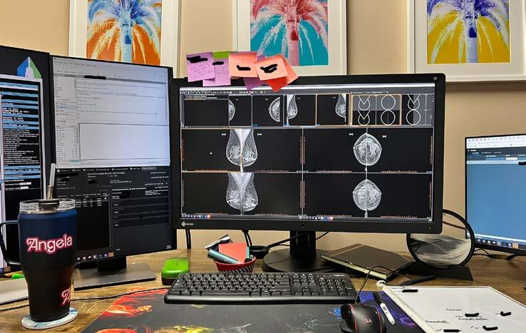
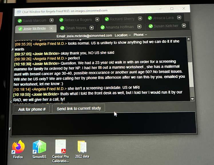
The job of a remote radiologist is not easy. Our remote radiologists cover up to 12 sites a day, with cases scheduled independently at each site. Radiologists must be able to manage multiple cases at the same time while ensuring that technologists complete their cases in the allotted time. This workload can be very challenging even with a good system. Not all radiologists can make the transition to managing remote diagnostic cases. Successful radiologists must have an organized process to make sure they are not mixing up cases or forgetting to dictate reports. I write my list of patients for each site on a wipe-off board in the morning (Figure 1). I place a check mark next to a patient’s name when the technologist sends me the images and then erase the name once I have dictated the report. I also dictate as I go, so I have the history, mammographic findings, and requested ultrasound images noted in the report when additional images are sent back to me for review. I don’t have to figure out who the patient is, what is going on, or what I wanted done, because it is all documented. This system saves time and limits mistakes.
Figure 1
Angela Fried, MD
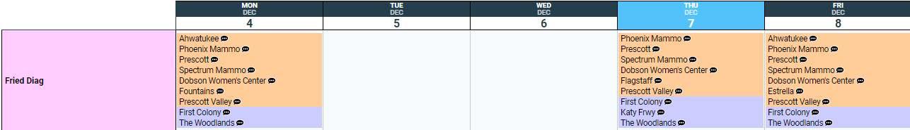
Our practice has developed programs to streamline the process. Our scheduling system assigns each radiologist their sites for the day (Figure 2) so the radiologists and technologists know who they will be working with that day. The physician relations team is also available to reassign cases to different radiologists to manage overbooking or emergencies, if needed. Technologists communicate with radiologists through our messenger system in the picture archiving and communication system (PACS). They send a link to the case and a short blurb describing the clinical history and indication. Instructions are then sent back to guide the workup (Figure 3). We have an online protocol available to the technologists. Each radiologist has a page in the protocol that outlines what they want done in a standard set of situations. This allows technologists who have not previously worked with a radiologist to review the radiologist’s expectations. We also tailor clinical schedules to individual radiologists. More efficient radiologists can cover more sites and more cases. Radiologists who are less efficient aren’t pushed to read more than they are capable of or comfortable with. We also have access to other breast radiologists for consultations through the messenger system in the PACS.
Ultrasonography is clearly the biggest concern for radiologists looking to start a remote diagnostic program. Dedicated breast sonographers are critical to a successful program. The technologist-radiologist relationship is very important, so we
try to schedule our radiologists consistently to cover the same sites on a regular basis so trust can grow over time. Giving technologists detailed instructions and descriptions of what you are looking for is helpful. Solving problems may involve using cine clips, describing landmarks, using three-dimensional ultrasonography, suggesting strategies to optimize image quality, or explaining to the technologist what you would do if you could actually scan the patient yourself. We also have lead technologists who can help troubleshoot issues if needed. Most importantly, though, when referring a patient for remote ultrasonography, radiologists should have a plan in case no ultrasound correlate is found, just as if they failed to find a correlate on in-person realtime examination.
None of this happened overnight. Our remote radiologist program grew from just two remote radiologists to 30 radiologists over 10 years. This time period allowed us to assess our performance as we grew, to receive and respond to feedback from procedural radiologists performing the breast biopsies, and to assure quality for our program. We developed strategies to improve after assessing weaknesses in the system, and we continue to look for ways to minimize errors and streamline processes. This model has allowed our practice to continue to grow and to recruit more highly qualified breast radiologists in the current job market.
Figure 2
Figure 3
MQSA Final Rule 2023: A Team Approach to Understanding the Changes
By Robyn Hadley, RT(R)(M)
Recent headlines include “FDA Updates Mammography Regulations in MQSA Final Rule,” “Major Updates Coming to Mammography Quality and Certification Standards,” and “Effective date: 9/10/2024.” All of these headlines refer to an upcoming update to the US Food and Drug Administration (FDA) Mammography Quality Standards Act (MQSA) guidelines. What regulations were updated? What changes will be necessary for a facility to implement? The value of MQSA regulations is in understanding the objective and purpose of regulations and using that knowledge to establish programs with better or best practice quality standards. The following information and task list outline the updates and suggest action steps that facilities can take to prepare for the deadline and ensure compliance.
Lead Interpreting Physician
The lead interpreting physician is the individual ultimately responsible for ensuring that the quality assurance program meets the required standards. The information provided here is intended to be helpful guidance that may be passed along to additional designated quality assurance personnel to use during the transition process. This can also be a great resource for discussions with all team members regarding adhering to and complying with the regulations while also exploring new ideas for implementing best practices.
FDA’s Final MQSA Rule Update 2023
What has changed and why? The FDA’s objectives are to improve delivery of mammography services, strengthen communication, provide information for patients and clinicians to make informed decisions, update technological standards, ensure the availability of qualified personnel, and improve mammographic interpretation through essential feedback with required metrics for the mammography medical outcomes audit analysis. The changes made by the FDA are summarized in the following three categories. Some changes within the final rule are minor revisions pertaining to specific verbiage modifications, whereas others are new or specifically revised changes warranting action.
Updates Addressing Changes in Technology
• Image retention, transfer, and release of copies. Facilities must retain original mammographic images and have them available for transfer upon request. Facilities shall implement policies and procedures to minimize record loss. Transfer of mammograms
and reports must take place within 15 calendar days of receiving the request. Providing timely results to patients is of utmost importance; therefore, image access and availability are essential. Minimizing loss of records is aimed at increasing the impact of clinical care.
Changes to Enhance Quality Standards
• Image interpretation. Mammograms must be submitted for interpretation in the mammographic modality in which they were originally produced and images must be retained in the original modality in which they were produced. To meet record retention requirements, images cannot be scanned, copied, or digitized. The purpose of submitting images in the original mammography modality is to prevent possible negative implications associated with visualization of normal and abnormal breast tissue due to image quality.
• Accreditation application after three failures. No accrediting body shall accept an application for accreditation from a facility that has failed to become accredited after three consecutive attempts until one year after the date of the most recent failure. This rule will prohibit facilities from switching accrediting bodies to avoid the one-year exclusion after three failed accreditation attempts. A one-year waiting period is believed to be adequate for a facility to complete necessary corrective action such as mandatory trainings, personnel changes, and so forth.
• Facility certificate suspension. A facility’s certificate may be suspended or revoked due to failure to comply with requests by the FDA, the state certifying agency, or the accrediting body for records or information. This includes clinical images for an additional mammography review or requests for records documenting personnel qualifications.
• Digital accessories. The final ruling states, “All devices used in mammography must have met the applicable FDA premarket authorization requirements for medical devices of that type and intended use.”
• Additional mammography review and patient-physician notification Updates added the state certification agency as an entity that
Continued on page 12>
Robyn Hadley, RT(R)(M)
may initiate an additional mammography review. The FDA and state certification agency can notify patients and their physicians individually or through mass media when a facility is unable to perform a required patient-physician notification.
• Facility closure. Before a facility closure, the facility must arrange for access to mammography images and reports for patients and health care professionals.
• Retention and release of personnel records. A facility must provide personnel with copies of their MQSA qualification records. The FDA recognizes that employees need to have access to their personnel records for a period of time upon leaving a facility. Facilities are required to keep an employee’s personnel records for no less than 24 months from the date of the employee’s departure and be able to provide these documents to former employees for at least a 24-month period. The records must also be available for review during inspections occurring during that 24-month period. The goal of this requirement is to preserve access to mammography services and minimize delays in hiring new personnel. Upon facility closure, copies of MQSA qualification records must be provided to personnel.
Changes to How Results Are Reported, Categorized, Retained, and Transferred to Patients and Clinicians
• Mammography report and results letter information.
Mammography reports must include the facility name and location (at minimum, the city, state, zip code, and telephone number). Facilities may choose to add additional information such as email address, records storage site, or additional information about the health care network or organization. At a minimum, the letters must include the name of the patient; name, address and telephone number of the performing facility; and a breast density assessment.
• Report assessment categories. The final rule updated the explanatory language in the “benign” final assessment category and added three new assessment categories to the existing categories in regulation. The two “incomplete” assessment categories are taken from the 2003 approved alternative standard 11: “incomplete: need additional imaging evaluation” and “incomplete: need prior mammograms for comparison.” “Postprocedure mammogram for marker placement” is almost identical to the 2003 approved alternative standard 12. This category also helps facilities identify and exclude this assessment category from the medical outcomes audit data.
• Reporting time frame. For mammography reports with a final assessment of “suspicious” or “highly suggestive of malignancy,” the report to the clinician and the result lay letter to the patient must be sent within seven days of final interpretation. The time frame for sending all mammographic reports and results lay letters is 30 days from the date of the examination.
• Breast density notification. All facilities must use a national dense breast reporting standard for all patients for mammography reports to health care professionals and result lay letters to patients. Both reporting standards state the tissue density and recommend that patients talk with their clinician about breast density and their individual situation. At a minimum, the standard language (below) must be included and cannot be altered. Facilities may choose to add additional information. Most importantly, for facilities in states that already require specific density notification, this information can be included but must be distinctly separate from the FDA’s required language. Facilities need to comply with all federal, state, and local requirements. The intention of the final rule is to ensure that patients are given a consistent baseline of information regarding their breast density.
- Not dense: “Breast tissue can be either dense or not dense. Dense tissue makes it harder to find breast cancer on a mammogram and also raises the risk of developing breast cancer. Your breast tissue is not dense. Talk to your provider about breast density, risks for breast cancer, and your individual situation.”
- Dense: “Breast tissue can be either dense or not dense. Dense tissue makes it harder to find breast cancer on a mammogram and also raises the risk of developing breast cancer. Your breast tissue is dense. In some people with dense tissue, other imaging tests in addition to a mammogram may help find cancers. Talk to your provider about breast density, risks for breast cancer, and your individual situation.”1-3
• Medical outcomes audit metrics. The mammography medical outcomes audit report must contain specific information collected and calculated by facilities. At a minimum, the metrics required to be reported are positive predictive value, cancer detection rate, and recall rate.
- Check the MQSA Policy Guidance Help System regularly for updates and changes: Policy Guidance Help System | FDA
- Densebreast-info: https://densebreast-info.org/
• Breast density results lay letters: Research your state and local guidelines and determine the language your facility will use in addition to the federal requirements. Refer to the FDA standard language along with the FDA Final Rule 900.12 and specific state requirements.1
To save lives and minimize the impact of breast cancer. .....
• Breast density mammography report: Ensure that mammography reports contain the specific breast density language. Refer to the ACR BI-RADS Atlas and the FDA Final Rule.
- “The breasts are almost entirely fatty.”
- “There are scattered areas of fibroglandular density.”
- “The breasts are heterogeneously dense, which may obscure small masses.”
- “The breasts are extremely dense, which lowers the sensitivity of mammography.”
• Reporting
- Review your mammography reports to ensure that proper verbiage is used for the assessment categories.
- Review your mammography reports to ensure that adequate facility and patient information is included on both the reports and results lay letters.
• Policies and procedure review and update: Ensure that your policies and procedures are up to date and reflect all changes in the final ruling. Be mindful of time requirements.
- Record retention: how and how long?
- Reporting and review time frames for all assessment categories
- Reporting time frame for all mammography examinations with updates for reports with “suspicious” and “highly suggestive of malignancy” categories
- Time frame requirements for image retention and release
- Facility closure guidelines pertaining to patient record transfer and personnel records availability
- Personnel record retention and release with appropriate time frame listed
• Medical outcomes audit: Ensure that the required metrics (positive predictive value, cancer detection rate, and recall rate) are being calculated accurately and reported.
Regulatory compliance tasks may be designated to one or two individuals within your organization, but upholding the rulings is ultimately the responsibility of all team members. Doing so is essential to keeping best practice at the core of high-quality imaging. In the midst of these changes, it is imperative to remember that caring for our patients and protecting their precious moments of life is the “why” that drives the attention to regulatory compliance.
References
1. Mammography Quality Standards Act. Federal Register. Accessed March 5, 2023. https://www.federalregister.gov/documents/2023/03/10/2023-04550/ mammography-quality-standards-act#print
2. Densebreast-info. Accessed March 2023. https://densebreast-info.org/ Accessed March 5, 2023.
3. Berg WA, Seitzman RL, Pushkin J. Implementing the national dense breast reporting standard, expanding supplemental screening using current guidelines, and the proposed Find It Early Act J Breast Imaging. 2023;5(6):712-723. doi:10.1093/ jbi/wbad034
Incorporating Hybrid Breast Imaging Work in a Large Academic Practice (continued from page 8)
A key factor to the success of remote diagnostic breast care was found to be the sonographer’s skill. Therefore, we enhanced the training of all of our ultrasound technologists, educating them in a similar way as first-year residents so they could confidently review mammography and tomosynthesis images, understand the features of various findings, locate a finding, and confidently search that location for an ultrasound correlate. This additional training has been highly valued by our technologists, has enhanced their job satisfaction, and has facilitated overall excellence in our patient care.
In short, remote diagnostic breast imaging has several potential advantages. First, it may significantly improve care in underserved areas by allowing patients in these areas to have access to expert breast radiologists located in other regions. Second, it creates the opportunity to offer radiologists needed autonomy and wellness by giving them a choice about their day-to-day work and improves organizational resiliency by increasing potential recruitment to any organization. Third, it empowers our technologists to contribute to the care of our patients more fully, which fosters growth and development.
References
1. Milch HS, Grimm LJ, Plimpton SR, et al. COVID-19 and breast radiologist wellness: impact of gender, financial loss, and childcare need J Am Coll Radiol 2021;18(7):1017-1026. doi:10.1016/j.jacr.2021.02.022
2. Parikh JR, Sun J, Mainiero MB. Prevalence of burnout in breast imaging radiologists J Breast Imaging. 2020;2(2):112-118. doi:10.1093/jbi/wbz091
3. Ganeshan D, Rosenkrantz AB, Bassett RL Jr, Williams L, Lenchik L, Yang W. Burnout in academic radiologists in the United States Acad Radiol 2020;27(9):1274-1281. doi:10.1016/j.acra.2019.12.029
4. Recht MP. Work from home in academic radiology departments: advantages, disadvantages and strategies for the future Acad Radiol. 2023;30(4):585-589. doi:10.1016/j.acra.2022.11.019
5. Mango VL, Stoeckl EM, Reid NJ, et al. Impact of high neighborhood socioeconomic deprivation on access to accredited breast imaging screening and diagnostic facilities J Am Coll Radiol. 2023;20(7):634-639. doi:10.1016/j. jacr.2023.04.006
6. Nguyen DL, Ambinder EB, Myers KS, Oluyemi E. Addressing disparities related to access of multimodality breast imaging services before and during the COVID-19 pandemic Acad Radiol. 2022;29(12):1852-1860. doi:10.1016/j. acra.2022.03.017
The Canadian Society of Breast Imaging
By Jean Seely, MD, FRCPC, FSBI, FCAR
It’s hard to believe that the new year is upon us. This is a time of hope, and like most of you, the Canadian Society of Breast Imaging (CSBI) is working to make the world of breast imaging a better place. The Inuktitut word that is used by the first Indigenous Canadian governor general, Mary Simon, is ajuinata, pronounced “aye-yoo-ee-nah-tah.” The word means that when you are confronted with adversity or things that are difficult, you keep going and you don’t give up, and you make a commitment to continue to make changes.
Times are challenging: we are dealing with staff shortages of radiologists and of mammography, ultrasound, and magnetic resonance imaging (MRI) technologists. There is a nationwide shortage of health care professionals, with one in five Canadians having no regular clinician. 1 Record levels of burnout and increasing volumes of work with inadequate staff contribute to staff member resignations and increasing wait times for imaging. 2
Despite these hardships and global health care challenges, CSBI is doing well. Its 210 members are working to connect radiologists, technologists, patients, and health care professionals across the country, forming a community that is working to sustain and build the workforce and increase advocacy for breast imaging. We are stronger together and work to support each other.
The CSBI Board of Directors now includes Dr. Supriya Kulkarni, FSBI (president-elect); Dr. Paula Gordon, FSBI (University of British Columbia); Sue Peters (patient advocate); two new radiologist members, Dr. Zina Kellow (Dalhousie University, Halifax) and Dr. Silma Solorzano (McGill University, Montreal); and two new trainee members, Dr. Alice (Jijun) Wang (University of Calgary, postgraduate year 3 radiology resident) and Dr. June (Huijang) Wang (University of Michigan, postgraduate year 5 radiology resident). They join Dr. Mona El Khoury (Centre hospitalier de l’Université de Montréal), Dr. Raman Verma (University of Ottawa), Christie Barbesin, MRT (The Ottawa Hospital), Dr. Kaitlin Zaki-Metias (University of Michigan,
radiology resident), Dr. Sri Sannihita Vatturi (University of Ottawa, radiology resident), Dr. Jean Seely (University of Ottawa), and Amy Smith, BComm (executive director).
We are truly grateful for the contributions of former board members Dr.
Nancy Wadden, Dr. Charlotte Yong-Hing, and Dr. Carolyn Flegg.
This past fall, CSBI hosted these excellent educational initiatives:
• Shortened Breast MRI Protocols (October 26, 2023), in collaboration with Siemens, a live breast webinar to enhance the quality and capacity for breast MRI with ACR-accredited and Canadian Association of Radiologists–accredited breast MRI protocols, by Dr. Seely
• The Second Annual Collaborative Patient and Expert Information Webinar (November 11, 2023), by Dr. Gordon, Dr. Sehdev, and Dr. Seely, moderated by Dr. Appavoo, with guest speaker Ellyn (advocate and breast cancer survivor)
• CSBI Hands-On Virtual Mammography Workshop (November 18, 2023), in collaboration with DetectedX, by Dr. Seely, Dr. Kulkarni, and Dr. Rickard (Australia)
• CSBI Breast Imaging Career Night (November 16, 2023), by Dr. Zaki-Metias, Dr. Vatturi, Dr. Scott-Moncrieff, and Dr. Solorzano
Regarding breast imaging advocacy, we have some good news. New Brunswick announced that in early 2024 it will start to include women in their 40s in their screening program, a new change since the program began in 1996. And for the first time since the Ontario Breast Screening Program was established in 1990, the provincial government has approved self-referral for women in their 40s, albeit only every two years, starting in the fall of 2024. This means that 305,000 women in Ontario in their 40s will have access to earlier detection for breast cancer. It is difficult for many of our American colleagues to understand
To save lives and minimize the impact of breast cancer. .....
Jean Seely, MD, FRCPC, FSBI, FCAR
the limitations imposed by the Canadian Task Force (CTF) guidelines of 2011 and 2018, which recommend against women in their 40s undergoing screening mammography. Screening practices under these guidelines have led to higher rates of advanced breast cancer and lower rates of stage I breast cancer diagnosed among women in their 40s compared with women in their 50s.3 For the first time, the Ontario government is following other jurisdictions such as British Columbia, Nova Scotia, Yukon territory, and Prince Edward Island and will not follow the outdated CTF guidelines. Important work must continue to provide access to screening among women aged 40 to 49 years in the remaining provinces: Quebec, Manitoba, and Saskatchewan. Alberta recently updated their guidelines to include women aged 45 to 49 years.
As noted in the Summer 2023 SBI newsletter, the Public Health Agency of Canada announced that the CTF will be updating its breast screening guidelines. Work began in the summer and for the first time included patients, radiologists, and experts in oncology. However, we have since learned that experts and stakeholders are not allowed to vote, and a strong antiscreening bias by the two cochairs refuting the strong science for screening has been noted on public record.4
On December 6, 2023, the Canadian House of Commons Standing Committee on Health, which studies issues related
to Health Canada, invited Dr. Gordon, Jennie Dale (Dense Breasts Canada), and Dr. Anna Wilkinson (general practitioner oncologist) to discuss the importance of the CTF update. They responded eloquently to the antiscreening defense of the Canadian National Breast Screening Study by Dr. Steven Narod. A recording of the meeting is publicly available: https://www. ourcommons.ca/committees/en/HESA/StudyActivity?studyActivityId=11695303
Ajuinata is apt; let’s keep working, moving forward, and making positive changes as we look forward to connecting and sharing our achievements at the SBI/CSBI symposium in April in Montreal!
References
1. Duong D, Vogel L. National survey highlights worsening primary care access CMAJ. 2023;195(16):E592-E593. doi:10.1503/cmaj.1096049
2. Cao DJ, Hurrell C, Patlas MN. Current status of burnout in Canadian radiology Can Assoc Radiol J. 2023;74(1):37-43. doi:10.1177/08465371221117282
3. Wilkinson AN, Billette JM, Ellison LF, Killip MA, Islam N, Seely JM. The impact of organised screening programs on breast cancer stage at diagnosis for Canadian women aged 40-49 and 50-59 Curr Oncol. 2022;29(8):5627-5643. doi:10.3390/curroncol29080444
4. Thériault G, Reynolds DL, Grad R, et al. Debunking myths about screening: how to screen more judiciously Can Fam Physician. 2023;69(11):767-771. doi:10.46747/ cfp.6911767
ACADEMIA OR PRIVATE PRACTICE: AN ENDURING CONUNDRUM, PART 2
By Wenhui Zhou, MD
After years of dedicated training, the choice between academic and private practice is a pivotal decision that radiologists often grapple with because it shapes the trajectory of their professional lives. These distinct paths offer unique opportunities and considerations, spanning areas such as research, teaching, compensation, and autonomy. Individual priorities, including work-life balance, career growth, financial factors, and personal fulfillment, play a decisive role in this choice.
We are dedicating a two-part Member-in-Training series to shed light on this topic. For this second article, we sought advice from mid-career and expert radiologists from academic and private practice settings. Their valuable insights and experiences will help readers navigate the appeals and drawbacks of private practice and academic radiology and offer guidance in the pursuit of professional satisfaction and success.
Dr. Brittany Dashvesky is a breast radiologist and assistant professor at Stanford University School of Medicine. She completed her breast imaging fellowship in 2018. She served as the director of breast imaging at a private practice group in northern California and as medical director of breast imaging for Providence/ St. Joseph Health for four and a half years before assuming her current academic position.
Dr. Steven Poplack is a breast radiologist and a professor at Stanford University. He completed his residency and fellowship training in 1993 and has since had a distinguished career in education, research, and patient care in the field of breast imaging.
WZ: What led you to choose either academia or private practice for your career as a radiologist? What factors influenced your decision?
BD: After completing my breast imaging fellowship, I joined a private practice in Sonoma where I assumed a leadership role in the breast section. The opportunity to work with hospital administration to improve and expand the breast program was exciting to me. This practice struck the right balance between
clinical demands and work-life balance. I valued autonomy and the ability to shape the breast section according to my vision. However, as the COVID-19 pandemic affected our practice’s sustainability, I more recently made the decision to transition to academia to pursue my passion for teaching and research.
SP: I’m not sure I chose academics; rather, it chose me. My father was a radiologist who had begun his career in academics and transitioned to private practice. In anticipation of joining his private practice I did cross-sectional and breast imaging fellowships in my fourth year of residency. (In 1992 there were few postresidency fellowships.) Concurrently there was a wave of reorganization in medicine, prompting many smaller community practices to merge, including my dad’s practice, which resulted in a hiring freeze. I expanded my job search to include academics, thinking that would position me for an easy jump to private practice. After several unsatisfying job interviews (in both academics and community practice) I was offered a job in academics to develop a stereotactic biopsy program, which was new technology at the time. I accepted the offer because I trusted the chair, liked the attitude and demeanor of my future radiologist colleagues, and was attracted to the job description that included general radiology, allowing me to maintain a broad skill set and develop subspecialty expertise in breast imaging (almost exclusively mammography back then).
Can you share any personal anecdotes or experiences that highlight the unique aspects of academic and private practice radiology?
BD: There are unique challenges with each. In private practice you may have to recall from distant training how to do a hysterosalpingogram, lumbar puncture, thoracentesis, or joint injection. Basic general procedures and participation in evening or overnight calls are usually expected. When considering a private practice, it is important to know expectations around which cases you will interpret and which cases are read by subspecialists. For newly minted fellowship-trained breast
Wenhui Zhou, MD
radiologists, you will be able to enjoy being an expert in your field but may need to find a mentor to help you through cases outside your comfort.
In contrast, a career in academia generally precludes you from reading studies outside of your subspecialty and blesses you with no general radiology call. I am often struck by the sheer complexity of cases in academia, such as the occasional fourplus wire localization or multisite bilateral magnetic resonance imaging biopsy. With fewer cases and trainee-prepared dictations, there is time to engage/teach trainees and hopefully spark a passion for breast imaging. Academia also brings a robust network of renowned colleagues to provide second opinions or to collaborate with on research endeavors.
SP: While still intending to transition to community practice, I had two serendipitous experiences that solidified my career in academic practice. First, I was invited to serve as the consulting radiologist in a new mammography-pathology registry, ultimately one of the five founding member registries of the Breast Cancer Surveillance Consortium. This experience introduced me to public health research and working with a diverse team of nonradiologist researchers. About a year later I was approached by a computational scientist to provide clinical expertise for a Department of Defense grant submission to develop five novel breast imaging platforms. Collaborating with a group of biomedical engineers, who thought and worked differently than I, was particularly intriguing. These two early experiences opened my eyes to the breadth and vitality of research and to the possibility of contributing at the societal level in addition to helping individual patients.
Can you share your insights into the overall culture, leadership structure, and work environment of academia and private practice?
BD: In private practice, the culture revolves around efficient patient care delivery and building strong relationships with referring providers. Partnership is usually based on time at the practice. Radiologists work largely independently. This is balanced by social lunches and regular group meetings, which can be pretty rowdy, particularly when picking vacation dates, allocating bonuses, or venting about policy changes and call schedules.
In academia, the culture fosters collaboration among radiologists [and] emphasizes multidisciplinary coordination of care, teaching, and research pursuits. You’re often with a team of residents and fellows, overseeing their procedures and exams, as opposed to performing them yourself. There’s a policy or protocol for most things that can help guide your decision-making process. Leadership structure can vary, with section chiefs, department chairs, or academic committees guiding decision-making processes, including promotions, which are based on individual achievements and demonstrated regional or national recognition.
SP: From a leadership perspective, academics seems more structured than community practice. This structure can make it harder to have your voice heard at the highest levels but also contributes to a comforting sense of boundaries and clearer picture of who to turn to first. For the most part academic practice is collaborative. There is less (though not zero) focus on relative value unit productivity and more of a team approach to getting the work done. There is more capacity to share difficult cases and learn from knowledgeable colleagues. The academic work environment seems lower key than many community practices. There seems to be more opportunity for appreciating “great cases” and focusing on the service we provide to patients. On the flip side there is more tension related to academic productivity. Most importantly for me, there is less attention to the business of radiology. In academic practice, I feel that I can attend to various responsibilities and be spared from having to consider the financial bottom line.
Can you comment on work-life balance and professional growth opportunities in academia and private practice?
BD: In private practice, there is often a focus on efficiency, resulting in a higher daily volume of cases. The workload can vary depending on the practice. Some practices offer a shorter work week and more vacation time, while others may require more hours with commensurate compensation. In terms of professional growth, private practice often offers a clear path to partnership, where you can have a voice in practice management and decision-making.
In academia, the pace is often more measured, allowing for dedicated time for research and teaching commitments. Ample time is provided to attend/present at conferences and allow for professional development. In terms of professional growth, there is a structured path for professional growth and advancement, with opportunities for promotion based on research, teaching, and contributions to the field.
SP: To my mind, work-life balance is dependent on personal choices and [has] less to do with the practice environment. I think it is essential for radiologists at all experience levels to understand what they value most and find a way to structure their job (or find a job) that fits those values. There are always demands to commit to more. That commitment can bring work fulfillment at the expense of life balance, while emphasizing balance can bring greater nonwork satisfaction, potentially at the expense of job satisfaction.
Continued on page 19>
EUSOBI Annual Scientific Meeting 2023: A Summary by the EUSOBI Young Club
By Sarah Hickman, MD, PhD; Iva Biondic Spoljar, MD; Paola Clauser, MD, PhD;
Marianna
Fanizza, MD;
Elisabetta Giannotti, MD; Machteld Keupers, MD; Maria Adele Marino, MD; Simone Schiaffino, MD; Thiemo van Nijnatten, MD, PhD; Mirjam Wielema, MD; Michael Fuchsjäger, MD
The 2023 European Society of Breast Imaging (EUSOBI)
Annual Scientific Meeting took place in the city of 100 bell towers and home of paella, Valencia, Spain, in September 2023, achieving record attendance of 1737 in-person attendees. The Valencia Conference Centre was a hive of activity for attendees to meet friends, colleagues, and industry partners; listen to interesting presentations in the main hall and small satellite rooms; and view the 142 e-posters accepted for this year’s conference.
The conference once again covered popular topics within breast imaging and provided a productive environment to discuss topics such as different approaches for BI-RADS 3 lesions, breast cancer staging, hot topics in breast oncology, advances in imaging techniques to characterize lesions, and closing the global breast cancer divide. The potential role of abbreviated breast magnetic resonance imaging and personalized breast cancer screening continued to be topics of interest at this year’s meeting. The new BI-RADS lexicon for each modality was covered in detail; updates are expected early next year. The conference welcomed visitors from around the globe, including Radiology Editor in Chief Professor Linda Moy, who provided her insights into breast imaging in 2040, and speakers from Colombia, who explored triple-negative breast cancer and implant complications.
Educational sessions for clinical learning included an image interpretation quiz (resulting in a draw between the Spanish and European teams) and unusual cases presented by a multidisciplinary team panel (a surgeon, pathologist, and breast radiologist) providing valuable insights into complex case management. The importance of breast radiologist wellbeing was also highlighted with a dedicated session on how to stay happy as a breast radiologist. Artificial intelligence (AI) continues to dominate the discussion about the future of breast
imaging, risk stratification, and personalized screening. The use of AI in breast cancer screening was a center of discussion with the recent publication of two prospective trials in Sweden.1,2 However, there is ongoing deliberation as to the best method of adopting AI into the clinical workflow of each screening program.
Congratulations to this year’s dignitaries and award winners! Professor Francesco Sardanelli was awarded the Gold Medal for his outstanding contribution to the field of breast imaging. The most quoted breast imaging article published in European Radiology was an article by Lång et al titled “Identifying Normal Mammograms in a Large Screening Population Using Artificial Intelligence.”3 The most quoted article in Insights Into Imaging was an article by Trimboli et al titled “Do We Still Need Breast Cancer Screening in the Era of Targeted Therapies and Precision Medicine?”4 The 2023 EUSOBI Young Club (EYC) young research grants were presented to Dr. Pötsch and Dr. Rizzo, and the Carla Boetes award was presented to Dr. Bitencourt.
The EUSOBI conference dinner was held in the stunning City of Arts and Sciences, lit by a pink glow for the occasion. The EYC hosted a full program of activities for its members, including a morning workshop, practical laboratories, lunches, and an evening event with an inspirational talk from the chair of the European Society of Radiology Patient Advisory Group, Caroline Justich. The EYC lounge was an excellent meeting space for colleagues from all over Europe.
This year’s conference was a brilliant celebration of the collaborations and connections within the breast imaging community. The meeting hosted a broad range of talks and demonstrated the ongoing enthusiasm within the community to continue to improve clinical practice and patient care. We look forward to the 2024 meeting in Lisbon, Portugal, from October 3 to 5.
Sarah Hickman, MD, PhD
References
1. Dembrower K, Crippa A, Colón E, Eklund M, Strand F; ScreenTrustCAD Trial Consortium. Artificial intelligence for breast cancer detection in screening mammography in Sweden: a prospective, population-based, paired-reader, noninferiority study Lancet Digit Health. 2023;5(10):e703-e711. doi:10.1016/s25897500(23)00153-x
2. Lång K, Josefsson V, Larsson AM, et al. Artificial intelligence-supported screen reading versus standard double reading in the Mammography Screening with Artificial Intelligence trial (MASAI): a clinical safety analysis of a randomised, controlled, non-inferiority, single-blinded, screening accuracy study. Lancet Oncol. 2023;24(8):936-944. doi:10.1016/S1470-2045(23)00298-X
3. Lång K, Dustler M, Dahlblom V, Åkesson A, Andersson I, Zackrisson S. Identifying normal mammograms in a large screening population using artificial intelligence Eur Radiol. 2021;31(3):1687-1692. doi:10.1007/s00330-020-07165-1
4. Trimboli RM, Giorgi Rossi P, Battisti NML, et al. Do we still need breast cancer screening in the era of targeted therapies and precision medicine? Insights Imaging. 2020;11(1):105. doi:10.1186/s13244-020-00905-3
Member-in-Training Column: Academia or Private Practice: An Enduring Conundrum, Part 2 (continued from page 17)
For trainees who are undecided about whether to pursue an academic or private practice career in radiology, what advice would you offer to help them make an informed decision?
BD: Identify your unique needs/limitations and apply broadly to understand the demands/expectations and benefits of each. On interviewing, hopefully you find a group that you connect with. Seek guidance from mentors who have experienced both paths and see how each aligns with your personal aspirations. Also, as with everything, a job that is a great fit now may not be the right fit in 10 years. Stay engaged with SBI and maintain contacts.
SP: I would advocate for trainees that are uncertain to consider academics first. From a confidence perspective I think it is harder to transition from community practice to academic practice. Starting from private practice, there is a tendency to feel less equipped to function as “the expert” in the academic setting. For trainees who have considered both options and are beginning in academics, I think it is important to structure your academic practice in a way that allows you to maintain a broad skill set. In that way, if you decide to move to community practice you will remain comfortable interpreting a wide range of exams and be more attractive to a wider range of practices.
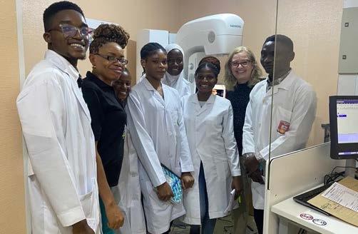
RAD-AID Nigeria Breast Imaging Update
By Adeleye Omisore, MD; Rachael Akinola, MD; Mobolaji Jaiyesimi, MD; Jeff Reiner, MD; Cindy Thornton, RT(M); Dolores Brown Smith, RT(M); Erica Pollack, MD; Farouk Dako, MD; Victoria L. Mango, MD, FSBI
Nigeria is a West African nation with about 218 million people and is the most populous country in Africa.1 Breast cancer is the most common cancer and the most common cause of cancer deaths among women in Nigeria.2 Late-stage presentation is common; more than 80% of Nigerian women with breast cancer present with stage III or IV disease, with an average tumor size of 10.5 cm.3 The incidence of breast cancer in Nigeria has increased in recent years to 52 to 64 cases per 100,000 women, a threefold increase compared with four decades ago.2 Timely access to quality breast imaging and image-guided biopsies are among the multiple factors contributing to great disparities in late-stage presentation and poor overall survival compared with North America.
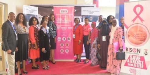
Our collaborative efforts to improve breast cancer outcomes in Nigeria through breast imaging incorporate the Breast Imaging Society of Nigeria (BISON), RAD-AID International, the African Research Group for Oncology (ARGO), and the Memorial Sloan Kettering Cancer Center Global Cancer Disparities Initiatives program, with a multidisciplinary focus on education, clinical care, and research.
BISON was founded in 2018 and consists of approximately 75 members, primarily radiologists, who are devoted to improving breast imaging services in Nigeria. BISON strongly believes in empowering women through education to be aware of their own breast health to make informed decisions. In addition to patientrelated education, BISON provides ongoing educational opportunities to radiologists in Nigeria to promote the field of breast imaging, educate members about breast imaging, and foster community involvement among breast radiologists. BISON has been instrumental in creating locally applicable, resource-appropriate breast imaging guidelines in Nigeria. BISON, in collaboration with Memorial Sloan Kettering Cancer Center, has also tremendously improved the use of tablet-based ultrasonography for imageguided biopsy by training multiple radiologists across Nigeria. This has resulted in improved diagnostic accuracy, encouraging local surgeons to trust and rely on image-guided biopsy for diagnosis.
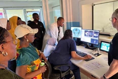
The Global Cancer Disparities Initiatives program and ARGO, a consortium of 30 Nigerian hospitals, celebrated their 10th anniversary in September 2023 with their annual symposium at Obafemi Awolowo University Teaching Hospitals Complex (OAUTHC) in Ife, southwestern Nigeria. The symposium had about 200 participants who were multidisciplinary cancer care professionals and trainees
from multiple centers in Nigeria. The participants included surgeons, radiologists, pathologists, medical oncologists, nurses, genetics counselors, technologists, statisticians, and informaticists. Nigerian radiologists participated in hands-on ultrasound-guided biopsy skill and wire localization workshops. Didactic lectures covered a range of breast imaging topics, including early detection strategies, bridging disparities through technological innovations, contrast-enhanced mammography, tomosynthesis, and axillary imaging. Interdisciplinary teams from Nigeria and North America also participated in multidisciplinary tumor board conferences to discuss further management of challenging breast cancer cases.
In addition to in-person teaching efforts, RAD-AID offers free educational materials to radiologists and technologists from ARGO sites through their learning center platform. This versatile online platform has high-quality educational content covering all subspecialties of radiology and includes numerous recorded educational lectures by SBI members and RAD-AID volunteers. Through generous donations from the ACR, the learning center platform also provides access to ACR Case in Point and RadExam educational materials, which incorporate subspecialty and resident-level examination questions.4,5
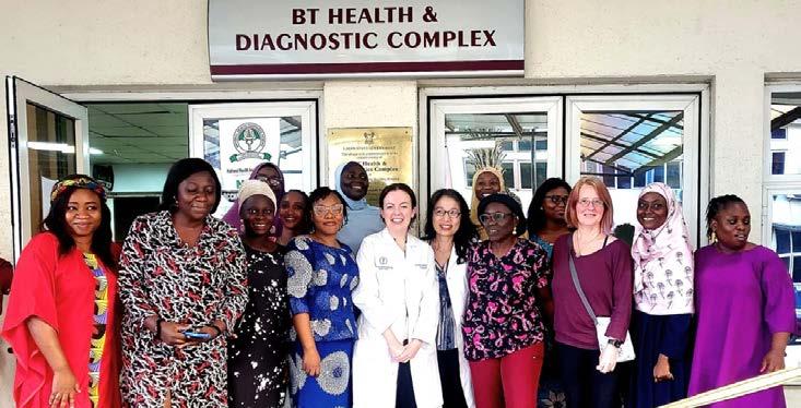
Through the support of RAD-AID International and the RAD-AID and American Society of Radiologic Technologists Foundation Outreach Fellowship, we have expanded breast imaging education outreach initiatives to include breast imaging technologists at University College Hospital Ibadan, Lagos State University Teaching Hospital (LASUTH), and OAUTHC. Technologist training has focused on mammography and breast magnetic resonance imaging (MRI), including mammographic positioning, breast imaging basics, mammography quality control and quality assurance, breast MRI positioning, MRI artifacts, and sequence optimization. Technologists also participated in hands-on training for mammography and breast MRI positioning. These efforts have helped introduce and establish the use of breast MRI at LASUTH, impacting the treatment of breast cancer patients at the institution.
In addition to building our ongoing educational programs, we look forward to our upcoming collaborations on teleultrasonography
Victoria Mango, MD, FSBI
initiatives to provide real-time breast imaging consultations remotely between Nigeria and the United States. Interdisciplinary collaborations will be further fostered through our regular radiologypathology conferences via Zoom. We are also working on deploying breast ultrasonography artificial intelligence at LASUTH and OAUTHC in 2024. Our long-term goal is to significantly decrease breast cancer–related disparities through our collaborative education, training, and research initiatives.
The RAD-AID breast imaging team is eager to welcome new volunteers with expertise in any aspect of breast cancer care who are interested in promoting high-quality care to underserved patients. Attending-level physicians and radiologists in training are welcome to apply, as are physician assistants, technologists, nurses, physicists, and informatics specialists. We invite you to learn more
on the RAD-AID website (www.rad-aid.org), sign up at https://portal.rad-aid.org/survey/general-volunteer-survey, or email breastimaging@rad-aid.org with inquiries. Remember to indicate that you are an SBI member when you sign up!
References
1. World Bank open data. World Bank. Accessed December 22, 2023. https://data. worldbank.org/
2. Cancer tomorrow. International Agency for Research on Cancer. Accessed February 4, 2023. https://gco.iarc.fr/tomorrow
3. Olasehinde O, Alatise O, Omisore A, et al. Contemporary management of breast cancer in Nigeria: insights from an institutional database Int J Cancer 2021;148(12):2906-2914. doi:10.1002/ijc.33484
4. Case in point. American College of Radiology. Accessed December 22, 2023. https://www.acr.org/Lifelong-Learning-and-CME/Learning-Activities/CaseinPoint
5. RadExam. American College of Radiology. Accessed December 22, 2023. https://www.acr.org/Lifelong-Learning-and-CME/Learning-Activities/RadExam
Breast MRI training at LASUTH.
Mammography technologist training at OAUTHC.
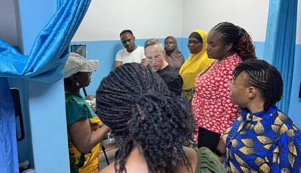
Hands-on ultrasound-guided breast biopsy sessions at LASUTH.
ARGO symposium.
LASUTH radiology department.
BISON annual meeting and scientific conference, December 2023.
On Monday, November 27, 2023, seven members were inducted as fellows of the SBI at the 2023 Radiological Society of North America conference in Chicago. The fellowship distinction is granted to SBI members who have demonstrated excellence in the discipline of breast imaging. Only 5% of SBI members have been awarded this honor. Get to know our latest round of honorees here.
Hannah L. Chung, MD, FSBI
Dr. Chung is a breast radiologist at University of Colorado Anschutz Medical Campus. She has both communitybased private practice and universitybased academic practice experience. She received her diagnostic radiology residency training at the University of Utah, Salt Lake City, and her breast imaging fellowship at the Iris Cantor Breast Imaging Center at the University of California, Los Angeles. She completed a nuclear medicine fellowship at the University of Utah before her radiology residency. In mid career, Dr. Chung entered academic practice interested in resident and fellow education and mentorship and in further defining the radiologist’s role in advancing the personalized care of each patient. Even in different academic environments, the observed variations in the practice and management of all aspects of breast imaging, including assessment and management of lymph nodes, high-risk lesions, and interventional procedures, indicate an ongoing need to better define best practices. Dr. Chung’s publications reflect her experiences and data-driven observations across these settings.
Brian N. Dontchos, MD, FSBI
Dr. Dontchos is an associate professor of radiology at the University of Washington and the clinical director of breast imaging at Fred Hutchinson Cancer Center. Before his time at the University of Washington, he served as the service chief of breast imaging at Massachusetts General Hospital. He graduated cum laude from the University of Pittsburgh School of Medicine in 2008 and was elected to the Alpha Omega Alpha and Gold Humanism honor societies during medical school. He completed his diagnostic radiology residency and breast imaging fellowship at the University of Washington. Dr. Dontchos is passionate about clinical implementation of creative workflows and new technologies and has led work in mitigating patient disparities through same-day imaging protocols.
Laura Heacock, MS, MD, FSBI
Dr. Heacock is an associate professor and director of breast magnetic resonance imaging (MRI) in the Department of Radiology at New York University (NYU) Langone Health. Dr. Heacock earned her undergraduate degree at Wellesley College, a master of science degree at Georgetown University, and her medical degree at New York Medical College. She completed her radiology residency and served as chief resident at New York Langone Health, subsequently completing a breast imaging fellowship and joining the NYU faculty in 2017. Dr. Heacock’s work on incorporating ultrafast and abbreviated breast MRI into clinical workflow led to a Radiological Society of North America Seed Grant in 2017. Her research interests include translational and multiparametric breast MRI, the use of multimodal artificial intelligence for breast cancer detection and risk prediction, and large language models/generative artificial intelligence in improving patient care.
Megan Kalambo, MD, FSBI
Dr. Kalambo is an associate professor in the Department of Breast Imaging at The University of Texas MD Anderson Cancer Center and clinical medical director of breast imaging for the Houston-area locations. She obtained her medical degree from Pennsylvania State College of Medicine and completed her residency training at The University of Texas McGovern Medical School at Houston. Following her residency, she completed a dedicated breast imaging fellowship at MD Anderson in 2012 and joined the breast imaging department as a faculty member in 2013. In 2021, she began serving as clinical medical director of breast imaging for the Houston-area locations, overseeing breast imaging clinical operations at the West Houston, Woodlands, and League City breast centers. She has a keen interest in community-based academic radiology practice, clinical operations, safety, and quality improvement. She also has a passion for teaching residents and fellows.
Supriya Kulkarni, DMRD, DNB, FSBI
Dr. Kulkarni is an associate professor and division head of breast imaging at the University of Toronto Department of Medical Imaging. She is a breast imaging radiologist within the Joint Department of Medical Imaging and has over 23 years’ experience with breast imaging, working in hospitals with patients in multiple capacities, including advocacy within the Ontario Breast Screening Program, the largest organized screening program in Canada. She is internationally renowned for her teaching and education and has over 150 didactic lectures, numerous visiting professor engagements, and outstanding teacher and mentorship awards for postgraduate education and contribution to cancer education. She is the president-elect of the Canadian Society of Breast Imaging and currently serves as a board member and the educational director.
Randy C. Miles, MD, MPH, FSBI
Dr. Miles serves as the chief of breast imaging at Denver Health, with oversight over the breast division’s clinical, research, and educational programs. Dr. Miles earned his medical degree from Mayo Clinic College of Medicine, where he was awarded the Miller Award for Humanitarianism related to his work leading medical mission trips to Haiti and the Dominican Republic. He also completed his master of public health degree from Harvard School of Public Health during medical school, where he was selected as a Zuckerman Fellow. He completed his diagnostic radiology residency training at the University of Illinois, Chicago, where he served as chief resident, and completed his breast imaging fellowship at the University of Washington Medical Center/Seattle Cancer Care Alliance. Dr. Miles has received numerous research grants, has published over 50 academic papers in breast imaging, and has led numerous educational efforts involving trainees at all levels, both nationally and internationally.
Lisa A. Mullen, MD, FSBI
Dr. Mullen is an assistant professor in the Division of Breast Imaging in the Johns Hopkins Department of Radiology in Baltimore, Maryland, where she has been the breast imaging fellowship director since 2013. She is a board member of the Breast Imaging Fellowship Consortium. Dr. Mullen is also involved in breast imaging curriculum development and delivery for radiology residents in international low-resource settings, including Tanzania and Zambia. Her research interests include applying artificial intelligence to breast imaging and improving breast imaging clinical care and patient access. She has authored or coauthored over 50 publications on these topics in peerreviewed journals. Dr. Mullen is active in legislative advocacy, both at the state and national levels, as the current president of the Maryland Radiological Society and as an ACR councilor representing Maryland. She has served on several SBI committees and received a 2022-2023 Journal of Breast Imaging Editor’s Recognition Award as an exceptional reviewer.
THE PATIENT'S PERSPECTIVE
JoAnn Pushkin
By Danielle Sharek, MD
Ms. JoAnn Pushkin is the executive director of DenseBreastInfo.org, a website centered on developing comprehensive, medically sourced educational resources to objectively inform patient and health care professional discussions around breast density. Ms. Pushkin has coauthored educational courses, articles, and grants and given talks and interviews on the topic of breast density. Her advocacy efforts were prompted by her own personal experience of self-detecting a palpable breast cancer that was masked by dense breast tissue on mammography (https://densebreast-info.org/about-us/our-team/). This interview has been edited for clarity and length.
DS: Please tell me about yourself and your background.
JP: I spent most of my career in stock research with a side consulting business in marketing/writing. At age 45 I was diagnosed with invasive breast cancer. At nearly the same time, in early 2006, a lifelong dream came true and a children’s story I wrote was published. I had hoped to focus on writing for children full time. While my dream to write was realized, my published work is now entirely on the topic of breast density.
How were you diagnosed with breast cancer?
Several weeks after a “normal” mammogram I felt a lump during a self-exam. Though large enough to feel, it did not show on a diagnostic mammogram, but that same day it was easily seen on an ultrasound. I learned I had breast cancer, I learned it was missed due to dense tissue, and I learned I had dense breasts, all within 10 minutes of each other.
How did you feel when you learned of the news?
At first diagnosis, numb. Then devastated. Then incredulous. Then angry. Then motivated to effect change.
After a recurrence 5 years later, I remember thinking, in that split second before my body physically reacted to hearing the devastating news, “Well, I am apparently very good at growing breast cancer.”
What was your treatment process?
Did you face any treatment obstacles? How did you overcome them?
Surgeries, chemo, more surgeries. Despite a mastectomy, a recurrence 5 years later, and then radiation.
What motivated you during your diagnosis and treatment process?
My daughter was 15 when I was diagnosed. I remember negotiating with the universe, “Please let me at least get her off to college and then you can have me.”
What did you learn from your experience?
That patients need to be informed about tests they are being sent for—and should specifically ask what the benefit and realistic limitations are for those tests in their personal situation.
If I had known I had extremely dense breasts and that cancer detection on mammography was so compromised due to it, I certainly would have initiated a conversation about supplemental screening with my provider. But you can’t have a conversation about something you aren’t even aware that you need to. That lack of knowledge effectively denied me the opportunity for an early-stage diagnosis.
How has this diagnosis impacted your life? How have you used your diagnosis to impact others?
After learning that Connecticut had just enacted a law to provide women some limited information about breast density, in 2010 I initiated efforts for a New York state “inform” law, a federal law for national notification, and an FDA/MQSA [Food and Drug Administration/Mammography Quality Standards Act] rule change to require that standardized information about breast density be included in the patient lay letter.
Danielle Sharek, MD
New York’s law was the first to actually inform women if they have dense breasts and served as a template to many other state laws that followed. On the national level, the FDA did update its dense breast reporting requirement, and that goes into effect in September 2024. I am also currently a consumer representative to the FDA National Mammography Quality Assurance Advisory Committee.
And to address educational needs created by density informing and supplemental screening discussions, in 2015 I cofounded, with Dr. Wendie Berg and rad tech Cindy Henke, the DenseBreast-info.org website. This medically sourced educational resource on the topic is clearly needed; in 2023 alone, the website hosted 810,000 visits.
Are there any lessons that you think the breast imaging community can learn from your experience?
Nearly every state inform law was fought for by a woman who was screened annually by mammography, did not know she had dense breasts, detected a lump, learned it had been missed on the mammogram due to density, and was floored to learn that this “missed-due-to-density” cancer was not a rare occurrence to medical providers. When everyone a patient is relying on for medical direction doesn’t share this one very important thing that results in a cancer detected that is no longer early stage, it results in a feeling of betrayal and crisis of faith in the medical system.
You have worked very hard to advocate for women with dense breasts. What achievement is most meaningful to you, and what are your plans for future work in the field of breast cancer?
Three achievements:
• Website: DenseBreast-info.org is now the world’s leading website on the topic. The educational website provides medically sourced, peer-reviewed, evidence-based educational content for patients and providers. Our medical advisory board consists of internationally recognized, published experts in breast imaging and obstetrics/gynecology.
• National reporting standard: The FDA reporting rule going into effect in September 2024 creates a single baseline level of dense breast information for women across the United States and solves the dilemma of varying levels of “inform” created by 38 individual inform laws. Though I was relieved, it was a bittersweet accomplishment (see my March 2023 public statement).
• Find It Early Act: The new FDA reporting standard will mean that all women will be told whether their breasts are “not dense” or “dense.” And for those who have dense breasts, that “…other imaging tests in addition to your mammogram may help find cancers.”
But once women are informed that they have dense breasts or are at higher risk and may benefit from supplemental imaging to complete breast screening, can they afford it? For those individuals for whom a mammogram screening is not enough, expanded insurance coverage for breast imaging beyond the mammogram is needed. The next legislative front is continuing work on state insurance laws and the federal Find It Early Act (FIEA). The FIEA would ensure all health insurance plans cover screening and diagnostic breast imaging with no out-of-pocket costs for women with dense breasts or at higher risk for breast cancer, close loopholes inherent in state insurance laws, and cover all private and public plans, including TRICARE and Veterans Health Administration plans.
A statement from Executive Director JoAnn Pushkin
It's been over ten years since my request to the FDA to consider the requirement that women be informed about their breast density after their mammogram, and over ten years since an FDA advisory committee agreed that women certainly should be.
Too many patient "density inform" advocates are no longer with us to join me in an exhale of relief that all U.S. women will now be told about their breast density. That their tragedies - of hidden cancers finally found when no longer early stage - may be prevented from wreaking havoc on other families.
So, while we all share a hallelujah! that this has finally come to pass, to quote songwriter Leonard Cohen, for some of us, "it's a cold and it's a broken Hallelujah." But a hallelujah it is.
Sustaining Relationships in the Remote Imaging Environment
By Sarah Jacobs, BS, RT(R)(M)(CT)
It is no secret that there is a shortage of dedicated breast radiologists in almost every state in America.1 In addition to the breast radiologist shortage, as of this writing there are over 4500 job postings for mammography technologists in the United States. This shortage has increased burnout and decreased job satisfaction over the last several years. Facilities have had to redefine their workflows and processes to cope with the workforce shortage.
One method used to combat these shortages and provide care to mammography patients is the implementation of a remote or hybrid reading system through internal or external telemammography services. Since the COVID-19 pandemic began, our industry has seen a dramatic increase in the interpretation of examinations through telemammography. Before the pandemic this method of teleradiology interpretation was reserved for overnight and on-call shifts2 rather than for routine mammography examinations. Using telemammography has proved to be beneficial in many aspects, including increasing access to breast radiologist expertise, improving outcomes in breast cancer diagnosis, and providing timely interpretation and treatment for patients. This is accomplished without negatively impacting workflow.3 Additional benefits of telemammography services include a potential decrease in burnout and increase in job satisfaction.
While telemammography has many benefits, there are also some challenges that should be considered when implementing a telemammography program. Challenges may include implementation cost, liability risks, and the possibility of radiologist resistance to using these services as routine standard of care.3 One specific challenge that must be considered is how the group will build and preserve connection, camaraderie, and the team approach to breast imaging between radiologists and technologists.
When radiologists are reading in remote or off-site settings through telemammography, there is a lack of face-to-face interaction between the radiologist and the technologist, patient, and other clinicians. Verbal interaction may also be lacking
when the technologist and radiologist communicate through virtual options such as messaging platforms. A study by Quraishi published in 2020 evaluated the telemammography experience by using the ACR Engage platform. The study demonstrated that 28% of respondents reported less rapport with other physicians when using telemammography services.4 Generally, telemammography is practiced in isolation, which may contribute to a lack of connection and rapport.
With more facilities and breast imaging centers gravitating toward remote reading systems, combatting this lack of connection is crucial to the success of a telemammography program. Effective communication involves focusing on feedback and sociability.
Focusing on Feedback
According to the ACR Task Force on Teleradiology Practice, communication is critical to ensuring overall quality and patient safety.2 It is imperative to establish a solid working relationship based on trust between the radiologist and the technologist to minimize communication barriers. Reliable communication is particularly critical for diagnostic breast imaging examinations, in which direct feedback may be necessary during the examination itself (eg, while the patient is in the examination room).2 “Communication by any means must be timely. Failure to implement a responsive communications system for addressing RTs’ [radiology technologists’] questions and concerns can lead to a number of adverse events, including failure to diagnose a condition because of an inappropriate examination and unnecessary radiation exposure from an unnecessary study. Failure to have an adequate communications system in place prevents RTs from fully complying with their obligation under principle 6 of the American Registry of Radiologic Technologists’ code of ethics, which requires RTs to ‘obtain pertinent information for the physician to aid in the diagnosis and treatment of the patient.’” 2
Sarah Jacobs, BS, RT(R)(M)(CT)
Feedback is an essential aspect of the Mammography Quality Standards Act Enhancing Quality Using the Inspection Program. Radiologists should promote accountability by offering effective feedback. Feedback helps technologists feel empowered and trusted to willingly offer their opinions or comments to improve systems and processes. Feedback provided in the form of appreciation or individual coaching can be beneficial.
• Appreciation should be the first and most often used method of delivering feedback. Radiologists may find it beneficial to use appreciation when they desire to see more of a specific behavior.
• Coaching should be used with the intent of asking for a change in behavior. Coaching inspires action and can help technologists find the courage to adapt, improve, and learn.
• Consider implementing employee rounds to measure the engagement, satisfaction, and strength of your imaging teams. Use virtual options, on-site resources, or personnel to assist.
Focusing on Sociability
It is becoming more common for radiologists to work from home or off-site, where they are isolated from their team. The same can be true for mammography technologists. Some facilities may only have 1 or 2 technologists working at a time, often confined to 1 examination room, limiting interaction with other members of their team. When technologists have never spoken to their lead interpreting physician on the phone or met them in person, this lack of communication could create an environment in which technologists are less likely to ask questions and suggest improvements, more likely to increase patients’ radiation exposure with unnecessarily repeated images, and less likely to provide imperative information and patient history that may be of utmost importance to the interpreting radiologist.
According to the Centers for Disease Control and Prevention, social connectedness is the degree to which people have and perceive a desired number, quality, and diversity of relationships that create a sense of belonging and being cared for, valued, and supported.5 By nature, we are all social individuals to varying degrees, and we thrive on connectedness for survival.6
When most of our time each day is spent in our working environment, it is important that our environment offer positive working relationships with colleagues, whether they are working off-site in a remote location or in the examination room down the hall. Positive working relationships improve job performance and also contribute to a sense of belonging and a feeling of involvement in quality care for patients. Consider these tips for creating a higher level of sociability:
• Offer digital photo and biography introductions of radiologists and technologists to promote recognition and identification. Include general personal information such as hobbies and special interests.
Encourage team educational activities, such as virtual lunch and learns.
• Pick up the phone and call a colleague, allowing them to hear your voice, rather than sending a digital message.
• Have protocols in place when verbal or digital communication is necessary.
• Ensure continuous open lines of communication.
Using a telemammography program for breast imaging can be efficient and helpful in many facilities. However, it is imperative that leaders emphasize maintaining connections in this type of technology-driven environment. Efforts should be placed on maintaining relationships and connections with technologists in different locations. A voice on the other end of a phone call relays a smile, enthusiasm, authenticity, and generosity. The sound of a person’s voice can brighten someone’s day and turn a daily challenge into favorable optimism. Making every effort to promote teamwork, connection, and sociability is incredibly challenging yet has the potential to be the most impactful aspect of telemammography for team members and patients.
References
1. Three ways remote breast radiologists help improve your practice. DocPanel. June 9, 2022. Accessed December 4, 2023. https://www.docpanel.com/three-waysremote-breast-radiologists-help-improve-your-practice/
2. Silva E 3rd, Breslau J, Barr RM, et al. ACR white paper on teleradiology practice: a report from the Task Force on Teleradiology Practice J Am Coll Radiol 2013;10(8):575-585. doi:10.1016/j.jacr.2013.03.018
3. Chung HL, Parikh JR. Telemammography: technical advances improve patient access in breast care. J Breast Imaging. 2020;2(2):152-156. doi:10.1093/jbi/wbz088
4. Quraishi MI, Rizvi AA, Heidel RE. Off-site radiology workflow changes due to the coronavirus disease 2019 (COVID-19) pandemic J Am Coll Radiol 2020;17(7):878-881. doi:10.1016/j.jacr.2020.05.008
5. How does social connectedness affect health? Centers for Disease Control and Prevention. Accessed December 4, 2023. https://www.cdc.gov/emotionalwellbeing/social-connectedness/affect-health.htm
6. Holt-Lunstad J, Steptoe A. Social isolation: an underappreciated determinant of physical health Curr Opin Psychol. 2022;43:232-237. doi:10.1016/j. copsyc.2021.07.012
Understanding Breast Implant–Associated Large Cell Lymphoma: Insights for Radiologists
By Aneka Khilnani; David Sun, MD; Anita K. Mehta, MD
The widespread use of breast implants for cosmetic and reconstructive purposes requires breast care physicians to be familiar with potential complications. As breast imaging radiologists, we frequently encounter imaging findings related to mastitis with or without abscess, postoperative seroma, fat necrosis, and implant rupture. More recently, the world of breast implants has been tainted by controversies due to a rare yet concerning link between textured implants and breast implant–associated anaplastic large cell lymphoma (BIA-ALCL), a T-cell non-Hodgkin lymphoma.1 Textured implants, often crafted from polyurethane, were introduced to reduce issues like implant migration and capsular contracture.1 Despite the low incidence of BIA-ALCL, the widespread historical use of textured implants underscores the significance of BIA-ALCL as a substantial health concern. In this article we review BIA-ALCL and highlight the clinical and imaging findings essential to the breast imaging radiologist.
Incidence
The US Food and Drug Administration (FDA) first suggested an association between ALCL and breast implants in 2011. Since then, determining the exact incidence of BIA-ALCL has been challenging, with estimates ranging from 1 in 7000 to 1 in 30,000 individuals with current or previous textured implants.2 Due to limited worldwide implant registries and potential underrecognition, the actual risk may be higher.2 Although most cases occur in cisgender women, sporadic cases in transgender individuals have also been reported.2
Etiology
Although all reported cases of BIA-ALCL are associated with current or past exposure to textured implants or tissue expanders with a textured surface, the precise pathogenesis remains unclear. Suspected factors include genetic influences, bacterial biofilms promoting inflammation and T-cell activation, and chronic inflammation.3 The risk of BIA-ALCL does not seem to vary according to the intended purpose of the implant (reconstructive or cosmetic) or the implant filling (saline or silicone).4 On average, patients receive a diagnosis 8 to 10 years after placement of the initial textured implant, with a range of 7.5 to 11 years. While some cases have been reported within a much shorter or longer time frame, recent time-to-event analysis has demonstrated an increased risk of developing BIA-ALCL with longer exposure to the textured device.4
BIA-ALCL presents in two forms5:
In situ: fluid buildup around the implant with cellular growth confined to the fibrous capsule
• Infiltrative: a mass that can invade surrounding tissues
Patients with BIA-ALCL typically present with peri-implant effusions occurring about a decade after textured implant exposure. Initial clinical presentations vary but include the following5:
• 66% Present with a seroma.
• 8% Present with a palpable mass.
• 7% Exhibit both a mass and a seroma.
• 18% Display other manifestations, including capsular contracture, axillary lymphadenopathy (confers a worse prognosis),5 skin lesions, and B symptoms.
Diagnosis
Currently, no standardized imaging guidelines exist for diagnosing or staging BIA-ALCL, leading to global use of various radiologic techniques. Initial evaluation for implant-related swelling or pain often begins with breast ultrasonography, which has high sensitivity (84%) for detecting peri-implant fluid but limited specificity.6 Peri-implant collections can have various causes, including infection, inflammation, implant rupture, seroma, hematoma, malignancy, and idiopathic factors.6
BIA-ALCL typically presents as a peri-implant effusion with inflammatory changes in the surrounding breast tissue and occasionally with irregular capsule contours.4 When ultrasonography results are inconclusive, breast magnetic resonance imaging (MRI) is valuable for assessing the integrity of the implant, peri-implant effusion, and (if contrast agent is
Anita K. Mehta, MD
David Sun, MD
administered) mass components. Chest computed tomography with contrast is particularly useful for detecting locally advanced, mass-forming disease and assessing local-regional staging. When an effusion is present, fine-needle aspiration is recommended for cytologic analysis, immunophenotyping, culture, and protein assessment (Figures 1 and 2).4 To use flow cytometry to evaluate for the presence of lymphoma, specimens collected by fineneedle aspiration should be stored in RPMI medium, which the pathology department can provide.
Differential Diagnosis
Late seroma, a rare benign fluid buildup occurring more than a year after breast augmentation or reconstruction, can result from various causes, including hematoma, infection, implant rupture, capsule bleed, and unknown factors.7 Its symptoms can mimic those of BIA-ALCL, making differentiation challenging with radiologic tests. Timely fluid and cytologic analysis are crucial for proper patient management.8
Patients with mass-forming lesions may have various potential diagnoses, such as primary breast cancer, lymphoma, extranodal marginal B-cell lymphoma, BIA-ALCL, or metastases. Confirming the diagnosis often requires histologic confirmation through percutaneous or excisional biopsy.8
Textured Implants and the Current Market
Globally, around 84% of reported BIA-ALCL cases have been associated with current or prior exposure to Biocell textured implants or tissue expanders made by Allergan.9 While this may in part be due to Allergan’s large market presence, it is worth noting that the FDA recalled the Allergan Biocell textured implant in 2019 after finding that the risk of BIA-ALCL was six times higher with Allergan Biocell textured implants than with other textured implants sold in the United States.9 Certain textured implants and tissue expanders produced by other manufacturers and Allergan’s other product lines are believed to have a lower risk of BIA-ALCL and were not subjected to recall. These alternative brands remain available for purchase in several countries, including the United States.
Recommendations
Given the established risk of BIA-ALCL and the recall of Allergan Biocell implants, those with breast implants often have questions about managing textured implants. If patients experience symptoms like breast swelling, a mass, changes in implant shape, or other concerning signs, they should undergo a diagnostic evaluation, beginning with breast ultrasonography.10 For individuals diagnosed with in situ BIA-ALCL, standard treatment includes removing the implant, cancerous tissue, any associated mass, and the scar capsule around the implant. Those with infiltrative BIAALCL may require further interventions beyond implant removal, including systemic therapy.5,10
For asymptomatic patients with textured implants, there are currently no screening recommendations for BIA-ALCL. The
FDA and the American Society of Plastic Surgeons advise against preemptive removal of textured implants in asymptomatic patients, including those with recalled Allergan implants, as the risk of BIAALCL does not appear to change with total capsulectomy.10 Total capsulectomy carries its own surgical risks, including bleeding, changes in breast shape, and wound-related issues like skin necrosis.11 Therefore, the risks associated with proactive implant removal or exchange may outweigh the potential benefits.
Conclusion
The association between textured breast implants and BIA-ALCL has ignited significant concerns, with breast imaging radiologists assuming an indispensable role in diagnosis. Despite challenges in determining the precise incidence, it is imperative to maintain vigilance for signs and symptoms of this disease. Ongoing research endeavors seek to identify higher-risk cohorts, elucidate the exact pathogenesis and possible genetic predispositions such as sequence variations in JAK1 and STAT3 genes, and optimize treatment strategies for patients with advanced disease.10 The evolving information on BIA-ALCL underscores the importance of informed discussions and collaborative efforts in navigating this complex issue and ensuring the best treatment for our patients.
References
1. Quesada AE, Medeiros LJ, Clemens MW, Ferrufino-Schmidt MC, Pina-Oviedo S, Miranda RN. Breast implant-associated anaplastic large cell lymphoma: a review Mod Pathol. 2019;32(2):166-188. doi:10.1038/s41379-018-0134-3
2. de Boer M, van Leeuwen FE, Hauptmann M, et al. Breast implants and the risk of anaplastic large-cell lymphoma in the breast JAMA Oncol. 2018;4(3):335-341. doi:10.1001/jamaoncol.2017.4510
3. Rastogi P, Riordan E, Moon D, Deva AK. Theories of etiopathogenesis of breast implant-associated anaplastic large cell lymphoma. Plast Reconstr Surg. 2019;143(3S A Review of Breast Implant-Associated Anaplastic Large Cell Lymphoma):23S29S. doi:10.1097/PRS.0000000000005566
4. Sharma B, Jurgensen-Rauch A, Pace E, et al. Breast implant-associated anaplastic large cell lymphoma: review and multiparametric imaging paradigms Radiographics. 2020;40(3):609-628. doi:10.1148/rg.2020190198
5. Leberfinger AN, Behar BJ, Williams NC, et al. Breast implant-associated anaplastic large cell lymphoma: a systematic review JAMA Surg. 2017;152(12):11611168. doi:10.1001/jamasurg.2017.4026
6. Rupani A, Frame JD, Kamel D. Lymphomas associated with breast implants: a review of the literature Aesthet Surg J. 2015;35(5):533-544. doi:10.1093/asj/ sjv016
7. Clemens MW, Medeiros LJ, Butler CE, et al. Complete surgical excision is essential for the management of patients with breast implant-associated anaplastic large-cell lymphoma J Clin Oncol. 2016;34(2):160-168. doi:10.1200/ JCO.2015.63.3412
8. Bengtson B, Brody GS, Brown MH, et al; Late Periprosthetic Fluid Collection after Breast Implant Working Group. Managing late periprosthetic fluid collections (seroma) in patients with breast implants: a consensus panel recommendation and review of the literature Plast Reconstr Surg. 2011;128(1):1-7. doi:10.1097/ PRS.0b013e318217fdb0
9. Ionescu P, Vibert F, Amé S, Mathelin C. New data on the epidemiology of breast implant-associated anaplastic large cell lymphoma Eur J Breast Health 2021;17(4):302-307. doi:10.4274/ejbh.galenos.2021.2021-5-6
10. Longo B, Di Napoli A, Curigliano G, et al. Clinical recommendations for diagnosis and treatment according to current updated knowledge on BIA-ALCL Breast 2022;66:332-341. doi:10.1016/j.breast.2022.11.009
11. Santanelli di Pompeo F, Sorotos M, Clemens MW, et al. Mortality rate in breast implant surgery: is an additional procedure worthwhile to mitigate BIA-ALCL risk? Aesthetic Plast Surg. 2023;47(3):914-926. doi:10.1007/s00266-022-03138-5
Continued on page 30>
Understanding Breast Implant–Associated Large Cell Lymphoma: Insights for Radiologists
(continued from page 29)
Images are courtesy of Icahn School of Medicine at Mount Sinai Medial Center.
Figure 1. A 75-year-old woman with a history of left breast cancer and left breast mastectomy presented for MRI after a recent diagnosis of right breast cancer. Contrast-enhanced MRI demonstrates a complex enhancing fluid collection in the intracapsular space exerting mass effect on the implant. Findings were suspicious for BIA-ALCL. Second-look ultrasonography was performed to guide aspiration and biopsy.
2. Targeted ultrasonography of the lateral left breast demonstrated a complex fluid collection with possible hypoechoic solid components surrounding the implant, as seen on MRI. Ultrasound-guided aspiration of the fluid and biopsy of the solid component were performed. Pathologic analysis results yielded a seroma with no evidence of BIA-ALCL.
Figure
SBI Committee Highlight: The SBI Social Media Committee
By Toma Omofoye, MD
With over 3.8 billion people on social networks worldwide, social media is no longer a trend but a way of life. SBI’s Social Media Committee aims to strengthen SBI’s image as a powerful, trusted professional organization, improve the public’s image of breast radiologists as professionals, and engage the breast imaging community in important discussions on new opportunities, research, professional development, and community building
As part of Breast Cancer Awareness Month, committee members used their social media skills to design a successful campaign. Using the hashtag #endtheconfusion, SBI members created and shared original content on social media platforms throughout October. In our content we explored several themes, including what breast radiologists do, what everyone should know about Breast Cancer Awareness Month, and myths about breast cancer. We targeted a variety of audiences, including patients, clinicians, trainees, and breast radiologists. The informal style and video format proved popular nationwide and internationally, garnering thousands of views on all social media platforms, including X (formerly known as Twitter), Instagram, TikTok, and Facebook. The content remains available online at SBI’s official social media accounts and can be accessed and shared by members of the SBI community.
In addition, the Social Media Committee partnered on social media with the Journal of Breast Imaging and other SBI groups, including the SBI Inclusion Diversity Equity Alliance and Membership Committee, to amplify their efforts during Breast Cancer Awareness Month. A collaboration with the American Roentgen Ray Society facilitated the sharing of breast radiologists’ perspectives during Breast Cancer Awareness Month. Additionally, members of the Social Media Committee appeared numerous times in the media (print, television, and online) to increase awareness and spread accurate information about breast cancer.
Whenever possible, we look to amplify the social media reach of members of the SBI. We encourage any breast imaging radiologists who are curious to embrace social media as a powerful tool to enhance their professional growth, collaboration, and impact.
Here is what the committee members have to say about why they use social media:
Norran Hussein, MD: I use social media because I enjoy it. I discovered a creative part of myself with social media and it took my awareness activities to a new level, where I can reach women anywhere in the world!
Jeff Reiner, MD: I use social media in breast imaging to stay connected to the breast imaging community; see and post what exciting new opportunities, developments, and events are happening; and raise awareness of breast cancer imaging for both my colleagues and the general public.
Anicia Mirchandani, MD: Social media enables me to connect with the greater radiology community, fostering the exchange of breast radiology information, and also serves as a platform to engage and encourage large-scale advocacy efforts.
Rachel Preisser, MD: I dipped my toe into social media engagement to better advocate for and connect with my patients, but I have found that having a social presence has so many additional benefits for radiologists; it has helped me to befriend many lovely colleagues and opened doors to professional opportunities and collaborations I never would have been exposed to if I hadn’t ventured out of the reading room. It is important for radiologists to have a presence in conversation on digital platforms—and it’s really easy to do!
Toma Omofoye, MD: I am passionate about using my platform to serve as a voice for breast health education connecting physicians and patients with trusted breast health experts and resources. As health misinformation rises, we need SBI experts to communicate positive, accurate information to patients. Social media is a crucial platform to amplify breast cancer awareness, promote screening, and save lives. I believe that social media can help members strengthen their relationships with the SBI and with each other.
Robyn Roth, MD: I have developed my popular breast health platform, @theboobiedocs, to educate and empower a younger audience about the importance of early detection and understanding your risk factors. I have turned this into a successful podcast and unlocked a new career path. Social media has opened many doors and allowed me to share my creativity and knowledge outside of the reading room; I am passionate about inspiring others to do the same.
Continued on page 32>
Toma Omofoye, MD
SBI Committee Highlight: The SBI Social Media Committee (continued from page 31)
Where to Find Us
You can find and follow members of the SBI Social Media Committee on your favorite social media platform and in person at the upcoming annual symposium in Montreal. If you’re interested in going deeper, keep an eye out for opportunities to join the committee.
Norran Hussein, MD
IG: dr.norranhussein
Youtube: Dr NorranHussein
Rachel Preisser, MD
IG: dr.rachelpreisser
X: RachelPreisser
Whether on the committee, as an SBI member, or trainee, we encourage you to join the conversation.
X:
Jeff Reiner, MD
X: DrJeffReiner
Tom Omofoye, MD
X: TomaOmofoyeMD
IG: TomaOmofoyeMD
Threads: TomaOmofoyeMD
Robyn Roth, MD
Tiktok: theboobiedocs
IG: theboobiedocs
X: theboobiedocs
Threads: theboobiedocs
Anicia Mirchandani, MD (PGY4)
aniciamirch Tiktok: drmirchi
IDEA (INCLUSION DIVERSITY EQUITY ALLIANCE) INSIGHTS
Creating an Inclusive and Welcoming Environment for Non-English-Speaking Patients: A Guide for SBI Members
By Haydee Ojeda-Fournier, MD, FACR, FSBI; Yara Feliciano, MD (on behalf of the Inclusion Diversity Equity Alliance)
Fostering an inclusive environment is paramount to ensuring the well-being of all patients, regardless of their linguistic and cultural backgrounds. This article delves into the significance of creating an inclusive and warm environment for non-English-proficient patients in a breast imaging setting, where the process of undergoing imaging for possible breast cancer can be particularly stressful. Specifically, we provide a Spanish guide with essential phrases for non-Spanish-speaking health care professionals to facilitate effective communication and make patients feel comfortable during visits to breast imaging centers. Radiologists must also be familiar with their institution’s interpreter protocols and requirements for interpretation services that are mandated by the federal government and major hospital organizations.
Current Language Landscape in the United States
The United States has no official language.1 However, most governments and businesses use English as their primary language, and multiple states list English as their official language. Spanish is the second most spoken language in the United States, with over 41 million people in the United States speaking Spanish at home. More than 350 languages, including sign language, are used in the United States. Limited English proficiency describes a person whose primary language is not English and who is unable to speak, read, write, or understand English effectively.2
In health care settings, language barriers can impede effective communication, hinder patient understanding, and ultimately impact the quality of care received. Patients with limited English proficiency experience significant issues in accessing health care and are least likely to receive preventive health care.3 Lower mammography screening rates have been documented in women with limited English proficiency.4 Health care professionals must bridge these gaps to ensure all patients receive equitable and compassionate treatment.
For patients undergoing mammography, ultrasonography, magnetic resonance imaging, and image-guided interventions, the experience can be emotionally and physically taxing. Some anxiety accompanies all these procedures, and a language barrier can exacerbate the anxiety. Creating an inclusive and inviting environment in this context requires sensitivity, empathy, and proactive efforts from health care
professionals to make Spanishspeaking and nonEnglish-speaking patients feel welcomed, understood, and cared for. Breast radiologists must encourage a culturally sensitive clinical environment to increase patient engagement and use of preventive services.
Guidelines for Interpreting
Title VI of the Civil Rights Act requires interpreter services for all patients with limited English proficiency receiving federal financial assistance, and failure to provide these services is considered discriminatory and illegal.2 The Affordable Care Act expanded on previous federal mandates requiring language services when more than 10% of the population speaks a language other than English.5 There are other national guidelines and standards for interpreting in health care settings to ensure effective communication and provide quality care for patients with limited English proficiency. The US Department of Health and Human Services Office of Minority Health offers national standards for culturally and linguistically appropriate services, providing recommendations for health care organizations to address the language needs of patients.6 The Joint Commission, an organization that accredits and certifies health care organizations, also has standards related to effective communication. These include providing interpreters when necessary, ensuring the availability of translated materials, and training staff members in culturally competent care.7
A certified medical interpreter is crucial when the clinician does not speak the patient’s language. All translation guidelines emphasize the importance of trained and qualified interpreters, maintaining confidentiality according to HIPAA (Health Insurance Portability and Accountability Act), respecting cultural differences, and promoting effective communication between patients and health care professionals. Using certified medical interpreters is associated with improved patient satisfaction, clinical care, and compliance with breast cancer screening.8 Table 1 shows tips for creating an inclusive environment.
Haydee Ojeda-Fournier, MD, FACR, FSBI
Yara Feliciano, MD
IDEA (Inclusion Diversity Equity Alliance) Insights: Creating an Inclusive and Welcoming Environment for Non-EnglishSpeaking Patients: A Guide for SBI Members (continued from page 33)
Table 1. Top Ten Tips for Creating an Inclusive Environment
Language access services
Cultural sensitivity
Ensure that interpreter services are readily available to facilitate communication between health care professionals and patients. Refrain from relying on ad hoc interpreters (eg, family, friends, or untrained staff members) because they may be unfamiliar with the significance of confidentiality and the use of appropriate medical terminology, potentially increasing the risk of medical errors.9 When professional interpreters are unavailable, trained bilingual staff members can provide interpretation services after adequate training to maintain accuracy and professionalism.
Understand that cultural norms, beliefs, and practices may influence patient preferences and responses. Approach patients with cultural sensitivity and respect.
Patient education materials Provide multilingual educational brochures, websites, and videos in English and Spanish to enhance patient understanding and to promote a culturally competent environment.
Clear communication
Use simple language, avoid medical jargon, and speak slowly to aid comprehension. When using an interpreter service, talk to and maintain eye contact with the patient, use visual aids whenever possible, and have the patient repeat the information to verify comprehension.10
5 Empathetic listening Take the time to listen to patients’ concerns and questions actively. This helps build trust and demonstrates genuine care.
6 Nonverbal communication Use nonverbal cues, such as nodding and smiling, to convey empathy and support. Decrease physical distance during the clinical encounter by sitting closer to patients.
7 Personal introductions Introduce yourself and your role in the patient’s care to establish a sense of familiarity and trust.
Family involvement
Privacy and dignity
If appropriate, involve family members, but be aware that health care institutions may not allow family members to serve as interpreters. Validate the importance of family by allowing enough time for the patient to discuss important health care decisions with family members. They can provide emotional support and ensure the patient understands the discussion. Do not place a family member in a position to deliver difficult news.
Maintain patient privacy during discussions and ensure that patients feel comfortable expressing themselves. This is particularly important when using video translation in which an interpreter may see and hear the patient.
Essential Spanish Phrases for Breast Radiologists
Non-Spanish-speaking health care professionals can significantly enhance patient comfort by familiarizing themselves with a few key phrases. Here are some phrases with phonetic transcriptions to facilitate communication:
Hello: “Hola” (ˈola)
• My name is: “Mi nombre es” (mi 'nom.bɾe es)
• Thank you: “Gracias” (ˈɡɾaθjas)
• You’re welcome: “De nada” (de ˈna.ð̞a)
• Please: “Por favor” (por faˈβ̞oɾ)
• Do you have any breast concerns: ¿Tiene alguna preocupación sobre las mamas? (ˈtje.ne alˈɣ̞u.na pɾe.o.ku.paˈθjon ˈso. β̞ɾe laz ˈma.mas)
• Have you noted breast changes: “¿Has notado cambios en las mamas?” (az noˈta.ð̞o ˈkam.bjos en laz ˈma.mas)
• Do you feel a lump: “¿Sientes un bulto?” (ˈsjεn.tes um ˈbul.to)
• Do you have nipple discharge: “¿Tiene secreción del pezón?” (ˈtje.ne se.kɾeˈθjon dεl peˈθon)
• Tell me if you feel pain: “Avíseme si siente dolor.” (aˈβ̞i.se.me si ˈsjεn. te ð̞oˈloɾ)
Nonverbal Cues
Nonverbal cues can convey empathy, understanding, and support, helping ease patients’ anxiety and enhance their overall experience. Gestures and nonverbal cues are a universal language that transcends linguistic boundaries. The most common types of nonverbal communication include haptics, gestures, facial expressions, body language, appearance, and eye contact. Some ways breast radiologists can use nonverbal communication are shown in Table 2.
In conclusion, creating an inclusive and inviting environment for non-English-speaking patients in a breast imaging setting is crucial to their overall health care experience. Health care professionals should continually be educated on culturally sensitive care to increase patient engagement, use of preventive services, treatment adherence rates, and overall health status. Understanding the significance of language access, learning key phrases in other languages, and practicing nonverbal cues can help promote effective communication with our diverse patient population. Breast radiologists can address disparities in breast cancer screening and survival by promoting a culturally sensitive environment in the clinic.
Audio QR code: Essential spanish phrases for breast radiologists.
Technique
Smiling
Eye contact
Nodding
References
1. Official language of the United States. USAGov. Updated December 27, 2023. Accessed September 5, 2023. https://www.usa.gov/official-language-of-us
2. Guidance to federal financial assistance recipients regarding Title VI and the prohibition against national origin discrimination affecting limited English proficient persons - summary. US Department of Health and Human Services. Accessed September 27, 2023. https://www.hhs.gov/civil-rights/for-providers/lawsregulations-guidance/guidance-federal-financial-assistance-title-vi/index.html
3. Green AR, Nze C. Language-based inequity in health care: who is the “poor historian”? AMA J Ethics. 2017;19(3):263-271. doi:10.1001/journalofethics.2017.19.3.medu1-1703
4. Jacobs EA, Karavolos K, Rathouz PJ, Ferris TG, Powell LH. Limited English proficiency and breast and cervical cancer screening in a multiethnic population Am J Public Health. 2005;95(8):1410-1416. doi:10.2105/AJPH.2004.041418
5. Limited English proficiency (LEP). US Department of Health and Human Services. Accessed September 5, 2023. https://www.hhs.gov/civil-rights/ for-individuals/special-topics/limited-english-proficiency/index.html
6. Ohta G. National standards for culturally and linguistically appropriate services (CLAS) in health and health care Hawaii J Med Public Health. 2015;74(7 Suppl 1):12.
7. Language access and interpreter services—understanding the requirements. The Joint Commission. Accessed September 5, 2023. https://www.jointcommission.org/ standards/standard-faqs/behavioral-health/rights-and-responsibilities-of-the-individual-ri/000002120/
8. Bell TS, Branston LK, Newcombe RG, Barton GR. Interventions to improve uptake of breast screening in inner city Cardiff general practices with ethnic minority lists Ethn Health. 1999;4(4):277-284. doi:10.1080/13557859998056
9. Brandl EJ, Schreiter S, Schouler-Ocak M. Are trained medical interpreters worth the cost? A review of the current literature on cost and cost-effectiveness J Immigr Minor Health. 2020;22(1):175-181. doi:10.1007/s10903-019-00915-4
10. Feliciano-Rivera YZ, Net J, Sanchez P, Wahab R, Mehta T, Yepes MM. Culturally competent care in the breast imaging clinic: Hispanic/Latino patients J Breast Imaging 2023;5(2):188-194. doi:10.1093/jbi/wbac074
Comments
A warm and genuine smile can go a long way in making patients feel welcome and at ease. A smile communicates friendliness, empathy, and a willingness to help.
Maintain appropriate eye contact to show you are actively engaged. Avoiding overly intense eye contact while maintaining a respectful gaze can help build trust.
Nodding in response to patients’ statements or concerns demonstrates active listening and understanding. It encourages patients to continue sharing their thoughts and feelings.
Open posture Stand or sit with an open, relaxed posture. Avoid crossing your arms, which can seem defensive or unapproachable.
Gentle touch (haptics) A gentle touch on the shoulder or forearm can provide reassurance and comfort, but be mindful of cultural differences and patient preferences regarding personal space.
Pointing and gesturing Use gentle and precise gestures to guide patients. For example, you can point to the imaging machine or demonstrate the positioning needed for the procedure.
Respecting personal space Be aware of the patient’s personal space and maintain a respectful distance. Invading personal space can lead to discomfort.
Empathetic facial expressions Express empathy through facial expressions, such as a concerned look or a compassionate expression, to convey that you understand the emotional aspect of the situation.
Patient-centric focus Orient your body and attention toward the patient, showing that they are your primary care focus. This is particularly important when an interpreter is present in person, on the phone, or via video call. Talk to the patient (not the interpreter) and look at the patient to acknowledge the translation. Of course, remember to thank the interpreter.
Encouraging gestures Use gestures encouraging patients to ask questions, such as an open palm gesture inviting them to share their thoughts.
Using visual aids Use diagrams, charts, or images to explain procedures.
Table 2. Nonverbal Communication Techniques
Legislative and Regulatory Update
By Amy K. Patel, MD
Hello Radvocates!
It’s been a very busy and lively legislative year. As we look toward 2024, we know the work is not done, but we also reflect upon legislation introduced, including victories.
Medicare cuts continue to plague our specialty regardless of practice type, and the need for long-term Medicare reform is paramount, arguably now more than ever. In the meantime, it’s important we continue to contact our lawmakers and respond to calls to action when any legislation arises that proposes to mitigate these continued cuts.
Recently, we had a call to action to contact lawmakers and urge Congress to stop the full 3.37% Medicare physician payment cut that was proposed in the Medicare Physician Fee Schedule final rule for calendar year 2024. Unfortunately, despite a yearlong effort led by the ACR and multiple other organized medical societies, Congress will not address the cuts before the end of the 2023 calendar year, and the −3.37% conversion factor reduction will likely go into effect beginning January 1, 2024. The ACR will continue to address this issue, and it is expected that Congress will also address it when they debate the next government-wide funding continuing resolution that is set to expire on January 19, 2024. Once Congress finally votes on this payment issue, we anticipate that claims will be retroactively adjusted to January 1, 2024.
Although Congress has not achieved a consensus yet, we are encouraged by several proposals to address these cuts. In November, the Senate Committee on Finance voted in favor of the Better Mental Health Care, Lower-Cost Drugs, and Extenders Act, which included a 1.25% increase to the 2024 Medicare physician payment conversion factor. In December, the House Committee on Energy and Commerce voted in favor of HR 6545, the Physician Fee Schedule Update and Improvements Act, which also included a 1.25% increase to the conversion factor. Also introduced by a bipartisan group of legislators and led by Representative Greg Murphy, MD (R, North Carolina),
was HR 6683, the Preserving Seniors’ Access to Physicians Act. If this piece of legislation is enacted, it will completely eliminate the scheduled 3.37% cut. It is still possible that one of these pieces of legislation will be voted on and finalized in January 2024.
Scope of practice and surprise billing continue to stay top of mind, and we are fighting vociferously to ensure we are victorious on behalf of our patients and profession as a whole at both the state and federal levels. Scope-of-practice bills have skyrocketed in each state, and interpretation of imaging studies continues to be threatened by professionals in medical subspecialties other than radiology, particularly advanced practice registered nurses and physician assistants. As a result, the American College of Radiology Association has devised a Scope of Practice Fund, and states can apply for funding to fight scope issues. To learn more about this fund and apply if your state is in need, go to https://www. acr.org/Advocacy-and-Economics/State-Issues/Scope-ofPractice/Fund-Criteria.
Breast imaging legislation continues to be dominant at the state and federal levels. This year we saw a substantial amount of legislation introduced at the state level, particularly regarding diagnostic breast imaging without copay or deductible and regarding supplemental screening with varying coverage regardless of risk for breast cancer. Nineteen states have now enacted some form of diagnostic breast imaging legislation with or without supplemental screening. At the federal level, the Find It Early Act was introduced. This act would eliminate copays and other out-of-pocket expenses for breast imaging diagnostic examinations as well as supplemental screening, regardless of insurance type. The Access to Breast Cancer Diagnosis Act, which includes the same coverage of breast imaging
Amy K. Patel, MD
examinations but with a narrower scope of insurance coverage, was also introduced.
This year we also witnessed the United States Preventive Services Task Force release a draft recommendation to lower the age of screening mammography to 40 years but unfortunately fall short of recommending annual surveillance in keeping with the recommendations of the ACR and other subspecialty groups. Additionally, the Mammography Quality Standards Act (MQSA) program, under the purview of the US Food and Drug Administration (FDA), released updated requirements that need to be enacted by August 2024 by breast imaging centers across the country. The requirements include a breast density reporting mandate. Every state will be required to notify patients of their breast density and the availability of supplemental screening, if desired, so patients can make the best-informed decisions for themselves. It will be important to turn to your state inspectors to know what will be required by your state in terms of language used, documentation, and reporting, particularly regarding the breast density component. Although the FDA and MQSA have released requirements, states will use that information to best guide radiologists and breast imaging centers, especially considering that a number of states have already enacted breast density notification laws.
We will continue to soldier on, regardless of congressional climate, to ensure that our patients have access to the care they deserve and that we are fairly reimbursed for the services we provide. We hope you will continue to join our ranks and recruit others in the process. It takes a village, and we cannot be successful at our radvocacy endeavors without each and every one of you, regardless of career level or practice type. It is a critical time in health care for us to get involved, whether by responding to a federal or state call to action, attending ACR’s Capitol Hill Day, or contributing to RADPAC, our profession’s political action committee that supports pro-radiology candidates. We need active involvement from all of you on these fronts, and we appreciate your ardent dedication and support. Here’s to a brighter 2024.
History of SBI
By Marc Homer, MD
Marc Homer, MD, FACR, FSBI
Continued on page
History of SBI (continued from page 39)
REGISTRATION IS NOW OPEN!
Reimagining Breast Imaging – Seeing Opportunities Together
Join us at the 2024 SBI Breast Imaging Symposium, April 11 – 14, 2024, hosted in partnership with the Canadian Society of Breast Imaging (CSBI). This annual event will feature global experts from both sides of the border, all gathered under one roof at the Montreal Convention Centre.
The SBI Annual Symposium is the world’s largest gathering in breast imaging. With a diverse range of offerings, there’s something for everyone.