CONTINUING EDUCATION
ProGlider™: clinical protocol Using clinical case reports, Dr. Peet J. van der Vyver outlines the clinical protocol for the use of the ProGlider™, a single file glide path rotary instrument to facilitate glide path enlargement before canal preparation
D
espite the fact that nickel-titanium manual and rotary instruments have revolutionized the field of endodontics (Kubde, et al., 2012), there is still a risk of instrument fracture as a result of cyclic or torsional fatigue (Sattapan, et al., 2000; Serene, Adams, Saxena, 1995; Ullmann, Peters, 2005; Plotino, et al., 2009). The preparation of a glide path prior to the introduction of rotary nickel-titanium instruments became a standard adjunct to ensure more safety during the use of these instruments (Mounce, 2004). According to West (2006, 2010, 2011a), a glide path can be defined as a smooth radicular tunnel from the canal orifice of the canal to the physiologic terminus of the root canal. In other words, the clinician should ensure that there is an uninterrupted pathway (glide path) for the first rotary instrument to passively follow the root canal up to the canal terminus. Some clinicians advocate that a glide path can either be created with hand files (manual glide path) or by using small rotary files (mechanical glide path). However, it is very important to realize that the initial phase of a glide path can only be discovered or created by using small stainless steel hand files (sizes 06-10 in a sequential manner) and not by the use of rotary “glide path” instruments (Nahmias, Cassim, Glassman, 2013). According to West (2011b), it is very important that clinicians understand the following observations regarding root canal anatomy: 1. Most root canal foramina are at least the diameter of a No.15 hand instrument. 2. All root canal systems are different. 3. Many root canal systems are already essentially smooth-walled tunnels, albeit some of them are much narrower than a No.10 hand file.
Dr. Peet J. van der Vyver is extraordinary professor at the Department of Odontology, School of Dentistry, University of Pretoria. His private practice is limited to endodontics in Sandton, South Africa. (Visit www.studio4endo.com for more information.)
42 Endodontic practice
Educational aims and objectives
This clinical article aims to outline the clinical protocol for the use of the ProGlider, a single file glide path rotary instrument, to facilitate glide path enlargement before canal preparation with ProTaper® Next instruments by means of clinical case reports.
Expected outcomes
Endodontic Practice US subscribers can answer the CE questions on page 48 to earn 2 hours of CE from reading this article. Correctly answering the questions will demonstrate the reader can: • Recognize clinical protocol for the use of the single file glide path rotary instrument, ProGlider. • Realize that certain types of root canal anatomy will affect the glide path and the file needed. • Identify how to secure a reproducible glide path in certain situations.
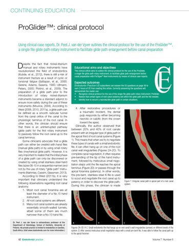
4. After restorative procedures or a traumatic incident, the dental pulp responds by either becoming necrotic or calcific (from the crown toward the apex). Clinically, the author observed that between 20% and 40% of root canals present with an irregular type of glide path in the apical third of root canal systems (Figure 1). This means that when we try to negotiate these types of canals with a small endodontic file, it can often hang up on one of the root canal wall irregularities (Figures 2A-2C). To complete canal negotiation, it often requires pre-bending of the tip of the hand instrument, followed by meticulous small negotiation steps until the file reaches the apical terminus (Figure 2D) or passes through the apical foramina (patency). In other words, the pre-bent, stainless steel K-file is used to scout and negotiate the root canal up to patency in order to discover the glide path. During this phase, the clinician is made
Figure 1: Irregular canal path in apical part of a root canal system
Figures 2A-2D: A-C. Small endodontic file that hangs up on root canal’s wall irregularities (arrows) on different levels of the system. D. After several meticulous small negotiation steps with a small pre-bent file, it was able to follow the canal path up to the apical foramen Volume 7 Number 6