CONTINUING EDUCATION
Minimally invasive endodontics using a new singlefile rotary system Drs. Peet J. van der Vyver, Martin Vorster, and Ove A. Peters discuss the design features of the TruNatomy™ instruments and present case reports to illustrate the clinical application and benefits of these instruments Introduction The long-term retention of root canaltreated teeth depends on many factors, but it has become evident that the most common reasons for extraction of these teeth are “large carious lesion” or “unrestorable tooth,” followed by “tooth fracture,” “periodontal disease” and last of all, “endodontically related disease” (Ng, Mann, and Gulabivala, 2010). Moreover, it is apparent that remaining structural integrity and the preservation of especially peri-cervical dentin are key factors that determine the long-term prognosis (relating to fracture resistance) in these teeth (Tang, Wu, and Smales, 2010). The term pericervical dentin was first described by Clark and Khademi (2010) and refers to an area roughly 4 mm coronal to the crestal bone and 6 mm apical to the crestal bone (Figure 1). According to Herbranson (2014), it appears to be critical dentin for tooth strength, and that should be conserved as much as possible to ensure long-term retention of the tooth. It is also the area of the tooth that is often destructed with access burs, Gates Glidden burs, and orifice shapers used for coronal enlargement of root canal systems. The fact that endodontically treated teeth are more prone to fracture is largely due to the structural loss during the shaping phase of endodontic treatment and not to dehydration. Studies show minimal dehydration effects from pulpal removal with Professor Peet J. van der Vyver, BChD, MSc, PhD — Studio for Endodontics, Restorative Dentistry and Dental Education (www.studio4endo.com) — is a part-time Lecturer at the University of Pretoria, Department of Odontology, School of Dentistry, University of Pretoria, Pretoria, South Africa. Martin Vorster, BChD, MSc, is a Lecturer/Dentist at the University of Pretoria’s School of Dentistry, Pretoria, Gauteng, South Africa (martin.vorster@up.ac.za) Department of Odontology, School of Dentistry, University of Pretoria, Pretoria, South Africa. Professor Ove A. Peters, DMD, MS, PhD, is Professor and Chair, Department of Endodontics, University of the Pacific Arthur A. Dugoni School of Dentistry, San Francisco, California, and University of Queensland Oral Heath Centre, Herston Qld, Australia.
22 Endodontic practice
Educational aims and objectives
This clinical article aims to discuss the design features of the TruNatomy™ instruments and present case reports to illustrate the clinical application and benefits of these instruments.
Expected outcomes
Endodontic Practice US subscribers can answer the CE questions on page 29 to earn 2 hours of CE from reading this article. Correctly answering the questions will demonstrate the reader can: •
Realize that respecting original canal anatomy, preserving dentin, and therefore maintaining the structural integrity of teeth should form an integral part of root canal shaping and preparation.
•
Define peri-cervical dentin.
•
Realize why peri-cervical dentin appears to be critical dentin for tooth strength.
•
Identify a way to preserve structural integrity and preservation especially of peri-cervical dentin.
•
Recognize some characteristics of TruNatomy instruments that can result in superior debridement while respecting original canal anatomy.
similar strength test results between vital and nonvital dentin (Sedgley and Messer, 1992, Papa, Cain, and Messer, 1994). Structural loss alone is, however, not the only cause for increased fracture incidence in teeth. The impact of irrigants, medicaments, as well as restorative procedures, and even physiological age changes should also be taken in account. Root canal therapy requires effective shaping in order to facilitate irrigation and disinfection of the canals. This should be done in such a conservative manner that the structural integrity of the tooth is respected, and dentin is preserved where possible. Peri-cervical dentin preservation has been reported as critical in the long-term survival, especially in molars with optimum function (Clark and Khademi, 2010). More advanced treatment options in endodontics (for example, magnification and more flexible nickel-titanium instrumentation) have therefore also shifted paradigms to a minimally invasive approach in both access cavity preparation as well as shaping of root canals in order to preserve dentin (Gluskin, Peters, and Peters, 2014). Recently, TruNatomy™ (Dentsply Sirona), a new generation of rotary files, was launched. TruNatomy files are prepackaged, presterilized rotary instruments designed to shape root canal systems to a continuously tapering
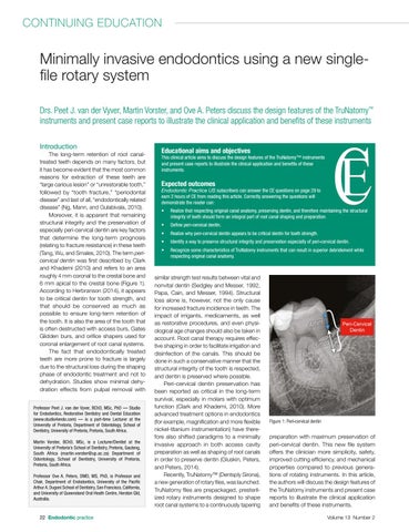
Figure 1: Peri-cervical dentin
preparation with maximum preservation of peri-cervical dentin. This new file system offers the clinician more simplicity, safety, improved cutting efficiency, and mechanical properties compared to previous generations of rotating instruments. In this article, the authors will discuss the design features of the TruNatomy instruments and present case reports to illustrate the clinical application and benefits of these instruments. Volume 13 Number 2