CONTINUING EDUCATION or be limited to the coronal or mid portion of the canal, or even be present only at the apical portion. The isthmus is an area difficult clean and decontaminate with mechanical preparation due to its perpendicular position to the file, and it can have a major impact in the success of a root canal treatment.5,8 In a clinical study,12 1,400 teeth from 618 patients were evaluated using cone beam computed tomography. The study demonstrated that 87.9% of mandibular molars have isthmus present (Figure 4). A high prevalence of isthmus areas means the practitioner must pay special attention during the preparation of mandibular molars. The use of ultrasonics and optical magnification is a fundamental tool to locate, identify, and clean this intricate anatomy. Besides the high percentage of isthmus areas in mandibular molars, another clinical difficulty is the presence of a third canal in the mesial root, the middle mesial canal. This canal is located between the mesiobuccal and mesiolingual canals. The entrance point for the middle mesial canal is very hard to find without the use of magnification; however, in studies using the operative microscope, the canal was found in 46% of the investigated molars.13 The instrumentation, either manual or mechanized, is restricted by the main canal diameter, having little or no effect on flattened areas and twists/curvature of the root canal. Specially designed ultrasonic instruments allied to ultrasonic cavitation can favor the treatment inside flattened regions — raising the percentage of touched wall areas by the activated irrigants and performing a thorough cleaning without promoting excessive loss of tooth structure.
2. Once the isthmus is visible through magnification, initiate its cleaning using a thin tip such as the E18D diamond-coated ultrasonic tip (Helse Ultrasonic, Santa Rosa de Viterbo, Brazil) or the UCT-1 diamond-coated ultrasonic tip (Vista Dental, Racine, Wisconsin) or the BUC-1A (Kerr Dental, Orange, California). 3. In addition to cleaning this area, we must look for the middle mesial
canal, which is generally located closest to the mesiolingual than to the mesiobuccal canal. 4. Once the entrance is found, prepare the canal with carbon steel hand files, since they have a proper stiffness for this situation. A No. 6 carbon steel file is ideal for initial exploration of the canal due to its greater stiffness compared to a stainless steel file of the same size.
Protocol to prepare the mesial root canals in mandibular molars (Figure 5) 1. After preparing the mesiobuccal and mesiolingual canals, remove the dentin over the isthmus using a diamondcoated ultrasonic tip. This procedure is best accomplished with a thick cone, pear, or sphere-shaped tip.
Figure 5: Micro-CT showing high prevalence of flattened canals and isthmus areas 30 Endodontic practice
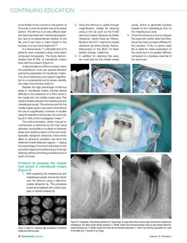
Figure 6: A. Diagnostic X-Ray kindly provided by Dr. Tiago Braga; B. Image taken after access surgery showing the mesiobuccal, mesiolingual, and distal canals already prepared; C. Middle mesial canal entrance located using an pear-shaped diamondcoated ultrasonic tip; D. Middle mesial canal after bio-mechanical preparation; E. Final X-ray showing preparation for a post in the distal root; F. Inverted X-ray image. Volume 10 Number 4