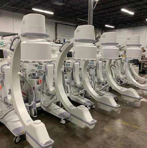
Kenneth Saltrick, President of Engineering Services in Twinsburg, Ohio, knows from his long experience that C-arm machines themselves are absolute workhorses.
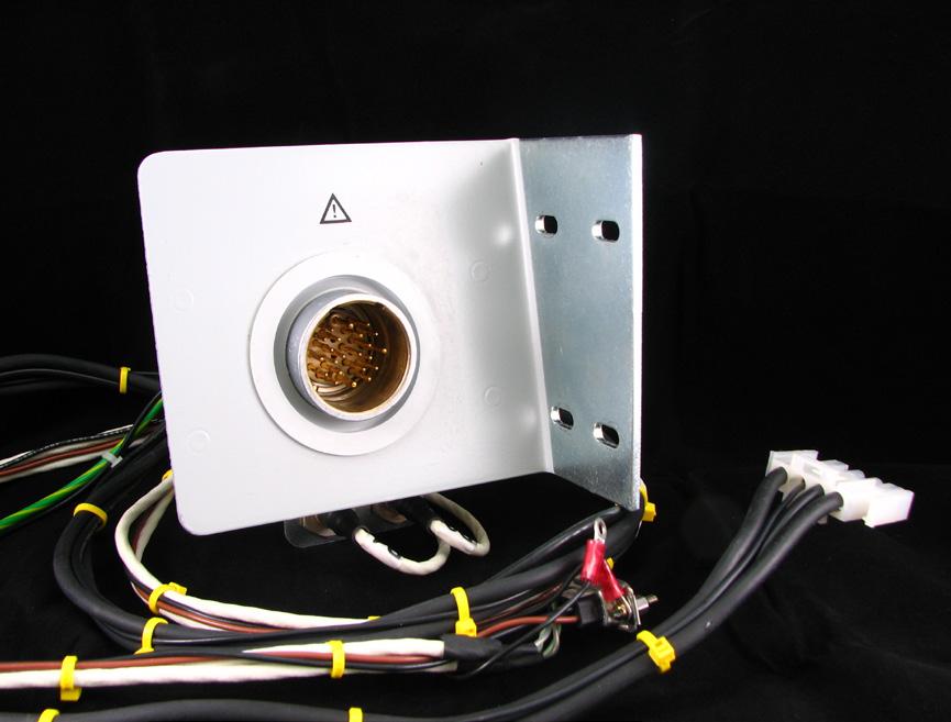
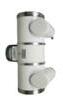
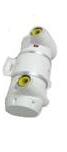
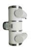
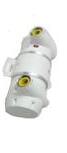
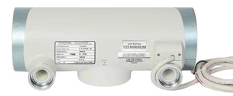
For customers looking to blend the gap between expensive OEM and unreliable used assemblies, WE have your solution.
Our complete repair contains a new cable assembly, utilizing all OEM cable and components with a harvested plate and connector housing as they are proprietary items. These completely repaired products will have a significant cost savings with build quality above new OEM products and carry a warranty of 180 days, which is untouchable in the market.
XperTIS
XperTIS
What is XperTIS?
What is XperTIS?
Tri-Imaging Solutions introduces a cutting-edge platform that enhances supply chain management, engineer performance, and system monitoring. It provides engineers with diagnostic tools and video tutorials, and streamlines parts ordering and tracking. Designed for efficiency, the platform minimizes downtime and optimizes operations, setting a new standard for reliability in medical imaging.
Tri-Imaging Solutions introduces a cutting-edge platform that enhances supply chain management, engineer performance, and system monitoring. It provides engineers with diagnostic tools and video tutorials, and streamlines parts ordering and tracking. Designed for efficiency, the platform minimizes downtime and optimizes operations, setting a new standard for reliability in medical imaging.
Objectives Solutions
Objectives Solutions
XperTIS proactively monitors system health, supports engineers in repairs, and enhances the supply chain process by giving teams seamless access to parts ordering and order tracking.
XperTIS proactively monitors system health, supports engineers in repairs, and enhances the supply chain process by giving teams seamless access to parts ordering and order tracking.
XperTIS offers step-by-step repair guidance, helping engineers troubleshoot efficiently while ensuring faster, more accurate parts ordering. This helps maximize uptime and minimize repair costs.
XperTIS offers step-by-step repair guidance, helping engineers troubleshoot efficiently while ensuring faster, more accurate parts ordering. This helps maximize uptime and minimize repair costs.
REVOLUTIONIZING
Medical Imaging Equipment Maintenance and Repair
Finally, the features you love most about your favorite ride share app, food delivery app, Angi, UpWork, or TaskRabbit are available for medical imaging equipment maintenance and repair.
Enter a Trace Ticket with one tap to broadcast your repair needs to a network of qualified technicians.
Review Bids to find the best service option for improved repair outcomes with less equipment down time, resulting in a lower overall cost.
Track progress, issue payments and rate services all in a single dashboard.
EFFICIENCY MADE EASY for Your Healthcare Teams
Speed
Instantly blast your service request to every qualified and vetted service provider
Take
Control of Tracking
From response times to uptime, you no longer have to rely on service companies to track their own activities and performance
Standardized Communication
Like your favorite personal apps, get in-app alerts when a service technician is on their way, arrived, waiting for a part, completed a job, etc.
Pay Through the App
Competitive bidding among service technicians allows you to get the best price and only pay and track one entity
FEATURES
DIRECTOR’S CUT
Sometimes, the most reliable people are the ones carrying the heaviest load – and never saying a word.
COVER STORY
AI can accompany a patient from ordering an imaging study to scheduling it, undergoing the exam, communicating and analyzing the results afterwards, and maximizing the impact of those findings.
RISING STAR
Jennifer Ellis is an imaging supervisor with Orlando Health Sebastian River Hospital who aspires to become a director of imaging services.
NOVEMBER 2025
IMAGING NEWS
Catch up on the latest news from around the diagnostic imaging world.
PRODUCT FOCUS
A look at products that can improve the patient experience in your facility.
EMOTIONAL INTELLIGENCE
Change occurring around us often means we must adapt the way we do things just so we can keep up.
MD
1155
Phone:
President John M. Krieg
Vice
Kristin Leavoy kristin@mdpublishing.com
Vice
Jayme McKelvey jayme@mdpublishing.com
Senior Account
Megan Cabot megan@mdpublishing.com
Editorial
Editorial
Beth Allen
David V. Buczkowski
Kimberly Love
Megan M. Parker
Dean Skillicorn
Jason Theadore
Art
Karlee Gower
Taylor
Alicia
Events
Kristin Leavoy
Webinars
Linda Hasluem
Digital
Cindy Galindo
Kennedy Krieg
Haley Harris
Accounting
Diane Costea
1.REGISTER
Register to view the webinars each month
2.WATCH
Watch recorded webinars on-demand
3.EARN
ARRT Category A CE credits pending approval
RISING STAR
JENNIFER ELLIS, ARRT; R.T. (R)
Je nnifer Ellis, ARRT; R.T. (R), earned a Bachelor of Science in H ealthcare Administration from Keiser University in Melbourne, Florida to go with an Associate of Science in Radiologic Technology. Her education continued on the job where she is now an imaging supervisor with Orlando Health Sebastian River Hospital.
She recently shared more about her education, career and goals with ICE Magazine
Q: WHERE DID YOU GROW UP? OR WHERE ARE YOU FROM?
A: I was born in Milford, Massachusetts, and lived in Woonsocket, Rhode Island, until I moved to Florida with my family in 1994.
Q: WHERE DID YOU RECEIVE YOUR IMAGING TRAINING/EDUCATION?
A: I received my education from Keiser University in Melbourne, Florida. I have an Associate of Science in Radiologic Technology and a Bachelor of Science in Healthcare Administration.
Q: HOW DID YOU FIRST DECIDE TO START WORKING IN IMAGING?
A: I have always been fascinated by the medical field and when my dad got sick it made me think about being in the medical field. Watching him in and out of the hospital and seeing how he was treated, either good or bad, made me decide to go to school to be in the medical field.
Q: WHAT IS THE MOST REWARDING ASPECT OF YOUR JOB?
A: Getting to meet all different kinds of people and hear their stories. Everyone has a story to tell.
Q: WHAT DO YOU LIKE MOST ABOUT YOUR POSITION?
A: That I am in a position to make a difference, whether it is for a team member or for the community.
Q: WHAT INTERESTS YOU THE MOST ABOUT THE IMAGING FIELD?
A: It never gets boring; there is always something different each day.
Q: WHAT HAS BEEN YOUR GREATEST ACCOMPLISHMENT IN YOUR FIELD THUS FAR?
A: Being nominated by administration for the Community Hero’s award and then being selected to be recognized on the Orlando soccer field as a Community Hero.
Q: WHAT GOALS DO YOU HAVE FOR YOURSELF IN THE NEXT 5 YEARS?
A: To continue my employment with Orlando Health and become a director of imaging services. •
FUN FACTS
FAVORITE HOBBY: Making things homemade, examples include homemade vanilla, my own chicken stock, canning, etc.
FAVORITE SHOW: “On Patrol Live,” or any live cop show.
FAVORITE FOOD: Potatoes, they are so versatile.
FAVORITE VACATION SPOT: Anywhere that I am with my family.
1 THING ON YOUR BUCKET LIST: To visit Italy.
SOMETHING YOUR CO-WORKERS
DON’T KNOW ABOUT YOU: I would give up my career to go live on a farm and raise animals and have my own fruit and vegetable garden.
FOCUS IN
KAREN MIHALIC, RT (R), (M), ARRT
As a self-proclaimed “geek,” Karen Mihalic, RT (R), (M), ARRT, serves as the director of imaging/admin team member for WVU Medicine Barnesville Hospital and WVU Medicine Harrison Community Hospital. However, the word “geek” can have negative connotations. She is more of a cool intellectual with a knack for critical thinking that empowers her to bring top-tier diagnostic imaging to the rural community the WVU Health System serves.
Karen’s love of technology and a desire to provide quality patient care started her on a radiology path.
“I found science/anatomy field very interesting, and wanted to find a profession that allowed me to be in the front line of patient care while being challenged with new technologies,” she shares. “I shadowed a radiology technologist and I was hooked from there on.”
Hooked but not stuck, Karen continued to grow as an imaging professional and care provider.
“I have a passion for critical thinking, and the imaging field offers a broad scope of opportunities across various modalities as technology continues to advance. For me, it is incredibly rewarding to see capital projects come to fruition – bringing industry-standard care to our small, rural facilities and allowing patients to receive high-quality services close to home,” Karen says. “I am grateful to be part of leadership teams at both sites that work collaboratively to ensure we consistently deliver the highest standard of care.”
“Building a successful team with technologists who are
committed to providing the highest quality imaging to our patients. I truly enjoy seeing staff who thrive as patient advocates,” Karen says when asked about her greatest accomplishment.
As an imaging leader, Karen is thankful for the lessons shared along her career journey. She also passes along her knowledge to the talented individuals she works with in West Virginia.
“I have truly valued the opportunity to grow in my roles at both facilities. Mentoring others and helping prepare them for future growth and opportunities has added a new dimension of pride in the accomplishments of my coworkers across the organization,” Karen says. “Over time, I have been challenged to adapt my leadership style – shifting toward a more flexible, supportive mentoring approach rather than serving solely as the primary problem solver – while fostering a culture of accountability and open communication. Navigating through some of the most difficult times, including the COVID-19 pandemic and the most significant staffing challenges we have ever faced, has strengthened my resilience and deepened my commitment to my team’s success.”
“I hold myself to a high standard, always mindful that I should never ask my staff to do anything I would not be willing to do myself,” she adds.
She is quick to add that several mentors empower her growth.
“I have been fortunate to be guided by exceptional mentors throughout my career,” Karen says. “Some of the greatest pieces of advice and mentorship came from several of my former CEOs:
• Richard Doan — “Any decision you make, you can learn from. At a critical time, make the best informed decision you can with the knowledge you have. If it turns into a learning experience, be sure to store the lesson learned.”
• David Phillips — “Be accountable and always be willing to do more.”
• Stacey Armstrong — “Strive to approach situations with fair, level-headed guidance and support. Ensure those
KAREN MIHALIC
RT (R), (M), ARRT
1. What is something most of your coworkers don’t know about you? I do have a small amount of “geek squad” mentality; being how deeply embedded our modalities are in advanced technology, I enjoy learning new software and become fluent enough to train others.
2. What is one thing you do every morning to start your day? Be grateful for the opportunity that is before me for that given day.
3. Best advice you ever received? “If you change the way you look at things, the things you look at change.” Wayne Dyer
4. Who has had the biggest influence on your life? My aunt passed away from breast cancer just as I was beginning my career in imaging. This personal experience deepened my empathy and
around you are strong, capable leaders in their own right.”
I now have the privilege of mentoring new leadership team members and providing guidance to my leads at both sites – encouraging them to grow in their roles and continue developing as leaders.”
Looking ahead, Karen’s love of tech keeps her informed and she sees AI as a tool that will impact diagnostic imaging as well as the entire healthcare delivery process.
“Artificial intelligence (AI) has become an increasingly significant component of medical imaging, growing at an exponential rate. The ability to transfer large volumes of data instantly, enhanced quality measures supported by advanced software, new screening tools designed for improved abnormality detection, and patient-centered workflows are all innovations rapidly transforming our field,” she explains. “The fact that patients can now access their results almost immediately through platforms like MyChart – something unimaginable just 20 years ago – reflects how dramatically both patient access and staff workflows have evolved, even within the past five years. The pace of change in our profession is truly remarkable.” •
compassion in delivering patient care. In response, I established the Barnesville Hospital Breast Health Foundation to support underinsured and uninsured patients diagnosed with breast cancer. I initiated fundraising efforts within our communities to sustain this mission. Now, 24 years later, we continue to provide vital assistance to those in need during challenging times.
5. What would your superpower be? Planning and executing; seeing plans come to fruition.
6. What are your hobbies? Family, travel, reading, locking into a good series on TV/Netflix, etc., new experiences
7. What is your perfect meal? Something Hungarian that I would have learned the recipe from my grandmother. Making those dishes reminds me of Sunday dinners at my grandparents house when I was younger, and a way to honor them.
Karen Mihalic (front) is pictured with the Harrison Community Hospital imaging team.
ICE Debut
INTERSOCIETAL ACCREDITATION COMMISSION (IAC)
The IAC is a nonprofit organization in operation to evaluate and accredit facilities that provide diagnostic imaging, therapeutic and interventional procedures, thus improving the quality of patient care provided in private offices, clinics and hospitals where such services are performed. The IAC provides accreditation programs for vascular testing, echocardiography, nuclear/PET, MRI, diagnostic CT, dental CT, carotid stenting, vascular interventional, cardiac electrophysiology, cardiovascular catheterization and image-guided procedures. The IAC programs for accreditation are dedicated to ensuring quality patient care and promoting healthcare and all support one common mission: Improving healthcare through accreditation. Committed to its mission through a rigorous peer review process, the IAC has granted accreditation to more than 14,000 sites since its inception in 1991.
A nationally recognized, CMS-approved nonprofit organization, IAC exists solely as an accrediting body and is not a membership society.
IAC Director of Marketing/Communications Tamara Sloper recently shared more details and insights regarding the organization.
Q: How does your company stand out in the imaging space?
SLOPER: As the only CMS-approved accrediting body that provides a clinical peer review of case studies (with pathology) for diagnostic quality, report accuracy and report completeness, IAC provides the most comprehensive review process in the accreditation industry. For many facilities, simply meeting minimum standards is not enough;
they want to go the extra mile and be evaluated at a high level. Facilities accredited by IAC demonstrate a meaningful commitment to quality and patient safety; one that patients and referring physicians can rely on.
IAC continues to grow and experience success through the multi-specialty, intersocietal collaboration of a vast array of physicians, technologists, sonographers, physicists and numerous other medical professionals upon which it was founded 35 years ago. These individuals represent more than 40 medical specialties that serve as sponsoring organizations, contributing to IAC’s multi-stakeholder efforts. This multi-stakeholder involvement ensures that the accreditation standards are built by and for the specialties they serve, reflecting a consensus among experts. This collaborative model allows the IAC to develop and revise standards that are relevant and effective for each of the modalities accredited.
Q: What is on the horizon for IAC?
SLOPER: The IAC Standards are continuously reviewed and updated to ensure they are reflective of current practice and societal guidelines. Acknowledging the ongoing, rapid evolution of artificial intelligence (AI) in medicine, the IAC Board of Directors established an AI Task Force in 2024. Developed through the collective expertise of the task force, a guidance document was created. The document, approved by both the IAC Board of Directors and each division board, was published April 1, 2025, as an addendum to each set of IAC Standards. The guidance document serves as a recommendation for IAC-accredited facilities utilizing AI technology.
The Artificial Intelligence (AI) Guidance Document was created to assure the quality and safety of care delivery when using AI applications for direct-patient care (clinical*) purposes, each facility should create and follow policies
and procedures that address:
1. Training for personnel who use AI;
2. Security of AI software, updates, HIPAA considerations, etc.;
3. AI for Quality Improvement (if applicable);
4. Appropriate use for each AI application; and 5. Governance (authority to make decisions regarding AI implementation).
*Clinical use of AI includes image acquisition, image processing/enhancement, image interpretation, report generation, risk assessment of prognosis, patient history, identification of critical values/results and equipment quality control.
Q: Is there anything else you would like ICE Magazine readers to know?
SLOPER: IAC utilizes a rigorous clinical peer review process to ensure that quality and safe practices are established for improved patient outcomes. The process seeks to advance appropriate utilization, standardization and quality of diagnostic imaging, interventional and therapeutic
procedures. Feedback from surveys conducted among IACaccredited facilities demonstrates that participation in the process has a positive impact on various quality metrics. Improvements in report standardization, adherence to guidelines, test standardization, report completeness, identification of deficiencies, improved staff knowledge, report timeliness and image quality, as reported, contribute significantly to better patient outcomes.
In closing, IAC’s customer service model is designed to be a partnership with facilities, focusing on education, support and continuous quality improvement. Those working on their applications appreciate the IAC staff’s strong clinical background and that they are readily available via phone, email and online chat to answer questions and provide guidance throughout the accreditation process. IAC understands that this level of accessibility is especially advantageous for busy imaging professionals who need application support while managing their patient care responsibilities. •
For more information, visit Intersocietal.org.
Clock Off THE
KEITH IRWIN, GENERAL MANAGER, INDUSTRIAL INSPECTION AND CONSULTING
BY MATT SKOUFALOS
At Industrial Inspection and Consulting (IIC) of Norton Shores, Michigan, general manager Keith Irwin and a team of imaging professionals use industrial radiography to provide an array of services, mostly for clients in production and manufacturing businesses.
Imaging studies at IIC are conducted with cabinet-and-vaultstyle Nikon industrial X-ray and CT scanners that can resolve an image down to three microns. The technology is used to examine inconsistencies in structural castings and component-level, manufactured elements, as well as to certify that products are constructed within established tolerances. The IIC laboratory is ISO17025-accredited, and its personnel are certified by the American Society for Non-Destructive Testing and National Aerospace Standards for certification and inspection.
“We love and are good at what we do,” Irwin said. “We have a lot of companies that depend upon our services.”
For all the work IIC is contracted to perform – like studies on structural castings for brake pads, or imaging leaky condiment bottles to shore them up before mass production – the company has also evolved a bespoke service for the collectibles industry. IIC can perform imaging studies to help verify the authenticity of high-ticket items before they’re sold at auction, or to certify that a purchase that’s already been made is legitimate. It’s believed to be the first laboratory of its type to offer the service.
“There had been chatter online for almost 20 years, when we tried to look back in time on the Internet, about whether it’s possible to X-ray or even CT scan collectibles,” Irwin said. “The general consensus was, ‘no.’ There’s even evidence of other labs that were trying.”
“As a contract inspection lab, our specialty is being able to say ‘yes’ to pretty much any inquiry that we get,” he said. “We deal with a lot of the very difficult tasks that people don’t think are possible.”
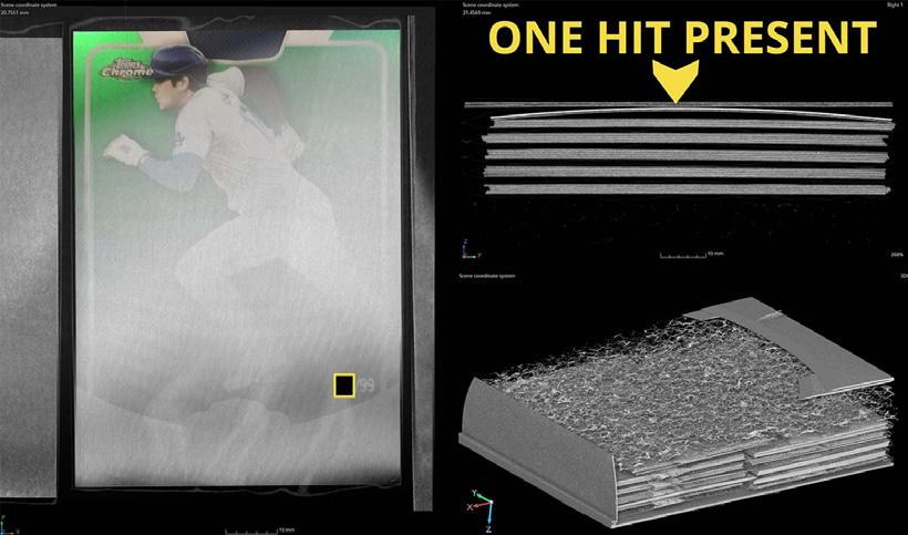
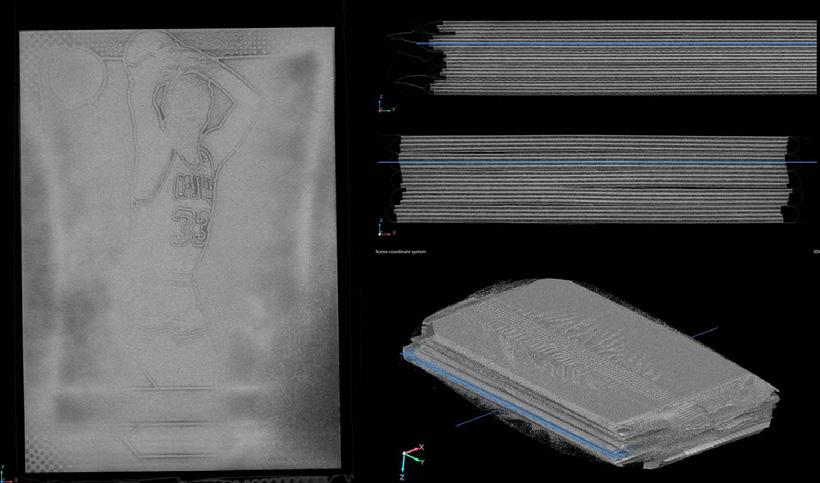
For a company that prides itself on establishing linear defects in contact lens cases, or measure the thickness of polymer coating on manufactured materials within 10 microns or less, IIC took up the challenge to demonstrate just how fine of a detailed distinction its equipment could deliver.
“That’s how the idea of a Pokémon card came up,” Irwin said. “It’s just a layer of ink on a cardstock. We tested it, and it worked very well. We put it online as a case study, and it went viral. That virality led to hundreds of thousands of hits to our website, with many requests.”
IIC staff spent “several weeks not sleeping” to determine how to image trading cards as a service, Irwin said. It was a novel application for their imaging equipment, and the team was eager to meet the challenge.
“It took us a while to perfect the technique and a while to monetize it,” he said. “It was a lot of experimentation. Anybody who goes and buys this equipment and the software can figure it out; we were just the first to try, and we tried hard.”
Beyond sweat equity, any laboratory studying the collectible market must not only be able to capture usable data with its imaging equipment, but also to read the subtle differences in that data, “looking inside those gray values and figuring out what it is,” Irwin said.
“Every card is a little different, even if it is the same product line,” he said. “After a lot of practice and experience and time reviewing that we can do it, [we realized] ‘Wow, this works.’
“I remember the first time we found a high-value card, a six-figure card, and it was like a bomb dropping in here,” Irwin said. “It was a lot of excitement.”
Irwin said his staff are completely disconnected from the
collectability of the materials they study, and in the time since the service took off, all of them have made formal attestations to not collect or resell the products with which they are tasked to image.
“We’re an unbiased, accredited laboratory,” Irwin said. “To us, the work is all the same; scanning aerospace castings or providing authentications for auction houses.”
“We didn’t know anything about cards; we know there’s a lot of value there,” he said. “We are not going out and buying a bunch of collectibles, scanning them, and using our equipment for that purpose, because there could be a perceived bias or interest.”
“There was a lot of concern and criticism about that tampering with high-value products, but there are also many things people can do to secure their items: they can weigh their packs or photograph the seals, which act as unique product fingerprints,” Irwin said.
IIC has reviewed “the full variety” of collectibles, from packs, boxes, and cases of cards, to cases of vintage toys, Irwin said. Sometimes X-rays can be used simply to verify that the original product is in the box, and that it hasn’t been resealed with its contents removed.
Famously, Internet celebrity Logan Paul was an alleged victim of such a scam. After having spent $3.5 million on what he’d believed was an authenticated, sealed case of Pokémon cards, he opened them to discover GI Joe trading cards instead.
“We’ve found a lot of fake and fraudulent boxes, and helped people in their disputes,” Irwin said. “There are several metrics that we can grade against. Is the
right number of packs in there? The right number of cards in packs? Inconsistencies? Are the seals appropriate?”
“We provide a report to that customer or auction house,” he continued. “We place a coded sticker on that, a matching sticker on the product, and then it’s traceable. Some of these boxes that we get are worth $50,000 to $100,000. All of that is shown on our online registry.”
Another time, an IIC client purchased three, antique Gameboy cartridges at auction, and, when the company imaged them, found the contents had been replaced with other materials that would simulate the weight of the cartridges.
ing because they must be exposed to hours of radiation to get a reading, Irwin said, but they’re not going to be damaged in the process.
“There is a value-added approach for people who have their collectibles but don’t want to devalue them,” Irwin said. “A lot of our customers are people who have a random collectible sitting on their shelf that’s old, and they don’t want to open it, because then it’s worth nothing, but the contents themselves could be life-changing. If you pull a Charizard out of a 1999 Pokémon pack, that card could be worth $400,000.”
IIC product scans are priced upon the hourly work rate at the business, and not upon the perceived value of the collectible. The service charges $75 to scan a pack, whether it would sell for $20,000 or $500, and bulk rates are available. Boxes of cards are more time-consum-
“There are times when we’re trying to expose [an item for] as long as possible to get very good averaged data,” he said. “We have correction measures and reconstruction parameters that we apply, but we are trying to get the best raw data as possible.”
Although IIC has received a great deal of press attention for its involvement in the collectibles market, its core line of business remains in the industrial and manufacturing sectors. Irwin hopes to add larger, higher-exposure equipment to the lab, the better to test items at the forefront of technological change. If a few more trading card studies help the business get there, they’ll take the work. •
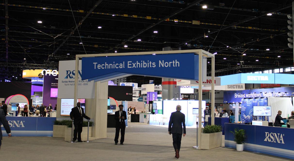
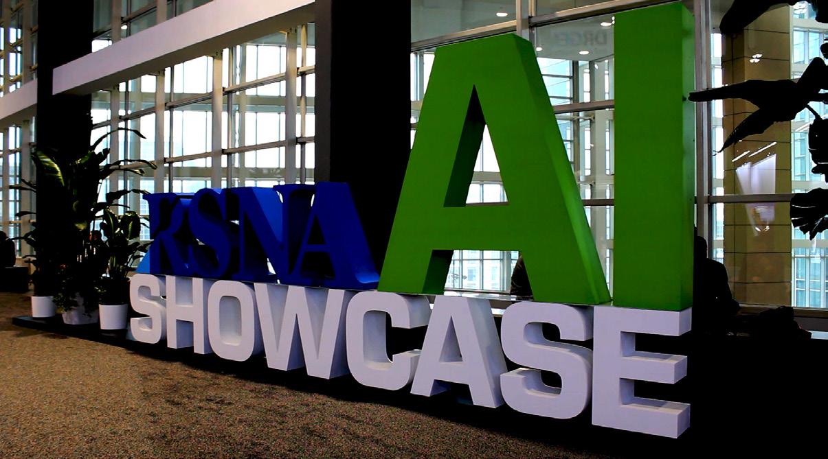
GUIDE TO RSNA 2025 TECHNICAL EXHIBITS FEATURE LARGEST RADIOLOGY AI SHOWCASE
OA K BROOK, Ill. | The Radiological Society of North America (RSNA) has announced Technical Exhibits highlights at RSNA 2025: Imaging the Individual, the Society’s 111th Scientific Assembly and Annual Meeting, taking place at Chicago’s McCormick Place Nov. 30-Dec. 4, 2025.
Spanning the North and South Halls of McCormick Place, Technical Exhibits will occupy over 415,000 feet of show floor space. More than 660 companies have registered to exhibit thus far, and more than 100 are exhibiting at RSNA for the first time.
“Exhibitors come from all over the world, bringing the latest technology for every facet of medical imaging,” said John P. Jaworski, RSNA’s assistant executive director of meeting services & corporate relations.
“We’re excited to host the largest exhibition in radiology, including the largest exhibition in clinical artificial intelligence.”
The AI Showcase in the South Hall houses the latest in AI software and solutions across a 45,000-squarefoot show floor. Attendees can view AI Theater presentations, explore RSNA-led AI research and education initiatives and talk face-to-face with exhibitors about
how AI can improve clinical workflows. The Radiology Reimagined exhibit hosts a variety of 30-minute interoperability demonstrations that show new ways AI can be integrated into practice. More than 200 companies plan to demonstrate AI and machine learning solutions within the Showcase and at their exhibit booths.
Also in the South Hall, the 3D Printing and Mixed Reality Showcase invites attendees to interact with the latest 3D medical printing and virtual reality technologies. Meanwhile, in the North Hall, the First-Time Exhibitor Pavilion introduces newly exhibiting companies from regions across the globe, offering products and services for every specialty and need.
Publishers, radiology associations and educational institutions have a presence in Educators Row, while Recruiters Row – a must-stop for job seekers – will house 55 recruiting companies. Attendees can have a free professional headshot taken in Recruiters Row for a fresh update to online profiles and resumes.
To help navigate the offerings at RSNA 2025, the new “RSNA Road Trip” feature gives attendees an interactive way to engage with highlights of the Technical Exhibits. Attendees will earn points at each Road Trip destination and qualify to enter a raffle to win RSNA swag.
Some of the featured destinations include a variety
of new RSNA Parks. The Bark Park offers a chance to interact with therapy dogs in a calming atmosphere, while Sustainability Parks demonstrate OSCAR, AI-enabled recycling units that advise which trash items go in which bin. Puttology sports a three-hole mini-golf course, and Recharge Parks offer comfortable seating, worktables and charging stations to refresh attendees and devices alike.
During the new Exhibit Hall Social on Tuesday afternoon, attendees can unwind and network with exhibitors and colleagues with food, beverages and interactive experiences designed by participating exhibitors.
Exhibiting companies have scheduled corporate symposia, vendor workshops, lunch-andlearns and AI/Innovation Theater presentations throughout the week.
Technical Exhibits will be open Sunday, Nov. 30, through Wednesday, Dec. 3, from 10 a.m. to 5 p.m. CT.
View the RSNA 2025 exhibitor list by category, showcase, first-time status and more.•
Follow #RSNA25 for the latest meeting updates.
Imaging News
A LOOK AT WHAT’S CHANGING IN THE IMAGING INDUSTRY
GE HEALTHCARE EXPANDS ACCESS TO CLOUD-ENABLED ENTERPRISE IMAGING SOLUTIONS IN AWS MARKETPLACE
GE HealthCare recently announced via a press release that its Genesis portfolio, with cloud-enabled software as a service (SaaS) solutions for enterprise imaging, is available in Amazon Web Services (AWS) Marketplace, providing more ways for hospitals and health systems to adopt the Genesis software suite and to help accelerate their digital transformation.
their digital transformation,” said Scott Miller, CEO of solutions for enterprise imaging, GE HealthCare.
The Genesis portfolio is designed to:
The enterprise imaging solutions, Genesis Storage, Genesis VNA, and True PACS SaaS, are the first of GE HealthCare’s solutions to be offered in AWS Marketplace, a digital catalog of software listings that make it easy to find, test, buy and deploy software that runs on AWS.
“We are committed to delivering enterprise imaging solutions with our cloud strategy. Our Genesis portfolio can help improve patient care through accessible, sharable, and secure patient data while simultaneously improving clinical and operational efficiencies. Now, with its availability in AWS Marketplace, we are providing more ways for our customers to adopt these next-generation solutions so they can accelerate
• Streamline workflows for healthcare professionals and caregivers
• Facilitate interoperability through centralized patient data storage and access
• Enhance user experiences for both IT and clinical teams
• Support secure, scalable deployment in the cloud
“The Genesis portfolio is built on AWS, which is architected to be the most secure cloud computing environment available. As GE HealthCare’s strategic cloud partner for almost 10 years, AWS supports GE HealthCare’s goal to triple its cloud-enabled offerings by 2028 while driving momentum in digital transformation to help enhance patient care,” the press release states.
IMAGE GENTLY LAUNCHES FAMILY-FRIENDLY CAMPAIGN
The new Image Gently Family-Friendly Campaign offers a free online “plug-and-play” module to educate medical students and early-career providers about informed, team-based radiation safety and dose optimization strategies to help ensure appropriate, optimally performed pediatric imaging.
“With the Family-Friendly Campaign, family physicians, radiologists, and other healthcare providers are working together to achieve a better collective understanding of interactions for medical radiation safety and dose optimization,” said Christopher William Bunt, MD, FAAFP, family medicine physician, module co-director for the Family-Friendly Campaign. “Informed providers make better ordering decisions, can help advocate for care with their healthcare teams and help patients understand their imaging needs.”
“The Family-Friendly modules are wonderfully concise and helpful to all medical providers involved in pediatric imaging in any capacity,” said Delaney Walden, MD, pediatric resident, Family-Friendly module creation team when a medical student. “The new module clearly summarizes and explains best practices for imaging children, how to prioritize imaging modalities and discuss risks and benefits with parents. This helps us as providers have more productive shared decision-making conversations.”
The interactive module uses a radiation risk framework to close the health literacy gap, encourage evidence-based practice, empower learner-directed change through motivational interviewing and employ shared decision-making to promote a respectful healthcare environment.
“Radiologists, referring providers, medical physicists and radiologic technologists all need to contribute to appropriate and optimized imaging,” said Donald Frush, MD, FACR, chair of the Image Gently Alliance (imagegently.org). “The imaging team can help referring providers to decide which imaging should be done and when, understand examination doses and dose management, and weigh the relative benefits of these often lifesaving exams.”
Upon completion of the module, which simulates imaging decisions for a common pediatric case of acute abdominal pain, learners should be able to accurately:
• Explain radiation, radiation risks and risk mitigation strategies using layperson terminology.
• Use clinical decision support, or critically appraise available literature, to guide medical recommendations.
• Describe how to engage with healthcare teammates to utilize available evidence-based recommendations.
• Demonstrate shared decision-making with patients and their families.
“Medical imaging and radiation therapy professionals are on the front lines of patient care and serve a crucial role in making sure patients receive high quality radiologic care,” said Heather Moore, Ph.D., R.T.(R), chair of the board of directors for the American Society of Radiologic Technologists. “A better understanding of dose optimization and imaging appropriateness, and the ability to relay this information to patients, can strengthen our partnership with patients and help achieve better care.”
The Image Gently Family-Friendly Campaign module was created by an interprofessional, interdisciplinary team made up of family medicine and pediatric radiology physicians, radiologic technologists, medical physicists, health profession educators and medical students.
“Medical physicists ensure that imaging equipment is installed, operates and delivers doses appropriately to ensure quality images that enable physicians to make the right diagnosis,” stated M. Mahesh, PhD, FAAPM, FACR, president of The American Association of Physicists in Medicine (AAPM). “The Family-Friendly Educational Campaign, through a clinical setting of medical imaging and radiation use, brings multiple healthcare provider specialists together to make care more informed and collaborative decisions to improve the patient experience.”
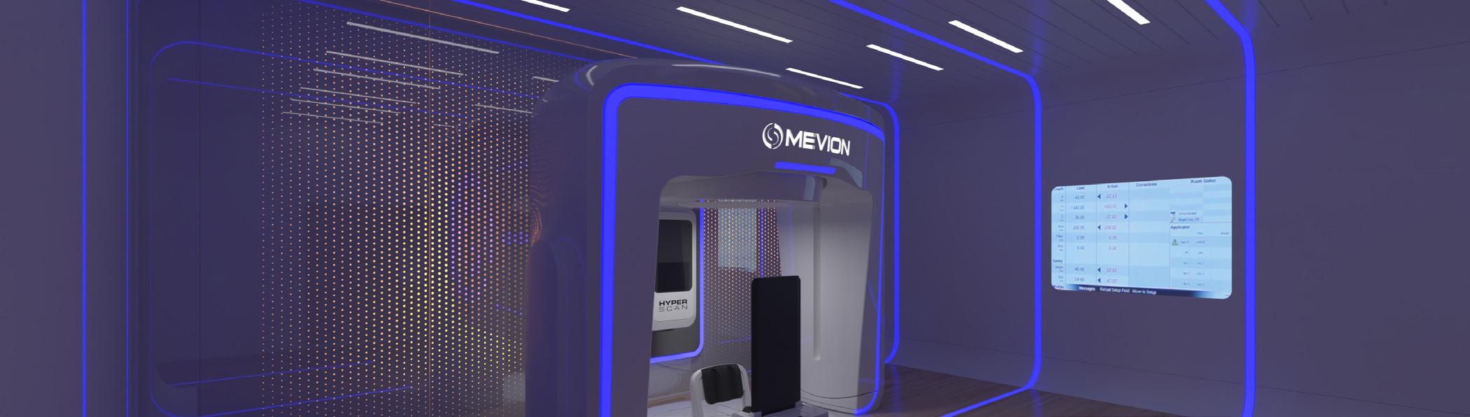
FDA CLEARS MEVION S250-FIT PROTON THERAPY SYSTEM
A new press release states that the U.S. Food and Drug Administration (FDA) has granted 510(k) clearance for the MEVION S250-FIT Proton Beam Radiation Therapy System, marking a significant milestone in expanding access to proton therapy.
The MEVION S250-FIT is the first and only proton therapy system designed to seamlessly fit into a standard radiation therapy vault. By leveraging existing infrastructure, the MEVION S250-FIT system lowers barriers for hospitals and cancer centers to adopt proton therapy, accelerating access and bringing advanced, high-quality proton therapy closer to patients worldwide.
The MEVION S250-FIT is currently under installation at Stanford Health Care and BayCare Health System, with additional installations beginning at Atlantic Health System and the University of Nebraska Medical Center. This
momentum underscores how top-tier institutions are embracing the MEVION S250-FIT system as a future-ready, quality-driven solution, reaffirming Mevion’s role as the trusted partner of leading cancer centers.
“The FDA clearance of the MEVION S250-FIT marks the beginning of a new era in proton therapy. For more than two decades, Mevion has redefined proton therapy through audacious innovation – from pioneering the world’s first compact, single-room system to now enabling proton therapy to fit into existing radiation therapy vaults, expanding access for patients worldwide. I am incredibly proud of the Mevion team and our partners at Leo Cancer Care for making this achievement possible and look forward to the impact of this innovation as centers like Stanford bring it to patients,” said Tina Yu, CEO and president of Mevion Medical Systems.
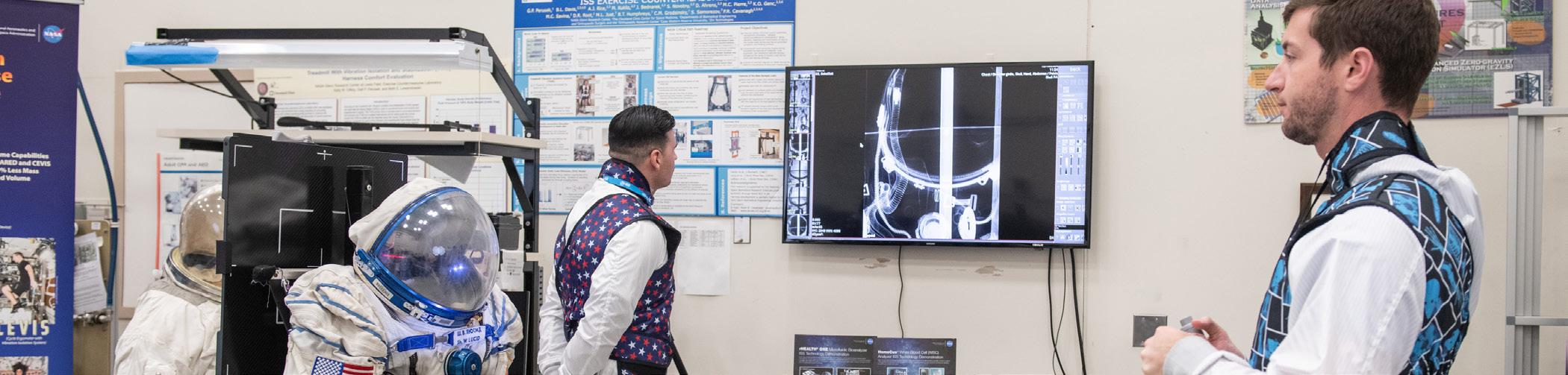
MINXRAY’S IMPACT WIRELESS X-RAY SYSTEM AMONG FINAL 3 FOR NASA MISSIONS
MinXray’s Impact System, a complete wireless digital X-ray system, is one of three portable X-ray units in the second round of testing by NASA for future human exploration missions. The system that is selected by NASA will be used to address the challenges of long-distance space travel to the Moon, Mars and beyond.
The Impact System is well-suited for long-term space travel due to its portability, battery power and capabilities for full-body imaging. Its compact sizing also makes it an ideal solution given the limited room available on a spacecraft, according to a press release.
It was recently utilized as part of the Fram2 mission, where it captured several radiographs in April of this year, most notably the first human X-ray image ever captured in space.
“The Impact system has proven its capabilities in the most extreme environments from the base camp at Mount Everest to the deserts of Africa to high-elevation research centers in the rainforest of Papua New Guinea,” said MinXray Director of Global and Military Sales Mike Cairnie. “The Fram2 mission was another step toward proving diagnostic images can be captured in any conditions, a goal that is shared by the scientists at NASA as they look to expand medical care capabilities in the final frontier.”
Researchers are expected to make a final selection at the end of 2025 and test the chosen system aboard the International Space Station in 2027 or early 2028.
ULTRASOUND AI SECURES U.S. PATENT FOR AI-DRIVEN
CLINICAL VALUE DETERMINATION
Ultrasound AI Inc., a pioneer in artificial intelligence for medical imaging, has announced that the United States Patent and Trademark Office has issued U.S. Patent No. 12,369,883, “Artificial Intelligence System for Determining Clinical Values through Medical Imaging.” The patent protects the company’s proprietary system for determining current or future clinical or laboratory values directly from non-invasive medical images such as ultrasound.
The company’s other significant patents are for Premature Birth Prediction, which permits methods for using ultrasound images to quantitatively predict premature birth and estimate gestational age.
The new grant adds to Ultrasound AI’s growing portfolio in obstetrics and broader clinical prediction, and comes on the heels of the company’s recently published PAIR (Perinatal Artificial Intelligence in Ultrasound) Study,
which reported high accuracy predicting time-to-delivery using only ultrasound images – R² up to 0.95 for term births and 0.92 across all births, based on more than two million images. Together, the study and the patent underscore Ultrasound AI’s progress in transforming obstetric care and beyond with scalable, image-only AI.
With today’s issuance, Ultrasound AI now holds four U.S. patents and two international patents.
“Securing this latest patent is more than a legal milestone – it further validates our long-term vision that standard ultrasound can unlock powerful, predictive clinical insights in a fast and cost-effective manner at the point of care,” said Robert S. Bunn, founder and president of Ultrasound AI. “By protecting methods that determine lab and clinical values from imaging alone, we’re paving the way for safer, faster, and more accessible care worldwide.”
QUEEN’S HEALTH SYSTEMS & SIEMENS HEALTHINEERS
FORM VALUE PARTNERSHIP
Siemens Healthineers and The Queen’s Health Systems, Hawai‘i’s largest private health care provider and its largest private employer, have entered into an eight-year value partnership to support the expansion and upgrade of diagnostic imaging offerings at Queen’s to enable more timely access to urgent and routine services for patients across Hawai‘i.
By adopting advanced, AI-enabled imaging systems, including magnetic resonance (MR), computed tomography (CT), positron emission tomography (PET), single-photon emission computed tomography (SPECT) and X-ray, Queen’s is working to lessen the impact of nationwide radiology staffing shortages on employees and patients. With faster, more efficient scans and post-scan image review, patients will be better able to remain within the Queen’s systems for imaging services, enhancing their continuity of care.
The new equipment and technology are intended to support Queen’s ongoing efforts to provide a higher quality of care and fulfill its mission of improving the health and well-being of the people of Hawai’i.
“We are pleased to partner with Siemens Healthineers
with the goal of optimizing care for our patients utilizing advanced imaging solutions,” said Darlena Chadwick, executive vice president and chief operating officer of The Queen’s Health Systems. “We are always looking for innovative ways of providing care and this partnership helps us continue investing in leading-edge resources for our patients.”
The Queen’s Health Systems has more than 9,500 employees and more than 1,800 affiliated physicians and providers in its statewide network. The health system is made up of six hospitals.
“We are proud to partner with The Queen’s Health Systems in delivering advanced technologies as they navigate the challenges of operating a major health care system across the Hawaiian Islands,” said John Kowal, president and head of the Americas at Siemens Healthineers. “Their investment in new imaging equipment reflects a strong commitment to elevating patient care and delivering high-quality service.”
AMX SOLUTIONS OFFERS
COMPLETE REBUILDS OF GE AMX PORTABLE X-RAY SYSTEMS
AMX Solutions, a company specializing in portable AMX4+ X-ray systems, is offering a full rebuild service designed to restore units inside and out, extending their useful life for healthcare facilities.
Unlike standard refurbishing, the company said its approach involves a complete rebuild from top to bottom, ensuring units look and function like new.
“There is a world of difference between simply refurbishing and actually rebuilding your AMX unit,” said Lee Ready, president of AMX Solutions.
The rebuild process includes installation of new components such as OEM paint, hardware, GE logo and function labels, a nine-battery set with test block, custom reinforced top cover, rebuilt collimator with front panel and cable, caster wheels with an updated bearing design, a main wiring harness, HV cables, rotor and arm brake cables, drive belts, a hand switch with hanger, bumper pads, wear strips, noise suppressors, filters, grounding cables and a hospital-grade cord reel.
Rebuilt items include the vertical column, horizontal arm, drive wheels, drive motors and drive handle assem-
bly. Tubes are tested, cleaned, sanded and repainted.
AMX Solutions has been in the imaging industry for more than 20 years. The company’s technicians perform all rebuilds, painting and service is done 100% in house. In addition to complete rebuilds, AMX Solutions offers individual replacement parts and batteries for AMX4+ and Optima units.
“Our mission is to let the industry know that AMX Solutions is still here, with the same team who built our reputation for quality service and customer care,” Lee Ready said. “We want to rekindle old partnerships and create new ones while continuing to provide the same level of excellence people have come to expect from our brand.”
AMX Solutions remains family-owned and operated, led by Ready along with his sons, Campbell and Leland. •
Contrast Injector Training
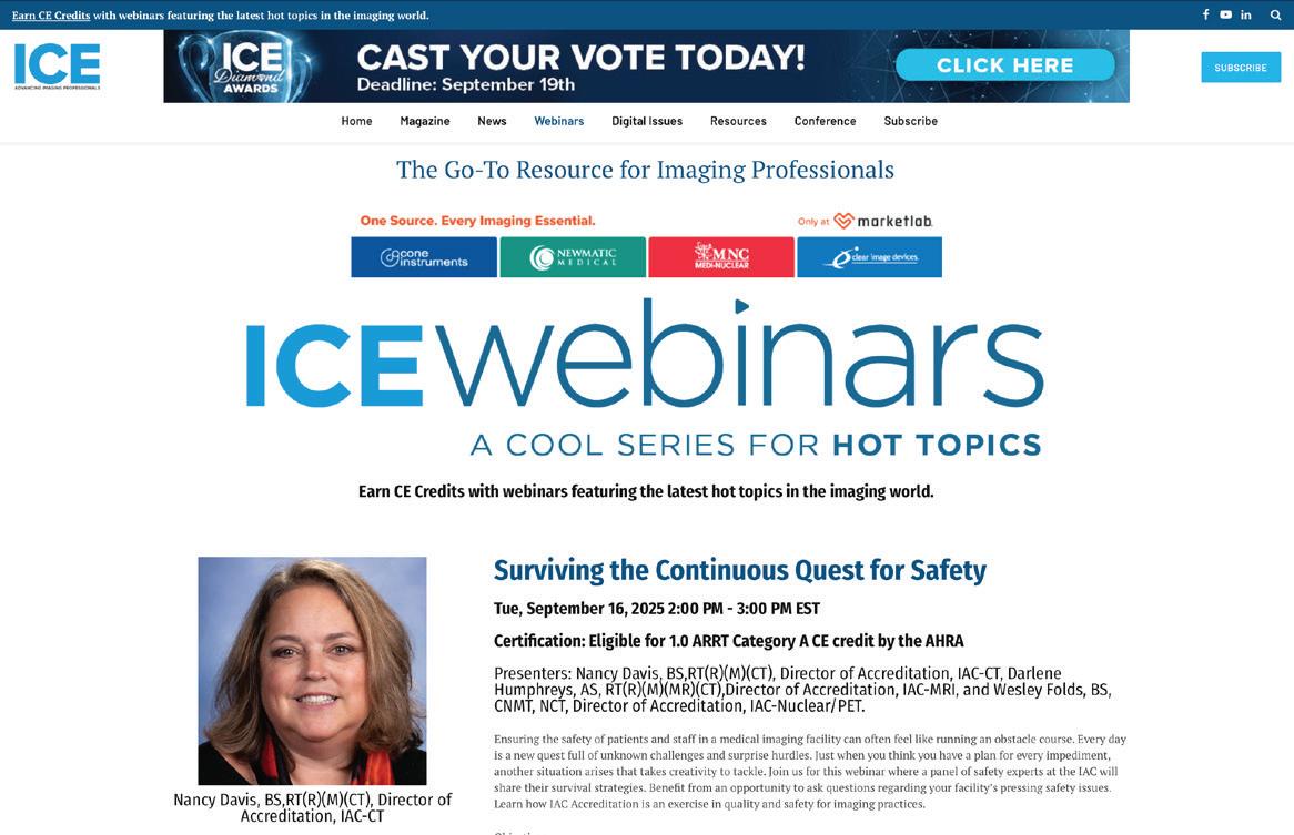
WEBINAR EXPLORES CONTINUOUS QUEST FOR SAFETY
STAFF REPORT
Ensuring the safety of patients and staff in a medical imaging facility can often feel like running an obstacle course. Every day is a new quest full of unknown challenges and surprise hurdles. Just when you think you have a plan for every impediment, another situation arises that takes creativity to tackle.
In the ICE webinar “Surviving the Continuous Quest for Safety” a panel of safety experts from IAC shared their survival strategies. The panel of experts was made up of Nancy Davis, Darlene Humphreys and Wesley Folds.
Attendees were able to benefit from an opportunity to ask questions regarding their facility’s pressing safety issues. The experts also explained how IAC Accreditation is an exercise in quality and safety for imaging practices.
The objectives of the webinar included:
• Understanding the safety challenges within a multimodality imaging practice
• Analyzing the current safety practices at a facility
• Creating strategies that will address departmental and interdepartmental safety concerns
ICE Webinars would like to thank the Intersocietal Accreditation Commission for sponsoring the webinar. Since 1991 IAC has operated as a nonprofit, accrediting facilities that provide vascular testing, echocardiography, nuclear/PET, MRI, diagnostic CT and intervention-based procedures under its mission of Improving health care through accreditation. More than 14,000 IAC-accredited sites have implemented standardized and optimized processes, experienced cost reductions and most importantly, continuously improved their patient outcomes.
More than 100 individuals registered for the session and a recording of the webinar is available for on-demand viewing at ICEwebinars.live. Jen Sturm, a certified nuclear medicine technologist with Novant Health won an ICE Magazine gym bag during the webinar!
Attendees were asked, “Excluding CE credits, why do you attend ICE webinars?”
“To keep learning!” said Amanda Hedges, lead mammography technologist, Sutter Health.
“To be more informed about other modalities, recent radiology studies, and updates to the field of imaging,” said Latasha Traylor, supervisor, Children’s of Alabama.
“I value the opportunity to connect with peers in the field and hear diverse perspectives from thought leaders and practitioners,” said Calin Corciova, associate professor, medical bioengineering faculty.
“Keep up to date on new information and technology,” said Stephanie Voigt, director women’s imaging & practice support, Consulting Radiologists Ltd.
“Easy to take part. Educational and informative,” said George Konstantulakis, assistant chief technologist, Jewish General Hospital.
“I am a student, and my professor recommends all of us to attend these kinds of webinars for extra knowledge. Also, this webinar was related to one of the courses which I am taking so I decided to attend,” said Axi Patel, a student at St. Clair College. •
For more information, including upcoming webinars, visit ICEwebinars.live.
Earlier this year, GE HealthCare reported on “How Consumerism is Transforming MRI Departments.”
“In today’s healthcare landscape, patients are no longer passive recipients of care,” the report stated. “They’re taking an active role, searching for convenience and a comfortable experience. This trend, known as healthcare consumerism, is reshaping the priorities of MRI departments to focus more on improving the patient experience.”
The article reported how patients are changing the game for MRI departments, and how upgrading existing MRI can help a facility address patients’ demands and remain competitive.
Think about how you choose a restaurant or a hotel. You look for convenience, quality service, and a good value. Patients are now applying those same standards to healthcare. They want faster and more convenient scheduling, shorter scan times and an overall more comfortable experience.
An Accenture survey found that 50% of patients would switch providers for better service, and 70% prefer online scheduling. This shift is forcing MRI departments to adapt and prioritize the patient experience. In 2023, the top priority for MRI departments was to improve patient satisfaction with their MRI experience, according to a IMV 2023 MR Market Outlook Report.
Patients want quick appointments and fast results. A Deloitte study revealed that over 60% of patients are frustrated with healthcare wait times. Some MRI departments are responding by offering online scheduling and by investing in advanced systems that allow facilities to see more patients per day, accommodating for the growing demand and reducing scheduling delays.
Consumers expect efficiency, and MRI departments are responding by adopting advanced imaging techniques and AI-powered systems to help reduce scan times and improve image quality. This means that patients spend less time in the scanner and can help improve the time it takes to get results.
Many patients find MRI scans uncomfortable, even anxi-
ety-inducing. The reported incidence of premature termination or failure of MRI examinations can be as high as 14.5%, and the reported incidence of anxiety related reactions during MRI reaches 37%, according to studies.
To improve comfort, MRI departments are upgrading to machines with wider bores, investing in noise reduction features, and using lighter, more comfortable coil technology. These improvements can significantly enhance patient satisfaction as well as attract more patients.
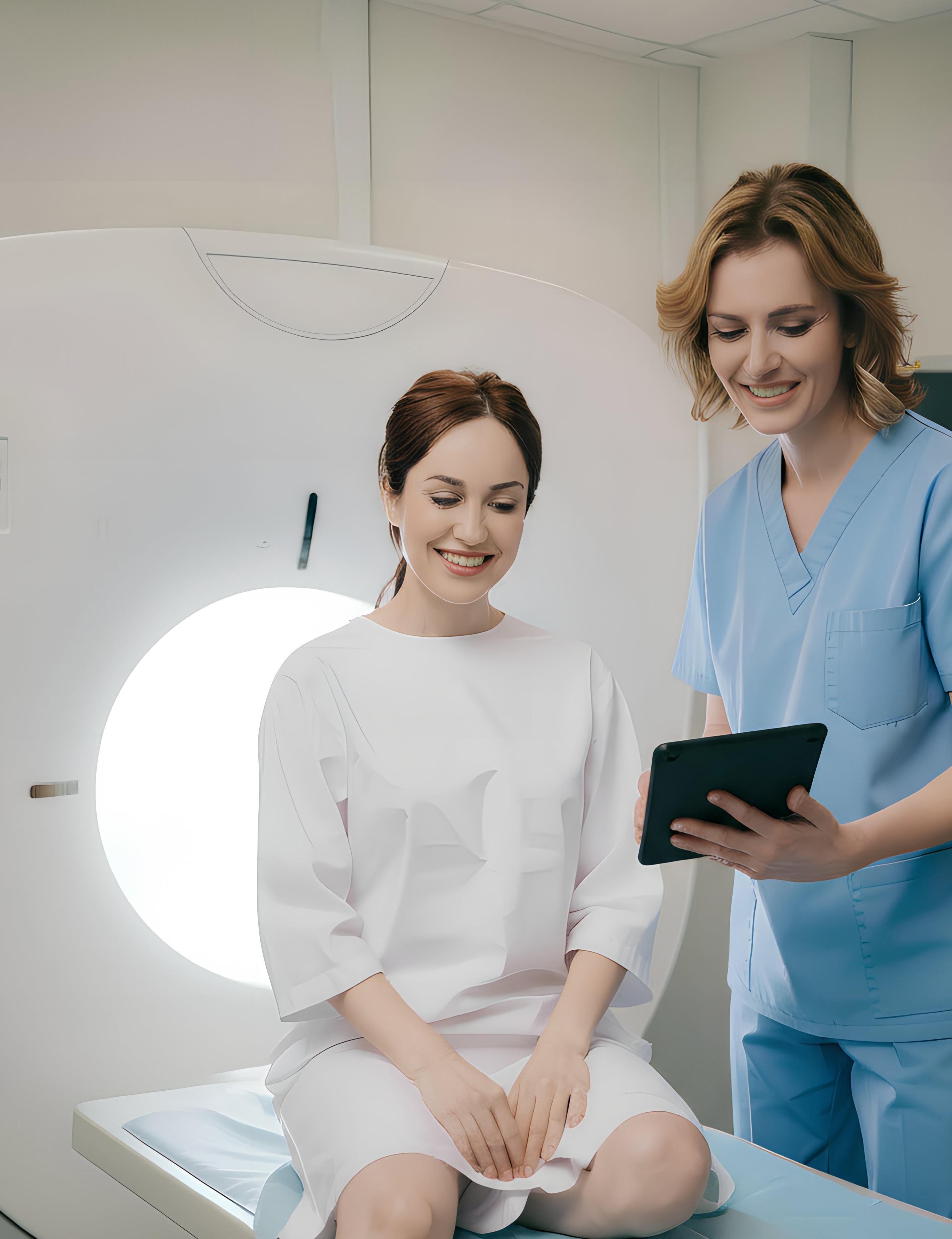
Precedence Research reports that The global diagnostic imaging market size was estimated at $47.81 billion in 2024 and is anticipated to reach around $76.69 billion by 2034, expanding at a CAGR of 4.84% from 2025 to 2034. The rising awareness of early disease detection is expected to boost the market growth.
Recent developments featured in the report include:
• In November 2024, Detection Technology, a pioneer in X-ray detector solutions, unveiled a comprehensive portfolio of flat-panel X-ray detectors at the RSNA 2024 exhibition to advance medical imaging. This unique medical flat panel lineup includes 20 solutions that combine high frame rates with exceptional image quality at low doses and a wide dynamic range. The newly enhanced portfolio is optimized for a range of medical applications, including image-guided surgery, wireless radiography, fluoroscopy, oncology and dental imaging.
• In October 2024, Clemson University collaborated with Prisma Health and unveiled a new 3T functional magnetic imaging (fMRI) machine at Prisma Health Oconee Memorial Hospital. This next-generation, non-invasive scanning technology provides faster, higher-quality medical imaging than previously available on the community hospital campus.
• In September 2024, Olympus Corporation announced the launch of VISERA S, an all-in-one imaging platform with stroboscopy. The new video platform integrates advanced diagnostic capabilities, including Narrow Band Imaging (NBI) technology. It is designed to improve ENT diagnostic efficiency and patient experience. •
Product Focus
Patient Experience
1
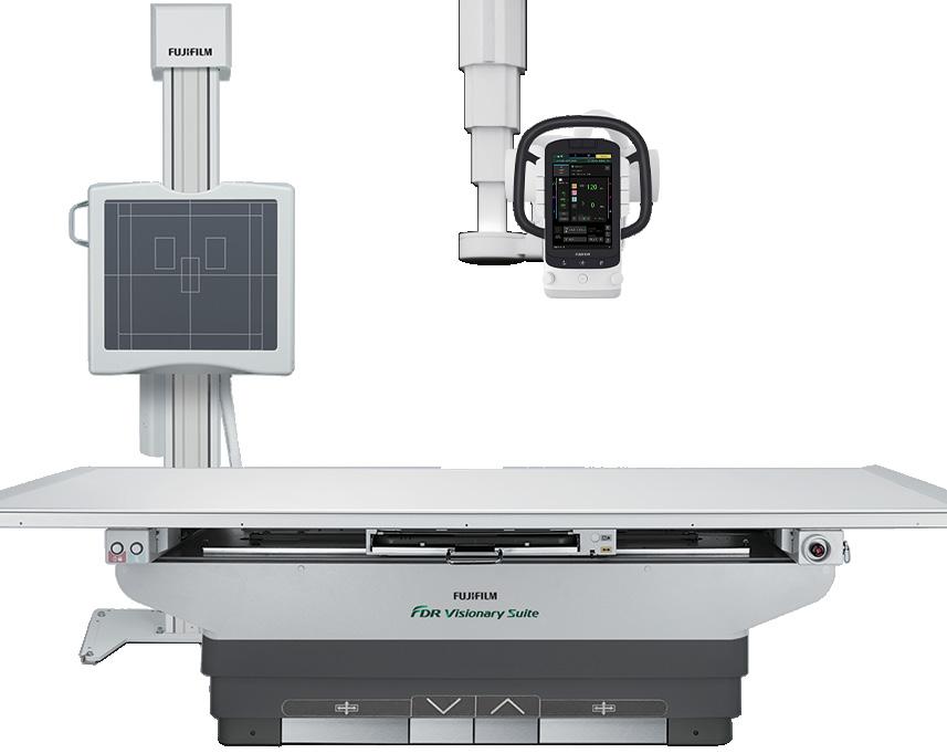
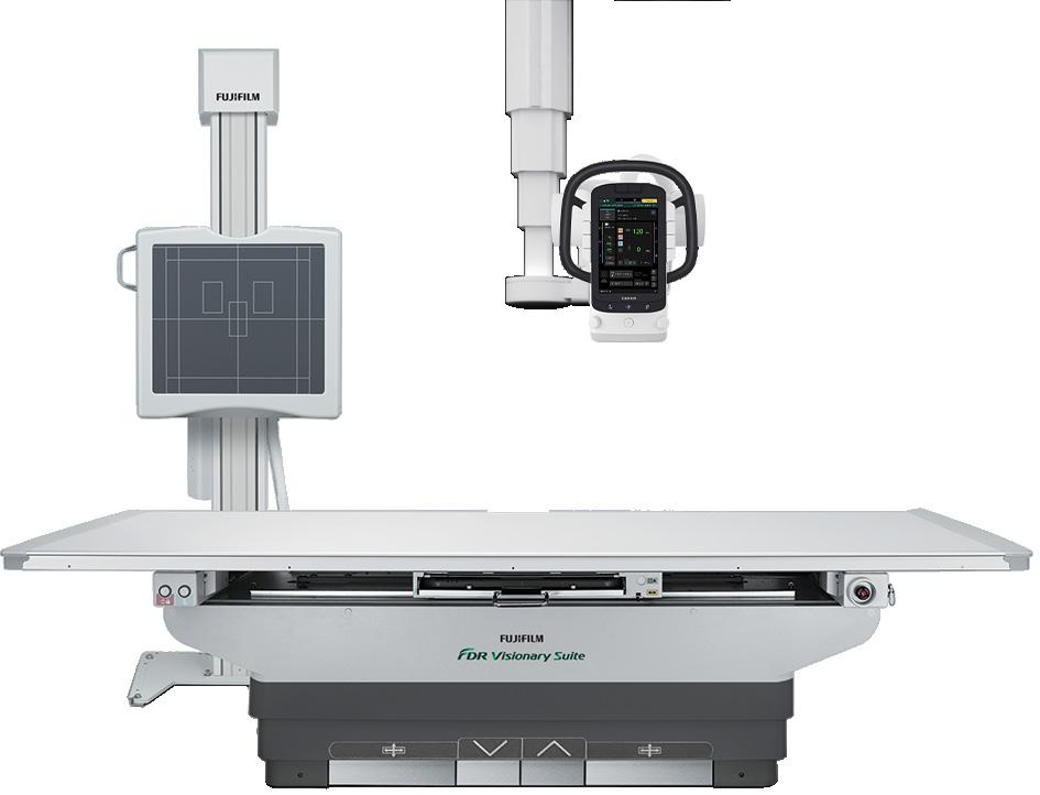
FUJIFILM HEALTHCARE AMERICAS CORPORATION
FDR Visionary Suite Digital Radiography Room
FUJIFILM Healthcare Americas Corporation has launched several advanced automated functions for its FDR Visionary Suite digital radiography room. Optimized to support high-volume imaging in hospital radiology departments and imaging centers, the automation features are designed to enhance workflow and improve the patient and technologist experience for a wide range of general radiology exams. In addition to the system’s automation features, the system is designed with patient comfort in mind. FDR Visionary Suite accommodates
*Disclaimer: Products are listed in no particular order.
patients of all sizes and patients with mobility challenges, as it features a large tabletop and stroke with motorized elevation, and a weight capacity of 649 pounds. Additionally, the system automatically captures long length images of up to 63 inches upright and 47 inches supine. Misalignments caused by patient movement can be corrected through automatic motion correction software, and images can be captured in as little as 20 seconds, for a better patient experience.
PRODUCTS
SIEMENS HEALTHINEERS
The Magnetom Free.Max 0.55 Tesla (0.55T) magnetic resonance imaging (MRI) scanner from Siemens Healthineers is the world’s first 80 cm wide-bore MRI system. Its wide, tapered bore and 705-pound patient table extend access to severely obese and claustrophobic patients, offering them an improved patient experience. Deep Resolve’s artificial intelligence-based deep learning algorithms enable re -
duced scan times and less patient sedation, in addition to delivering sharper, higher-resolution images. The SkyView siting option routes all cabling through the floor rather than the ceiling, giving an appearance similar to a computed tomography system to further reduce claustrophobic feelings for the patient.
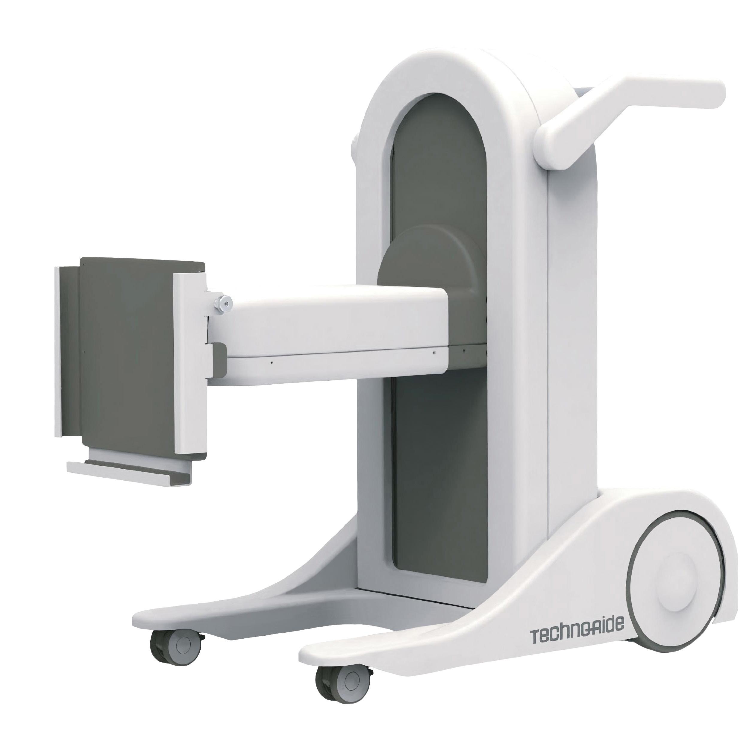
TECHNO-AIDE Sidekick 3
The Sidekick represents a significant advancement in medical imaging technology as the industry’s first remote-controlled mobile panel positioning system. This innovative device employs precision actuator-driven technology to execute vertical and horizontal positioning, as well as panel tilt and pivot adjustments with exceptional accuracy. The system delivers enhanced workflow efficiency through remote positioning capabilities, reducing procedure time and minimizing patient repositioning while improving infection control by maintaining appropriate distance during panel adjustments and reducing direct contact in the sterile field.
Technologists benefit from ergonomic advantages by eliminating awkward manual positioning within confined
patient spaces. The Sidekick features an actuator-driven multi-axis positioning system with wireless remote-control operation, a locking caster system for secure positioning, and chemical-resistant surfaces compatible with hospital-grade disinfectants. Its compact design is optimized for space-constrained environments, making it ideal for operating rooms, orthopedic suites, chiropractic practices, and urgent care facilities where precise imaging positioning and workflow efficiency are critical. The Sidekick addresses key challenges in diagnostic imaging by combining technological innovation with practical clinical requirements, ultimately enhancing both technologist productivity and patient care quality.•
INSIGHTS
DIRECTOR’S CIRCLE
ICE Magazine gathered insights from radiology directors and imaging leaders for a roundtable article focused on the patient experience in imaging. Insiders were able to share their thoughts regarding patient experience to highlight strategies, successes and challenges across the industry.
Participants include:
• AdventHealth Executive Director of Retail Services Health Parks Joel George, MBA, MSN, RN;
• Children’s Hospital Los Angeles Executive Director of Imaging Mario Pistilli, DBA, FACHE, FAHRA, CRA, CNMT.
• Lexington Medical Center Director of Imaging Services Wesley Harden, CRA, FAHRA
Q: HOW DOES YOUR DEPARTMENT CURRENTLY APPROACH IMPROVING THE PATIENT EXPERIENCE? DO YOU HAVE SPECIFIC PROGRAMS, INITIATIVES OR BEST PRACTICES IN PLACE?
DENNIS: Yes, we have a dedicated committee that focuses on radiology patient experience and service training for the staff. We started the committee last year and have seen improvements with our patient experience scores over the last year. The committee has team members from each of
our hospitals and every modality is represented. We provide “passport THANK YOU cards” to our patients after their visit. The cards serve as a good reminder for who took care of them during their visit along with other information, such as how to obtain their images. My role has recently changed into a radiology program director focusing on patient experience, as this is such a large focus within our organization.
GEORGE: In our AdventHealth Health Parks, patient experience is embedded into the strategy and daily operations of care delivery. Rather than viewing it as a standalone initiative, we draw on proven practices from consumer-obsessed and hospitality-driven brands to shape the way patients access and engage with care. Our Health Parks model integrates multiple services under one roof primary care, imaging, labs, sports medicine rehab, and specialty care creating a seamless “one-stop” patient journey. We utilize consumer journey mapping to identify friction points and design purposeful moments of ease and delight, ensuring patient flow and team connectivity are both optimized. We have also implemented hospitality training for frontline teams, emphasizing anticipatory service, personalized welcomes and thoughtful farewells. These practices are operationalized through centralized check-in systems, coordinated workflows and consistent measurement of patient
MARIO PISTILLI
JAMIE
DENNIS ADVENTHEALTH
JOEL GEORGE LEXINGTON MEDICAL CENTER
CHILDREN’S HOSPITAL LOS ANGELES
satisfaction. The result has been stronger loyalty, high team engagement and growth fueled by organic reviews and referrals to prove that patient experience, when embedded into operations, becomes a sustainable advantage.
HARDEN: We focus on communication, compassion and care. We want to make sure we are letting the patient know what is going on, how long it will take and then give them the chance to ask questions and be engaged in their care. Our teams really do a great job of providing that compassionate care and making sure we are doing all we can to make the experience a positive one.
PISTILLI: We use Press Ganey to administer a patient satisfaction survey and track and trend results. Based on the results, we choose one question to focus on improving for the year based on its score and correlation to overall satisfaction. Once chosen, the leadership team and staff brainstorm ideas on action that might improve that particular aspect of patient experience. So, the initiatives in place change yearly in terms of focus but the action items, if they work, we keep.
Q: HOW DO YOU MEASURE PATIENT EXPERIENCE IN YOUR DEPARTMENT? WHAT METRICS OR STORIES HAVE BEEN MOST IMPACTFUL IN SHOWING IMPROVEMENT?
DENNIS: We use Press Ganey, a national survey group. We have held classes over the last year focusing on training radiology leadership on how to interrupt the scores, and how to interrupt comments and best practices to share with team members. We have also held dedicated meetings with modality teams providing feedback based on patients’ comments and partnering with the modality to see how we can best solve some of the recurring issues. We report out scores weekly for what has been accumulated during the month to-date and if the score has gone up or down from the prior month.
GEORGE: We take a dual-lens approach to measuring patient experience combining traditional industry
EMORY HEALTHCARE
benchmarks with real-time consumer feedback.
• Surveys: We utilize validated tools such as Press Ganey and Forsta surveys to track likelihood-to-recommend, provider communication, care coordination and overall satisfaction. These standardized measures allow us to benchmark against national performance and ensure we are meeting clinical and service quality expectations.
• Likelihood to Recommend: Among these metrics, likelihood to recommend (LTR) has been especially impactful. It not only reflects satisfaction with a single encounter but also signals the patient’s confidence in making us their long-term healthcare partner.
• Google Reviews: Equally important, we place a strong emphasis on organic Google reviews. In today’s consumer-driven healthcare environment, these reviews serve as a public trust score and the most visible reflection of the patient experience. We actively monitor and respond to reviews, learning from both praise and constructive feedback. The consistency of our 4.8+ star ratings across multiple Health Park locations with over 5,000 organic reviews is a direct validation that hospitality-driven practices resonate with patients and their families.
HARDEN: We use Press Ganey surveys to measure how we are doing and focus on the voice of the customer. Whether positive or negative, we want to hear how we are doing so we can be sure our efforts are focused in the right place. It is always nice to hear positive comments as it lets us know what we are doing well. The negative comments we view as opportunities to make adjustments to improve.
PISTILLI: By survey and data tracking.
Q:
HOW DOES IMPROVING PATIENT EXPERIENCE BENEFIT STAFF, THE DEPARTMENT AND THE LARGER HEALTH SYSTEM?
DENNIS: Improving the overall patient experience has a direct impact on the overall employee experience, in a positive way. Typically, the patients help us to identify problems
WESLEY HARDEN
or gaps based on their comments. By fixing some of those issues, it usually helps resolve an issue the staff might have been experiencing as well. Sometimes I think the patients’ voice is louder than the staff’s voice for bringing up concerns. We don’t know what to fix unless the concern is raised.
GEORGE: Improving patient experience is not just about the patient, it transforms the entire care environment. For staff, a well-designed experience reduces friction, improves workflows and fosters pride in their work. When teams see patients leaving happier, more at ease and more connected, it directly fuels engagement, lowers burnout and reinforces the “why” behind their calling in healthcare. A culture of hospitality-driven service gives staff the tools and permission to go beyond tasks and focus on meaningful human connection. For the larger healthcare system, improved patient experience has strategic impact. Positive encounters build loyalty, increase likelihood-to-recommend and strengthen brand reputation. Seamless care journeys keep referrals in-network, improve retention and support financial stewardship by maximizing utilization of services within the system.
HARDEN: Let me just state the obvious first. People want to go back to places where they had a good experience. They also want to tell their friends about the good experience they had but will also tell about the bad ones too. Probably more so. So yes, improving the patient experience does have an element of that to it. However, the real benefit in finding ways to improve the patient experience is in knowing you are doing the best you can to ensure that patient has the best outcome. There is a sense of pride in knowing the efforts you are putting in make a difference in not just the patient but their family’s lives as well.
PISTILLI: It benefits staff as our staff is very mission driven and they truly want our patients to have the best experience possible. When we get validation of that from our patients through the satisfaction survey then the team feels good that our patients are well cared for. The department benefits as we can put our whole focus on the patient care that needs to happen and not into service recovery. The institution benefits through driving trust, customer loyalty and good word of mouth.
Q: WHAT ARE THE BIGGEST CHALLENGES YOUR DEPARTMENT FACES IN IMPROVING PATIENT EXPERIENCE? HOW ARE YOU WORKING TO OVERCOME THESE CHALLENGES?
DENNIS: We have some locations in our healthcare system that are hospital based and those locations see a mixture of patients: inpatients, emergency room and outpatients. These areas tend to see more challenges with improving patient
experience for outpatients. There might be a patient scheduled but if an emergent patient comes through the emergency department, they could get delayed, causing dissatisfaction with the patient experience. This certainly limits our ability to stay on a specific schedule depending on the location. The staff do a great job of communicating with the patients if there is a delay and outpatient appointments are limited at some location schedules based on ED trends for volume. To mitigate the limited schedule at some of our hospital settings, we have extended our evening hours and are offering weekend hours at most of our outpatient locations. This increases our appointment availability and allows more convenient times for our patients to receive their imaging.
GEORGE: Integrating imaging into a one-stop shop care model creates tremendous value for patients, but it also comes with unique challenges:
• Capacity & Scheduling Balance: Imaging demand is highly variable. Managing capacity to serve same-day walk-ins, urgent add-ons from primary care, and scheduled specialty orders all under one roof requires sophisticated scheduling systems and flexible staffing models.
• Technology & Space Optimization: Imaging equipment requires significant square footage, shielding and infrastructure. In a multi-specialty setting, the challenge is balancing high-tech imaging suites with the need for exam rooms, rehab space, and other services, while still creating a seamless flow for patients.
• Workforce & Training: Recruiting and retaining technologists who are not only clinically excellent but also hospitality-trained is essential. Ensuring consistent service standards across modalities (MRI, CT, ultrasound, X-ray) can be complex.
• Consumer Expectations: Patients increasingly expect quick access, comfortable environments and transparent communication of results. Meeting those expectations while balancing safety, quality and throughput is an ongoing challenge.
HARDEN: I think one of the biggest challenges we face is the ever-increasing volumes combined with the staffing shortages that make it really challenging for frontline staff to maintain the personal experience sometimes. No matter how much we talk about taking time to ensure the patient in front of you feels like they are the only one, it is really tough to put aside the thought of those you know are waiting. We are trying to be creative in finding ways to address the staffing shortage and are making progress, but these can take time to realize. Working with schools to accept more students and creating pathways for progression to advanced modalities quicker are a couple of initiatives we have started. There are others, but the real focus has to be on the staff
we have now and how we can help them get through those busy times. Our staff are phenomenal and have done an excellent job, but we try to make sure they know how much they are appreciated. Food is always a good pick me up!
PISTILLI: I think many places are challenged as we are with time, staffing and complexity. There is lots of communication that needs to happen from scheduling an appointment to finally walking that patient out the door and a break in communication anywhere in that chain can sour the patient experience. We are working to overcome this through including the entire team in the patient experience process. We also reinforce that every team member is responsible for that patient’s experience regardless of their role so trying to minimize the “that’s not my patient” attitude or “that’s not my job.” We also reinforce to focus on the patient in front of you not how many more there are – you can only image one patient at a time so be present in the moment and focus on doing your best in that moment – easy to say and hard to do.
Q: WHAT ROLE DOES EQUIPMENT, SCHEDULING SOFTWARE, AI OR THE PHYSICAL ENVIRONMENT OF YOUR DEPARTMENT PLAY IN SHAPING PATIENT EXPERIENCE?
DENNIS: The physical environment plays a big role in our patient experience for a lot of our locations and based on what the patient’s expectations might be. Our locations for imaging are throughout all of the Atlanta area, known for heavy traffic all the time. Each location is different and has a different layout, thus it can be a little difficult finding the radiology department. This is a constant struggle for all our locations. We have been working with the operating units based on the patient’s comments on ways to improve the wayfinding capabilities for our patients. The system did launch a new wayfinding app that provides a “yellow brick road” to exactly where one wants to go, it is now a matter of making sure everyone knows about it.
GEORGE: Technology and design are as critical to patient experience as the care itself. In imaging and one-stop shop models, they play distinct but interconnected roles. Intelligent scheduling platforms help balance demand between walk-ins, urgent add-ons and pre-booked appointments. AI-powered tools can predict peak times, optimize equipment utilization and minimize downtime – all of which translate into reduced wait times and greater convenience for patients. The built environment is a silent driver of experience. In my prior role as regional director of imaging, I was blessed to be able to integrate calming themes and tools like MRI video goggles to reduce anxiety and improve patient comfort. At the same time, thoughtful placement of equipment within a Health Park supports efficient patient flow, minimizes bottlenecks, and keeps the experience intuitive and stress-free.
HARDEN: I think the physical environment can play a role in the patient experience. We get many comments about how clean the facility looks and the grounds. We also see lots of comments about the comfort of the waiting room. These are always challenging as what is just fine for one person may not meet the standards of another. So, we try to focus on cleanliness and making sure the patient is not spending too much time there. Also, making sure they are kept informed of delays does help when they do have to wait. Scheduling can also impact the patient experience. If they have to hold for long periods of time or no one answers the phone this can frustrate patients. We have put efforts into allowing electronic communications about scheduling and I see a real opportunity to use AI and other scheduling tools to make improvements on this process.
PISTILLI: All of those play huge roles in the patient experience. The easier we make every step of the process then the more satisfied our patients will be. AI and software will increasingly allow us the ability to communicate better and in real time and allow processes to be streamlined.
Q: WHAT ELSE SHOULD ICE MAGAZINE READERS KNOW REGARDING THIS TOPIC?
GEORGE: The vision for hospitality embedding in healthcare is that it should be as intuitive and seamless as the best consumer experiences we see in consumer-obsessed hospitality brands even outside of healthcare such as highend hotels or restaurants. AdventHealth is an incredible organization that makes consumers feel whole and welcome through our service standards that drive patient experience. A one-stop shop model is not simply about convenience it is about reimagining care delivery so that patients feel known, supported and cared for across their entire journey.
HARDEN: I think it is important for imaging leaders to share their experiences and ideas around this topic as the more information that is out there the better we all can become. By sharing what you have done may help another leader find an answer to a problem they were struggling with. Together we are stronger.
PISTILLI: I think ICE readers should know that giving a great patient experience is still possible even in staff constrained, stressful, busy environments. It takes the entire team coming together and being willing to support each other to make it happen. •
• Register for free and apply to any of our listings
• Browse our 350+ open positions across the United States
• Get directly connected with industry hiring managers
WHY SHOULD HIRING MANAGERS POST WITH US?
• A talent network of 3600+ actively looking biomedical and imaging professionals
• A variety of listing and advertising options
• Social media, print, and eNews promotion
“HTMjobs.com is a remarkable website that can be used by any personnel looking for employment, internships, or just a good read from their Career Center . The site offers job postings from employers in the HTM industry, as well as resources for job seekers to help them prepare for their job search, including resume tips, interview advice, and more.”
AI and the Patient Experience
By Matt Skoufalos
Just as the advancement of medical imaging technologies has evolved nearly every aspect of healthcare delivery in the modern era what it previously could achieve, so too has the refinement of artificial intelligence (AI) and AI-powered solutions compounded those gains, paving the way for new growth in the systems that allow healthcare practitioners access to the inner workings of the body.
AI technologies like natural-language search, machine learning, and vast data-analysis tools have found ready utility in the world of medical imaging, facilitating the heavy computing needs of image-capture, processing and data storage systems that underpin the operations of medical imaging systems. The earliest promised returns on the synthesis of these technologies have been realized in improved radiologist workflow, patient scheduling and image refinement processes. Discovering applications through which they may be leveraged in the future – namely, in improvements that can play a more direct role in the patient experience – involves taking a closer look at how AI-powered processes presently are deployed, and in what ways they may be.
Jason Polzin, general manager for MR applications platform and research technologies at GE HealthCare, agreed that much of the advancement of AI-powered technologies in the medical imaging space has focused on the experiences of the radiologists and technologists that most frequently interact with imaging devices themselves.
Polzin pointed out, however, that as much as AI computing advancements that improve image quality, reduce length of scan times and automate patient positioning supports the work of imaging professionals, they also can improve the patient experience during
an exam by streamlining those various interactions into a smoother process.
Moreover, he said, “where there’s overlap” in the realized gains of AI technologies for a variety of stakeholders, “that’s best.”
“The best use of AI is where it hits administrators, clinicians and patients,” Polzin said. “A lot of what these AI technologies do is reduce recalls and rescans. When they have to come back a couple days later, that’s very disruptive.”
“Shortening the amount of time also makes the exam better for the patient because the less time they’re on the table, the better an experience it is for them,” he said. “It’s less time being anxious and having to hold still.”
Likewise, AI-powered intelligent protocoling that gathers personalized patient data from electronic medical records (EMR) and prior examinations helps inform automated processes onboard the imaging devices themselves about what studies may be performed based on prior experiences. Whether those solutions are integrated within the platforms that vendors create themselves, or added as an after-market enhancement, the value they add in the clinical domain all distills into a more seamless patient experience.
“When we consider workflow, the more integrated the solution, the easier it is to adopt, and for the scan operator to use,” Polzin said. “It’s critical that the on-device, uses [of AI technologies] are available. This includes developing systems that are compatible with third-party solutions being leveraged for their deep learning and AI expertise
in the clinical domain.”
“We want this to be as seamless a workflow as possible,” he said. “We spend as much time developing these AI technologies to make the clinician’s job easier, as we do working to ensure seamless integration for the radiologist, the technologist and the patient.”
Polzin’s colleague Erdogan Cesmeli, GE HealthCare chief strategy, marketing, and commercial officer of molecular imaging and computed tomography, said it’s easiest to contemplate the myriad ways in which AI-enhanced technologies support the patient experience by regarding the patient journey from a holistic perspective. AI processes can accompany the patient from ordering an imaging study to scheduling it, undergoing the exam, communicating and analyzing the results afterwards, and maximizing the impact of those findings.
“[The patient] starts out seeing their generalist, who may be referring them to their cardiologist or radiologist, and then get scheduled for CT or MR,” Cesmeli said. “During that process, we get their demographics, some of which are used with our images.”
AI can aggregate detailed patient data like height, weight, age and gender to support optimization of imaging protocols applied at the device level during an imaging exam. Technologists and physicians benefit from access to the menu of those options, and ultimately must decide which is best to use for which patient; however, access to that combined knowledge base can help to optimize the image reconstruction, Cesmeli said.
“More and more, with AI technologies, patients come to the room, they are positioned, they are scanned, and before they probably dress up, the images are analyzed, and sent to PACS,” he said. “Radiologists are then reading them and sending a report to the referring physician, who shares the results with the patient.”
“We are more [concerned]
COVER STORY
about [what happens] when the patient shows up to the room where our equipment is, and [how] the data goes to the reader or physician preparing the report [afterwards],” Cesmeli said.
“AI is a little bit like art when you are doing the reconstruction,” he said.
Optimizing patient data for imaging protocols can help expedite the process of undergoing an imaging study; optimizing the reconstructive analysis that helps radiologists and referring physicians process the results of these studies can have another spillover effect on workflow; namely, freeing up some of the time spent analyzing those reports.
“Deep-learning-based [image] reconstruction has really taken the industry by storm,” Polzin said. “The images
accurate diagnosis.”
Beyond their diagnostic imaging applications, AI-powered processes can also support imaging-guided patient treatments, like theranostics and radiation oncology. Both disciplines involve highly advanced applications of medical-imaging-guided processes that can benefit from AI computing solutions to refine and advance the effectiveness of the treatment processes.
In theranostics applications, PET-CT (Positron-Emission Tomography-Computed Tomography) provides detailed anatomical information that illustrates how tissue functions at a molecular level, the better to destroy cancerous material with a targeted radiation dose. Similarly, when delivering radiation
Cesmeli described strategic acquisitions like these as “not completely outsourcing” the AI functionality that underpins these processes but allowing GE HealthCare to be “the face and quality check of a major vendor in this space.”
“Partnership is a necessity, especially for those narrow-but-focused companies,” he said. “[Our customers would like] for us to be able to [provide] the platform so that we can easily include these [functions]. Instead of trying 10 different packages, they would like to have one they can trust, and they can do the risk assessment.”
Ultrasound imaging is another area in which the application of AI-powered processes can help to evolve and refine
When AI-powered image reconstruction, analysis and transmission to PACS processes are automated through technologies that are onboard an imaging system, they can eliminate hours of inefficiencies that free up healthcare professionals for other uses of their time, and potentially help alleviate burnout. – Erdogan Cesmeli
are just easier to interpret and easier to look at, [so] a radiologist doesn’t have to spend as much time interpreting the images. The other thing is that some of these clinical decision support tools help clinicians to interpret the exams more quickly, consistently and confidently.”
When AI-powered image reconstruction, analysis and transmission to PACS processes are automated through technologies that are onboard an imaging system, they can eliminate hours of inefficiencies that free up healthcare professionals for other uses of their time, and potentially help alleviate burnout, Cesmeli said.
“The whole point is about giving back time to the reader, the physician, the radiologist so they are less prone to make mistakes, and patients can get results back faster,” he said. “Remove the burden on the reading physicians so they can get to the point with minimized risks and can provide the
oncology care, AI-powered processes can help automate treatment planning, support clinical decision-making, and deliver personalized care specific to any patient’s individual anatomy.
“If [physicians] see that there’s no response [to the treatment], they stop trying that treatment,” Cesmeli said. “There’s optimization from the patient point of view. We are in collaboration with the pharmaceutical companies, using AI to personalize some of those treatments even better.”
On the other hand, some AI-driven advancements are evolving so rapidly that manufacturers like GE HealthCare recognize the need for specialized, third-party integrations. After acquiring AI developer MIM Software in 2024, GE HealthCare was able to integrate its own cardiac imaging systems with advanced PET and SPECT (Single Photon Emission Computed Tomography) post-processing and analysis tools from MIM.
a mature, ubiquitous technology to improve patient wait times, limit repeat scanning, support image quality, and even help physicians to recapture revenues that may previously have been lost due to oversights in patient documentation.
As POCUS (Point Of Care UltraSound) continues to expand its reach in a space that is also facing a continued staffing shortfall, AI computing can help to automate certain aspects of a modality that relies greatly upon the manual skills of its users. It’s a process that Cesmeli said resulted from an outgrowth of manufacturers finding ways to automate cross-disciplinary functions among MRI and CT technologists.
“One of the things that we are doing is finding out how to make our user interfaces more intuitive, so that an MR tech can also do CT scans, and vice versa,” he said. “We think AI is going to be useful in that area versus the discrete layout.”
“Ultrasound is real-time imaging, so in that case, guided by the AI, [a sonographer finds] the point of interest, or doesn’t,” Cesmeli continued. “I think it’s very clever. It’s less so that we have an interventional mode for performing a biopsy, and more that we have another device that gives the same feedback loop whether to do the biopsy or not.”
Other emergent research focuses on the idea that AI-powered processes could help automate back-end reporting and data aggregation that improves the patient experience. A July 2025 study published in Scientific Reports describes how a research team, led by Oluwatosin Ogundare from the Department of Information and Decision Sciences at California State University-San Bernardino, developed a theoretical “AI Affinity Score” model to predict how AI integration can “maximize their experience of care.”
“Typically, the ultimate goal of the customer satisfaction survey is profit maximization for the service provider, whereas an assessment of care aims to cater to an optimization of the psychological factors within the context of the healthcare ser-
General Manager, MR Applications Platform and Research Technologies, GE HealthCare
vice,” Ogundare wrote. “One might argue that a comprehensive study of the patient’s experience of care should assess how much of the human qualities of empathy, understanding, gentle touch, etc., is lost in the AI integrated healthcare.”
The study concludes that AI-based therapy can be most effective for patients who trust it as a segment of their care delivery, accounting for differences in age, education and other socioeconomic factors.
If more closely integrating AI with patient care leads to better health outcomes or improved delivery of care, it stands to reason that patient satisfaction could also improve correspondingly. However, Ogundare, et al., noted that those outcomes would be best driven by an understanding of patients’ confidence in them.
Another study from the JANUS Group of Barcelona, published in the English edition of Medicina Clinica in February 2025, talks about how AI could improve outpatient care by incorporating “socio-health information on each patient to better personalize their clinical care.” Researchers believe this could include tracking health indicators through wearable devices, self-administered questionnaires, and other data sources to engage the patient in shared decision-making.
That study concludes with the following statement:
“In this sense, it is considered that AI should evolve from its current, relatively ‘narrow’ applications in healthcare (performing specific tasks repeatedly at the back of the medical consultation) to a broader and more flexible perspective that allows the current healthcare system (based on primary and specialized care) to be transformed into one that also includes the patient as a key player in the implementation.”
“There’s a psychology to this,” Cesmeli said. “People have different preferences [for the use of AI processes in different environments. Attitudes reflect that] it’s OK if artists use AI for music, but not for a politician to prepare their speech.”
“We hear from customers that they don’t want us to fully automate everything,” he said. “They want to automate the boring stuff, but leave the fun stuff so they can spend more time with their patients, and in quality [areas].”
The bottom line in all of these processes, as Polzin said, is that whether AI is employed in the calculus or not, “physician trust is critical” to the patient experience. •
Jason Polzin,
MIM Software’s Contour ProtégéAI+
Auto Positioning
AIR Recon DL
BEYOND THE SCAN:
AI-POWERED MAMMOGRAPHY TO AID WITH EARLIER DETECTION AND PEACE OF MIND
Traditional breast cancer imaging is faced with critical screening and diagnostic challenges – from workflow bottlenecks and reading inconsistencies to clinician burnout and patient accessibility. The stage is set for desperately needed innovation to propel us into the future of breast cancer care.
That’s where Hologic has stepped in with its breakthrough combination of Genius AI Detection PRO and the Envision mammography platform. Together, they directly tackle these industry challenges. By engaging cutting-edge technology, they can deliver improved processes and results while also supporting more accessible and comfortable screening for patients.*
TACKLING FALSE POSITIVES
False positives and positioning errors cost more than just time. Not only do they slow down workflow and require additional imaging, but they can also negatively impact patients.
False positive results remain concerningly common, occurring in 10-12% of mammograms in women 40-49 years.1 These results can create anxiety for patients, discouraging some from receiving potentially life-saving supplemental imaging. In fact, research found that the number of women returning for a follow-up screening dropped from 77% to 61% after a false positive finding required an additional mammogram.1 These false positives can also lead to emotional distress, additional costs and time wasted for patients, so it’s critical to prioritize reducing them.
Hologic’s Genius AI Detection PRO solution can help. With its integrated 2D/3D deep learning algorithm, the software supports improvements in cancer detection performance.2 The platform’s ability to read priors for mammography images can increase specificity, in addition to driving additional reduction in false positive recall due to analysis of temporal changes.3
ADDRESSING POSITIONING CHALLENGES
The largest percentage of MQSA-related quality failures is attributed to poor positioning.4 Nearly half of those posi-
tioning failures are attributed to inadequate visualization of the posterior tissue and/or the pectoralis major muscle, and sagging of the breast.4 Positioning is also the leading mammography deficiency reported by ACR5 and a significant issue for radiologists and technologists.
Only portions of the breast that are included on the mammographic image can be evaluated for signs of cancer,3 making proper positioning critical for accurate detection. Better patient positioning, and other technologist-associated factors, have been associated with earlier detection and fewer missed cancers.6
The Envision mammography platform offers a solution to help: Tilt positioning. This unique feature enables a more comfortable and accessible imaging experience.** In fact, 10-15% of the patient population has limitations that may result in sub-optimal imaging7, putting them at an increased risk of potentially missed cancers. With improved patient positioning thanks to the Envision platform’s Tilt technology, there may be greater breast tissue capture compared to the standard of care.7
MAXIMIZED EFFICIENCY IN AND OUT OF THE EXAM ROOM
As the radiology industry faces a workforce shortage driven by issues such as burnout, inefficiency and increased de-
Transformative
mand for imaging9, AI stands to play a key role in improving workflow so providers can focus on the most important part of their job: the patient.
Genius AI Detection PRO solution is designed to tackle these issues and give clinicians valuable time back in their day. The perceived fatigue with using Genius AI Detection PRO solution was significantly lower than the perceived fatigue without it,10* with the solution supporting a 24% reduction overall in reading time and perceived fatigue.10
The solution includes various features that contribute to a reduction in perceived fatigue and reading, while also improving administrative task efficiency. These include an intuitive case score, which allows radiologists to organize cases and allocate time based on case complexity, and time-saving pre-reporting capabilities that automatically send findings to the radiologist’s report.
The Envision mammography platform also offers time-saving features that help improve efficiency in breast imaging. Envision delivers a fast 3DTM scan time of no more than 2.5 seconds11, up to 10x faster than competitors and the fastest scan time on the market.12 The system’s reduced scan time is designed to minimize compression time and patient motion, compared to systems with longer scan times.13
These time-saving mechanisms are critical. Technologists report that a typical screening patient spends about 50 minutes at their imaging facility, with about half of that time spent on positioning and image acquisition.8
Additionally, the Envision platform is designed to track more than 700 new analytics parameters across the subsystems of the gantry, compared to Hologic’s Selenia Dimensions and 3Dimensions. This new tracking allows for predictive service potential, saving users from unnecessary downtime.14
SUPPORTING EARLY DETECTION FOR ALL PATIENTS
There is a key demographic that is often left behind in breast screening: patients with limited mobility. Screening mammography compliance rates are lower in populations who may face limitations or challenges with positioning during imaging than in women without mobility-restricting cases15, specifically 47% lower among women who are dependent on a wheelchair.15
Technologists report being unable to position patients as effectively as they would like in 10% of screening mammograms,8 facing particular difficulties when that patient has mobility issues. The challenges with positioning a wheelchair-bound patient can add more than 10 minutes to their
screening mammogram,8 negatively impacting efficiency, patient time and care.
The Envision platform could be the key to helping improve the standard of care. Its Tilt technology may help with the positioning of patients with limited mobility, possibly resulting in greater tissue capture.7 With the Envision platform, Hologic tackles image quality for patients with limited mobility and challenges the status quo with an inclusive system that affirms that all patients are entitled to the best quality of care.
A FUTURE OF PRECISION AND COLLABORATION
The fusion of AI with next-generation mammography technology is paving the way for a future where breast cancer detection is faster, more accurate and more patient-friendly. Hologic’s Genius AI Detection PRO solution and Envision mammography platform work hand-in-hand to bolster health centers and radiology departments, supporting clinicians and patients alike. By combining the power of advanced AI and mammography technology, they enhance the screening and diagnostic process from start to finish. •
1. Diana L. Miglioretti, Linn Abraham, Brian L. Sprague, et al. Association Between False-Positive Results and Return to Screening Mammography in the Breast Cancer Surveillance Consortium Cohort. Ann Intern Med.2024;177:1297-1307. [Epub 3 September 2024]. doi:10.7326/M24-0123
2. S. Pacilè, et al. (2023, May). Application of artificial intelligence to mammography-tomosynthesis combined images for breast cancer screening. [conference presentation]. SBI 2023
3. K240301 510(k) Summary.
4. Bassett, Lawrence & Farria, Dione & Bansal, Swati & Farquhar, Marybeth & Wilcox, Pamela & Feig, Stephen. (2000). Reasons for Failure of a Mammography Unit at Clinical Image Review in the American College of Radiology Mammography Accreditation Program1. Radiology. 215. 698-702. 10.1148/radiology.215.3.r00jn32698.
5. Albus, K. (n.d.). Frequent deficiencies (revised 8-2-2024). American College of Radiology. https://accreditationsupport.acr.org/support/solutions/articles/11000047152-frequent-deficiencies-revised-5-2-20226. Anubha Wadhwa, Julie R. Sullivan, Mary Beth Gonyo, Missed Breast Cancer: What Can We Learn?, Current Problems in Diagnostic Radiology, Volume 45, Issue 6, 2016 ISSN 0363-0188, https://doi.org/10.1067/j.cpradiol.2016.03.001.
7. Miller, L. (2014, September). What every mammography technologist would like their radiologist to know about: Our patients. Mammography Education, Inc.
8. Based on an internal survey of 58 Patients and 7 Technologists. Inspired Health, November 2023. Unpublished internal Hologic data on file.
9. Hudnall, C. E. (2024, July 3). Burnout Fueling Workforce Woes. American College of Radiology. https://www.acr.org/Clinical-Resources/Publications-and-Research/ACR-Bulletin/Burnout-Fueling-Workforce-Woes
10. S. Pacilè, et al. (2024). Evaluation of a multi-instant multi-modal AI system supporting interpretive and noninterpretive functions. Accepted for publication in the Journal of Breast Imaging. https://doi.org/10.1093/jbi/ wbae062
11. VER-12082 (1.0)
12. Competitor publications.
13. Smith, A. Improving Patient Comfort in Mammography. Hologic WP-00019 Rev 001 (2017).
14. SRS-01327; AT_02-REQ-17382
15. Olsen, J., Pohlman, S., & Shames, J. (2024). HSR24-155: Disparities in Real-World Screening Mammography Compliance Rates by Body Mass Index and Mobility-Restricting Diagnosis—A Commercial Claims-Based Analysis. Journal of the National Comprehensive Cancer Network, 22(2.5), HSR24-155. Retrieved Jul 30, 2025, from https://doi.org/10.6004/jnccn.2023.7164
PACS/IT/AI
BY MARK WATTS
TTHE DIGITAL DOCTOR IS IN: YOUR GUIDE TO SPOTTING
AI FAKE MEDICAL
he reel on my social media automatically loaded after I watched a Go-Pro video my friend had posted. It was a famous actor, his golden voice familiar to millions, standing behind a podium. “He has discovered a new product to clear the lungs of excess fluid,” a caption read. It was presented as a major announcement from the press secretary of the United States. This was amazing news – that after a long career, the actor was turning his efforts to medical research.
I reported the reel to the social media team. But will that be enough to stop the misinformation?
I have been researching medical AI for years, best model, deployment, governance. This reel violated all the best practices that help us build trust with the public and yet there it was.
In the age of information overload, the biggest health challenge we face isn’t a lack of data – it’s a tidal wave of it. With a click, we can access medical research, health forums, and advice from around the globe. This power is double-edged: the right information can empower us to make better health decisions, while misinformation can lead to dangerous choices and unnecessary anxiety. Navigating this online “jungle” is a skill we all need to master.
Misinformation isn’t always a dramatic conspiracy theory; it can be a slick website, a compelling headline or a seemingly innocent social media post. To help you arm yourself, here are some key principles for assessing medical news, websites and even the health apps on your phone.
TRUST YOUR GUT, THEN VERIFY
That email from a “credible-looking” source promising a miracle cure or a revolutionary discovery? Take a moment
to pause. If your instinct tells you something isn’t quite right, listen to it. Instead of clicking on the link, open a new browser tab and search for the claim and the source. A quick three-second search could save you from a world of trouble.
Consider the recent spread of misinformation during the COVID-19 pandemic. False claims about a horse dewormer called ivermectin being a cure for the virus spread like wildfire on social media. People were being told to self-medicate with a drug not approved for this use, with potentially serious health consequences. A quick search of the FDA, CDC and WHO websites would have immediately revealed that these claims were unfounded and dangerous. Remember, a groundbreaking medical discovery won’t be announced exclusively in a chain email or on a niche social media reel.
THE ‘TOO GOOD TO BE TRUE’ TEST
If you read about a health breakthrough on a single website and it sounds too good to be true, it almost certainly is. Major medical advancements are not kept secret. They are published in reputable scientific journals and reported on by multiple, credible news organizations. Cross-referencing is your best friend. A single website’s interpretation of a study is just one perspective. For a more accurate understanding, seek out the original abstract of the study and read the conclusion yourself.
This is not a new phenomenon. History is filled with medical hoaxes that promised miraculous results with no scientific backing. For centuries, “bloodletting” was believed to cure a host of illnesses, from fever to pneumonia, but it was nothing more than a dangerous, ineffective practice that likely hastened the deaths of many. Today’s version of this is the single, isolated “breakthrough” story that appears on a low-traffic blog, often promising a cure for a major disease with a natural remedy. If the biggest breakthrough in the history of medicine has only been
reported on one site, it’s not a breakthrough – it’s a hoax.
DON’T JUST TAKE THEIR WORD FOR IT
Reputable health companies and medical organizations are transparent. If a company claims its product is “FDA-approved” or backed by a specific study, they should provide a clear link or reference. Don’t be afraid to verify these claims yourself. You can search the FDA’s website for product approvals or use research databases to find the study they’ve cited. A lack of available links is a major red flag.
BEWARE THE BLINKING LIGHTS
It seems obvious, but it’s worth repeating: be suspicious of flashy websites and aggressive pop-up ads. If a site tries to grab your attention with flashing signs, animated advertisements, or a “clickbait” headline, it’s likely more interested in your data or your wallet than in your health. These visual cues are often a signal of unreliable information.
Simplifying The Imaging Equipment Ownership Experience
WHERE TO FIND RELIABLE INFORMATION
In a world overflowing with fake news, relevant and trustworthy information can easily get buried. The key is to turn to sources that meet both medical and scientific standards. Major medical organizations like the World Health Organization (WHO) and the Centers for Disease Control and Prevention (CDC) along with reputable university health systems and well-respected public health agencies are your best bet.
This is the beginning of a long new period where real looking reels produced on familiar social media outlets will become commonplace. The term “Sloshing” is being used to describe this event.
Your health is too important to leave to chance. By becoming a more critical consumer of online information, you can transform the digital “data jungle” into a powerful tool for your well-being. •
Mark Watts is an experienced imaging professional who founded an AI company called Zenlike.ai.
Image One Medical is the only Engineer-Owned medical equipment service group that is fully dedicated to Florida’s amazing base of Imaging Centers, Hospitals, and Cancer Treatment Centers. We have a mission: Self perform on every aspect of our business. Specialize on specific modalities: Pet CT, CT & MRI, and Focus in a key geographic region.
Fort Myers I Fort Lauderdale I Tampa I Orlando
FLORIDA BASED
Dealer and servicer of PET CT, CT and nuclear medicine
• Equipment service: full coverage plans
• Equipment sales: installation, relocation and project management
• Mobile coach construction, refurbishment, maintenance and management
and GE
THE DANGERS OF I/O DEVICES
BY CHRISTOPHER WRIGHT
In the repair industry, one thing is certain, the user is the greatest stress test of a system. With this in mind, it should be no surprise that user input can have some devastating effects to the functionality of a system.
Let’s first discuss what qualifies as a user input on a medical device. The common ones we all think of are keyboards, trackball and USB. Any device, however, that can interface with the computer can be considered a user input. For instance, an HDMI port might only be perceived as an output for the system. This, however, is not the case. HDMI is an input/output (I/O) device and allows the system to interface with an external monitor or television and negotiate audio devices and display assignments. This query for information is a playground for problems. If the system identifies an external audio source, it can lose the default assignment. This is noticeable by the loss of audio from the primary speakers. A similar phenomenon can occur with the display assignment. This symptom manifests as improperly scaled resolution or a blank touchscreen. The typical method to resolve this issue, once it has occurred, is to reload software thus reinstating the default assignment. This can cost precious time and labor at best and
may end with having to purchase software, loss of options, and rescheduled patients.
So, if these “I/Os” are so dangerous, why have them at all? I/Os allows the user to project their image onto another screen for others to view. It can allow for an external mouse to be plugged in if the trackball is not working. It gives the service technician the ability to export backups and load software. All these reasons make external I/ Os an unfortunate necessity.
I/Os are also not exempt from the ever-present march of technology over time. I/Os used to be very simple in their approach and would not interface with any devices. This allowed them to be very stable and robust at the cost of data throughput. Many video interfaces would not carry audio and were limited to only a one-way signal.
The release of HDMI in 2002 and mass adoption into the medical field in the early 2010s did not account for the additional cross traffic that would occur. It was seen as a direct replacement for DVI and VGA instead of a different medium entirely. HDMI utilizes the extended display identification data (EDID) on a device in a process called handshaking. This handshake can grab the default assignment from the system if the EDID has a feature called Consumer Electronics Control (CEC). This gives the display the ability to change
This is an example of the Rear IO of a system to illustrate what one might expect to see on a modern ultrasound.
A phone being hooked up to an ultrasound machine can potentially cause software corruption.
video and audio assignments at the manufacturers will. In addition to this, the large number of smart TVs flooding the market requires almost all new models to be hooked up to the Internet to function. This allows manufacturers to push updates and change settings after sale. One of these settings includes the CEC. This means your TV might be working fine with your system and as soon as they push an update, without warning, your system no longer plays audio or gives a main display. There are also several standards of HDMI that make older systems particularly unreliable when identifying a destination properly.
This issue is getting woven into more than just video output though. USB type A can also cause issues of its own. Let’s say a sonographer is trying to export a patient while the network is down, or a service technician is trying to grab a backup of the system during routine preventative maintenance. When they plug in a USB thumb drive, the system must interface with the device to determine the size, partition scheme, even the brand name. When this is done, the possibility exists for the device being misidentified, prompting OS level errors. These errors can be a nuisance or even prevent the application from loading entirely. Also,
there is a common issue where any system that has software loaded via USB will halt when a non-OEM USB is plugged in during the boot process. USB type A can also be used with cellular devices. Phones can transmit data and audio over USB causing problems. Any sonographer looking to charge their phone off the USB port of the machine risks the whole operation.
The addition of USB-C into newer systems, typically post 2016, also allows for video signals to be carried as well, causing a larger likelihood of loss of assignment.
How can we prevent these issues from biting us? Unfortunately, there is no catch all answer or patch disk that is going to fix these design flaws. Remember to be conscious of what devices are being plugged into the system and the risks inherent in doing so. Check to verify standard backups are made on systems in the event software does need to be reloaded. Make sure software is on hand for systems that have these outputs in use. If you do come across one of these symptoms, hopefully, this will help you diagnose the issue faster and get the system back up and running as soon as possible. •
For more info, visit advancedultrasound.com.
YOU CAN LEAD WITH EQUITY & NEVER SAY ‘DEI’
Th ese days, the term “DEI” feels different.
A few years ago, bringing it up in a meeting showed initiative. Now, it can feel like a hand grenade. In today’s climate, with federal programs being cut and equity offices dissolving, those three letters can spark tension and fear.
I’ve felt it, and maybe you have, too.
But here’s the thing to remember: The work still matters. The people still matter.
We may not use the acronym as often, but the mission – making our department fairer, more inclusive and more effective – is as critical as ever.
So, how do we lead this work without saying “DEI”?
I’ve been leaning into five simple, no-buzzword strategies that any leader can use, especially in a department as diverse and dynamic as imaging.
1. SPEAK IN OUTCOMES, NOT IDEALS
Instead of focusing on abstract concepts like “diversity,” I ask concrete questions.
• “Who’s burning out?”
• “Why are we losing good staff after six months?”
• “Are we doing everything we can to retain top talent?”
These are equity questions, but they’re framed around performance, retention and results. That’s language everyone can get behind.
2. AUDIT SYSTEMS, NOT PEOPLE
Unfairness often hides in our systems, not in individuals. This isn’t about placing blame; it’s about making our processes smarter and fairer.
• “Who always gets the least desirable shifts?”
• “Who is consistently offered leadership opportunities, and who is overlooked?”
• “Are our policies applied the same way for everyone?” By questioning the systems themselves, we can create lasting change.
BY NICOLE DHANRAJ DEI
3. RETHINK ‘QUALIFIED’
Some of the best hires I’ve made didn’t have perfect resumes. They had grit, empathy and a willingness to learn. By broadening our idea of what “qualified” means, we can open the door to amazing people who might have been overlooked, such as military veterans, second-career parents, and techs who started in transport or front-desk roles.
4.
BUILD MENTORSHIP INTO YOUR CULTURE
You don’t need a formal program to make an impact. Mentorship can be as simple as noticing someone’s potential and saying:
• “I see leadership in you.”
• “Want to sit in on this next meeting with me?”
• “Let me walk you through how I handled that.”
When done right, mentorship becomes part of the air we breathe.
5. LET
YOUR ACTIONS DO THE TALKING
You don’t have to post a mission statement or host a town hall to create equity. Just focus on your daily actions:
• Make sure every voice is heard in meetings.
• Step in when something feels wrong.
• Be transparent about how decisions are made.
• Follow through on your commitments. These things go further than any acronym ever could. This moment might feel tricky. But leading with equity doesn’t have to be loud, political or performative. It just needs to be intentional.
Call it team culture. Call it good leadership. Call it fairness. You don’t need to say “DEI” to build a place where people feel like they belong.
And that’s what makes people stay. •
Nicole
Dhanraj, Ph.D., SHRM-SCP, PMP, GPHR, CPSS, CRA, R.T(R)(CT)(MR), is an experienced imaging director.
In December 2025, we will announce the eight imaging directors or managers selected by our ICE audience for their outstanding contributions.
HOW DO YOU ASSESS THE QUALITY OF IMAGING & REPORTING SERVICES PROVIDED BY YOUR FACILITY?
COLLECT | MEASURE | IMPROVE
BY WESLEY FOLDS BS, CNMT, NCT
Ad ministrators and imaging managers are familiar with the ever-present need to assess and improve the quality of care provided to patients. Diagnostic imaging is particularly prone to human factors that can lead to variations from image acquisition to reporting. When armed with Quality Improvement (QI) data, managers are empowered to actively seek out and implement changes that can lead to continuous improvement. The ability to identify specific areas for improvement, understand the impact of current processes and make data-driven decisions ultimately results in better patient outcomes.
It is clear that measuring image quality and reporting leads to continuous improvement, resulting in positive impact on diagnoses and treatment plans. That said, whether a facility provides echocardiography, vascular testing, nuclear/PET, MRI and/or CT, in today’s environment of staffing shortages and increased patient loads, it can be easy to fall behind on the performance of incremental QI initiatives. Having the right tools in place can make the process much less daunting. Explaining the goal of QI initiatives can be as simple as summarizing them to the team
as “How are we doing?” and “How can we do it better?”, along with a facility-wide agreement that quality improvement is a never-ending process.
Leadership has a strong role in establishing a culture of quality to facilitate the success of QI initiatives. Championing the QI efforts, by supporting an environment where staff feel empowered to identify problems and propose solutions, can make all the difference.
QI initiatives in health care are based on a cyclical process of collecting data, measuring performance against established benchmarks and implementing targeted improvements to achieve desired outcomes:
• COLLECT: The very first component, data collection, proves to be an obstacle for many. Keeping data collection straightforward and user-friendly can help minimize stress. A consistent format to record the data must be established and the data collection should include all variables relevant to what is being measured. It is optimal to over-gather data, rather than having to go back and collect more. Data separates what is thought to be happening from what is really happening.
• MEASURE: Effective measurement of quality improvement metrics is critical to drive meaningful change toward positive outcomes. Data analytics should focus on aspects that have the
greatest impact on patient outcomes and operational efficiency.
• IMPROVE: The intentional periodic evaluation and transparent communication of QI data to all stakeholders will support a culture of quality, leading to continuous improvement.
IAC QUALITY IMPROVEMENT (QI) SELF-ASSESSMENT TOOL
While you have likely heard of the Intersocietal Accreditation Commission (IAC) as an accrediting body, did you know we offer a Quality Improvement (QI) tool? Whether your facility participates in the IAC accreditation process or not, the tool is accessible to all and free to use.
The IAC Quality Improvement (QI) Self-Assessment Tool simplifies the internal review process by focusing on case studies and providing a standardized workflow for assessing four quality metrics:
• test appropriateness;
• technical quality and safety;
• interpretive quality; and
• report timeliness and completeness.
The tool allows facilities to critically assess their imaging studies and reports, providing a data-driven, objective measure of QI status and progress. It helps facilities visually benchmark findings (through charts and graphs), track QI progress and generate quantitative reports that pinpoint areas for improvement, ultimately enhancing patient care.
Results of a study published in the July/September 2019 issue of Quality Management in Health Care, “Usefulness of the Intersocietal Accreditation Commission (IAC) Quality Improvement Self-Assessment Tool After 1 Year”, summarized respondents’ perceptions of the effectiveness and usefulness of the tool. There was
“Everyone in health care really has two jobs when they come to work every day: to do their work and to improve it.”
- Paul B.
Batalden,
MD
Professor
Emeritus of Pediatrics, Community and Family Medicine and The Dartmouth Institute for Health Policy and Clinical Practice, Dartmouth Medical School
a high level of agreement that the tool is easy to use (91.8%), encouraged critical thinking (90.3%) and the activity was worthwhile (89.6%). In addition, IAC often hears reports of satisfaction from users of the QI tool. They remark on characteristics such as the comprehensiveness of the tool, enabling all aspects from image quality to reporting to be assessed on a single platform. Many report finding value in the scores received through the tool, enabling ease in monitoring and progress tracking.
As an added benefit, the use of the IAC QI Tool can be utilized as a Centers for Medicare & Medicaid Services (CMS) Merit-based Incentive Payment System (MIPS) Improvement Activity to satisfy a component of the MIPS Improvement Activity score.
CONCLUSION
Regardless of the tools we use, when QI data is used to integrate improvements such as standardized processes, making data-driven decisions and fostering commitment from staff, we’re all doing our best to not only care for our patients, but to offer that care at the highest level possible. •
For more info, visit intersocietal.org.
Wesley Folds, BS, CNMT, NCT, is the Interim Director of Accreditation for the IAC Nuclear/PET accreditation program. Previously serving as an IAC Nuclear/ PET application reviewer, he joined the IAC as a Clinical Specialist in 2022. Mr. Folds is an experienced nuclear medicine technologist and has overseen quality improvement (QI) projects and radiation safety training in previous positions.
DIRECTOR’S CUT
BY NICOLE DHANRAJ
WHAT RADIOLOGY LEADERS NEED TO UNDERSTAND ABOUT VETERANS
Before you even meet the veteran technologist on your team, they’ve likely already navigated three job moves, two deployments, and a dozen sleepless nights wondering if their spouse is coming home. They show up on time, stay late and never make excuses. Not because life has been easy – but because they’ve had to make hard things look easy for years.
In radiology, we value reliability.
But sometimes, the most reliable people are the ones carrying the heaviest load –and never saying a word.
That’s why this Veterans Month (also known as National Veterans and Military Families Month), it’s time we widen our lens. Because service doesn’t stop at the person in uniform.
It echoes through families, through frequent moves, through delayed careers, through the quiet strength that shows up every day in scrubs and lead aprons.
IT’S NOT JUST ABOUT THE UNIFORM
Behind every veteran is often someone else who served – differently, quietly and without a ceremony.
• The technologist who followed their spouse from base to base, licensing and relicensing.
• The receptionist who held it together through four deployments.
• The scheduler whose child has been “the new kid” six times before middle school. These aren’t just staff members. These are survivors of systemic transition. And many of them work right beside you – holding a department together while managing a life most people don’t understand.
A DAY IN THE LIFE OF A MILITARY FAMILY IN IMAGING
Meet Jenny.
She’s your newest CT technologist. Last month, she was finishing night shifts at a trauma center in Texas while helping her husband prepare for retirement after 20 years in the Army. She’s worked in five different hospitals across four states over the past decade – not because she wanted to move, but because the military told them it was time. Every move meant starting over:
• Re-licensing.
• Learning new protocols.
• Adjusting to new PACS systems.
• Reintroducing herself to another team who didn’t know she had 15 years of experience. You don’t see any of this on her resume. What you see is a professional, efficient, steady presence. The kind of person who never complains, always learns fast, and offers to cover the holiday shift – because she’s
used to making sacrifices.
Jenny doesn’t need pity. She needs recognition.
WHAT YOU MIGHT NOT SEE (BUT SHOULD UNDERSTAND)
Veterans and military families bring traits that radiology departments thrive on: calm under pressure, resilience, flexibility and loyalty. If they seem “different” from the average employee – it’s because they are.
They’ve had to rebuild their lives over and over. They’ve learned how to work through chaos. And they’ve become experts at holding it together – for everyone else.
WHAT MANAGERS CAN DO TO LEAD WITH AWARENESS
You don’t need to have served to support someone who has. You just need to lead with curiosity, not assumption.
• Look Beyond the Resume Gaps: If a candidate or staff member has scattered dates or multiple employers across different states, ask about their story – not just their history.
That “gap” might’ve been a move to Germany. That job shift might’ve been during deployment recovery.
And that’s not instability. That’s adaptability.
Ask: “How did you manage change across different departments or facilities?”
Say: “I’d love to hear how your past roles shaped the way you work today.”
• Offer Context, Not Just Compliance: Military families are used to structure. What they’re not used to is ambiguity. Civilian hospitals often assume people “figure it out.” But veterans and spouses thrive in systems with clear roles, responsibilities and expectations.
Do: Provide onboarding that goes beyond checklists –explain department dynamics, unwritten norms and “how things really work around here.”
Don’t: Assume silence means disengagement. Many are watching first, contributing second.
• Respect Boundaries: Veterans may not talk about their service. Military spouses may avoid sharing personal life stressors. It’s not secrecy – it’s protection. They’ve been trained not to make things about them.
Instead of asking: “Did your spouse see combat?”
Try: “How’s your family adjusting to life here?” or “Is there anything we can do to support your transition?”
When you ask from a place of respect, you create safety. And safety builds loyalty.
WHY THIS MATTERS IN RADIOLOGY
Radiology is fast, high-pressure and full of moments that require clear thinking under stress. Sound familiar? That’s
what veterans and military families bring: mission-first mentalities, discipline without ego and the emotional endurance to show up even when life is heavy. But that strength can be missed if you only see the surface – or worse, assume it doesn’t need support.
You don’t need a veteran hiring initiative to start. You need veteran awareness. And you already have people on your team worth seeing more clearly.
VETERANS MONTH BELONGS TO MORE THAN VETERANS
This November, yes – honor the veterans in your department. But also honor the tech who covered her spouse’s deployment while raising three kids. The nurse who started over again and again. The team member who moved every two years, rebuilt everything, and still shows up to work early.
These aren’t side stories. They’re survival stories. They’re the roots of strength in your department.
So don’t just say, “Thank you for your service.”
Say, “I see what you’ve carried.” Say, “I see what you’ve rebuilt.” Say, “I’m glad you’re here.”
Because when one person in a military family serves, the whole family serves. And when we, as leaders, honor that truth – we don’t just lead better teams. We become better humans.
VETERANS MONTH REFLECTION: WHO’S ON YOUR TEAM?
As you consider the stories behind your colleagues this month, take a moment to reflect:
• Do you know if any of your team members have a military background – or a spouse, child or parent who served?
• Have you ever worked with someone who started over multiple times and never said a word about it?
• What assumptions might you’ve made about someone’s resume gap, communication style or silence in meetings?
• Who in your department has shown resilience not just in work – but in life?
Veterans Month is a time to honor service – but it’s also a time to look closer. Because sometimes the people who’ve carried the most have asked for the least.
Let’s lead with awareness, not assumption.•
Nicole Dhanraj, Ph.D., SHRM-SCP, PMP, GPHR, CPSS, CRA, R.T(R)(CT)(MR), is an experienced imaging director.
PROVEN PRINCIPLES TO HELP NAVIGATE CHANGE
EMOTIONAL INTELLIGENCE
BY DANIEL BOBINSKI
Thriving organization know that what got you here won’t necessarily get you there. In other words, change occurring around us often means we must adapt the way we do things just so we can keep up.
Over the past few years, we’ve witnessed unprecedented shifts in how business is done. But even without the recent changes, if you’ve been in the workplace for any length of time, you know that adaptation and modification is inevitable. Therefore, how managers and leaders guide their teams through transitions can make the difference between surviving and thriving.
For years I’ve been a fan of the work of psychologist Jeffrey Schwartz and executive coach David Rock. They translated brain research and neuroscience into practical applications for how people handle change. From their findings, three things stand out to me as vital for increasing the likelihood of implementing effective change:
• Help people focus on the big picture
• Set a high expectation for people to share their “a-ha” insights
• Increase attention density (the amount of attention devoted to a subject over time) Allow me to elaborate. Focus means providing a big picture of the change desired by the organization. The earlier it is communicated, the better. Big picture
means providing a broad vision of the new direction without getting bogged down in details.
Setting a high expectation refers to leadership communicating a belief that people will have breakthrough ideas for how to best achieve the new big picture. This creates a freedom to provide personal input, which leads to better buy-in for adapting to the coming change.
Increasing attention density means don’t stop at one and done. In other words, don’t just announce that change is coming and leave it at that. Keep discussing the focus and the expectation. Make the big picture a frequent part of workplace conversations. Then, acknowledge, celebrate and encourage ongoing dialogue on the ideas people offer for making the change a reality.
That’s the overview. Now for some shoe leather. The following six principles expand on the recommendations of Rock and Schwartz and will help any organization in effecting change.
1. Stay Engaged. Whether you’re a high-profile superstar or an unsung hero, the work you do contributes to a common good. If you recede from an upcoming change, important connections and communication lines can fade away. Instead of withdrawing from change, refocus and think “excellence.” Press for being world class. In every aspect of your job, ask your-
self, “If someone else were looking at my attitude about this change, would they consider it to be excellent?”
Beyond an internal attitude of staying involved, we can also create cross-functional teams or an innovation committee. Their purpose should be to help us stay plugged into the transformation process.
2. Keep Considering the Bigger Picture. Since your workplace is more than just your own workstation, you’ll expand your value to the organization by looking at how change is occurring at all levels. Even the picture outside your organization should be considered. Think about it. Gen Z is entering the workforce, AI is transforming operations, and supply chains are more fragile. Also, customers are increasingly expecting personalized, on-demand experiences.
We should also consider changes in our individual industries. Thus, your big picture thinking can be improved by staying active in professional associations, reading industry publications, attending virtual conferences, and even leveraging sites such as LinkedIn to keep up with real-time insights.
3. Talk and Listen. We are better able to interpret the events around us if we stay in tune with others about what’s happening. This means seeking out and considering others’ perspectives.
This starts with the people in our work area, but extends out to hearing from people in other parts of the company, and even from other organizations. We can even inquire of people outside of our industry. Seeking their viewpoints can provide unique and fresh perspectives.
4. Look for Ways to Be of Value. Since change always brings new problems, we must resolve to be part of the solution. After all, problem-solving is part of every job, so let’s practice it. We can think of it this way: It’s one thing to identify a problem, it’s something else to solve it.
Another way to increase our value is to understand the internal strengths and weaknesses of our teams, as well as the external opportunities and threats. One way to accomplish this is through a SWOT Analysis. By identifying your team’s strengths
and weaknesses as well as the opportunities and threats, you can decide how to capitalize on your strengths and shore up your weaknesses to take advantage of the opportunities and mitigate the threats. From there, you can identify the skills needed to maximize the benefits of upcoming change.
5. Be Flexible. Change is inevitable, so look for ways to blend changes into your normal routine. Think in terms of creating new workflows or systems.
Naturally, an organization must maintain efficiency and effectiveness, but flexibility allows you to flow with the changes instead of fighting against them. This includes being flexible in both your attitudes and responsibilities. One phrase I teach my clients is, “Blessed are the flexible, for they shall bend and not break.”
6. Learn From Your Network. Since people outside your organization are probably facing changes themselves, they can serve as sounding boards. Exercise your connections and be willing to ask a lot of questions. A quote often attributed to Benjamin Franklin says, “We must all hang together, or most assuredly we shall all hang separately.”
Bottom line, how we approach change affects how it impacts us. We can work to accommodate it, or we can get overwhelmed as it accelerates around us. My recommendation? Stay focused on the upcoming big picture, eagerly expect “a-ha” moments for how to make the change work better, and make both of these part of your regular conversations. By doing these things, your chances of success in the face of change go way up. •
Daniel Bobinski, Th.D, is a best-selling author and a popular speaker at conferences and retreats. For more than 35 years he’s been working with teams and individuals (1:1 coaching) to help them achieve excellence. He was also teaching Emotional Intelligence since before it was a thing. Reach him by email at DanielBobinski@protonmail.com or 208-649-6400.
MEDICINE FOR THE SOUL:
NEW LANDMARK PROJECT ON WHOLE-PERSON HEALTH & FUNCTION
The National Institutes of Health (NIH) has launched an effort to advance research on whole-person health and create an integrated knowledge network of healthy physiological function. Whole person health involves looking at the whole person – not just separate organs or body systems – and considering multiple factors that promote health. For example, a multicomponent lifestyle intervention including healthy diet, physical activity and stress management may improve multiple and interconnected aspects of health including cardiovascular (e.g. blood pressure), metabolic (e.g. glucose metabolism) and musculoskeletal function (e.g. muscle strength).
“Biomedical research is largely organized around the study of specific organs and diseases. In contrast, we do much less research on health itself, which is an integrated process involving the whole person,” said Helene M. Langevin, M.D., director of NIH’s National Center for Complementary and Integrative Health, which leads the NIH-wide program.
The five-year research initiative will proceed in several stages, drawing from existing scientific knowledge to develop a complete, working model of healthy human physiology. It will build on the NIH Human Reference Atlas and the Human BioMolecular Atlas Program (HuBMAP) to connect the com-
plex anatomy and function of the body’s different organs and systems into a single “map.”
Future stages of the project will link common clinical measures, such as blood pressure, blood glucose and cholesterol, to major physiological functions. This initiative will also populate the framework with existing human data and ultimately build and test an interactive model of whole-person health.
“By organizing healthy physiological function into a whole-body knowledge network, researchers will be able to explore scientific questions about health in a new way,” said Dr. Langevin. “With our ability to acquire new scientific data at an increasingly dizzying speed, the importance of integrating and connecting new data to what we already know is greater than ever. The Whole Person Reference Physiome will lay a foundation for understanding the factors that drive declines in health and mechanistic pathways to health restoration.”
More information about the research program is available on the NIH Research Portfolio Online Reporting Tools (RePORT) website, here: https://reporter.nih.gov/search/NHCW3mdunUCF3ULUAvilYQ/project-details/11224772#description.
NCCIH conducts and supports rigorous scientific investigation into the fundamental science, safety, and effectiveness of complementary and integrative health approaches, as well as their roles in improving health and health care in a whole-person health framework. News releases, fact sheets and other NCCIH-related materials are available on the NCCIH website.•
ICE Break
What is XperTIS?
Tri-Imaging Solutions introduces a cutting-edge platform that enhances supply chain
engineer performance, and system diagnostic tools and video tutorials, and Designed for efficiency, the platform minimizes setting a new standard for reliability in medical
XperTIS
provides engineers with
streamlines parts ordering and tracking. Designed
What is XperTIS?
Tri-Imaging Solutions introduces a cutting-edge platform that enhances supply chain management, engineer performance, and system monitoring. It provides engineers with diagnostic tools and video tutorials, and streamlines parts ordering and tracking. Designed for efficiency, the platform minimizes downtime and optimizes operations, setting a new standard for reliability in medical imaging.
Objectives
WHAT IS XPERTIS?
Objectives Solutions
• $300 a month
• 1/6 page ad included in the marketplace
• Up to 5 categories in our ICEDEX
• Your company name, website and phone included in your ICEDEX listings
• Your company name in the alphabetical business index • Appear in the print and digital magazine
Objectives
Solutions
XperTIS offers step-by-step repair guidance, helping engineers troubleshoot efficiently while ensuring faster, more accurate parts ordering This helps
Tri-Imaging Solutions introduces a cutting-edge platform that enhances supply chain management, engineer performance, and system monitoring. It provides engineers with diagnostic tools and video tutorials, and streamlines parts ordering and tracking. Designed for efficiency, the platform minimizes downtime and optimizes operations, setting a new standard for reliability in medical imaging.
XperTIS proactively monitors system health, supports engineers in repairs, and enhances the supply chain process by giving teams seamless access to parts ordering and order tracking 10
XperTIS offers step-by-step repair guidance, helping engineers troubleshoot efficiently while ensuring faster, more accurate parts ordering. This helps maximize uptime and minimize repair costs