International Research Journal of Engineering and Technology (IRJET) e ISSN: 2395 0056

Volume: 09 Issue: 07 | July 2022 www.irjet.net p ISSN: 2395 0072

International Research Journal of Engineering and Technology (IRJET) e ISSN: 2395 0056
Volume: 09 Issue: 07 | July 2022 www.irjet.net p ISSN: 2395 0072
1PG Student, Department of CSIS, Rajagiri Scool Of Engineering & Technology, Kerala, India ***
Abstract - Deep learning based image segmentation has varying applications in this modern era. Automatically segmenting the Brain Tumor isanimportantoneamongthem. Manual segmentation of brain tumor is a very difficult task in the field of medical image processing. Moreover it is a time consuming process and chances for getting false results is high. Hence an accurate method for automatic brain tumor segmentation is necessary for the clicians to obtain a correct result and hence for the proper analysis , monitoring of the deseases. Unet is a CNN based architecture which is widely used for image segmentation process. Unet has its different modifications such as unet++, unet3+ . Unet 3+ having full scale skip connections is capable of extracting more features than other unet varients. Unet 3+ model provides a better accuracy and other metrics values compared to basic unet architecture in brain tumor segmentation.
Key Words: Unet, Unet++, Unet3+
Thequickincreaseindeeplearningalgorithmdevelopment suggeststhatdeepneuralnetworkshaveexcellentpotential for use in computer aided automated or semi automatic proceduresforclinicalfactprocessing.Convolutionalneural networks quickly improved, leading to models that could match or even outperform human level performance in several applications, including microscopic image segmentation. Deep learning knowledge based semantic segmentation has recently attracted a lot of attention. Medical picturesegmentationfrequentlyuseUNet,a deep learningnetworkwithanencoder decoderarchitecture.One of the crucial elements for accurate segmentation is the fusion of many scale variables. UNet++ was updated to resemble Unet by creating a structure with dense and stackedskipconnections.
However,itdoesn'tlookatmanyrecordsfrombroadscales, andtheremaystillbeatonneofroomforimprovement.The full scaleskipconnectionsadvantageisusedbytheUNet3+ paradigm.Thefull scaleskipconnectionsusefeaturemaps at various scales that provide low stage information with high degreesemantics.Inadditiontoboostingaccuracy,the UNet 3+ can decrease network parameters to increase processing performance. The changing shape and appearance of brain tumours in multi modal magnetic resonance imaging makes accurate segmentation of these lesionsachallengingmedicalimageanalysistask.Suchtypes ofbraintumoursmustbemanuallysegmented,whichtakes
a great lot of clinical skill and time and increases the possibilityofhumanerror.Additionally,themanualmethod lacks consistency and reproducibility, which has a detrimental impact on the outcomes and may, in the long run, lead to inaccurate analysis and treatment. A well designedU Netbasedarchitecturemaybeabletosegment braintumourseffectively.Sotheautomaticsegmentationof braintumourscanemploytheUnet3+model.Braintumor segmentation is an essential step in medical image segmentation.Automatic brain tumor segmentation will givesanaccurateresultandwillhelpsthecliciansforproper diagnosis.Moreover it saves the time and will reduces the chancesforgettingerrors.
Unet is a CNN based architecture having encoder and decoderpathwhichiswidelyusedforimagesegmentation process.Unethasitsdifferentmodificationssuchasunet++, unet3+ . UNet++ does not sufficiently examine the informationfromcompletescaleswhenusinglayeredand dense skip connections. UNet 3+ utilises full scale skip connectionstocombinehigh levelsemanticswithlow level semantics from feature maps at various scales. The suggested Unet3+ technique produces significantly better segmentation results than existing models and aids in accuracy improvements. An encoder decoder structure calledUnet3+hasconnectionsbetweeneachdecoderlayer aswell asbetween the encoder and decoderlevels.These full scale skip connections enable them to extract more characteristics and produce more precisely segmented results.
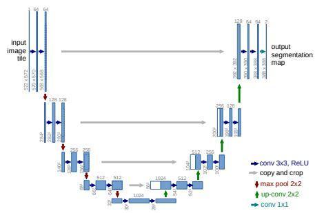
In order to make better use of the existing annotated examples,Ronneberger,Olaf,PhilippFischer,andThomas Brox suggested a project that presents a network and trainingtechniquethatheavilyreliesondataaugmentation. The design consists of a decoding path for accurate localisation and an encoding path for capturing context. Networkspeedisquick.OnamodernGPU,segmentingan image may take less than a second. This approach is built upon the so called "fully convolutional network," a more elegant architecture. This architecture is updated and extended so that it can function with a small number of trainingphotosandproducesegmentationsthataremore accurate. The fundamental concept is to use upsampling operators to replacepoolingoperators. Thus,theselayers raisetheoutput'squality.[1].
International Research Journal of Engineering and Technology (IRJET) e ISSN: 2395 0056
Volume: 09 Issue: 07 | July 2022 www.irjet.net p ISSN: 2395 0072
The dental panoramic image segmentation using unet architecturewasdiscussedbyS.Sivagami,P.Chitra,G.S.R. Kailash, and S. R. Muralidharan. In order to accurately segment dental panoramic x ray images, this method suggestsaUNetarchitecturethatmakesuseofconvolutional neural networks. This allows the dentist to identify any impactedteeth,locatethepreciselocationforplacingdental implants,andascertainthedirectionoftheteeth'sstructure. This method recommends a UNet architecture that uses convolutionalneuralnetworkstoaccuratelysegmentdental panoramic x ray images. Medical experts can identify and diagnose disease more precisely with the help of dental radiography pictures. Images obtained by radiographic procedureslikeX rays,CTscans,andMRIs.X raypictures are frequently complex in nature. It is challenging to accuratelydistinguishbetweenthevariousdentalportions whenthereisnoise.Thismakessegmentationaverydifficult task.Dentalimagesegmentationhelpsthedentistlocatethe best spot for dental implants, spot impacted teeth, and determine the direction of the teeth's structure. A more recent method is the segmentation of medically utilised imagesusingtheUNetarchitecturalmodel.[2].
AttentionUnet++approachforliverCTimagesegmentation wasdiscussedbyC.Lietal. Inorder toallowthefeatures recovered at various levels to be combined with a task related selection, this research suggested a layered attention aware segmentation network called Attention UNet++ that integrates attention mechanisms between nestedconvolutionalblocks.Here,thehierarchicalAttention UNet++ attention aware segmentation network was suggested.Livercancerisoneofthecancerswiththehighest death rates. It is imperative to develop an automatic liver segmentationmodelsincemanualsegmentationtakesalot oftimeandispronetomistakes,makingitchallengingfor medicalprofessionalstoidentifyandtreatliverlesions.The recommendedmethodincludesadeepsupervisedencoder decoder architecture and an improved dense skip connection.TheattentionmethodintroducedbyAttention UNet++ between nested convolutional blocks enables the combination of a task related selection with the features extracted at different levels. Furthermore, the addition of deep supervision increases the prediction speed of the prunednetworkatthecostofamodestperformanceimpact. The segmentation of the liver by UNet++ was quite successful.[3].
The article "UNet 3+: A Full Scale Connected UNet for MedicalImageSegmentation"waswrittenbyH.Huangetal. UNet 3+ utilises deep supervisions and full scale skip connections in this project. Full scale skip connections combinehigh levelfeaturemapswithlow levelfeaturesat variousscales.Fromthefeaturemaps,thedeepsupervision developshierarchicalrepresentations.Organsthatoccurat variousscalescanbenefitfromthesuggestedmethodology. ThesuggestedUNet3+candecreasenetworkparametersto increase computation efficiency in addition to improving
accuracy. Furthermore, it develops a classification guided module and a hybrid loss function to improve the organ boundary and lessen over segmentation in a non organ image,producingbettersegmentationoutcomes.[4].
J The authors of the research, "Optimized U Net for Brain TumorSegmentation,"areMichaFutrega,AlexandreMilesi, Michal Marcinkiewicz, and Pablo Ribalta. This study suggestedanimprovedU Netarchitectureforsegmenting brain tumours. Run a thorough ablation research to test: deep supervision loss, Focal loss, decoder attention, drop block, and residual connections in order to determine the bestmodelarchitectureandlearningschedule.Additionally, theidealUNetencoderdepth,convolutionalchannelcount, and post processing method were looked for. Automatic braintumoursegmentationisoneofthemostdifficultissues inmedicalimageprocessing.Amoreaccurate,dependable, and uniform method for disease identification, treatment planning, and monitoring would be made possible by developing a computational model that is capable of performing better than a trained human. Deep learning based semantic segmentation has recently attracted increasing attention. Medical picture segmentation frequently use UNet, a deep learning network with an encoder decoderarchitecture.[5].
It is essential for the diagnosis, follow up, and therapy planning of the condition to automatically segment brain tumoursusing3DMRIs.Manualdelineationtechniquesare expensive,time consuming,labor intensive,andsubjectto human mistake. Myronenko.A present a semantic segmentationnetwork basedonencoder decodersforthe segmentationoftumoursubregionsfrom3DMRIs.Duetoa smalltrainingdatasetsize,avariationalauto encoderbranch is added to reconstruct the input image itself in order to regularise the shared decoder and impose additional restrictionsonitslayers.Thesegmentationstrategyisbased on an encoder decoder CNN architecture, with an asymmetricallylargerencodertoextractimagefeaturesand asmallerdecodertoreconstructthesegmentationmask.An additionalbranchwasintroducedtotheencoderendpointin amannerreminiscentofautoencoderarchitectureinorder toreconstructtheoriginalimage.[6]
Auniquetwo stagecascadedU Netispresentedtosegment braintumoursubstructuresfromcoarsetofine.Thenetwork is trained end to end using the Multimodal Brain Tumor Segmentation Challenge training dataset.Experimental findings on the testing set show that, for the enhancing tumour,wholetumour,andtumourcore,respectively,With more than 70 teams competing, the strategy took first position in the segmentation task of the BraTS 2019 challenge.Multi modalmagneticresonanceimagesaresent tothefirststageU Net,whichapproximatesasegmentation map.ThesecondstageU netisfedtherawimagesandthe coarse segmentation map combined. A segmentation map with more network parameters may be provided in the
2022, IRJET | Impact Factor value: 7.529 | ISO 9001:2008 Certified Journal |
International Research Journal of Engineering and Technology (IRJET) e ISSN: 2395 0056
Volume: 09 Issue: 07 | July 2022 www.irjet.net p ISSN: 2395 0072
secondstage.Atwo stagecascadednetworkistrainedfrom beginningtoend.[7]
The field of deep learning is encouraging advances in biomedicalimaging,whichisakeyelementofmedicalcare and a driver of scientific advancement. While many applicationsusesemanticsegmentationtechniquestoenable picture analysis and quantification, designing the correspondingcustomisedsolutionsisdifficultandheavily dependent on the characteristics of the dataset and the available technology. Isensee, F., Ja¨ger, P.F., Kohl, S.A., Petersen,J.,Maier Hein,K.Hcreatedthedeeplearning based segmentation technique nnU Net, which adapts automatically to any new task, preprocessing, network architecture, training, and post processing. A set of fixed parameters,interdependentrules,andempiricaldecisions areusedtomodeltheprocess'smajordesigndecisions.nnU Netoutperformsthemajorityofcurrenttechniqueswithout the need for manual intervention, including highly specialised solutions on 23 open datasets used in internationalbiomedicalsegmentationcompetitions.[8]
Convolutionalneuralnetworks(CNNs)haverecentlybeen usedtosolveissuesinthefieldsofmedicalimageanalysis and computer vision. Despite their widespread use, most methods can only process 2D images, but the majority of medical data utilised in clinical practise is made up of 3D volumes. Fausto Milletari, Nassir Navab, Seyed Ahmad Ahmadiproposed a volumetric, fully convolutional neural network based method for 3D picture segmentation.CNN learnstopredictsegmentationfortheentirevolumeatonce after being trained end to end on MRI data showing prostate.Provide a novel goal function based on the Dice coefficientthatweoptimiseduringtraining.Inthismethod, canaddresscircumstancesinwhichtheratioofforeground to background voxels is significantly out of balance.Add more data using random non linear transformations and histogram matching to make up for the small number of annotatedvolumesthatareavailablefortraining.[9]
TheU Net3+segmentationnetworkproposedinthisstudy by Chuanbo Qin, Yujie Wu, Wenbin Liao, Junying Zeng1, ShufenLiang,andXiaozhiZhangisimproved.Itisbasedon stageresidual.Theencodersectionofthenetworkusesan encoderbasedonthestageresidualstructuretoovercome the vanishing gradient problem brought on by increasing network depth and improves the encoder's feature extraction capabilities, which are essential for full feature fusionwhenthenetworkisup sampled.Inordertoremove the impact of batch size on the network, filter response normalisation (FRN) layer was substituted for batch normalisation(BN)layer.TheIResUnet3+three dimensional (3D) model is built using the improved U Net3+ two dimensional(2D)modelwithstageresidual.[10]
AsymmetricU shapedefinestheU Netarchitecture,which is composed of two components, the encoder and the decoder. The input volume is converted into a lower dimensionalspacebythecontractingpath(encoder),which isthefirstcomponent.Themodularstructureoftheencoder ismadeupofconvolutionblocksthatrepeat.Twosmaller blocks of transformations make up each block. The input featuremap'sspatialdimensionsarefirstcutinhalfbyusing aconvolutionallayerwithkernelsof3x3x3andastrideof 2x2x2,followedbyinstancenormalisationandLeakyReLU activation with a negative slope if 0.01 is used. With the exceptionoftheconvolutionallayerhavingastrideof1x1x1, the subsequent feature map is modified using nearly the samesetofprocedures.
Thedecoderportionbeginsafterthefeaturemap'sspatial dimensions are changed to measure 2x2x2. Although the decoder also has a modular design, its objective is to decreasetheencoderfeaturemapinordertoimprovethe spatialdimensions.Thedecoder'sblockiscomposedofthree smallerblocks.Thefirstonedoublesthespatialdimensions ofthefeaturemapbytransposedconvolutionwithkernels and strides of 2x2x2. A convolutional layer with kernels 3x3x3andstride1x1x1,instancenormalisation,andLeaky ReLUactivationwithanegativeslopeif0.01arethenapplied totheupsampledfeaturemapandencoderfeaturemapfrom thecomparablespatiallevel.
International Research Journal of Engineering and Technology (IRJET) e ISSN: 2395 0056
Volume: 09 Issue: 07 | July 2022 www.irjet.net p ISSN: 2395 0072
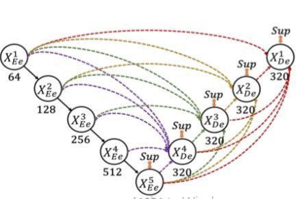
Encoderconsistsoffiveblocks.Eachblocksareconnected each other and pooling operations are applied.Also convolutional blocks are applied with corresponding filters.Thefifthblock isthe bottleneck layer.Decoderpart alsoconsistsoffiveblocks.Eachlayersareconcatenatedwith the previous layers along with encoding layers.Batch normalizationandreluisappliedtoeachlayersexceptthe last layer. The interconnection between the encoder and decoderaswellastheintraconnectionbetweenthedecoder sub networksarebothconvertedbythesuggestedfull scale skip connections. Both UNet with plain connections and UNet++ with nested and dense connections fall short of clearly learning the place and border of an organ by not exploringenoughdatafromfullscales.Eachdecoderlayerin UNet 3+ integrates smaller and same scale feature maps from the encoder and larger scale feature maps from the decoder, which capture fine grained details and coarse grainedsemanticsincompletesizes,toaddresstheflawin UNetandUNet++.
The feature map from the same scale encoder layers is received directly in the decoder, just like the UNet. As opposed to the UNet, a network of inter encoder decoder skip connections transmits low level detailed information fromthesmaller scaleencoderlayerusingnon overlapping max pooling, while a network of intra decoder skip connectionssendshigh levelsemanticinformationfromthe larger scaledecoderlayerusingbilinearinterpolation.We needtofurtherunifythenumberofchannelsandcutdown ontheunnecessarydatanowthatwehavefivefeaturemaps of the same quality. We had the idea that the convolution with64filtersofsize33mightbeagoodoption.tosmoothly combinedeepsemanticknowledgewithshallowexquisite information.Apply a feature aggregation process that consistsof320filtersofsize33,batchnormalisation,anda ReLU activation function to the concatenated feature map fromfivescales.
U Net3+modelexperimentationisdonetochoosethebest modelarchitecture.Agooddicescoreisprovidedbyunet3+ architecture, which increases segmentation accuracy. Complete avoidance of oversegmentation in complex backgroundsisaUNet3+skill.Comparingthisstrategytoall others, it is superior. Comparing Unet3+ to Unet's basic design, Unet3+ improved +'s version, which has intra and interconnectionsbetweenencoderanddecoder,isthebest methodforsegmentingbraintumours.Inordertomakethe mostoffeaturemapsatfullscalesforprecisesegmentation andeffectivenetworkarchitecturewithfewerparameters, UNet 3+ is utilised, which is a full scale linked UNet. In settingswithlittledata,itisa veryeffectivesegmentation methodthatperformsadmirablyinavarietyofbiomedical segmentationapplications.ItisanticipatedthatUNet3+will outperformallpriorstate of the arttechniquesandproduce accuratesegmentationresults.
[1]Ronneberger,Olaf,PhilippFischer,andThomasBrox."U Net: Convolutional networks forbiomedical image segmentation.arXiv2015."arXivpreprintarXiv:1505.04597 (2019).
[2]S. Sivagami, P. Chitra, G. S. R. Kailash and S. R. Muralidharan,"UNetArchitectureBasedDentalPanoramic Image Segmentation," 2020 International Conference on WirelessCommunicationsSignalProcessingandNetworking (WiSPNET),2020,pp.187 191
[3]Lietal.,"AttentionUnet++:ANestedAttention AwareU Net for Liver CT Image Segmentation," 2020 IEEE InternationalConferenceonImageProcessing(ICIP),2020, pp.345 349.
[4]H.Huangetal.,"UNet3+:AFull ScaleConnectedUNetfor Medical Image Segmentation," ICASSP 2020 2020 IEEE International Conference on Acoustics, Speech and Signal Processing(ICASSP),2020,pp.1055 1059
[5]MichałFutrega,AlexandreMilesi,MichalMarcinkiewicz, Pablo Ribalta "Optimized UNet for Brain Tumor Segmentation"arXiv:2110.03352[eess.IV],2021]
[6]Myronenko,A.:3DMRIBrainTumorSegmentationUsing AutoencoderRegularization.In:Crimi,A.,Bakas,S.,Kuijf,H., Keyvan, F., Reyes, M., van Walsum, T. (eds.) Brainlesion: Glioma, Multiple Sclerosis, Stroke and Traumatic Brain Injuries. pp. 311 320. Springer International Publishing, Cham(2019)
[7] Jiang,Z.,Ding,C.,Liu,M.,Tao,D.:Two StageCascadedU Net: 1st Place Solution to BraTS Challenge 2019 SegmentationTask.In:Crimi,A.,Bakas,S.(eds.)Brainlesion:
Glioma, Multiple Sclerosis, Stroke and Traumatic Brain Injuries. pp. 231 241. Springer International Publishing, Cham(2020)
[8]Isensee,F.,Ja¨ger,P.F.,Kohl,S.A.,Petersen,J.,Maier Hein, K.H.:nnU Net:aself configuringmethodfordeeplearning based biomedical image segmentation. pp. 1 9. Nature Methods(2020)
[9]FaustoMilletari,NassirNavab,Seyed AhmadAhmadi:V Net: Fully Convolutional Neural Networks for Volumetric Medical Image Segmentation. International Conference on 3DVision(3DV)(2016)
[10] Chuanbo Qin1, Yujie Wu1, Wenbin Liao1,2, Junying Zeng1*, Shufen Liang1 and Xiaozhi Zhang: Improved U Net3+ withstage residual forbrain tumor segmentation Qinetal.BMCMedicalImaging(2022)
International Research Journal of Engineering and Technology (IRJET) e ISSN: 2395 0056 Volume: 09 Issue: 07 | July 2022 www.irjet.net p ISSN: 2395 0072 © 2022, IRJET | Impact Factor value: 7.529 | ISO 9001:2008 Certified Journal |