


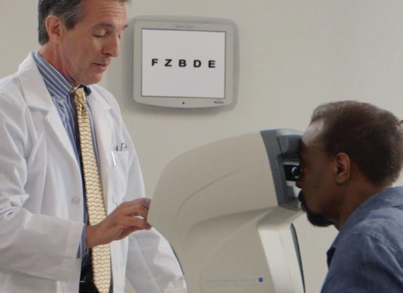
HOWARD LARKIN REPORTS
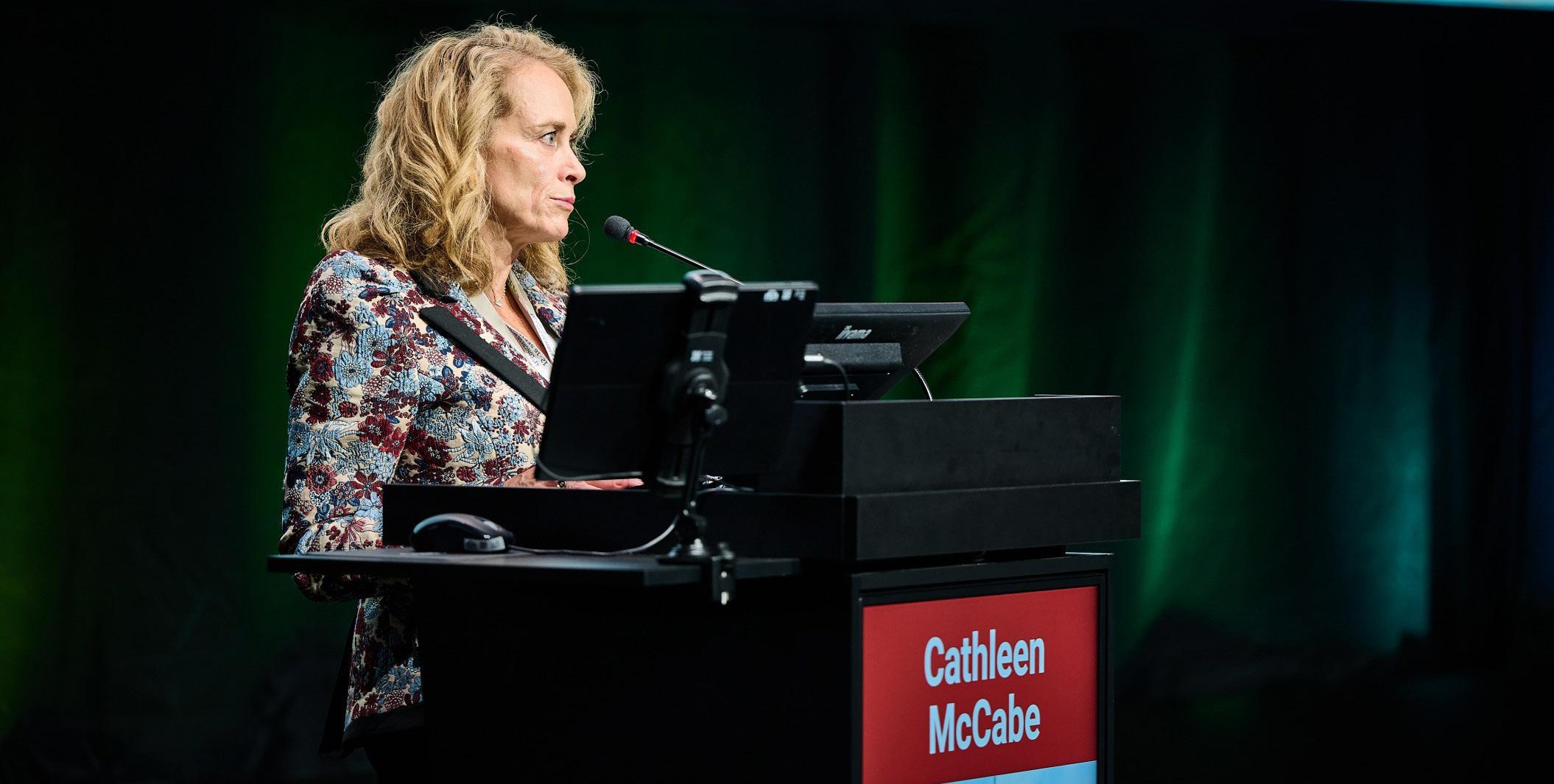
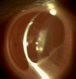
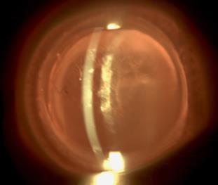
In two large-scale multi-centre trials involving 661 patients, the enVista Envy (Bausch + Lomb) full vision range trifocal intraocular lens (IOL) provided corrected distance visual acuity comparable to a monofocal IOL while adding about two lines of intermediate vision and four lines of near vision with continuous range of focus. Presenter Cathleen M McCabe MD also reported that dysphotopsias were low for the uniquely designed IOL optic, and no serious adverse events or secondary interventions were reported.
The IOL’s design contributes to its ability to deliver a continuous range of vision and low dysphotopsias, Dr McCabe said. It consists of a biconvex aspheric optic with a 1.2mm central zone with -0.15 microns of spherical aberration across its full dioptre range. Add powers are +1.6D for intermediate and +3.1D for near vision. The lens provides 0.2 logMAR or better visual acuity continuously across 4.0D of depth.
The anterior diffractive surface is apodized, with 22 steps in the central 4.5mm. The diffractive steps gradually decrease in height toward the periphery. This allows for an asymmetric distribution of light, with an even distribution in photopic conditions with 2-3mm pupil and distance vision prioritised in mesopic conditions with 3.5mm pupil or larger.
Proprietary technology reduces the profile of the diffractive edges, enhancing visual clarity and minimising glare and halos. The lens also comes with toric corrections available down to less than 0.75D cylinder.
“When we are looking at a diffractive IOL, we know how important it is to minimise residual refractive error,”
Dr McCabe said. Half-step cylinder corrections make this possible for patients with small amounts of visually significant astigmatism. The enVista platform also has good rotational stability.
The real proof of enVista’s technological prowess can be found in patient outcomes. And the enVista Envy delivered strong results in these trials, Dr McCabe reported.
Mean distance corrected monocular vision measured 0.03 logMAR for the Envy, which was judged non-inferior to a comparable monofocal IOL at -0.01. Monocular distance-corrected intermediate vision was significantly better for Envy at 0.12 compared with 0.35. The difference for distance-corrected near vision was even greater, with 0.15 (about 20/28) for the Envy compared with 0.54 (about 20/70) for the monofocal lens. Nearly 92% of Envy patients saw 20/40 or better near compared with about 13% of monofocal patients. Binocular results were even better for the Envy, with 97% seeing 20/40 or better.
Dysphotopsia outcomes were also strong. About 90% of patients reported little or no glare or starbursts, and more than 80% reported minimal halos. About 2-3% reported severe glare or starbursts, and 6% severe halos. Patient satisfaction was also high, with 93% of Envy patients completely or moderately satisfied with their vision, Dr McCabe noted.
Cathleen McCabe, MD serves as Chief Medical Officer at Eye Health America.
Most patients achieved spectacle independence, and 96% said they would recommend to family and friends.
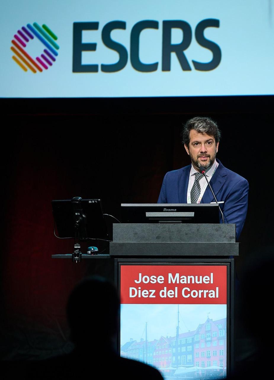
In a post-market observational study of the Tecnis PureSee (Johnson & Johnson Vision) extended depth of focus intraocular lens (EDOF IOL), patients reported excellent distance and intermediate vision and functional near vision without glasses along with low levels of bothersome visual symptoms, said Jose Manuel Diez del Corral Belda MD.
Interim results of the ongoing study revealed high levels of spectacle independence and high satisfaction with recommendations of the lens to family and friends, Dr Diez del Corral said. Indeed, more than 60% of patients said they wore glasses “none” or “a little” of the time, and nearly three-quarters said they were “completely” or “mostly” satisfied with near vision.
“Johnson & Johnson are being modest when they say [the IOL provides] ‘functional near vision,’” he said Dr Diez del Corral, who practices in Madrid, Spain, and is a medical advisor to Johnson & Johnson.
Built on the proven Tecnis platform, the PureSee is a refractive presbyopia-correcting IOL that extends the depth of focus with increased tolerance for residual refractive error while maintaining low levels of visual symptoms comparable to a monofocal IOL.
Puresee’s posterior optic refractive surface provides a continuous increase in power that enables a consistent depth of focus extension from distance to near. An aspheric anterior surface corrects for spherical aberration, and a high Abbe number reduces chromatic aberration, contributing to a low dysphotopsia profile, Dr Diez del Corral noted.
The interim study results reported involved 238 patients who were bilaterally implanted with the Tecnis PureSee toric or non-toric IOLs at several clinics and seen three months after surgery. Visual outcomes were quite good.
Mean binocular uncorrected visual acuity measured -0.02 logMAR, with intermediate at 0.12 and near at 0.25. With distance correction, these numbers shifted slightly to -0.05 distance, 0.11 intermediate, and 0.22 near. Overall, 87% of patients had uncorrected distance vision of 0.0 or better, rising to 94% corrected. All patients achieved 0.2 logMAR or better distance vision both uncorrected and corrected. Monocular results were similar, Dr Diez del Corral said.
Similarly, 73% of patients achieved distance-corrected binocular intermediate vision of 0.1 or better, and 32% reached that level for near vision. Three-quarters of patients achieved distance-corrected near vision of 0.3 or better.
Most patients also reached spectacle independence, at 85% overall. More than 95% used glasses “none” or “little” for distance, 93% for intermediate vision, and 62% for near. More than 90% said they had no or few visual symptoms such as glare, haloes, or starbursts. Overall, 95% said they were completely or mostly satisfied with their vision, and 96% said they would recommend the lens to friends and family.
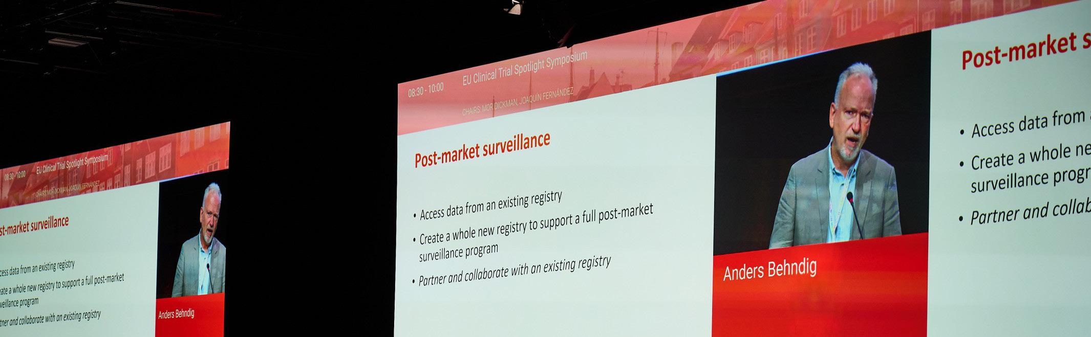
Compliance with regulatory requirements is a big motivator for surgeons and clinics to participate in post-market surveillance of drugs and devices. But detecting adverse events in the real world, comparing new products or treatments with the standard of care, and updating clinical guidelines if some patient groups benefit more than others are more substantive reasons, said Prof Anders Behndig MD, PhD.
Registries such as those run by national societies and the ESCRS are good vehicles for conducting such surveys, and more collaboration among clinicians, governments, and industry will help. “Unleashing collaboration sounds very good,” Prof Behndig said.
He pointed out that the US Food and Drug Administration defines post-market surveillance as “the active, systematic, scientifically valid, collection, analysis, and interpretation of data or other information about a marketed device.”
“The key words here are ‘scientifically valid,’” Prof Behndig said. “This is all about real-life data from real patients being operated on in real procedures,” so consistency and accuracy of reporting are critical.
Registries can be useful, Prof Behndig said. Sweden has had a cataract registry since 1992, and many other countries have added them since. They can collect and analyse large volumes of data to answer questions about how devices are used in the real world, the typical target population, and how a product performs in sub-populations by age, gender, ethnicity and geography.
Such data have led to important advances in patient safety, including the use of intracameral antibiotics to prevent endophthalmitis and continuously improving refractive outcomes. “[We have questions like] how does a particular IOL perform in very high hyperopes? It is a very small group of patients, and an individual clinic or clinician can never answer such a question,” Prof Behndig noted.
But gathering data on new products or procedures from existing registries is not always possible, simply because they may not be collecting the right information. Creating a whole new registry is possible and widely practised for post-market surveillance, but it is expensive and time consuming. “Probably the best way to go about this is partnering and collaborating with an existing registry,” Prof Behndig said.
The ESCRS registries initially were funded by the EU and now are supported by the society. The format of the EUREQUO and other ESCRS registries is now used for ESCRS clinical trials, Prof Behndig noted. “In time we will gather good scientific data … that can be useful in post-market surveillance.”
The ESCRS registries have created an industry task force to explore collaboration with academic and industry leaders. One goal is to co-create a future-proof operating model for the ESCRS registries rather than pursue purely company-specific goals. Other goals are to standardise datasets, share data, conduct studies and initiate dialogue among regulators, companies, patient advocates, and registry holders.
Possible concerns include privacy and security, competition, generalisability and data quality, and cultural challenges among different organisation types. But these can be addressed, Prof Behndig said.
“The goal is a new form of collaboration beyond the public-private partnership model. Together we can exchange data and use the registries to create public value,” he concluded
Anders Behndig is a professor in the Department of clinical Sciences/Ophthalmology at Umea University Hospital in Sweden
We’re willing to bet most eye care professionals don’t realize just how prevalent Demodex blepharitis is.
In fact, ~54% of eye care patients in Europe may have Demodex blepharitis (DB).1
Reference: 1. Data on File. Tarsus Pharmaceuticals, Inc.
LEARN HOW DB CAN FLY UNDER THE RADAR AT

The 2025 ESCRS Annual Congress marks the 35th anniversary of LASIK. The first LASIK procedure was performed by Greek surgeon Professor Ioannis Pallikaris in June 1990 and has since been performed at least 40 million times. He later developed epi-LASIK and has made countless contributions to refractive surgery, including co-developing the Tracey Technologies raytracing device and presbyopic intraocular lenses. He has published more than 200 articles and authored several textbooks. Prof Pallikaris has received many awards for his work, including an American Academy of Ophthalmology Lifetime Achievement Award and the European Society of Cataract Refractive Surgery (ESCRS) Binkhorst Medal.
“I originally developed the LASIK technique in an effort to solve problems in my own clinic,” said Prof Pallikaris, a former ESCRS president. “I never thought it would spread worldwide in the way that it did.”
Prof Pallikaris spoke with incoming ESCRS President Burkhard Dick, Prof Miltos Balidis, and EuroTimes Editor-in-Chief Sean Henahan about the birth of LASIK during a video interview conducted at the 2025 ESCRS Winter Meeting in Athens.
“He transformed a painful operation to a painless operation,” said Prof Balidis, former president of the Hellenic Society of Intraocular Implant and Refractive Surgery. “This made corneal refractive surgery very popular. He also continued to improve the procedure, making it safer and simpler, reshaping the future of refractive surgery. To this day, he is still thinking of ways to improve the results of refractive surgery. He thinks if you have ideas, don’t be shy, try to make them happen. That is the way ahead.”
Before LASIK, photorefractive keratectomy (PRK) was the leading corneal refractive surgery. Prof Pallikaris wanted to solve some of the problems associated with PRK, such as epithelial healing problems, pain, regression, and haze. He sought to address these issues by preserving the epithelium and performing the ablation in the corneal stroma, leading him to conceptualise the hinged flap to maintain a stable connection with the stroma and, to some degree, the subepithelial nerve plexus. In 1990, he and colleagues published the first paper using the acronym LASIK, after presenting the concept publicly for the first time at the ESCRS Winter Meeting in Zurich in 1989.
Prof Pallikaris has always emphasised the importance of a well-balanced life.
“If you want to maintain a long career, you need to maintain a good private life,” he said. “I am a farmer—I grow my own food, make my own wine and olive oil. I balance my time between technological research and natural life. [Strive to] always be optimistic. We are human beings who can use our abilities to solve our future problems.”
Prof Pallikaris is married to Varvara Terzaki Pallikaris, a social reformer in Crete, whose support he credits for his success. They have three children: two sons and one daughter. Together, they founded the “University of Mountains”, a not-for-profit organisation providing eye care to people in villages and mountainous regions who lack access to healthcare. He credited his wife with making him “more sophisticated and intellectual”.
The video interview with Prof Pallikaris can be viewed in the Heritage Archive section of the ESCRS website.

Combining the proven power of Scheimpflug imaging with the precision of ultra high-resolution OCT enables the detection of previously unseen corneal pathologies with unprecedented clarity.
Diagnose earlier. Treat smarter. Care deeper.
EPITHELIUM
BOWMAN’S LAYER
STROMAL LAYER
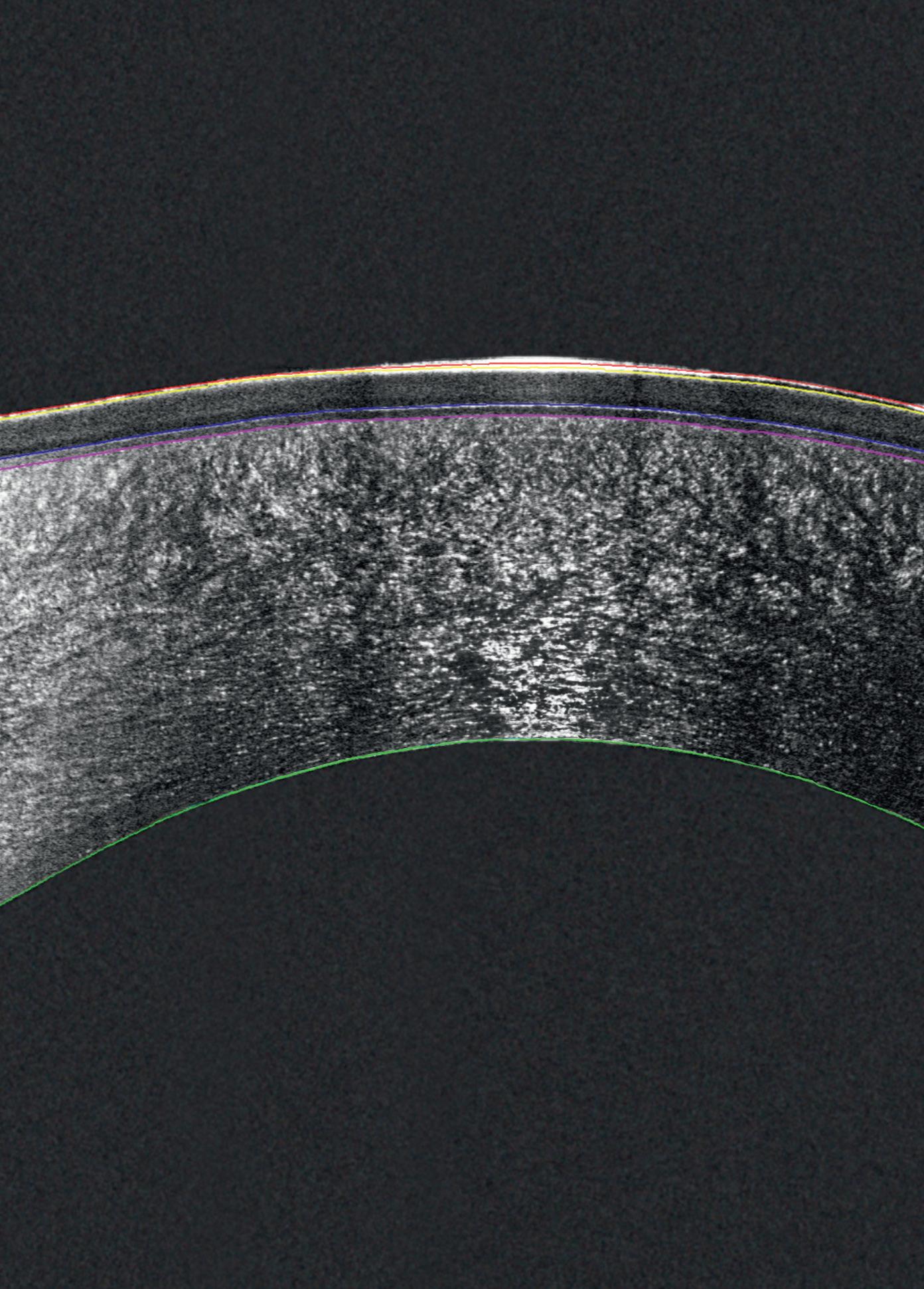
The Pentacam® Cornea OCT can increase the confidence that your diagnosis is correct.
New book provides an eyewitness account of the evolution of cataract and refractive surgery.
REVIEW BY SEAN HENAHAN
Anew book by Patrick Condon M.Ch., FRCS., FRCOphth, looks at the history of cataract and refractive surgery in the modern era from the perspective of his own career. Dr Condon began his ophthalmology career not long after Harold Ridley’s first intraocular lens implant in 1950. He is a pioneering ophthalmic surgeon and a founding member of the ESCRS who knew many of the key innovators in ophthalmic surgery.
Beginning with Harold Ridley’s first intraocular lens implant in 1949, Eye Healthcare—90 Years History of Progress documents the advances in cataract and refractive surgery by two successive generations of ophthalmic surgeons (Dr Condon and his father) working as consultants in the public sector services of the UK and the Republic of Ireland. The book recounts the founding of the European Intraocular Implant Council (EIIC) by Cornelius Binkhorst in 1982 and the remarkable progress of its successor, ESCRS, since 1992, during which time cataract surgery has advanced dramatically.
All royalties from the sale of this book are directed to the Ridley Eye Foundation charity.
The book covers the discovery of phacoemulsification in the US by Charles Kelman and its subsequent development by Eric Arnott in the UK and others throughout Europe. This advancement was the opportunity for eye surgeons to introduce small incision “keyhole surgery” for cataract patients.
The book also recounts the early years of ESCRS and its success in furthering cataract and refractive surgery on a pan-European level for a period of 30 years. This period saw the development of the Annual Congress and Winter Meeting and was also the genesis of many major research projects essential to the safety of the cataract procedure, one of which was the Endophthalmitis Study.
During this time, the European and US journals were integrated to form the Journal of Cataract and Refractive Surgery. Meanwhile, EuroTimes, under the leadership of editor-in-chief John Henahan, catapulted the society onto the international stage of major scientific and professional organisations.
The book also addresses many other developments in eye healthcare, such as the monitoring of diabetic retinopathy, the ongoing care of glaucoma patients, and the prevention of eye injuries in industry and sports.
In acknowledgement of Sir Harold Ridley’s legacy of the first intraocular lens implantation, all royalties from the sale of this book are directed to the Ridley Eye Foundation
charity. The Ridley Eye Foundation is dedicated to relieving cataract blindness among the relatively inaccessible population of Nepal living at high altitudes in the Himalayas.
Dr Condon’s many accomplishments include the ESCRS Grand Medal of Merit Award, the UKISCRS Choyce Medal Lecture, the ESCRS Ridley Medal Lecture 2005, and the ESCRS Heritage Lecture 2021. The latter can be viewed athttps://www.youtube.com/watch?v=cKOSSfwm_hg
Having finished his book, which he has been preparing for some years, Dr Condon said he is looking forward to returning to his other life passion, playing jazz piano. He is an accomplished player who has entertained at many earlier ESCRS and ASCRS events.
The book is available in the ESCRS member’s area during the Congress and on amazon.com.


Don’t let dry eye get in the way of patients’ satisfaction with their surgery - take control with Lacrifill, the hyaluronic acid canalicular gel that delivers six months of relief with a single treatment.1–3
✔ FDA-cleared
✔ CE-marked
✔ One syringe, one patient


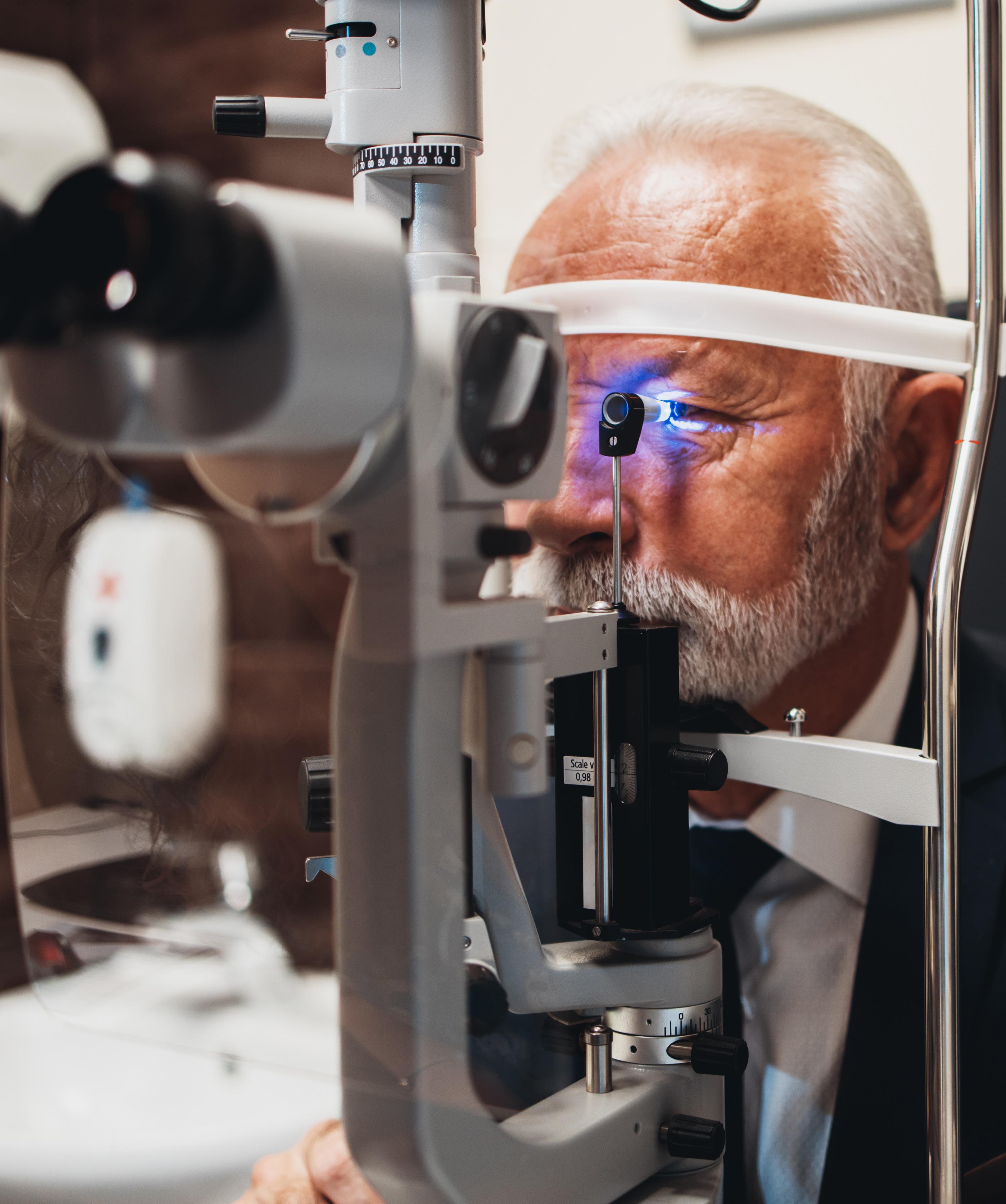
Careful screening for ocular surface disease is key to ensuring good surgical outcomes.
TIMOTHY NORRIS REPORTS
According to the 2024 ESCRS Clinical Trends Survey, 25% of cataract patients present to the operating theatre with clear signs of ocular surface disease.
“We have to keep in mind that there are many more patients suffering from this disease,” said Ramin Khoramnia, MD, PhD, FEBO during a “Modern Presbyopia Correction” symposium at the ESCRS Annual Congress in Copenhagen. “In fact, about 60% of the routine cataract patients are asymptomatic dry eye patients, and finding those patients is the duty we must fulfil.”
Looking at the Clinical Trends Survey, Dr Khoramnia observed how 66% of respondent ophthalmologists are very likely to postpone surgery in a patient with moderate dry eye. This mean that even though the biometry is not giving accurate data, 34% of surgeons are not postponing anyway. This can lead to worse outcomes, he added, suggesting that it might be important to prioritise patient assessment over surgery.
The ESCRS Clinical Trends Survey also revealed that 75% of respondents believe that mild to moderate dry eye significantly affects keratometry and IOL calculations. Even the best surgeon implanting the best lens will never get good outcomes if the data taken prior to surgery is not compiled in an orderly fashion, Dr Khoramnia said. Taking good and consistent preoperative measurements through multiple readings is therefore paramount, including ensuring the stability of the ocular surface. If it is not the surgeon who takes the exams directly, their staff must be trained to take multiple readings and confirm reproducibility, Dr Khoramnia said.
Data validation is also very important. According to Dr Khoramnia, there are some red flags that need to be taken into consideration: more than 1 Dioptre of difference between the eyes in average K should lead to further examination, as well as average K greater or less than 47 dioptres cylinder more than 2.5 Dioptres, ACD higher than 4.2 mm or lower than 2.0 mm, and axial length higher than 300 mm and lower than 22 mm.
Careful patient selection for the right kind of lens is also very important, Dr Khoramnia noted. Alcon’s Vivity lens is one of his IOLs of choice, thanks to the “Wavefront Shaping” technology that leads to greater spectacle independence with lower photic phenomena.
“It's very important that we screen the ocular surface in every patient, even if the patients are asymptomatic, and optimise the ocular surface,” Dr Khoramnia said. “We need to confirm a very stable and repeatable keratometry, and look for the right lens choice,”. “We really must optimise the surface to ensure an accurate power calculation and to achieve better vision quality. If we do all that, then the patient's satisfaction can be as high as possible.”
Ramin Khoramnia, MD, PhD, FEBO is Director of Ophthalmology at the Carl Gustav Carus University Clinic of Dresden, Germany


Digitalisation reduces errors and saves time, making a surgeon’s life easier.
NORRIS REPORTS
Doctors strive to make their patients happy, but a happy patient may not be a sign of a perfect surgery, according to Andrea Janekova, MD, FEBO, FEBOS-CR.
“Like an iceberg, only 10% of the whole volume is visible, with many steps that need to be done, hiding underneath,” she said during the Customized Patient Planning IME Symposium. “Any kind of mistake and any wrong measurement can damage an otherwise great result.”
As an example, many parameters during biometry can give wrong values if not performed correctly, thus inserting a mistake into the very beginning of a long process. For this reason, said Dr Janekova, patient flow is very important in clinical practice.
Across different kinds of clinical settings, time is the common denominator, especially time spent with patients. Adjusting to technology can be time consuming, but in the long run this leads to simplification of procedures and workflow and therefore better outcomes.
Digital biometry plays a key role in this process. Dr Janekova noted that in the past she had to perform measurements manually, but it is now possible to measure cataracts with biometers with a higher level of accuracy. It also saves time, as digital biometry can be integrated with advanced IOL formulas.
Corneal imaging is also a very important part of the workflow, Dr Janekova said. An inaccurate keratometry is a major source of postoperative refractive error, and therefore the precision of a digital measurement can be essential to producing correct IOL power calculations. Moreover, corneal imaging allows a thorough evaluation of the regularity of the corneal surface, allowing detections of keratoconus, ectasia and corneal scarring that would otherwise jeopardise surgi-
cal outcomes. Finally, corneal imaging can help in detecting corneal pathologies, finding the subtle or subclinical signs of corneal conditions that would be essential for the correct choice of IOL.
Dr Janekova presented the case of a 50-year-old woman with scutellar posterior cataract in both eyes and slight hypermetropia. The patient wanted spectacle independence due to her many daily activities that required near and intermediate vision. Dr Janekova chose the AT ELANA 841P lens thanks to its trifocal design.
The preoperative planning needs to start even before the patient enters the clinic, Dr Janekova said. There is a non-medical aspect that needs to be considered—the communication between the patient, the receptionist and the nurses. This can make an impact on patient satisfaction even before they reach the examination phase.
In her practice, Dr Janekova makes sure all measurements, such as tear film and visual acuity, are carefully taken before surgery. The patient is sent to a consulting phase and interviewed about their needs, their work and other daily life activities. Remote access workflow is also greatly appreciated, offering the possibility of obtaining biometric data even if the surgeon is not in the clinic.
According to Dr Janekova, all the digital planning and diagnostics are advantageous and save time. “This is something that can make your everyday life much easier, especially when dealing with higher volumes of patients,” she said.
Andrea Janekova, MD, FEBO, FEBOS-CR is Head Physician of the Cataract, Refractive, and Vitreoretinal Surgery Center Prague Eye Centre, Czech Republic


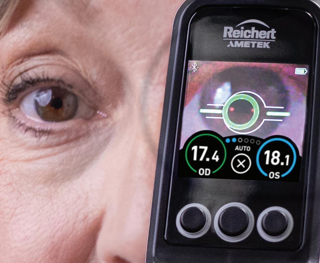


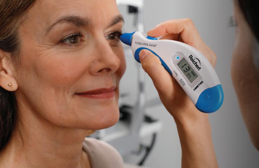
Experience the all-new Reichert® Tono-Vera® Tonometer featuring the patented, auto-measuring ActiView™ Positioning System, for fast, e ortless, objective, and repeatable rebound tonometry measurements. For nearly two centuries, Reichert has empowered a more e cient exam and shared in your passion to deliver exceptional patient outcomes.
DEMO AT ESCRS #C1.102
LEARN MORE AT REICHERT.COM


LEARN MORE ABOUT REICHERT’S COMPREHENSIVE FAMILY OF TONOMETRY AND REFRACTION DEVICES:


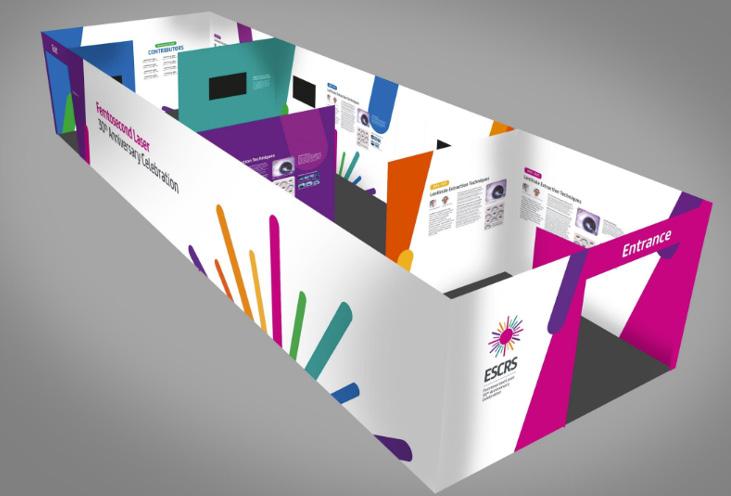
Femtosecond laser playing an ever-expanding role in ophthalmic surgery.
AUTHOR NAME
The 2025 ESCRS Annual Congress in Copenhagen celebrated the 30th anniversary of femtosecond laser eye surgery with an interactive exhibition detailing the remarkable evolution of this essential tool.
A reception in the ESCRS membership lounge included a live interview with some of the pioneering ophthalmic surgeons who developed these remarkable applications. Guests included Dr Ron Kurtz, co-founder of IntraLase and current president and CEO of RxSight, maker of the light-adjustable lens; Dr Tibor Juhasz, co-founder of IntraLase, current CEO of ViaLase, and professor of biomedical engineering at the University of California, Irvine; and Dr Burkhard Dick, professor of ophthalmology and chairman at Ruhr University Eye Hospital Bochum and incoming president of ESCRS.
A serendipitous event at the University of Michigan, US, in 1993 eventually led to the development of a new tool for ocular surgery. During an experiment at the College of Engineering, a graduate student’s eye was accidentally exposed to a series of stray femtosecond laser pulses. The resulting retinal burns were noted to be highly localised, eventually
sparking interest in the laser’s potential for medical applications. Collaboration with the university’s Center for Ultrafast Optical Science (CUOS) ultimately included Gérard Mourou (Nobel Prize in Physics, 2018), Dr Kurtz, Dr Juhasz, and others, with an initial focus on potential applications for corneal surgery.
By 1995, this team was able to demonstrate that femtosecond lasers could be used to perform lamellar resections of the cornea with minimal damage to the surrounding tissue.

A serendipitous event at the University of Michigan (United States) in 1993 eventually led to the development of a new tool for ocular surgery.
In subsequent reports, the team demonstrated flap creation (for LASIK), keratomileusis (for lenticule extraction), and other potential surgical procedures as alternatives to procedures that used the mechanical microkeratome.
Between 1997 and 2001, a team at IntraLase led by Drs Kurtz, Juhasz, Christopher Horvath, and others developed the first commercial femtosecond laser platform to enhance the safety, accuracy, and precision of LASIK procedures. The femtosecond laser was shown to create a more precise and predictable flap that was thinner and more uniform.
Today, the role of the femtosecond laser continues to expand to FLACS, SMILE, CAIRS, LIRIC, and glaucoma procedures, demonstrating how a lot of dedicated research can lead to transformative advancements in patient care.
European surgeons played an important part in the development of femtosecond eye surgery. Dr Zoltan Nagy performed the first femtosecond laser cataract procedure, and Dr Imola Ratkay-Traub performed first femtosecond laser LASIK procedure.
The interview will appear in the Heritage Archive section of the ESCRS website.
Low to moderate irregular astigmatism is not an exclusion criterion for binocular implantation of EDOF IOLs.
LAURA GASPARI REPORTS
Low to moderate irregular asymmetric astigmatism does not affect the visual performance of an extended depth of focus (EDOF) IOL, according to a study presented by Daniel Schartmüller, MD.
“Modern IOLs aim for the best possible visual acuity and presbyopia correction, and the current opinion is that presbyopic IOLs depend on the regularity of the cornea,” Dr Schartmüller said. For this study, AcrySof Vivity IOL (Alcon) was considered to see if it performs well in eyes with low to moderate anterior surface irregularities or asymmetries. The overall diameter of the Vivity IOL is 13 mm, and the optic diameter is 6 mm.
The study involved 28 patients with bilateral age-related cataract and with irregular astigmatism exhibiting an anterior root mean square (RMS/a) ≥ 0.3 µm/mm2 in the central 3 mm zone measured at the OCT. Eyes with astigmatism more
Modern IOLs aim for the best possible visual acuity and presbyopia correction, and the current opinion is that presbyopic IOLs depend on the regularity of the cornea.
than 75D received a toric IOL. As Dr Schartmüller reminded the audience, both eyes were implanted with the Vivity EDOF lens, as advised by the manufacturer. At 6 months post-operative follow-up, the researchers performed binocular best corrected distance visual acuity
(BCDVA), binocular distance corrected intermediate visual acuity at 66 cm and monocular defocus curves, IOL tilt and decentration. Pre-operative mean RMS/a was 0.0037. Dr Schartmüller reported that the defocus curve is like those they see in the Vivity IOL. The monocular defocus curves of the Vivity IOL showed an enhanced depth of focus of 0.16 logMAR at -1.5 D defocus in eyes with corneal irregularities.
Binocular distance visual acuity showed very good results with a mean of -0.08 logMAR. Only minimal deviations of decentration and tilt compared to preoperative values were observed at the measurement at the anterior segment OCT.
The study revealed that low to moderate irregular astigmatism is not an exclusion criterion for binocular implantation of EDOF IOLs. “We wanted to prove that, even though you have an irregular eye, you could have binocular, functional, good vision, and the mean was below zero, meaning we had a very good distance visual acuity, and the distance corrected intermediate visual acuity for both eyes was below 0.1,” Dr Schartmüller concluded.
Daniel Schartmüller MD is an ophthalmologist and researcher at Medical University of Vienna, Austria

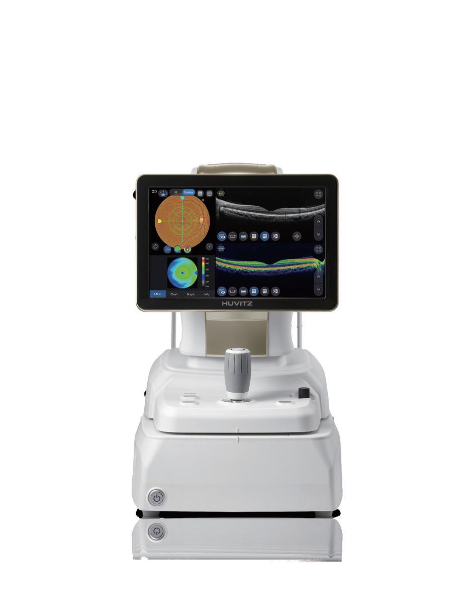
The MR-6000 delivers a smart combination of five different eye examinations and a Dry Eye observation app. Along with the advantage of automatic alignment, this means that the MR-6000 speeds up your workflow and makes it more efficient.
Dry-eye application
Blink analysis, tear meniscus height, hyperemia, and meibomian gland observation all help to assess dry eye.
Optional feature:
Tear stability analysis system (TSAS)
16 Mire rings examine an area 8 mm in diameter. A number of topography maps, including Fourier analysis, provide a wide range of options for visualising corneal shape.
A majority believe that dropless will be the standard of care for some or most patients without ocular comorbidities.
LAURA GASPARI REPORTS
The dropless after cataract surgery (DACS) regimen currently is not widely used for patients undergoing routine, uncomplicated cataract surgery, but it is expected to play a relevant role in the future, according to a study presented by Johannes Birtel, FEBO, MBA on behalf of the ESCRS Young Ophthalmologists for Sustainability (YOFS) working group.
The use of post-op eye drops presents some challenges, including treatment burden, non-adherence, and a negative impact on the environment. Although topical antibiotics are now often replaced by intracameral administration, postoperative inflammation is typically managed with steroid and/ or non-steroidal eye drops. However, these anti-inflammatory drops can be safely replaced by a dropless approach after cataract surgery. The presented study aimed at evaluating current practice patterns and attitudes of cataract surgeons toward DACS.
Cataract centres in 19 countries, mainly in Europe and the US, were considered for the purpose of the study. A cross-sectional survey questionnaire with 23 items was developed and distributed by the ESCRS Young Ophthalmologists for Sustainability working group. The questionnaire evaluated aspects such as postoperative management of routine cataract patients and experiences with and perspectives on DACS. A Likert scale ranging from 1 (strongly disagree)
to 5 (strongly agree) was used to assess the barriers perceived by surgeons in implementing and performing DACS in their routine cataract practice.
The questionnaire was completed by 208 respondents. Most participants perform 250-500 cataract surgeries annually, and 92% use intracameral antibiotics routinely. While more than 90% of participants have heard of DACS, only 19% have ever performed it, and 75% of those perform DACS currently. Surgeons without current DACS experience perceive many barriers and challenges, including an assumed higher risk of inflammation (2.9 vs 1.8), increased intraocular pressure (2.7 vs 2.4), and perceived lack of evidence on the matter (2.9 vs 1.6). However, 76% of surgeons stated that patients would likely prefer not to use eye drops.
Overall, centres performing DACS are primarily based in the US. Generally, most respondents believe that DACS will be the standard of care for either some (28%) or most (39%) patients without ocular comorbidities. This demonstrates that, although the DACS approach is not often implemented, it is recognised as advantageous from various viewpoints.
Dr Johannes Birtel is a consultant ophthalmologist at the University Medical Center Hamburg-Eppendorf, Hamburg, Germany
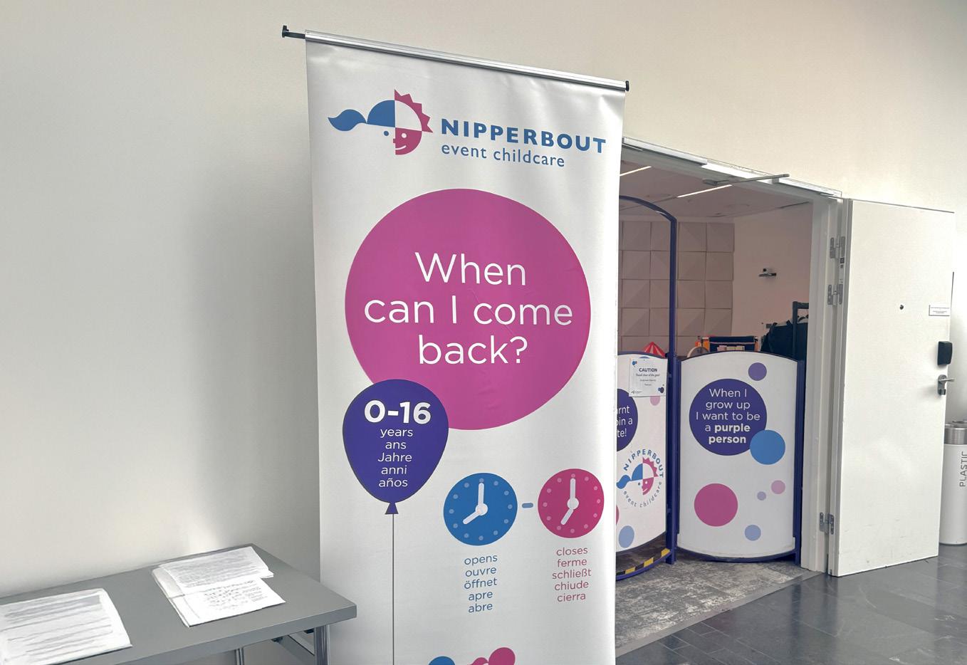
ESCRS is providing a childcare service during the 2025 Annual Congress in Copenhagen. The service is available at a cost of €10 per day for registered Congress attendees.
The childcare service is available for children aged 0–12 years only. Children may stay for up to a maximum of 4 hours at any one time. Parents must take their children out of the setting for food and fresh air and are welcome to return to the crèche after 1 hour.
The hours for the service are as follows:
• Sunday, 14 September, 08:00 to 20:30
• Monday, 15 September, 08:00 to 18:00
• Tuesday, 16 September, 08:00 to 15:00
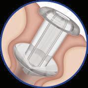
TEMPORARY PUNCTAL OCCLUSION UP TO 3 OR 6 MONTHS
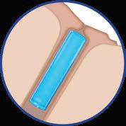
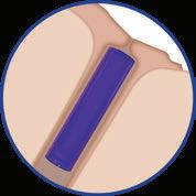
SOFT PLUG® EXTENDED DURATION
Absorbable canalicular occluders that are an effective option for transient dry eye symptoms before or after surgery, seasonally, etc. and for patients seeking to experience non-permanent options of punctal occlusion. Up to 90 days Up to 180 days
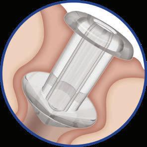
LONG TERM PUNCTAL OCCLUSION
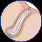
FORM FIT® HYDROGEL CANALICULAR PLUGS
Made of a safe and biocompatible hydrogel material that expands into a soft, pliable, gelatinous material when it contacts tear film.
SOFT PLUG® SILICONE PUNCTUM PLUGS
The distinctive low-profile cap combined with a softer, flexible silicone maximizes patient comfort. Flow Control Option also available*

To order/learn more or to see if this product is available in your country, please contact international@oasismedical.com
SEAN HENAHAN REPORTS
Dry eye is an issue in every area of eye care, especially the cataract, refractive, corneal, and glaucoma specialties. Attendees at this year’s Congress have many opportunities to learn what’s new in the diagnosis and management of dry eye disease (DED), with 1 Masterclass, 3 entire sessions, 43 abstracts, and 85 presentations devoted to the topic.
Research presented in a poster session today found that more than half of the general population in the US and Europe experience DED, yet only 20% of European patients and 17% of US patients were diagnosed, and they can wait years for professional help.
Dr Piotr Wozniak, refractive surgeon and dry eye specialist at Optegra Eye Clinics in Warsaw, Poland, and a lecturer and clinical instructor at Cardinal Stefan Wyszyński University in Warsaw, told the Congress:
“Results from our studies reveal a substantial group of
patients suffering without help. The European questionnaire explored why people don’t seek treatment. Many see dry eye as a normal part of ageing and something to endure. As a medical doctor, I find this particularly concerning because a simple eye drop could offer significant relief—but many people aren’t even asking for help.”
Dr Wozniak presented findings from a survey of 2,003 adults in the US conducted in April 2024, and from an ongoing, international arm of the study conducted by Bausch + Lomb with more than 5,000 adults in the UK, France, Germany, Poland, and Saudi Arabia called the ‘Needs Unmet in Dry Eye: Symptoms, Treatment and Severity’ (NESTS) study. In June 2025, the NESTS international arm surveyed 2,580 adults in the general population and 2,572 dry eye patients.
“In the NESTS study, we found that 58% of the general population reported experiencing dry eye symptoms, yet only one in five has received a formal diagnosis from a healthcare provider,” he said. “The large size of this study makes these results robust.
“The study explored the patient journey in detail. What stood out was that up to one-third of patients had experienced symptoms for more than five years before seeking professional help. NESTS also found that around half of patients experience symptoms every single day. The delay in seeking treatment is concerning, especially since dry eye is a progressive disease and early intervention can prevent a vicious cycle of inflammation.”
Other results from NESTS showed that 60% of dry eye patients waited at least four months before seeking help, and 20% waited more than a year before talking to healthcare providers.
Many stopped driving at night (17.0%), no longer wore makeup (14.8%), or reduced their use of heat or air conditioning (15.2%) due to their uncontrolled dry eye symptoms. One in three (34.0%) reported that their symptoms had worsened over the past year, and only 9.0% said there had been an improvement.
“These findings highlight the widespread impact of dry eye disease on quality of life, showing many people suffering silently,” Dr Wozniak said.
“We need to educate patients and the public on the causes, consequences, and treatment options for dry eyes, as well as the importance of regular eye checks. In addition, we must support healthcare professionals in distinguishing between different types of dry eye and matching treatments appropriately. One person’s dry eye can be very different from another’s.”

Better experience builds better practices.
Your reception desk receives a phone call just as a patient walks up. Does your associate ask the patient to wait while they take the call or put the call on hold to check in the patient?
“Both answers miss the mark,” says patient experience expert Matt Jensen. Separating the two functions is a better solution. This may take a little reorganisation, but the investment in improving operations leads to a more effective practice.
Refining operations is the first step in what Jensen, a certified Experience Economy Expert, calls the OPERA approach to marketing: better Operations lead to a better Experience that prompts Referrals, which are then reinforced with Advertising.
Using this framework has a transformational impact. As CEO of Vance Thompson Vision in the US, Jensen helped build a local practice into a regional enterprise with multiple locations and more than 400 employees. Jensen now leads Mend Join Make, an ophthalmic marketing agency and operations consultancy based in Sioux Falls, South Dakota, US.
As Jensen puts it, “work is an experience, and every business is a stage.” Businesses that understand their role in developing a quality experience are able to stand out. He recommends thinking of a favourite hotel or restaurant—the sights, the smells, the feeling—and bringing the practice environment up to that standard. Setting the stage involves everything from prompt and friendly service to excellent clinical services, to making sure the welcome area and restrooms are tidy and inviting.
The OPERA approach can also change the entire advertising framework. “Advertising is a tax you pay for being unremarkable,” Jensen said. When operations are solid, advertising acts as a support, not the only acquisition channel. Advertising’s core function becomes bolstering the goodwill established with patients and referring practitioners.
Jensen presented his comprehensive approach to practice marketing by building organically on effective operations during the ESCRS Leadership, Business, and Innovation Bootcamp.


JOHN HENAHAN WRITING
Each year, young ophthalmologists are invited to participate in the John Henahan Writing Prize, responding to an essay prompt provided by the medical editors of EuroTimes. Anuj Kodnani MBBS’s essay scored among the top three in a very competitive field.
Applicants responded to the following prompt: Diversity, equity, and inclusion (DEI) programmes, however well-intentioned, stir a variety of responses in the corporate and political worlds and in the scientific and medical spheres. What DEI and unconscious bias issues are present in the current culture of ophthalmology training, practice, and clinical research? What are the potential benefits of addressing these issues for patients and ophthalmologists? What kind of meaningful changes need to happen to move beyond ‘talking the talk’ to ‘walking the walk’?
“The eye sees only what the mind is prepared to comprehend.” – Henri Bergson
In a discipline that celebrates clarity of vision, it’s ironic that we sometimes fail to see our own blind spots. Diversity, equity, and inclusion (DEI) in ophthalmology is not just a matter of optics—it’s a matter of outcomes. Whether in the clinic, the classroom, or the research lab, unconscious biases subtly distort the lens through which we view talent, treat patients, and design studies.
As an Indian ophthalmologist who has trained in both London and the US, I’ve experienced this first-hand. While my fellowships were academically enriching, they also came with a sense of cultural distance. In clinical discussions, I was often the only non-Western fellow in the room. Teams rarely reflected the diverse communities they served. It made me ask: in a global specialty, why is the representation within our training and leadership so limited?
This isn’t just about optics. It’s about patients. Data shows diverse medical teams foster trust and improve outcomes, especially for underrepresented groups. And yet, women in ophthalmology still face subtle—but damaging—barriers: fewer surgical opportunities, being mistaken for nurses, or having their leadership ambitions quietly dismissed. Similarly, Black and Asian populations continue to be underrepresented in trials, even though they bear a disproportionate burden of diseases such as glaucoma and diabetic retinopathy.
Clinical research also reflects this inequity. Despite the global burden of blindness being highest in low- and middle-income countries, most randomised trials are designed, funded, and published in the Global North. These studies then dictate global guidelines, often ignoring socioeconomic or cultural nuances. As a result, interventions may be technically sound but practically ineffective or even inappropriate in local contexts.
Even our DEI initiatives sometimes feel performative. Diversity panels filled with the same voices. Committees with no power. Tokenism is easy. Transformation is hard.
So how do we ‘walk the walk’?
First, we widen the pipeline. Let’s reach out to underrepresented students early—with mentorship, shadowing,
and scholarships. Diversity must begin before the residency interview.
Second, we commit to transparency. Let’s publish anonymised data on applicant demographics, pay gaps, and promotions. If we track visual acuity so precisely, why not track equity with the same rigour?
Third, we embed bias training into real clinical life. Not just box-ticking modules. We need reflective storytelling, real mentorship, and debriefs on microaggressions. We train our eyes to spot retinal tears—why not train our minds to spot systemic ones?
Fourth, research must reflect reality. Funders and journals should prioritise diversity in trial recruitment and reward innovation that addresses health disparities.
And finally, leadership must reflect the world we serve. That means rethinking who gets the microphone, who gets promoted, and whose voices get heard.
This is not about lowering standards. It’s about understanding that the current standards may have been built to exclude. Just as we correct distorted corneas to sharpen sight, we must correct distorted systems to sharpen fairness.
My vision is of an ophthalmology community where a hijab-wearing woman, a first-gen student, a queer Black doctor, and a disabled surgeon feel not just included, but empowered; where diversity is not performative—but profound.
Until then, DEI must not be an agenda item. It is our collective responsibility.
Declaration: This essay was developed without the assistance of AI tools.

Need a quick introduction or refresher about a surgical procedure? Have a tip to share about a technique or approach you use that makes surgery easier?
The ESCRS 100 is the place to go. It’s a library of short (roughly 100 seconds), high-quality instructional videos about all fields of cataract and refractive surgery.
More than 40 videos have already been created, and additional videos are being uploaded each month.
Current videos include the following topics:
• Glass Rod technique to remove Soemmering's ring remnants
• Primary posterior capsulorhexis in the presence of a posterior capsule plaque
• Endoilluminator-assisted cataract surgery
—PUT THE ESCRS 100 VIDEO SERIES ON YOUR LIST OF MUST-WATCH EDUCATIONAL RESOURCES ! ESCRS
Emphasising good preoperative communication and setting clear expectations are the best ways to make patients happy.
TIMOTHY NORRIS REPORTS
Avast number of intraocular lenses are available to surgeons, and each one can create different outcomes for the patient, making the right choice for the right patient a very difficult task. But according to the ESCRS Clinical Trends Survey, 63% of patients have no knowledge of what their refractive lens options really are.
“Looking at how confident we or our staff are at communicating and educating patients about the reimbursement of these procedures, 41% lack confidence,” Jonathan Yu, MA (Cantab), MB, BChir, Cert LRS, FRCOphth said during the Customized Patient Planning IME Symposium.
As an example, Yu noted the case of a cataract patient receiving a simultaneous vision IOL. The patient enjoyed a wide variety of activities, both outdoors and indoors, that she began to limit as her vision deteriorated. She has primary angle closure and had previous YAG peripheral iridotomies, and her IOP started increasing on treatment. Fortunately, she does not have glaucoma. She had a full visual field and the OCT is normal, but her angle closure is narrowing due to the increasing cataract.
It is important to understand and elicit what this patient would like to do, Dr Yu said. What is her level of outside activity? What is her preference for near and distant vision?
Does she mind using glasses? If this patient wants to achieve spectacle independence, it is important to ascertain what she is trying to achieve and her aversion to photic phenomena, because the lenses that can provide that can also create dysphotopsia.
According to the Clinical Trends Survey, 44% of physicians believe patient-doctor direct communication is the most efficient way to educate patients about the technologies available and what they are trying to achieve. Communication is key to letting the patient know the risks and benefits of each option and encourage them to ask questions. If there are misunderstandings or misalignments between the doctor and the patient, the result is likely to be an unhappy patient, and unhappy patients are the worst patients, Dr Yu remarked.
Having empathy and building trust, making sure patients are happy, discussing their activities, and understanding their desires and concerns regarding spectacle independence are of extreme importance to Dr Yu for a positive outcome. Moreover, patients need to be informed about compromises and tradeoffs and asked what they would accept—for example, are they ready to accept night vision symptoms or issues with low light vision? If information is shared properly, the patient will be better able to accept recommendations and appreciate the time spent on them.
“It is important to identify a patient's unique needs and goals, show empathy, build trust, communicate, and personalise recommendations in a clear and open fashion,” Dr Yu said. “Getting the happiest patients at the end is what we are all trying to achieve.”
Jonathan Yu, MA (Cantab), MB, BChir, Cert LRS, FRCOphth, is a full-time Consultant Ophthalmologist at the Manchester Royal Eye Hospital, United Kingdom
First study of its kind suggests when to wait, when to treat.
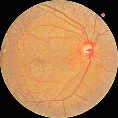
Artificial intelligence (AI) could help to predict which keratoconus patients need prompt treatment to stabilise their corneas and preserve their eyesight, report researchers from Moorfields Eye Hospital, London.
Shafi Balal MBBS reported the results of a new study in a free paper session on keratoconus clinics and diagnostics. The study involved a group of patients referred to Moorfields Eye Hospital NHS Trust for keratoconus assessment and monitoring, which included scanning the front of the eye with optical coherence tomography (OCT). AI was used to analyse 36,673 OCT images of 6,684 different patients along with other patient data.
The AI algorithm could accurately predict whether a patient’s condition would deteriorate or remain stable using images and data from the first visit alone. Using the technology, the researchers sorted two-thirds of patients into a lowrisk group who did not need treatment, and the remaining third into a high-risk group who required prompt cross-linking treatment. When information from a second hospital visit was included, the algorithm could successfully categorise up to 90% of patients.
“Our research shows that we can use AI to predict which patients need treatment and which can continue with monitoring. This is the first study of its kind to obtain this level of
accuracy in predicting the risk of keratoconus progression from a combination of scans and patient data, and it uses a large cohort of patients monitored over two years or more,” Dr Balal explained. “Although this study is limited to using one specific OCT device, the research methods and AI algorithm used can be applied to other devices. The algorithm will now undergo further safety testing before it is deployed in a clinical setting.
“Our results could mean that patients with high-risk keratoconus will be able to receive preventative treatment before their condition progresses. This will prevent vision loss and avoid the need for corneal transplant surgery with its associated complications and recovery burden. Low-risk patients will avoid unnecessary frequent monitoring, freeing up healthcare resources. The effective sorting of patients by the algorithm will allow specialists to be redirected to areas with the greatest need.”
The researchers are now developing a more powerful AI algorithm, trained on millions of eye scans, that can be tailored for specific tasks, including predicting keratoconus progression and detecting eye infections and inherited eye diseases.
Shafi Balal MBBS, MSc is based in London, UK.

The PRA aims to support and encourage independent clinical research in the field of cataract and refractive surgery. It can fund a variety of new initiatives, which may include— A novel research idea for the development of clinical trial studies; A non-interventional or observational study; or A natural history/epidemiological study.
An award of €25,000 is available for one grant. The competition is open to ophthalmologists up to the age of 45 (at the application deadline). Eligible participants must hold a full-time clinical or research position at a clinical or academic centre within the European region.
Applications are due by 31 October 2025.
A multidisciplinary lecture highlights the need for greater awareness of the condition amongst ophthalmologists.
ANDREW SWEENEY REPORTS
Dementia is a growing problem as global populations age, and ophthalmologists are on the front lines in combating the problem. It’s not a normal part of aging, its impact is severe on ocular health, and a multidisciplinary approach is required.
That was the message delivered in “Vision Problems in Dementia” as ESRS welcomed external experts to discuss this growing public health crisis. Renate Claassen MD began her presentation with a stark reminder of the scale of the problem: Roughly one-third of people living with dementia have a visual impairment, and half of people with dementia living in care homes have a visual impairment.
“It’s good to realise that dementia and visual impairment often coexist and worsen each other’s impact,” she said. “It’s essential to have a multidisciplinary approach, as this obviously impacts on day-to-day life.”
“Dementia can lead to social isolation, depression, and physical inactivity, the same as cataracts,” she continued. “You can imagine how having both of these conditions could interact. Dementia isn’t a normal part of aging, and it needs to be addressed.”
Dementia disrupts the visual signals the brain receives as its ability to process this information decreases. With less stimuli to process, patients can start to see faces where they don’t exist. Their episodic memory based on vision becomes compromised and they become significantly more likely to hallucinate.
The impact of these hallucinations in dementia patients is stark. Dr Claassen reported that hallucinations are present in 23% of cases of Alzheimer’s dementia, in 50% of patients affected by Parkinson’s disease dementia, and a staggering 80% of patients suffering from dementia with Lewy bodies.
A good example of how visual issues resulting from dementia can be observed is in the increased risk of falls. According to Dr Claassen, physical impairments like cataracts create the same relative risk from falls as cognitive
Dementia
disrupts the visual signals the brain receives as its ability to process this information decreases. With less stimuli to process, patients can start to see faces where they don’t exist.
impairments, a risk that continues to increase as patients age. While falls may seem like a relatively small concern for those younger in age, they can be catastrophic for the elderly, particularly those impaired with physical or cognitive problems.
“We know that people living with dementia have an eightfold higher risk of falls and also a higher risk of injuries due to these falls,” Dr Claassen said. “If falls result in a hip fracture, people living with dementia do way worse than people without dementia. They’re less likely to regain their walking ability, and they have a 67% higher risk of mortality.”
“Cataract surgery reduces the risk of falls in older patients by approximately a third,” she continued. “We can’t always treat cognitive problems, but we can try to improve visual ability.”
It was on that point that Dr Claassen concluded, issuing a rallying cry for clinicians from different disciplines to collaborate and create a system of holistic care for those suffering from dementia. Cataract surgery provides a vital opportunity to do so, she said, especially as it is a “potentially modifiable risk factor.”
Diagnosing dementia could fall within the purview of ophthalmologists if current research continues.
ANDREW SWEENEY REPORTS
It could be possible to intervene in Alzheimer’s disease as much as 20 years before symptoms begin to show—if ophthalmologists are able to identify and diagnose the biomarkers of the condition.
Dr Jurre Den Haan MD PhD’s presentation, “Retinal Biomarkers for Alzheimer’s Disease,” postulated this exciting idea, one that could revolutionize diagnosis and treatment for this disease. He reported that the disease can start more than two decades before the first symptoms present themselves, which he described as a “window of opportunity.”
“Alzheimer's disease is a complex disease, a complex interplay of proteins and amyloids leading to neurodegeneration,” Dr Den Haan said. “A clinical diagnosis starts with a syndrome diagnosis, which you can use biomarkers to support. You can check for neurodegeneration using an MRI or check the amyloids in the cerebrospinal fluid, but could this be done by examining the eye as well? I think so.”
Dr Den Haan described how optical coherence tomography (OCT) can be used to measure retinal layer thickness on a micrometer scale to achieve “better” results compared with an MRI. However, he said he wanted to find an alternative approach due to the large differences in retinal layer thickness between individuals.
Dr Den Haan then described how he hit “a fork in the road” during his research and decided to focus on studying posterior cortical atrophy Alzheimer’s (PCAD) versus typical Alzheimer’s disease cases (tAD) rather than focusing on examining the difference in biomarkers for early and late-onset Alzheimer’s. He made this decision because he felt “you would expect to see more retinal thinness as a result of retrograde atrophy.”
Describing the results of his study as “striking,” Dr Den Haan said that while he expected considerable variation between the PCAD and tAD patients when they were exam-

You can check for neurodegeneration using an MRI or check the amyloids in the cerebrospinal fluid, but could this be done by examining the eye as well? I think so.
flammation, examining the retina on a molecular level for disease-specific markers, and considering the applications of AI in OCT.”
In a final point, Dr Den Haan called for standardized patient enrolment in trials, as this will help to reduce retinal thickness layer variance. Support in this area would greatly help Alzheimer’s research, he said.
Dr Jurre Den Haan MD PhD is a resident in neurology at Amsterdam University Medical Center.
ined by MRI (which was subsequently confirmed), the difference in retinal thickness between the two groups was small to negligible. Concluding that this biomarker may not work for all patients, he began the search for another alternative.
“Retinal layer thickness does not discriminate between amyloid-proven, tAD and cases involving PCAD from control participants,” Dr Den Haan said. “Our group is now working on retinal biomarkers so that various proteins can be detected in the retina. To do this, we could use the help of ophthalmology to translate data from the pathological examination of retinal tissu, and see if we can get a disease-specific diagnosis for the retina. We’re also working on neuroin-
Asurvey conducted on behalf of ESCRS indicates a positive trend in the gender balance of conference speakers at the Society’s Annual Congress. While 27% of Congress presenters were female in 2022, this increased to 31% in 2023 and 34% in 2024. This year saw a slight dip to 33%, short of the 40% target.
“This does not diminish the progress made—the growth from 2022 is clear—but it does highlight the need to strengthen current initiatives and explore new strategies for 2026 to achieve a more balanced representation, in line with our strategy and key performance indicators,” the survey organiser noted.
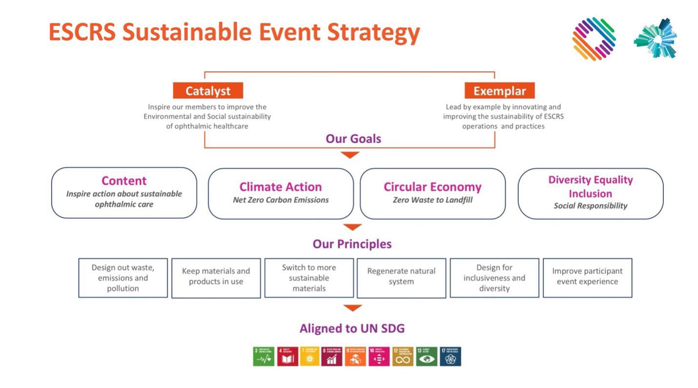
Gender equity is part of the larger sustainability campaign underway at the ESCRS. Through the Mission Zero Initiative in partnership with the Bella Center, the Global Destination Sustainability-Movement consulting group and suppliers, the goal is moving towards four key objectives: zero waste to landfill, net zero carbon emissions, and reaffirming a commitment to social responsibility.
Some milestones include:
• Promoted sustainability awareness, increased diverse representation, and supported inclusive education.
• Eliminated 90% of single-use plastics and achieved high recycling rates.
• Encouraged delegates to choose low-emission transport and served 50% locally sourced vegetarian or vegan meals.
• Raised female speaker representation to 40% and fostered community partnerships.
By joining these efforts, everyone at the Congress contributes to demonstrating how global medical events can lead to meaningful change.
Outside the Congress itself, the ESCRS is supporting critical causes, restoring ecosystems, and nurturing sustainability leadership within the ophthalmic community and beyond.
As part of the Mission Zero initiative to minimise waste to landfill at the conference, the ESCRS created a comprehensive approach to waste management that includes eliminating harmful materials, increasing recycling, and choosing smarter, more circular alternatives. For example, the exhibition floor coverings are a single-use, recyclable carpet that meets the strictest sustainability standards.
This decision directly supports the ‘Towards Zero Waste’ goals: to eliminate 90% of single-use plastics, polystyrene, and PVC; reduce landfill waste to less than 10% of total waste generated; and achieve a recycling rate of more than 60% across all waste streams. Results related to waste diversion and recycling rates will be measured and reported in November 2025 as part of a post-Congress sustainability performance review.
Copenhagen is known for its clean, high-quality tap water, which is sourced almost entirely from protected groundwater and enjoyed straight from the tap by residents and visitors alike. This reflects the city’s long-standing commitment to cautious water management and sustainable practices. To promote responsible water use and reduce single-use plastics, more than 20 water refill stations have been installed throughout the Bella Center.
Are you concerned about burning out early? Wondering what you can do to help promote equitable eye care for all patients? Looking for advice on building your career?
The ESCRS BoSS (Building Our Sustainable Society) initiative is addressing these questions by sponsoring a symposium and courses as well as a speed mentoring programme at the ESCRS Annual Congress in Copenhagen.
Check out the details below and make plans to attend.
BoSS Symposium:
Are you satisfied?
From burned out to burning bright
Date: 14 September
Time: 11:00–12:30
Location: Hall B2-M1 (300 seats)
(Held at the ESCRS membership booth)
BoSS Course:
Implicit bias
Date: 14 September
Time: 16:45–18:15
Location: Hall D2 (450 seats)
Speaker: Amy Johnson

Speed mentoring is a dynamic and interactive session where mentees have the opportunity to engage with experienced mentors in short, focused conversations. This format allows for the exchange of knowledge, guidance, and networking in a time-efficient manner. It also offers an excellent opportunity to build your professional network by meeting mentors and fellow mentees, fostering connections that could benefit your career for years to come.

BoSS Course:
Combatting unconscious gender bias in ophthalmology, industry, and research Date: 15 September
Time: 09:00–11:00
Location: Hall B1-M5 (300 seats)
Friday, 12 September, 14:00–15:00
Saturday, 13 September, 10:00–11:00
Saturday, 13 September, 14:00–15:00
Sunday, 14 September, 10:00–11:00
Sunday, 14 September, 14:00–15:00
Monday, 15 September, 10:00–11:00
Monday, 15 September, 14:00–15:00

Results are promising but more research is needed.
LAURA
GASPARI REPORTS
The Tecnis PureSee IOL (Johnson & Johnson) appears to perform well in eyes with a history of previous myopic or hyperopic laser vision correction, according to a study presented by Stephen Stewart, MBBS MA PgDipCRS CertLRS FRCOphth.
“The PureSee IOL from Tecnis is a novel refractive, extended, partial range of fields IOL,” Dr Stewart said. “The early reported results in virgin eyes are very good, but the results in eyes with a history of previous laser vision correction have not been reported yet.”
The study aimed to assess the visual, refractive, and quality of vision outcomes of the PureSee IOL in a challenging cohort of patients. It was a retrospective observational study conducted in the Cathedral Eye Clinic in Belfast, UK, that assessed 34 eyes from 20 patients with a history of previous laser vision correction. Of the 34 eyes, 22 had a history of hyperopic LASIK or PRK, while 12 had a history of myopic LASIK.
The patients underwent routine cataract surgery or refractive lens exchange from August 2024 to March 2025. A full ophthalmological assessment was performed, including biometry, corneal topography/tomography, and aberrometry. Micromonovision strategy was used, with non-dominant eye target up to -0.75 D. Primary outcomes measures such as unaided vision (distance, intermediate and near), manifest refraction, quality of vision questionnaire and spectacle independence questionnaire were recorded at the 3-month postoperative visit.
In terms of the visual outcomes, monocular mean visual acuity showed excellent distance, good intermediate, and some functional near vision. For the binocular mean visual
acuities, both myopic and hyperopic laser groups did well for distance, with all patients achieving 0.2 logMAR or better. Near vision was more variable, particularly for the hyperopic cohort. As for the refractive outcomes, patients underwent delayed, sequential, bilateral cataract surgery with a oneweek interval between the first (dominant) and the second eye.
In all these cases, refractive prediction accuracy within ±0.50 D is slightly lower than typically expected in virgin eyes, with a more pronounced decrease observed in post-myopic eyes compared to post-hyperopic ones, likely due to the higher rate of regression commonly seen in hyperopic patients. As for the defocus curves and quality of vision outcomes, both groups had similar defocus curve shapes, with the hyperopic patients exhibiting a greater area under the curve. More than half (57%) of post-hyperopic patients had complete spectacle independence, while myopic patients reached 50%, with good results in their quality of vision.
Although good results were achieved, Dr Stewart cautioned that this study involved a small group of patients, so there is a need for further investigation of the subject, with a more robust analysis.
Dr Stephen Stewart, MBBS MA PgDipCRS CertLRS FRCOphth is an ophthalmologist at the Cathedral Eye Clinic in Belfast, UK

Ophthalmologists and researchers (MD and/or PhD) as well as experienced ophthalmic nurses are encouraged to apply for the new ESCRS Sustainability Research (SURE) Award, which will fund projects that investigate meaningful, practical ways to promote environmental responsibility in ophthalmic care.
Two awards will be granted; each award will provide up to €10,000 per project. Applicants must be active ESCRS members at the time of application and must hold a current full- or part-time clinical or research position at a clinical or academic institution. Early-career researchers and young ophthalmologists are especially encouraged to apply.
The application period closes 01 November. Award recipients must submit an article to a peer-reviewed journal within six months after the research period concludes. The article should be made open access if accepted and submitted to the Journal of Cataract & Refractive Surgery in the first instance.
Get funding for projects to promote environmental responsibility in ophthalmic care.
Do not automatically rule out these lenses!
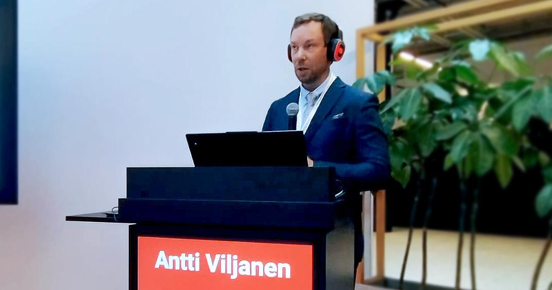
Off-label use of the Alcon Acrysof® IQ Vivity® EDOF IOL can give good uncorrected distance and intermediate visual acuity in non-pristine eyes with an epiretinal membrane, according to a study presented by Antti Viljanen, MD. The study evaluated visual acuity at different distances in patients with epiretinal membrane after a successful cataract surgery or refractive lens exchange surgery.
“Patients with non-pristine eyes want to have spectacle freedom,” he said.
The study was conducted in Turku, Finland with one single surgeon. It involved 17 eyes from 9 patients (5 male, 4 female) with a mean age of 63 years, an asymptomatic epiretinal membrane, and no reported distortion in preoperative check-up. Epiretinal membranes were found in 14 eyes through preoperative OCT imaging. Toric IOLs were used when applicable and micromonovision was targeted. A Barrett lens factor of 1.94 was used.
The first post-surgery visit was at one month and yearly afterward. Main outcome parameters were LogMAR uncorrected and corrected visual acuity at distance (UDVA 6 m), intermediate (binocular UIVA 63 and 100 cm) and near (binocular UCNVA 40 cm). ETDRS type charts with rulers were used.
The results were good—preoperative corrected visual acuities were logMar 0.00/0.04, right/left eye, with spherical refractive error range -3.0 - +2.9D/-6.8 - +3.3D, mean
near add was 2.2D, and mean implanted IOL power was 20.5D/21.3D. At one year, none of the patients had distance-corrected spectacles and 5 of 9 did not need reading glasses. Binocular UDVA was logMar 0.00. UDVA was logMar 0.05/0.12. CDVA was logMar 0.01/-0.01. Mean anisometropia was 0.26D. UCNVA at 40cm was logMar 0.32. UIVA was logMar 0.09 at 63cm and logMar 0.00 at 100cm. No halos or distortions were reported.
Dr Viljanen briefly presented the case of a patient who received her diagnosis four years before the implantation of the Vivity IOL lens, which occurred in 2020. Five years after the operation and presenting at every follow up, she has good vision and normal visual acuity and is not using glasses for reading because of the micromonovision.
Dr Viljanen said the important thing as is to encourage patients to come to follow-ups, even after years of surgery. “Remember patient counseling and what you promise,” he said. “Ask your patients to come to every follow up, even afterwards, and do not automatically rule out the use of premium IOLs in these cases.”
Antti Viljanen, MD is an ophthalmologist at Silmaasema Silmasairaala, Turku, Finland


Would you like to help shape decisions about ESCRS educational offerings? Are you interested in receiving a free delegate registration to the 2026 ESCRS Annual Congress in London?
If you answered yes to both questions, keep reading!
This year marks the 11th year of the ESCRS Clinical Trends Survey. The survey’s goal is to obtain a wide variety of perspectives on key issues facing ophthalmology today. The ESCRS leadership will use your confidential feedback to determine education needs based on current clinical opinions and practice patterns.
In less than 20 minutes, you can make a meaningful contribution to ESCRS programming; in return, you’ll be entered in a raffle to win one of four free 2026 delegate registrations to the ESCRS Annual Congress. You will also receive a preliminary results report prior to publication.
To participate in the survey, visit the ESCRS Survey Lounge next to the Exhibition Hall entrances of Halls C and E, near the ESCRS Booth & Member Lounge. You can also participate online by following the link below. The survey will close soon, so act now!
Scaffold-based system is easy to deploy and has short learning curve. SEAN HENAHAN REPORTS
Abioengineered, 3D printed endothelial keratoplasty implant could offer an alternative to conventional donor tissue in diseases where the corneal endothelium fails, such as Fuchs' dystrophy and bullous keratopathy, reported Dr Michael Mimouni in a free paper presentation.
“Do we even need innovations in the area of endothelial keratoplasty?” he asked. “We already have excellent techniques, including Ultrathin DSAEK and DMEK. These methods have limitations, with the main one being a lack of tissue availability. According to global reports, for every 70 patients who need a corneal transplant, there is one implant available.”
Surgical approaches to solving the tissue shortage include techniques such as DSO, quarter DMEK and hemiDMEK to leverage tissue. Newer technologies include artificial implants (such as ENDOART) and cell-based approaches, either scaffold-based or cell injection-based.
Dr Mimouni presented an update on a scaffold-based system called PVEK (Precise-BIovision endothelial keratoplasty). A monocellular layer of endothelial cells is printed at a density of more than 5,000 cells per square millimetre on an 8-micron-thin collagen tissue. Viability exceeds 90%.
It comes pre-stamped and preloaded in a Geuder cannula. After performing descemetorhexis, the implant is inserted through a small incision, unfolded, positioned, and held in place with a gas tamponade.
“This implant is very easily deployed with minimal manipulation,” Dr Mimouni reported. “It is much easier to unfold than a DMEK and reminiscent of an ultra-thin DSAEK.
We are able to produce 300 tissues from one donor.”
Dr Mimouni presented results of in vivo research with 64 rabbits. One-third received the implant seeded with human corneal endothelial cells, one-third the scaffold alone, and the remainder underwent sham surgery. All underwent unilateral descemetorhexis of the right eye. Subacute (4-week) and chronic (26-week) endpoints were assessed.
“In the sham group that underwent descemetorhexis only, we found severe corneal oedema followed by stromal scarring. In the scaffold-only group we saw that most animals developed subepithelial fibrosis and that there was a 46% detachment rate, with all those animals developing severe oedema. The overall average central corneal thickness was 630 microns. In the PVEK group, at 6 months we saw a 16% detachment rate with a significantly lower average central corneal thickness of 391 microns,” he reported.
Some of the potential advantages of PVEK, according to Dr Mimouni, include the ability to choose the size of the implant, the shape of the marking, and the density of the endothelial cells. Moreover, the learning curve is shorter than with DMEK since the transplant is very easy to deploy.
A Phase I clinical trial is in the planning stages.
Michael Mimouni serves as an attending physician and director of the Cornea Unit in the Department of Ophthalmology at Rambam Health Care Campus, Israel.
• Sham (control 1)
– Severe corneal edema
– Stromal scarring
– Average CCT: 761µm
• Scaffold only (control 2)
– Subepithelial fibrosis
– 6 mo. detachment: 46%
– Average CCT: 630µm
• PVEK
– 6 mo. detachment: 16%
– Average CCT (attached): 391µm
Animal 8046
CCT: 889µm
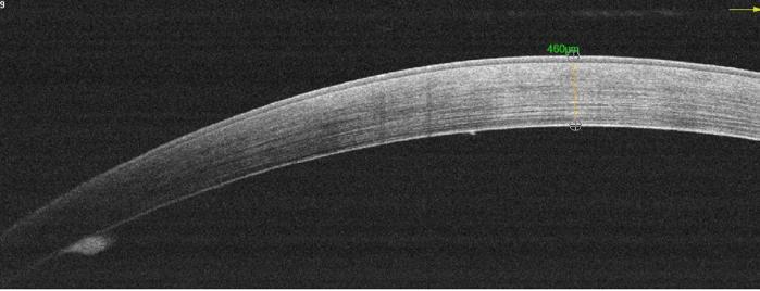
Animal 8023 CCT: 460µm
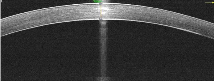
Animal 8085 CCT: 386µm
Are you up to date on the latest news about cataract and refractive surgery—new therapies and technologies, research findings, and trends in patient care?
ESCRS has you covered with its publications: EuroTimes magazine, the Journal of Cataract & Refractive Surgery, and brochures on topics ranging from presbyopia to the digital operating room to the future of refractive surgery. The latest issues of each are available at the ESCRS booth in the exhibition hall of the Bella Center.
Make sure you’re covered. Visit the ESCRS booth and pick up your copies today.
Memorable moments from the ESCRS 2025 Congress, capturing key connections and collaborations.


Don’t miss today’s IME Forum!
Join us today for this Independent Medical Education session on commonly encountered challenging phaco cases, ranging from white cataract, small pupil, and floppy iris to zonulopathy. Expect practical tips on how to optimise settings and learn from real-world case examples presented by leading surgeons.
Room B2–M5, Hall B2
Monday, 15 September 2025
13:00–14:00 CEST
Chaired by Professor Oliver Findl and Dr David Lockington, with faculty Drs Biljana Kuzmanović Elabjer, Jonathan Moore, Riccardo Vinciguerra, and Supriya Sriganesh, the programme features real-world case examples, interactive live polling, expert discussion, and insights from the latest ESCRS Clinical Trends Survey. Learn how to navigate complex cases through optimising phaco settings for difficult eyes.
The Congress finishes, as always, with the “Best of the Best” session. Featuring the best videos, papers, and posters, this session is a good way to aggregate everything that happened at the conference and hear thoughtful discussion of the latest developments and issues in cataract and refractive surgery.
Room D4
Tuesday, 16 September 2025
13:00–15:00 CEST