


Innovation, research and inclusion are paths to follow for success.
LAURA GASPARI REPORTS
The 43rd Congress of the ESCRS opened Saturday in Copenhagen with a nod to the Society’s commitment to innovation, connection and diversity at all levels, not just at the leadership level.
“This Congress is about connection, about learning, not just from the leading voices but from each other, because innovation happens when ideas, perspectives and cultures come together,” said ESCRS President Filomena Ribeiro, MD, PhD.
Delegates from more than 130 countries have gathered in Copenhagen to attend a vast programme of more than 200 sessions, cutting-edge symposia, wet labs, instructional courses and free paper presentations. These offerings demonstrate that new technologies, sustainability, gender balance and global representation are what continue to shape ESCRS as a scientific society.
Education at all levels is also a crucial part of ESCRS’s commitment to the profession. Residents, ophthalmologists, support staff and patients are all within the scope of ESCRS programming, as evidenced by education projects that were presented: the new Simulation Cataract Surgery Curriculum, the Refractive Guidelines and IOL classification, and the Patient Portal.
Simulation training is important in residency as it helps reduce errors in the operating room. A unified curriculum favours standardisation without fostering competition or unbalance. Standardisation also allows learners to improve their skills in each step of cataract surgery, as Sarah Maling, MD pointed out.
ESCRS is also a hub for evidence-based research and innovation. “ESCRS wants to be a leading community, a trusted and independent source of science education and
research in the field of cataract and refractive surgery,” said former ESCRS President Béatrice Cochener-Lamard. Projects like evidence-based guidelines for cataract and refractive surgery and the IOL classification scheme are ambitious, but they all aim to provide the best tools to clinicians, stakeholders and especially patients. The way is now open to work to strive for excellence and move from best practices to robust, evidence-based recommendation, Prof CochenerLamard noted.
Richard Packard, MD presented the new Patient Portal, an innovative resource to allow patients to better understand their treatment options. “Just imagine how much easier our job will be if our patients are well informed in advance, because they will have had access to accurate, comprehensive information,” he said. The Patient Portal includes detailed explanations, aftercare guidance, and a glossary of terms to help patients navigate medical language. Also, ESCRS has developed downloadable posters and slides in multiple languages for use in waiting rooms and consultation areas.
A highlight of the ceremony came when ESCRS President-elect Burkhard Dick, MD, PhD noted Prof Ribeiro’s accomplishments in leading the Society during the past year, praising her creativity, dedication, and keen eye for innovation as well as her commitment to building a sustainable and inclusive society. He praised her for her strong leadership that prepared ESCRS for a bright future.
“Filomena strengthened ESCRS and prepared it for a future of challenges, but also of opportunities and scientific excellence initiatives and also, of course, of building bridges,” he concluded.
From YOs to families, the ESCRS Annual Congress embraces full participation through inclusivity.
LAURA GASPARI REPORTS
The Annual Congress in Copenhagen offers many opportunities to make the experience increasingly accessible and enjoyable for everyone, particularly for young ophthalmologists (YO) and families, according to Laura Maubon MBBS.
This year’s programme is full of interesting topics and stimulating presentations. Equally noteworthy is the programme dedicated to young ophthalmologists, offering a comprehensive range of choices that can make the congress experience engaging, even for those just starting in the field. The main idea behind the YO programme is that young colleagues should not passively attend the ESCRS Congress but rather be an active and integral part of it.
As Dr Maubon remarked, the core and traditional part of the YO programme is the cataract symposium, which took place on Saturday. There, YOs refreshed their basics on cataract surgery and discussed topics such as PCR or iris management with senior colleagues. Videos submitted by young ophthalmologists themselves supported the entire programme, as this provided the opportunity to present and discuss their work on an international stage.
“We follow a similar framework each year, but all videos are new, unique, and actively engage the YO audience,” she said. “We find that the same surgical issues come up [repeatedly], so we make sure to comprehensively cover them.”

Dr Maubon is quick to point out the programme covers more than cataract surgery. A special symposium on multifocal and EDOF lenses and their application in clinical practice took place on Friday, while on Sunday afternoon, the spotlight will shift to a symposium dedicated to managing astigmatism in cataract surgery—clear proof that the ESCRS programme for YOs mirrors the current trends in ophthalmology.
Young ophthalmologists can also test their knowledge with practical learning activities. Following the success in Barcelona last year, the ESCRS Simulation Training Lab will be present at the Congress, as well as dry and wet labs accessible to all attendees.
A huge conference with such an extensive programme could be overwhelming for a young specialist. With so many people attending, many might believe there is often little opportunity to meet others, speak with senior colleagues, or exchange ideas. Some participants might find it intimidating to stand in front of such legends in the field, trying to talk or
give insights. For this reason, there are plenty of networking opportunities to facilitate communication between juniors and seniors, such as doing fun activities together.
The World Cafés offer an ideal solution, creating small, informal discussion groups where participants can get to know each other in a more relaxed setting—away from the fast pace of the main Congress—and perhaps even find valuable mentoring opportunities. The Arena Valhalla offers another fun way to connect with colleagues and exchange valuable opinions and ideas.
Dr Maubon encourages YOs not to be afraid of engaging in networking activities, adding many people are willing to cnnect, which can lead to unexpected opportunities.
We want to show we are embracing diversity and recognise the added value that different people can offer our ophthalmic community,
Building Our Sustainable Society (BoSS) activities are also a good way to connect with others. BoSS symposia topics include implicit bias, diversity, and sustainability. Other networking opportunities include speed mentoring sessions at the ESCRS membership booth and participating in the 5k Charity Run/Walk on Saturday morning.
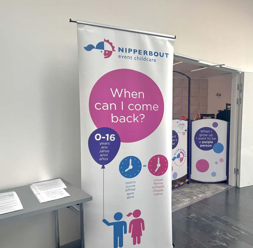
Following positive experiences in Barcelona and Athens, ESCRS is trying to become family friendly by offering a creche service that allows parents to attend sessions and symposia, even with children in tow. The childcare service has a low fee and is staffed by accredited professionals, ensuring that ophthalmologists who are also parents do not miss this opportunity and can balance their careers and family obligations. Kids benefit, too: they can get to know other children from all over the world and play in a multilingual and multicultural environment for a few days.
From YOs to well-known skilled ophthalmologists to parents, the ESCRS Annual Congress is for all, fulfilling one of its main goals: to create a welcoming environment for all people.
“This is what we are trying to do with the BoSS programme: we want to show we are embracing diversity and recognise the added value that different people can offer our ophthalmic community,” Dr Maubon emphasised. “We do not want the Congress to overwhelm anyone, and we offer streamlined programmes to help individuals target their learning objectives. We want the conference experience to be textured and layered: providing opportunities for everyone to learn, connect, and create their own narratives.”
Laura Maubon FRCOphth, MBBS, BMedSci, PGCert(Surg Ed) is a consultant ophthalmologist specialising in anterior segment surgery, ocular surface disease, and surgical education.

We’re willing to bet most eye care professionals don’t realize just how prevalent Demodex blepharitis is.
In fact, ~54% of eye care patients in Europe may have Demodex blepharitis (DB).1
Reference: 1. Data on File. Tarsus Pharmaceuticals, Inc.
LEARN HOW DB CAN FLY UNDER THE RADAR AT

Introduction
Type of Material
Hydrophobic and hydrophilic acrylic lenses: how and when to use them.
TIMOTHY NORRIS REPORTS
Intraocular lenses are not all alike; they can differ in many aspects. “Different IOL materials can have different characteristics in terms of surgical outcomes, quality of vision, flexibility, and especially biocompatibility and interaction with the ocular tissues,” said Tiago Monteiro, MD, PhD, FEBOSCR.
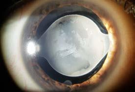
During his presentation in Copenhagen, Dr Monteiro noted that intraocular lenses can be divided into acrylic hydrophobic, acrylic hydrophilic, and acrylic hydrophilic with hydrophobic surface properties. Acrylic hydrophobic lenses are acrylic with low water content, so they have a higher contact angle and less surface wettability, Dr Monteiro said. Surface wettability is clinically relevant because it influences protein and cell adhesion to the lens.
Hydrophobic lenses are thinner, Dr Monteiro noted, meaning there is a higher level of flexibility in higher-power IOLs. Hydrophobic lenses also bring a lower incidence of posterior capsule opacification, therefore lowering the rates of YAG laser capsulotomy procedures. Moreover, Dr Monteiro said, there is a lower risk of IOL opacification and calcification.
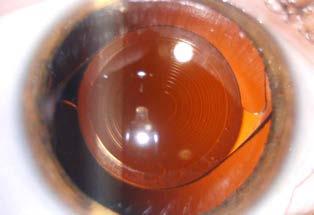
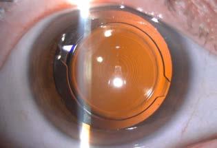
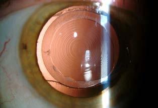
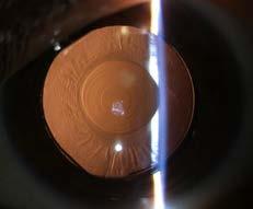
Hydrophobic lenses, on the other hand, come with some problems, mainly surface haze, glistening and nanoglistening, micro- and nanovacuoles of water that come up from the separation of water inside the polymer of the IOL. According to Dr Monteiro, different hydrophobic acrylic IOLs exhibit varying susceptibilities to glistening formation, reflecting differences in copolymer composition and manufacturing techniques. This created a clinical demand that pushed the industry to search for viable alternatives that can lower the incidence of glistening in their models of hydrophobic IOLs.
Acrylic hydrophilic lenses have a higher water content, with a higher surface wettability and lower contact angles. This results in fewer adhesions inside the capsular bag, making them theoretically easier to detach from the bag in case of a lens explantation, Dr Monteiro said.
Hydrophilic lenses have a lower refractive index and, therefore, less surface reflection. The lens doesn't shine inside the patient's eye, so patients with hydrophilic lenses normally don't have the “cat’s eye effect” that can be seen in some hydrophobic lenses, Dr Monteiro said. These lenses, however, have a higher incidence of posterior capsular opacification (PCO) and consequentially higher rates of Nd:YAG laser capsulotomies, as well as higher risk of IOL opacification and calcification.
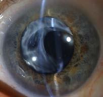
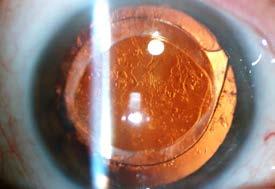
Opacification in hydrophilic lenses have been described as primary, in cases of calcium buildup inside the lens due to fabrication and packaging issues, or secondary—which is much more frequent—where there is calcium showing on the outer surface of the lens. This is normally due to clinical factors related to the patient (glaucoma and diabetes are risk factors) or procedures like DSAEK, DMEK and pars plana vitrectomy that use gas or air. Dr Monteiro recommended that hydrophilic lenses be avoided in patients with diabetes, glaucoma and uveitis, as well as in patients with a higher risk of developing PCO. “There are patients with Fuchs dystrophy, for instance, or patients with whom you are planning to do some kind of endothelial keratoplasty with the use of gas or air,” he said.
Tiago Monteiro is a professor (Associate) at Escola de Medicina da Universidade do Minho in Braga, Portugal.
5. Conclusion
IOL Material
Hydrophobic IOLs
higher refractive index and lower thickness, tend to be more flexible and easier to implant specially in high power IOLs lower rates of PCO and calcification, block the proliferation of A and E cells, reducing the risk of anterior capsule opacification and anterior capsule contraction syndrome
Hydrophilic IOLs
higher risk of posterior capsule opacification and a higher susceptibility to opacification of the lens optic lower refractive index, are generally thicker, show similar high -contrast sensitivity and possible lower incidence of negative dysphotopsia


Haag-Streit invites ESCRS delegates to join its distinguished faculty for a one-hour Satellite Symposium on how innovations in slit lamp microscopy can address emerging & future trends in eyecare health. The Symposium will also address the benefits of the new Elara 900, including its superior clinical observation, improved efficiency & optimized ergonomics.
Binkhorst Medal Lecture highlights need for further research on myopia.
LAURA GASPARI REPORTS
Today’s ophthalmologists have a considerable number of ways to correct myopia, but this year’s presenter of the prestigious Binkhorst Medal Lecture says we need further research and treatments.
“Be aware that in 2050 we will have 4 billion people myopic in the world,” said Thomas Kohnen, MD, PhD, FEBO. “That's maybe half of the population. Correction is not the treatment. It's a growing problem worldwide and without a cure, but we can do some things.”
Prof Kohnen noted there are risk factors that are well known by the ophthalmological community, such as not spending enough time outside, a lot of near work (especially with digital devices and more pressure in educational systems), staying in dim light, and, ultimately, genetics. He explained that the development of lens power over time shows a decline across all groups of ages, with a steeper drop indicating the onset of myopia. This suggests that the compensation is insufficient to counteract axial length growth, meaning there is no effective mechanism to compensate for myopia.
Ophthalmologists have some weapons to counteract myopia: first, by raising awareness among patients of the impact of lifestyle changes such as (especially for children) spending more time outside, as outdoor recess has been demonstrated to reduce myopia onset by 50%; also by using atropine 0.1%, which has been demonstrated to be effective in slowing myopia progression with minimal side effects; and recommending myopic defocus lenses, which have been shown to be effective in reducing myopia progression and axial elongation over 50% in two years. Prof Kohnen warned about the rebound process if treatment is stopped, showing that the lowest rates of it are for those using glasses.
Refractive surgery, such as PRK/TransPRK, LASIK, FemtoLASIK and KLEx, is another important option that
has seen significant evolution over the past decades. Prof Kohnen presented meta-analysis data showing high safety and effectiveness rates for PRK, LASIK and KLEx, with no superiority of any single procedure. There are now a lot of studies under way on the use of femtosecond laser and excimer laser. Phakic IOLs (angle-supported, iris-fixated and posterior chamber lenses), of which Prof Kohnen is a big fan, have expanded the possibilities for myopia correction, outperforming excimer laser in safety and satisfaction in moderate/high myopia cases.
One big issue facing the ophthalmological community is high myopia and the possible complications these patients can face even after refractive surgery and phakic IOL implantation, including cataract formation, ectasia, and retinal detachment. Prof Kohnen emphasized the importance of creating guidelines for refractive surgery and the need for continuous research and improvement, efforts in which ESCRS is heavily invested through new studies, an IOL classification project, and guidelines, not only for myopia but also for presbyopia. He recommended that ophthalmologists take a comprehensive approach when dealing with myopia. “Remember to treat the whole eye, not just a refractive error,” he said.
Prof Kohnen concluded with a call for reflection, quoting a 1934 article titled “Nationalism Against Science” that spoke of political shortsightedness as a threat to research and science. “Last week Richard Parrish sent me this and he wanted to republish this article to remind us of the near-sighted world we have,” he said.


Combining the proven power of Scheimpflug imaging with the precision of ultra high-resolution OCT enables the detection of previously unseen corneal pathologies with unprecedented clarity.
Diagnose earlier. Treat smarter. Care deeper.
EPITHELIUM
BOWMAN’S LAYER
STROMAL LAYER

The Pentacam® Cornea OCT can increase the confidence that your diagnosis is correct.
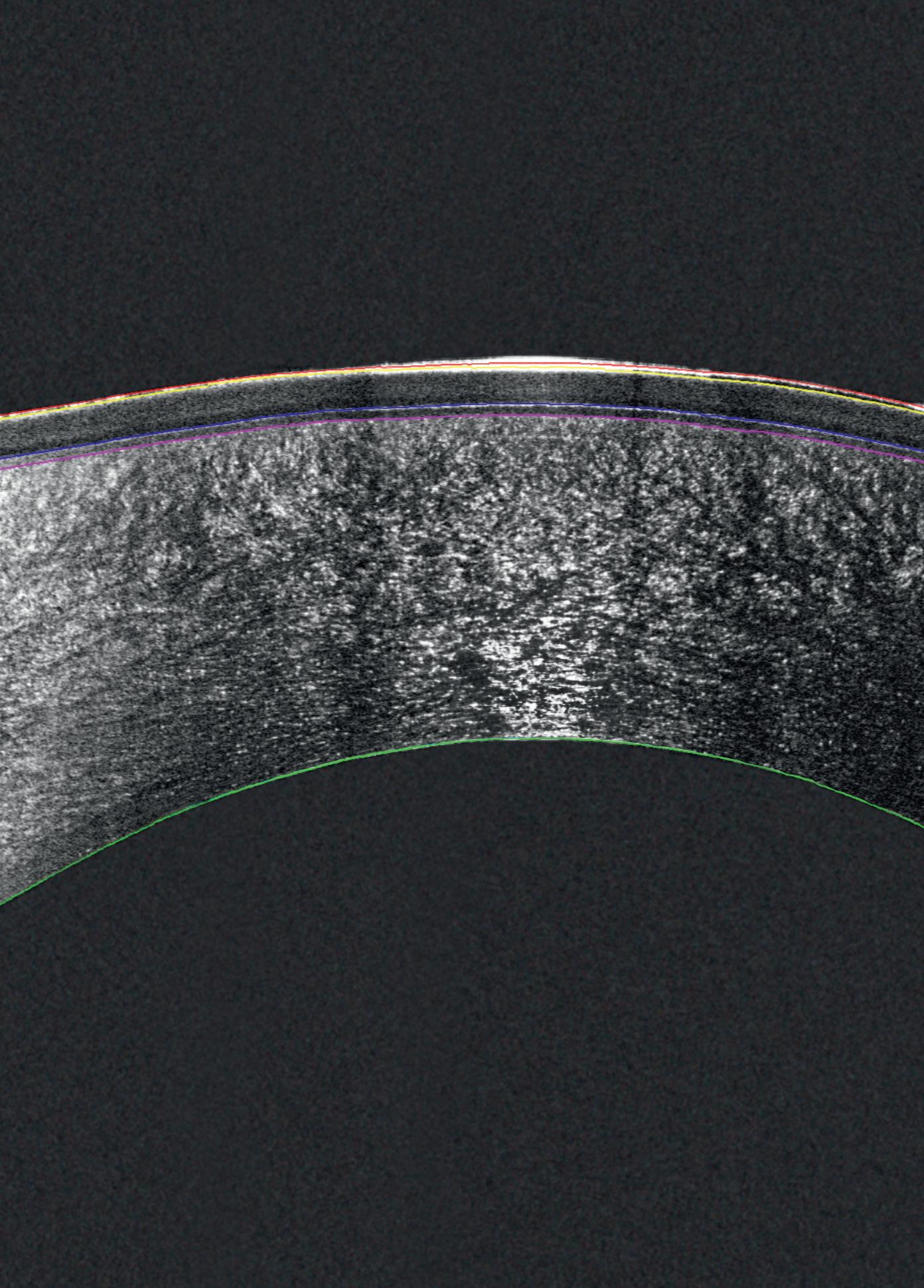
Using cultured human corneal endothelial cells for transplants could alleviate long waiting lists.
SWEENEY REPORTS
The sky is blue, gravity makes things fall down, we need more corneal tissue for transplants. Some things are so obvious they don’t need to be said.
But just because something is obvious doesn’t mean it cannot be changed. For example, corneal donor shortages are a perennial issue, but human corneal endothelial cell (HCEC) transplantation could be a way to alleviate them, according to Dr Shigeru Kinoshita.
In his presentation titled “Primary Cultured Cells for Cultured HCEC Transplantation,” Dr Kinoshita said HCEC transplantation therapy using young donor allogenic cells could mean that “conceptually, one pair of donor corneas could be used for 300 patients in a safe and speedy procedure.” The challenge is proving the efficacy of using cultured human cells in the process.
Dr Kinoshita reported that the drive to prove the efficacy of cultured HCEC transplantation began in 2013, and during the intervening years he has published several papers on the topic. In that time, he has found that “cultured cells do not show chromosomal abnormalities, and around 85% survive 10 years after transplantation.”
There are three scientific aspects to consider in fully utilising HCEC transplants, according to Dr Kinoshita. Mass production for cell cultures while ensuring their purity is an issue; self-organisation is another issue, as the cells must be able to “reach the cornea, and not the iris or lens surface.”
“What’s most important and interesting to me is rejuvenation by allogenic young cells not only for the corneal surface but the anterior chamber itself,” he said. “To find this out, we found Rho-kinase (ROCK) inhibitors to be effective.”
In a subsequent clinical trial, Dr Kinoshita divided patients into two groups: one with mature differentiated sub-population cells of less than 76.3%, and another with most cells reaching maturation of 94.6%. The results showed that group 2 exhibited significantly higher endothelial density compared with group 1, and cell shape was significantly improved even after three years post-op.
These results highlight the presence of rejuvenation, according to Dr Kinoshita. He added that the Japanese government has since approved Vyznova as a treatment for corneal endothelial failure, and that 180 HCEC transplants involving the use of this medication will take place in the country this year, up from just 25 in 2024.
HCEC transplantation is also frequently used by Dr Kinoshita’s university for a wide range of conditions. Bullous keratopathy is the most common condition, followed by glaucoma drainage bleb, failed graft, and Fuchs’ endothelial dystrophy.
Dr Kinoshita is now aiming at making cultured HCEC transplantation a “global standard medicine,” with phase 2 and 3 trials currently underway in Japan. Culture cells have been shipped to El Salvador, where 90 cases are currently underway as part of a phase 1 clinical trial. Meanwhile, phase 2 trials are ongoing in the United States and Canada.
Shigeru Kinoshita MD, PhD, is Professor and Chair of the Department of Frontier Medical Science and Technology for Ophthalmology at Kyoto Prefectural University of Medicine.


Don’t let dry eye compromise the surgery outcome - take control of your patients’ ocular surface with Lacrifill, the hyaluronic acid canalicular gel that delivers six months of improvement with a single treatment.1,2
✔ FDA-cleared
✔ CE-marked
✔ One syringe, one patient


How multifocal lenses are categorised based on the new IOL classification system.
TIMOTHY NORRIS REPORTS
The new ESCRS IOL classification proposed by the Functional Vision Working Group provides a new way to classify intraocular lenses, with multifocal, EDOF, trifocal and others now inscribed inside the Simultaneous Vision Lens group. But how are the different multifocal lenses fitting inside this group?
Oege Goslings, MD, PhD, analysed the different designs and explained which ones are now considered within the brand-new classification.
“Diffractive multifocals are an important part of the multifocal family due to the simultaneous vision created by multiple foci that provide multifocality,” Dr Goslings said. “Importantly, these lenses give pupil independence.”
Examples of diffractive multifocal IOLs in the market are the J&J Odyssey, Panoptix from Alcon, AT Lisa from Zeiss, BVI Fine Vision, and Gemetric by Hoya. According to Goslings, these lenses are popular in the Netherlands.
Refractive lenses like Ophtec Precizon Presby, Teleon Acunex Vario and LensTec Clearview, on the other hand, are more common in the US. These lenses are based on zones, not on diffraction, Dr Goslings said. There is a difference in refraction from the centre to the periphery in this type of lens, leading to pupil dependence. So, there's a different behaviour here, he remarked. Additionally, there's a new mechanism proposed by Rayner’s Galaxy IOL, where the optic zone is shaped like a vortex, creating depth of focus giving a full range of focus, Goslings said.
In the new classification, a cutoff was set at 2.3 Dioptres with a minimal visual acuity of 0.2 LogMAR, about 0.63
Snellen for far intermediate and near. Lenses that do not reach the 2.3 Dioptres mark are considered partial range of field (ROF), while lenses that are equal to or above the cutoff are considered full range of field. Based on this standard, Goslings observed how the Restor by Alcon can be considered partial range of field, whereas the Panoptix can be considered full ROF.
If you have the simultaneous visual lenses, how is it possible to differentiate between those individual lenses? Checking visual acuity at near and then comparing is the first step, Dr Goslings suggested. If the lenses have better near than intermediate, those are the Steep Transition lenses; if the defocus curve is straight, those are the Smooth Transition; those with a continuous visual acuity from near to distant are categorised as Continuous full range. According to Dr Goslings, AT Lisa by Zeiss and BVI’s Fine Vision fall into the Steep Transition category, Alcon Panoptix and Hoya’s Gemetric go into the Smooth Transition group, and J&J’s Odyssey, Rayner’s Galaxy, and Envy by Bausch+Lomb make it into the Continuous Range group.
“Full ROF Simultaneous Vision Lenses are better defined by their functional outcome, considering visual performance and presence of photic phenomena,” Dr Goslings said.
Oege Goslings, MD, PhD, is an ophthalmic surgeon at Elisabeth-Tweesteden Hospital, WGoslings@Hotmail.com
Green and clean
Innovative eco-design saves resources
Less waste and lower disposal costs
Swiss made Medicel quality


An alternative to cells derived from embryos could mean an improvement in patient outcomes.
ANDREW SWEENEY REPORTS
Stem cells are controversial in both the lay and medical communities, but what if there was a way to use them while avoiding the resulting ethical issues? Can ophthalmology have its cake and eat it, too?
In “IPSC-derived Corneal Endo and Epi Replacement,”
Jod Mehta PhD considered those questions. His presentation highlighted the potential of a new frontier in science that could achieve remarkably good patient outcomes.
Induced pluripotent stem cells (iPSCs) are a type of stem cell reprogrammed from a patient's ordinary somatic cells. As they aren’t derived from human embryo tissue, they avoid the many ethical issues in utilising this tissue. They also enjoy blood group compatibility and have human leukocyte antigen histo-compatibility as well.

iPSCs have applications in a variety of fields, ranging from disease modelling and cellular therapy to cultured meat production and wildlife conservation. What will be of particular interest to ESCRS Congress attendees is how iPSCs can be used in corneal endothelial and epithelial treatment.
Prof Mehta outlined how cells can be taken from several sources: unilaterally via a limbal biopsy, bilaterally, and from either a corneal transplant or oral mucosa if using an allogenic cell source. The resulting cells must then be sorted as the iPSCs will “spontaneously differentiate,” as four different layers of the cells undergo sequenced epithelial-mesenchymal assortment method (SEAM) formation. According to Prof Mehta, this can pose a challenge.
“There's not one single epithelial cell marker that's available in literature, so people use a combination of things,” he said. “You need a high level of purity before transplanting the cells into a patient. You’ve got to be looking at the surface markers. There’s no point in having a marker inside a cell that has to be then destroyed when you're basically looking at it. It has to be based on the surface.”
Clinical trials have shown the promise of iPSCs, and a 2024 study published in The Lancet found that after 52 weeks of treatment, corneal opacification had diminished in all treated eyes and corrective distance visual acuity had improved as well. In a trial involving monkeys, Prof Mehta reported that iPSC endothelial therapy had a noticeable impact in just two weeks, including an improvement in 1.6 to 1.2 better visual acuity and an average reduction in corneal thickness from 850 to 550μm without a change in intraocular pressure or tumor formation.
Concluding, Prof Mehta emphasized that the prognosis for iPSC-derived treatment is positive, but challenges do remain. Safety issues related to teratogenicity and mutation are concerns, as are regulatory issues that complicate scaling and manufacturing. Still, he is enthusiastic about its prospects.
“There is a good progression of clinical trials going around the world in multiple areas,” Prof Mehta said. “This is a very exciting field to look into.”
Jod Mehta MBBS, FRCOphth, FRCS(Ed), FAMS, PhD is a senior consultant at the Singapore Eye Centre

Agreement with US associations provides further insights into new IOL classification.
TIMOTHY NORRIS REPORTS
Anew system of classifying intraocular lenses aims at international standardisation and consensus, improving collaboration between the main international cataract and refractive societies of the world. Strongly based on literature and scientific data, this functional evidence classification is a clinically relevant system with mutually exclusive groups and subgroups that represents progression, according to Joaquín Fernández, PD, PhD.
Starting with a guest editorial in the Journal of Cataract and Refractive Surgery, the project developed within ESCRS and involved sister organisations such as the American Society of Cataract and Refractive Surgery, the American Academy of Ophthalmology and the Asia-Pacific Association of Cataract and Refractive Surgeons.
“We had our first meeting in Los Angeles with ASCRS where we achieved this consensus,” Prof Fernandez said. “The concept is to escape from opinion and ideas, all based directly on scientific evidence. We analysed 20 years of

literature in PubMed regarding simultaneous vision lenses before developing all the criteria, passing through a strict methodology.”
Cluster analysis was conducted using machine learning as an aid to check and allocate different groups. In a consensus with American colleagues, the term range of field was changed to the more academic concept of depth of field, divided into full and partial depths of field. According to Prof Fernández, it is time to move from the optical bench to the clinical level because it will be quite interesting to know the real performance of the lenses and the appropriate materials as well as collect and report data in clinics. It is also time, he said, to compare parameters like MTF to parameters like visual acuity and contrast sensitivity that we can collect from and with our patients.
After the cluster analysis, the team identified different criteria according to the depth of field and the defocus curve to check the range of the depth of field and the decrease in visual acuity from the intermediate to the near focus, Prof Fernández said. Two cur points were identified at 0.2 and 0.3 LogMAR, the latter having been added because ANSI and ISO do not distinguish the enhanced monofocal.
Thanks to the consensus reached with the US organisations, the ophthalmic community will be involved in the collection of patient-reported outcomes using new tools like the AIOLIS (developed by the American Academy of Ophthalmology). The AIOLIS is an incredible questionnaire that provides information on the symptoms, the frequency and the association with grading visions in terms of quality, satisfaction and spectacle independence, Prof Fernández said.
Functional evidence classification is evidence based, rooted in literature, clinically relevant and mutually exclusive, because the ISO classification is not mutually exclusive. In the ISO, all the full visual range are also multi focal and present progression.
“Our idea is focusing on the post market, trying to help the patient and the doctor to add decision making to the process and their own money,” Prof Fernandez said. “In the future, we would like to include direct community inclusion of other endpoints, such as contrast sensitivity and dysphotopsia.”
Joaquin Fernandez, PD, PhD, is secretary of ESCRS, CEO of Qvision, and medical director of the Andalusian Ophthalmology Institute at Vithas Hospitals, Spain.


Ongoing research into genes may mean doctors can halt disease progression.
ANDREW
GSWEENEY REPORTS
ene editing may seem like nothing more than copying and pasting DNA, but it holds considerable promise for ophthalmology, not least for the management of Fuchs’ endothelial corneal dystrophy (FECD).
In “Developing Gene-directed Therapeutic Approaches for Fuchs,” Dr Nihar Bhattacharyya provided valuable insights into the latest trends in gene editing and how it’s applied to the condition. She revealed there are two primary ways that genes affect the progression of the disease.
“Variants fall into two main categories: regulatory, where the disease variant affects the overall regulation and expression of the corneal endothelial cell, and mechanistic, where the disease variant affects the structural or functional component crucial to the role of the corneal epithelium,” Dr Bhattacharyya said.
“We focus on the regulatory gene TCF4, as it has by far the strongest risk factor for FECD via an encoded repeat expansion factor with a 50 times greater risk,” she noted. “In our research cohort of 1,500, 77% carried TCF4 as opposed to 5% in the general population.”
Excising out the repeat expansion part of the TCF4 gene may seem appealing, but this technique is still in the very early stages, according to Dr Bhattacharyya. Safety factors need to be considered, and there are also concerns about specificity—i.e., too much may come out during the process.
If the expansion factor of TCF4 is allowed to take place, however, options could exist downstream that could halt it. Somatic instability, representing the ‘when’ if inheritability represents the ‘if’, could be manipulated to delay disease onset via a therapeutic strategy. This would lead to RNA repeat-mediated toxicity therapeutics that could be targeted to arrest the growth of diseases like FECD.
Dr Bhattacharyya and her colleagues researched antisense oligonucleotide to sterically bind the RNA repeat sequences, reducing the toxic RNA foci that can facilitate the growth of FECD. Unfortunately, funding for the study ran out. They are now considering how to disrupt RNA aggregates by using small molecules.
Dr Bhattacharyya is presently working on models within the laboratory that would allow for the high throughput assay of small molecule libraries that would result in the disruption of the RNA foci. She is also researching therapy that could encourage cells to promote MLB1 expression and delete splicing factors.
Highly avant-garde possibilities are also being considered by Dr Bhattacharyya and her colleagues. TCF4 has over 90 RNA transcripts—most cells only have one or two—and there is real potential for examining these longer, protein producing RNA processes.
“Alteration of TCF4 RNA transcript usage would lead to wide-scale transcriptomic deregulation,” Dr Bhattacharyya said. “This would be the key in several signalling pathways that regulate cell state.”
“More research is needed into the potential impact of this process,” she summarised. “Given the age-related nature of FECD as a disease, slowing therapy holds great potential, and it can act as a model and proof of concept for other theories of repeat mediated diseases.”
Nihar Bhattacharyya MSc PhD is a research fellow at the Institution of Ophthalmology at University College London.


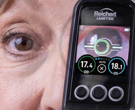


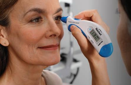
Experience the all-new Reichert® Tono-Vera® Tonometer featuring the patented, auto-measuring ActiView™ Positioning System, for fast, e ortless, objective, and repeatable rebound tonometry measurements. For nearly two centuries, Reichert has empowered a more e cient exam and shared in your passion to deliver exceptional patient outcomes.
DEMO AT ESCRS #C1.102
LEARN MORE AT REICHERT.COM


LEARN MORE ABOUT REICHERT’S COMPREHENSIVE FAMILY OF TONOMETRY AND REFRACTION DEVICES:


JOHN HENAHAN
Each year, young ophthalmologists are invited to participate in the John Henahan Writing Prize by responding to an essay prompt. Noha Fawky Abdelfattah MD’s essay scored among the top three in a very competitive field.
Applicants responded to the following prompt: Diversity, equity, and inclusion (DEI) programmes, however well-intentioned, stir a variety of responses in the corporate and political worlds and in the scientific and medical spheres. What DEI and unconscious bias issues are present in the current culture of ophthalmology training, practice, and clinical research? What are the potential benefits of addressing these issues for patients and ophthalmologists? What kind of meaningful changes need to happen to move beyond ‘talking the talk’ to ‘walking the walk’?
In a specialty defined by precision, clarity, and vision, ophthalmology has made extraordinary strides in science and technology. But beneath the surface of these advances lies a less visible challenge—our collective blind spot when it comes to diversity, equity, and inclusion (DEI). As a female ophthalmologist training in the Middle East, I’ve seen first-hand how unconscious bias shapes who gets to lead, who gets chosen, and who gets seen—not just in the operating theatre, but in our systems, language, and even the patients we serve.
I currently train and practice at Watany Eye Hospital in Egypt, a hub of excellence in our region. But despite the clinical sophistication, I still encounter an invisible obstacle: gender-based bias. I’ve had patients bypass me as a surgeon solely because I am a woman. I’ve walked into operating rooms where the unspoken assumption is that the lead surgeon must be male. And I’ve seen the hesitation in patients’ eyes—not because of my skill, but because of my gender.
This isn’t just a Middle Eastern issue. Around the world, women in ophthalmology still face slower promotion, unequal pay, and underrepresentation in leadership roles. And those from minority ethnic or socioeconomic backgrounds often face even steeper climbs. These aren’t isolated cases—they are symptoms of a system built on centuries of imbalance.
Yet, [through] these barriers, I was fortunate. I found a mentor who saw my potential before I could fully see it myself.
Professor Ahmed Assaf didn’t just teach me how to perform surgery. He taught me how to edit surgical films, how to explain complications with humility and confidence, how to make patients feel safe. He brought me to international conferences, pushed me to contribute to papers, and opened doors that would have otherwise remained closed. His mentorship didn’t just shape my skill—it shaped my belief in what I could become. Every young ophthalmologist deserves a mentor like that.
Bias in ophthalmology isn’t limited to career trajectories—it affects patients, too. One of my most unforgettable cases was a man with a single functioning eye, clouded by a dense brown cataract. He had been turned away by others because he couldn’t afford the surgery. His world had narrowed to
shadows and shapes, and yet, no one had offered him hope. I performed the surgery for free.
A week later, he returned beaming. For the first time, he could see his grandchildren’s faces. That moment reminded me why equity matters—not just in who gets to train, but in who gets to see.
It is far too easy for disadvantaged patients to fall through the cracks of our healthcare systems. Whether because of poverty, language, disability, or culture, countless patients are underserved, misdiagnosed, or delayed in care. And when the physicians who treat them do not reflect their diversity, communication gaps only widen.
These disparities echo into our research, where many clinical trials still lack representative samples. In global ophthalmology studies, patients from the Middle East, Africa, and South Asia are underrepresented. So are female patients, especially in interventional trials. Without diversity in data, our conclusions are biased before the first statistic is run.
Worse still, diverse investigators are often left out of leadership roles in research. We risk building the future of eye care on incomplete evidence—and leaving millions behind.
We often say that ophthalmology is about restoring vision. But real vision isn’t just about what the eye sees—it’s about what the mind perceives. Addressing unconscious bias and embedding DEI into our profession is not an ethical add-on; it is essential to clinical excellence.
When patients see themselves in their doctors, trust deepens. When diverse trainees are supported, the entire system benefits. When research reflects the full spectrum of humanity, our innovations gain power.
From words to action
To move from ‘talking the talk’ to ‘walking the walk,’ we must:
1. Reimagine training: Blind reviews, structured interviews, and mentorship programmes must be standard. Diversity should be seen as a strength, not a checkbox.
2. Confront clinical bias: Patients should choose surgeons
based on skill—not gender or ethnicity. Institutions must educate patients and empower all doctors equally.
3. Decolonize research: Funders and journals must demand inclusive trial design and diverse leadership. Global data should represent global people.
4. Create mentorship pipelines: Every young ophthalmologist deserves a Professor Assaf—someone who lifts them as they rise.
5. Measure and report: DEI metrics in hiring, publishing, and outcomes must be transparent and acted upon. What we do not measure, we cannot improve.
To the young ophthalmologist reading this: find a mentor who believes in you—and be that mentor for someone else. Challenge what is assumed. Advocate for who is excluded. Equity is not a destination; it is a discipline. In the end, we are a specialty of visionaries. Let us sharpen our lens—not just to treat cataracts, but to clear the cultural fog that holds our field back. Let us make it possible for every patient to see, and for every doctor to be seen.
Because true vision demands more than sight—it demands insight.
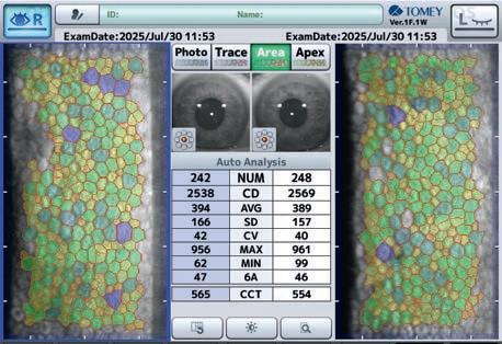
High-resolution specular microscope with full auto-alignment and capture in just four seconds per eye. Assess central and peripheral areas via fixation light, including Dark Area analysis. Captures over 300 endothelial cells across 15 mapped positions, with pachymetry and morphology metrics (polymegathism, pleomorphism) – all in one fast, user-friendly workflow.
Effortless operation & analysis
The complete version of the essay will appear in EuroTimes

+ L-count, Trace, Core method, Dark Area analysis
+ Counts more than 300 cells
+ Auto alignment per 1-2 sec
+ Auto capture of 16 images incl. CCT
+ Multi-fixation targets
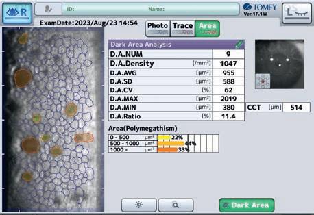
Dark Area analysis
+ Number of dark areas
+ Dark area density
+ Min. and max. size of dark area

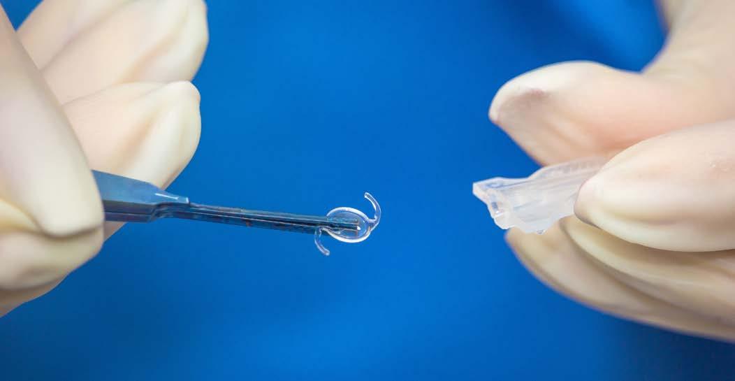
Introducing a toric IC-8 would further optimise visual outcomes for astigmatic patients.
HOWARD LARKIN REPORTS
Small-aperture intraocular lenses (IOLs) are effective in reducing irregular astigmatism. New research suggests that adding a toric correction may improve vision even more.
“The IC-8 Apthera (Bausch + Lomb) is a presbyopia correcting IOL that uses the pinhole effect to extend depth of focus, but it is also quite robust in treating cases of irregular astigmatism … and can tolerate regular stigmatism up to 1.5D,” said Grzegorz Labuz PhD. “When we add to this a toric surface, we can potentially expand the application or improve visual function in those patients.”
To test this hypothesis, Dr Labuz and colleagues conducted a bench experiment. A range of model eyes with simulated aberrated corneas were constructed based on surface elevation maps from Pentacam HR corneal topography data of 10 patients with irregular astigmatism.
The image quality of these models was then tested with a toric-only correction, a pinhole correction, and a combined pinhole and toric correction. Toric corrections ranged from 1.5D to 6.0D; the pinhole corrections were 1.36mm masks. Root mean square (RMS) of aberrations (lower is better) and the Strehl ratio of image quality (higher is better, with 1.0 the highest possible quality) were measured to evaluate optical function.
The results showed promise for the combined approach. Median Strehl ratio for the toric-only IOL was just 0.06 (0.05-0.19); for the pinhole-only IOL, it improved to 0.14 (0.10-0.39). The combined toric-pinhole IOL did much better, reaching 0.86 (0.63-0.92).
Similarly, aberration RMS dropped from a median of 0.42 (0.37-0.51) with the toric-only lens to 0.27 (0.17-0.36) with the pinhole IOL to 0.06 (0.05-0.11) with the combined IOL.
The study also found a correlation between projected and measured astigmatism in corrected eyes. This suggests
corneal topography data could help predict the potential effectiveness of the proposed correction and guide cylinder selection, Dr Labuz said.
“We can correct both the regular and irregular parts of the astigmatism,” Dr Labuz noted. The result is a greatly improved retinal image.
“Combining small-aperture and toric correction provides the greatest improvement in optical quality and potential visual function in these complex cases,” Dr Labuz concluded. “Introducing a toric IC-8 would further optimise visual outcomes for astigmatic patients; however, the sulcus-fixated IOL is currently the only available option.”
The study was conducted by researchers at the David J Apple Centre for Vision Research, Heidelberg University Clinic, and the University Hospital Carl Gustav Corus, TU Dresden, both in Germany.
Grzegorz Labuz PhD is associate professor of experimental ophthalmology and head of the Optics and Metrology Section at Heidelberg University.

JCRS working group aims for consensus on terms and treatment.
The diagnosis and treatment of pre- and postoperative astigmatism continues to be a challenge to ophthalmic surgeons. At the Copenhagen Congress alone, there are 3 sessions dedicated to astigmatism management in both cataract and refractive surgery, along with 134 separate presentations and 67 abstracts.
Recognising a need for more clearly defined terms of astigmatism, the Journal of Cataract & Refractive Surgery created a working group to establish a consensus. Led by Professors Douglas Koch and Thomas Kohnen, the group published its conclusions in the June issue of the JCRS.
The working group was determined to “(1) establish astigmatic reporting guidelines based on sound optical principles and (2) provide new astigmatism calculation tools that simplify the process and allow researchers to reliably calculate accurate and reproducible results.” The first paper defined astigmatism and provided information on its sources and measurement; discussed the population distribution of corneal astigmatism; explored the differences among corneal imaging technologies; and considered the impact of the ocular surface, irregular corneal astigmatism, posterior and internal sources of astigmatism, and refractive astigmatism. The second paper suggested the “recommended methodology for analysing astigmatic outcomes,” which the authors wrote was “based on the optics of corneal and lenticular astigmatism, as well as contemporary vector analysis and statistical approaches.”
The authors noted astigmatism as the difference between the orthogonal principal planes of minimal and maximal powers of a toric surface or lens. Corneal astigmatism is a common condition, and studies indicate 60% to 78% of individuals have more than 0.5 D of astigmatism, and 20% have more than 1.5 D.
As the magnitude of astigmatism increases, the incidence of against-the-rule (ATR) astigmatism is nearly constant, varying from 26% to 31%. By contrast, the incidence of withthe-rule (WTR) astigmatism increases as magnitude increases, from 26% to 64%, corresponding to a decreased incidence of oblique astigmatism. Corneal astigmatism also changes with age. Most population studies show an age-related shift in anterior corneal astigmatism from a vertical steepness (WTR) to a horizontal steepness (ATR).
Total corneal astigmatism is calculated through raytracing, using anterior and posterior curvature measurements
Studies indicate 60% to 78% of individuals have more than 0.5 D of astigmatism.
and corneal thickness. Various imaging technologies and algorithms help perform these measurements, including reflection (rings or single-point mires), Scheimpflug imaging, and optical coherence tomography. However, measurements from different devices are generally not interchangeable because distance differences from the centre can result in both magnitude and axis variations. Zonal keratometry (topography/tomography) over the patient’s mesopic entrance pupil samples a larger region of the central cornea, with the zone size customisable to each specific patient.
Astigmatism can be represented as a vector since it has both magnitude and direction. The magnitude is the absolute power difference between principal meridians, and the direction is defined by the meridian of greatest positive (or least negative) power. Since meridians extend in two directions from the centre, the angle must be doubled for Euclidean vector calculations. Using double-angle plots allows for vector algebra and statistical analyses, after which the angle is halved for single-angle results. JCRS will use double-angle plots to report astigmatic outcomes.
The vector prediction error (PE) is the key expression for evaluating surgically induced astigmatic change—defined as the vector difference between the postoperatively observed and refractive astigmatism predicted from preoperative measurements. Vector calculations can be performed independent of or relative to a reference meridian or axis. Double-an-

gle plots with convex polygons and statistical analysis for astigmatism data can be easily obtained with the wrap-up functions from the Wilcox-Holladay-Wang-Koch Statistical Software using the free R Project for Statistical Computing software or with Eyetemis software, also available online.
The articles, along with a video interview with Profs Koch and Kohnen, are available at https://journals.lww.com/jcrs.


QTY/BoxDuration
Provides ocular surface protection following surgery, injection and traumatic or non-traumatic corneal conditions.
Diameter: 14.05mm
Compound base curve: 9mm
Intended Use: Protect ocular surface following surgery, injection, traumatic, and non-traumatic corneal conditions.
•Comfort of a thin pliable collagen barrrier
700210Quick Shield, Less than 12 hrs
70121012 hrs non-patched, 24 hrs patched
70241024 hrs non-patched, 72 hrs patched
70721072 hrs non-patched
• Collagen certified suitable for use by EDQM
• Hydrates to 70% water content
• Lubricates with collagen while it gradually dissolves
To order/learn more or to see if this product is available in your country, please contact international@oasismedical.com

Clinical studies needed to confirm significance of in vitro findings
HOWARD LARKIN REPORTS
An in vitro study of the image quality produced by two extended depth-of-focus intraocular lenses (EDOF IOLs) found significant differences in resolution at different ranges of defocus, as well as glare in decentred and off-axis light conditions. The variations may relate to the different materials and designs of the two lenses, according to the researchers, who were supported by one of the lens manufacturers.
Both the Clareon Vivity (Alcon) and the Tecnis PureSee (Johnson & Johnson Vision) provide extended depth of focus and are made of hydrophobic acrylic materials but have different refractive indexes and Abbe numbers, with the Vivity at 1.55 and 37 and the PureSee at 1.47 and 55 respectively, noted Liliana Werner MD, PhD.
In addition, the Vivity optic covers the entire 6mm optical zone and the PureSee has a non-imaging area at the periphery of its optic. These design differences may affect visual performance, Dr Werner said. The goal of the study was to assess the optical quality and visual disturbances of these two marketed EDOF IOLs.
Differences in how the lens is implanted and calcification are just a few of the variables not seen on the bench.
The optical quality of the two IOLs was assessed using a modulation transfer function (MTF) bench. MTF was obtained in model eyes with corneal spherical aberration of 0.2 microns for the Vivity, and 0.28 microns for the PureSee. Halo was measured using high dynamic range halo imaging with and without decentration. A glare bench was used to measure glare.
The difference in predicted visual acuity at distance was not clinically significant, Dr Werner said. However, at low levels of myopic defocus, from about 0.3-1.5D, MTF resolution for the PureSee was 42% lower, she said. These results were consistent with a published study (1).
On-axis halo images were comparable for both IOLs when perfectly centred, Dr Werner reported. However, an asymmetric flare was observed for the PureSee at 0.3 and 0.5mm decentrations.
The PureSee also produced significant glare from off-axis light starting at about 35 degrees and increasing sharply to about 45 degrees. This may be due to the flat, non-imaging area at the edge of the PureSee platform, Dr Werner said. By contrast, the Vivity produced almost no glare out to 50 degrees off-axis, similar to a Clareon monofocal IOL.
Although these results suggest the PureSee may be more prone to glare and halos than Vivity, the differences seen on the bench may not manifest clinically, Dr Werner emphasised. Differences in how the lens is implanted, whether the edge is covered 360 degrees by the capsular bag, and calcification are just a few of the variables in vivo not seen on the bench. Therefore, clinical studies are needed to confirm the significance of these in vitro findings, she concluded.
The study was conducted with support from Alcon, for whom Dr Werner is a consultant; her coauthors are from Alcon Research, Fort Worth, Texas, USA. Dr Werner is from the John A Moran Eye Center of the University of Utah, Salt Lake City, USA.
1 Niknahad A et al. “Evaluation of Clareon Vivity and PureSee intraocular lenses: optical quality, depth of focus, and misalignment effects.” Scientific reports 2025; 15:26943.


Genetics can tell us the real story and better explain our origins and history.
LAURA GASPARI REPORTS
One of the greatest assets of a vast community like ESCRS is diversity. ESCRS comprises different people from different cultures speaking different languages and with different stories, all united in their commitment to ophthalmology. To know ESCRS members is a wonderfully enriching experience.
This year, ESCRS is staging its Annual Congress in Denmark. When we think about Scandinavia, our minds often turn to the Vikings. But how well do we really know the Vikings?
Not well, it turns out. Those we are used to seeing portrayed in the media are not the Vikings who walked the earth, as Prof Eske Willerslev of the University of Copenhagen pointed out in his TED Talk during the opening ceremony. So, how many of the stories of sagas and representations that we see in the movies are correct?
Prof Willerslev and his team conducted a large study on Vikings genomics, with interesting outcomes. In fact, Vikings were not just Scandinavians—they came from various regions, including Southern Europe and the Sami populations, and have a vast genetic diversity due to their extensive travel for trading that took them as far as Greenland.
Contrary to common belief, there were not epic raiding parties of Vikings like media show us.
Contrary to common belief, there were not epic raiding parties of Vikings like media show us, but rather small raids made by families, as an archeological site in Estonia revealed. Also, Prof Willerslev’s research found that Vikings were the first super-spreaders of infectious diseases, particularly smallpox.
As travelers, Vikings had a great impact on Europe, both culturally and genetically. Basically, everybody on our continent could have some Viking genes. “Being a Viking was a lifestyle, not an ethnicity,” he stated.
Are you up to date on the latest news about cataract and refractive surgery—new therapies and technologies, research findings, and trends in patient care?
ESCRS has you covered with its publications: EuroTimes magazine, the Journal of Cataract & Refractive Surgery, and brochures on topics ranging from presbyopia to the digital operating room to the future of refractive surgery. The latest issues of each are available at the ESCRS booth in the exhibition hall of the Bella Center.
Make sure you’re covered. Visit the ESCRS booth and pick up your copies today.
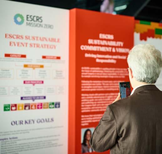
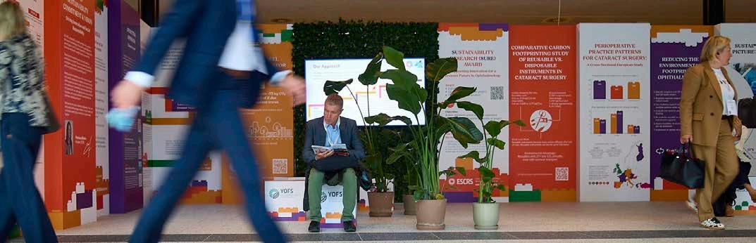
ESCRS members were asked to submit their favourite photos to spotlight sustainability-conscious innovation in ophthalmology, showing how they see sustainability in practice. The top ten photos are showcased at the 2025 ESCRS Annual Congress. Delegates will vote for the top three winners. The contest is part of the ESCRS strategy to promote sustainability awareness, increase diverse representation, and support inclusive education.
ESCRS is providing a childcare service during the 2025 Annual Congress in Copenhagen. The service is available at a cost of €10 per day for registered Congress attendees.
The hours for the service are as follows:
• Sunday, 14 September, 08:00 to 20:30
• Monday, 15 September, 08:00 to 18:00
• Tuesday, 16 September, 08:00 to 15:00
The childcare service is available for children aged 0–12 years only. Children may stay for up to a maximum of 4 hours at any one time. Parents must take their children out of the setting for food and fresh air and are welcome to return to the crèche after 1 hour.
ESCRS has assembled a team of ophthalmologists, data scientists, private equity investors, and clinical researchers to lead an effort to create high-quality, open-access data sets that can spark groundbreaking research in cataract, refractive, and corneal surgery.
The ESCRS Digital Health Special Interest Group plans to leverage the Society’s Digital Research Awards programme and host digital health symposia at the ESCRS Annual Congress to help further this effort. To jump-start this initiative, the group has assembled a project team to build a library of links to existing open-access image data sets relevant to cataract, cornea, and refractive surgery.
“We all know that in clinical medicine, if we want to analyse any intervention or new treatment, we need a lot of data,” said Magdalena Niestrata MD, PhD, who is leading the project group. “The more data we have, the more accurate the results. And what we’ve seen in ophthalmology is that, compared to the posterior segment, the anterior segment has relatively little data available.”
To establish a benchmark, Dr Niestrata and some colleagues conducted PubMed and Google searches to identify publicly available anterior segment image data sets. Of the 26 data sets they found, most were small and incompletely described, and half of the images were from normal eyes. The results of their research were published in a November 2024 article in the Journal of Cataract & Refractive Surgery.
The JCRS article prompted ESCRS to create the project group and establish a central, publicly accessible database for anterior segment data and images. The group hopes to entice researchers to upload their results and contribute to
the collective data pool, thereby enhancing the overall quality and quantity of available data.
To encourage ophthalmologists to use the data set library, the group has created an AI-powered bot that works in multiple languages and searches for data sets in response to spoken commands. “I continually text it, and it sends us alerts when it thinks there is a new article,” Dr Niestrata said. “It doesn’t eliminate the need for us, of course, but there is a lot we can use it to help with.”
Next steps for the project group include enhancing the bot to be more interactive and user-friendly and making the data sets more accessible to a global audience. The team also aims to include more diverse data sets, such as multimodal imaging, topography, biometry, and anterior segment OCT data, to provide a broader range of data for research and analysis.
The library can be found on the ESCRS website on https://www.escrs.org/special-interest-groups/digital-health/public-datasets.
Magdalena Niestrata MD, PhD is a Corneal Fellow at the East Sussex and North Essex Foundation Trust, UK.
Team building is key for success.
Building an effective team is important in any profession, but in ophthalmology it can be a real game-changer for both the practice and a personal career, according to Vincent Qin MD.
“Ophthalmology relies on coordinated care—from the front desk to technicians, optometrists, surgeons, and postoperative follow-ups,” Dr Qin said. “A strong team ensures smooth [handovers] and accurate patient data. Positive patient experiences build your reputation, increase referrals, and strengthen patient loyalty.”
Other advantages of a high-performance team include streamlining administrative tasks, which can reduce stress and burnout, and opportunities to share knowledge on new breakthroughs and technologies, Dr Qin said.
Critical skills for building an effective team, he said, include communication, emotional intelligence, delegation, and organisational and workflow design.
“An effective team is like the ‘optical coherence’ of a practice—it brings all the layers together so you can see the full picture and act with precision. In ophthalmology, it not only improves patient care and practice growth but also elevates your personal brand, career trajectory, and long-term job satisfaction,” Dr Qin added. He presents on team building at the ESCRS Leadership, Business, and Innovation Bootcamp on Sunday, 14 September, 09:00–16:00.
Vincent Qin MD, MBA, MPH, FEBO, SSL(Harvard) practices in Brussels, Belgium.
Clear communication and careful selection needed for a successful ROF IOL implantation.
TIMOTHY NORRIS REPORTS
There's no such thing as a free lunch in optics,” said Professor Paul Ursell, MBBS, MD, FRCOphth at the “Update in Presbyopia-Correcting IOLs” session on Saturday.
During his presentation, Prof Ursell described his clinical experience with partial range of field (ROF) IOLs, underlining how it is all about the trade-off. Trying to enhance one component to lens functionality means reducing something else, he said. In light of this trade-off, it is important to work out the best spot with each patient through assessment, transparent communication and compromise to obtain good results.
Prof Ursell then turned to the new classification scheme that categorises partial range of field (ROF) lenses as narrow, which considers monofocal IOLs; enhanced, which includes some monofocal plus IOLs; and extended, which includes some EDOF IOLs. According to Prof Ursell, this is a fair categorisation while still allowing for some nuances.
All of the lens manufactures are trying their best to differentiate and create innovation, but the principle of no free lunch still holds, he observed. In talking about the optics, he underlined the differences between diffractive and refractive optics designs, the addition of asphericity for optimisation of depth and image quality, and the balancing of light distribution between distant and intermediate. The reason these lenses exist is because trifocal lenses exhibit photic phenomena, Ursell said. The point of ROF is to minimise dysphotopsia while providing some reading vision.
ROF IOLs provide very good distance vision, monofocal-like, and an intermediate vision that would not be possible with a standard monofocal. What ROFs will not deliver is a reading vision as good as trifocal lenses, underlining that there is room for improvement. However, ROFs preserve contrast sensitivity and reduce disphotopsias.
According to Prof Ursell, the reason these lenses are appreciated is mostly for addressing monovision. If you look at all the patient satisfaction surveys and speak to your patients, the common thing is that they want to make their vision better. Looking at all the biometric calculations and papers, 80 to 85% of patients get within half a dioptre, which means 15 to 20% are half a dioptre out. Half a dioptre in reading is not comfortable, Prof Ursell noted. Getting a 100% prescription is not possible, but covering that little error can be important and give the patient spectacle independence.
Choosing the right lens is like choosing a car, Prof Ursell observed. Does it do the job? Will it break down? Is it affordable? Analyse the lens using this method. Patients may not care which brand of lens is implanted, but it still matters.
Patient selection is also very important, he said. According to Prof Ursell, the ideal patient should be a computer user with an active lifestyle who desires to reduce spectacle dependence but is OK with the occasional reading glasses. On the other hand, perfectionists, night drivers and patients with macular pathologies and severe dry eye should be avoided. Patients eligible for ROF lenses should expect an enhanced range and not to be spectacle-free, knowing that an adaptation period is normal and part of the process, he said.
“There's an element of care that's required to be given to people,” Prof Ursell said. “So make sure you've got a team that has the same conversation you're having, that doesn't send mixed messages. Watch out for the red flags, people with unrealistic goals. And just hold people's hands and walk them through.”

Would you like to help shape decisions about ESCRS educational offerings? Are you interested in receiving a free delegate registration to the 2026 ESCRS Annual Congress in London?
If you answered yes to both questions, keep reading!
This year marks the 11th year of the ESCRS Clinical Trends Survey. The survey’s goal is to obtain a wide variety of perspectives on key issues facing ophthalmology today. The ESCRS leadership will use your confidential feedback to determine education needs based on current clinical opinions and practice patterns.
In less than 20 minutes, you can make a meaningful contribution to ESCRS programming; in return, you’ll be entered in a raffle to win one of four free 2026 delegate registrations to the ESCRS Annual Congress. You will also receive a preliminary results report prior to publication.
To participate in the survey, visit the ESCRS Survey Lounge next to the Exhibition Hall entrances of Halls C and E, near the ESCRS Booth & Member Lounge. You can also participate online by following the link below. The survey will close soon, so act now!
Saturday’s Main Symposium did a deep dive into the new ESCRS Guidelines for Cataract Surgery released earlier this year after an exhaustive study and review process. The Symposium also provided an update on the soon-to-be-released ESCRS Guidelines for Refractive Surgery. The guidelines provide practical, evidence-based recommendations to improve care quality and decision making.
ESCRS formed a multidisciplinary guideline panel balanced to minimise potential bias from conflicts of interest. The panel prioritised clinical questions and outcomes according to their importance for clinicians and patients. The guideline-development process used a Grading of Recommendations Assessment, Development, and Evaluation (GRADE) approach and a GRADE Evidence to Decision framework— supported by a team of methodologists.
The panel agreed on recommendations concerning 31 questions for patient pathways for cataract management. Key recommendations of these guidelines include (according to level of evidence):
1. An intracameral injection should be used (e.g., cefuroxime 1 mg in 0.1 mL) at the end of the cataract surgery to lower the risk of postoperative endophthalmitis. (GRADE +++)
2. Topical anaesthesia appears to be the most used anaesthesia technique during cataract surgery, if suitable for the patient. (GRADE ++/+++) For further reducing pain during the cataract surgery, an additional intracameral lidocaine injection can be considered. (GRADE ++/+++)
3. Toric IOLs should be considered in eyes with a degree of corneal astigmatism of 1.0 D or more, with strong evidence for corneal astigmatism above 2.0 D, moderate
evidence for corneal astigmatism above 1.5 D, and may be beneficial above 1.0 D. (GRADE ++)
4. The selection of a specific target refraction highly depends on the selected IOL, expectations, and preferences of the patient. The patient and ophthalmologist should take the shared decision for IOL target selection. (GRADE ++)
5. The primary treatment options for CME after cataract surgery are topical NSAIDs or steroids. However, there is a lack of sufficient evidence to establish the optimal treatment approach for this condition. (GRADE ++)
6. Both conventional cataract surgery (CCS) and femtosecond laser-assisted cataract surgery (FLACS) can be used, as they are both safe and effective procedures. (GRADE +/++). They give comparable visual acuity and refractive outcomes and overall intraoperative and postoperative complication rates. (GRADE +/++)
The complete 275-page PDF document is available online at https://www.escrs.org/escrs-guideline-forcataract-surgery.
The Guidelines for Refractive Surgery have followed a similar methodology to the cataract guidelines, attempting to give the most weight to evidence-based conclusions supported by clinical trials as well as expert opinion.
Some of the provisional recommendations include using PRK for myopia to -6.0 D and hyperopia up to +3.0 D, with astigmatism up to 4.0 D. LASIK is recommended for higher corrections, up to -10.0 D for myopia, +5.0 D for hyperopia, and up to +6.0 D for astigmatism. KLEx could be used to treat myopia ranging from -1.0 to -10.0 D with astigmatism up to 2.0 D (or 5.0 D if cyclotorsion is compensated).


True innovation comes from good research. That’s the impetus for the ESCRS Pioneer Research Award (PRA).
The PRA aims to support and encourage independent clinical research in the field of cataract and refractive surgery. It can fund a variety of new initiatives, which may include—
• A novel research idea for the development of clinical trial studies;
• A non-interventional or observational study; or
• A natural history/epidemiological study.
An award of €25,000 is available for one grant. The competition is open to ophthalmologists up to the age of 45 (at the application deadline). Eligible participants must hold a full-time clinical or research position at a clinical or academic centre within the European region.
Applications are due by 31 October 2025.
Memorable moments from the ESCRS 2025 Congress, capturing key connections and collaborations.


Every year, the ESCRS features a session on ORBIS International, the international non-governmental organisation dedicated to the prevention of blindness and blinding eye diseases in underserved areas. The ORBIS symposium takes place tomorrow at 11:15 in Room D1.
This year’s session considers gender issues and the current efforts to improve access to women and girls around the world. The session also includes a discussion of the use of simulation ophthalmic surgery training in sub-Saharan Africa. Another presentation addresses the frequency and causes of distance vision in adults living in the Brazilian Amazon region.
Room D1
Monday, 15 September 2025
11:15–12:30 CEST
Join us tomorrow at the ESCRS Independent Medical Education Forum on Toric IOL surgery and take a deep dive into astigmatism management — from accurate diagnostics to intraoperative precision and management of postoperative surprises — with real-life case examples and insights from the latest ESCRS Clinical Trends Survey.
Room B2–M5, Hall B2
Monday, 15 September 2025
09:00–10:00 CEST
Chaired by Professor Filomena Ribeiro and accompanied by faculty Drs Daniel Chang, Andrzej Dmitriew, and Nino Hirnschall, the programme features expert talks, interactive live polling, and a closing panel discussion aimed to help you optimise preoperative diagnostics and manage toric lens power calculations and intraoperative alignment. Learn from the experts how to improve patient outcomes with toric IOLs.
Béatrice Cochener-Lamard chairs this international session featuring the very latest information on the use of femtosecond lasers in ophthalmic surgery. Look forward to hearing about the arrival of robotics in cataract surgery from Dagny Zhu, FLACS for complex cases from Soon Phaik-Chee, IOL power adjustment from Ron Krueger, capsulotomy-fixated IOLs from Gerd Auffarth, new uses in refractive surgery from Marcus Ang, and laser-assisted anterior keratoplasty from Mayte Ariño-Gutierrez.
Hall A1
Monday, 15 September 2025
08:15–10:15 CEST