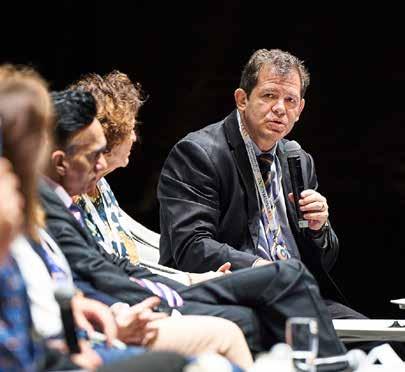
All Eyes on Innovation
Watching out for obstacles and opportunities THE INNOVATION


Watching out for obstacles and opportunities THE INNOVATION
It is my great pleasure to welcome you all to this year’s Congress in the beautiful city of Copenhagen! We opened with an action-packed Friday featuring iNovation Day, our Subspecialty Days (Glaucoma, Cornea, and Paediatrics), and the second edition of the Global Refractive Summit. This dynamic start has set the stage for the days ahead.
Throughout the meeting, you can look forward to an outstanding scientific programme covering every aspect of ophthalmic surgery. Each day will feature an unmissable Main Symposium, and we will have two prestigious named lectures: the Binkhorst Lecture, delivered by Dr Thomas Kohnen on Saturday, and the Heritage Lecture, presented by Dr Robert Osher on Sunday.
The Clinical Research Symposia, taking place today, are the scientific heart of the conference. A range of topics will be updated throughout the series, starting with “Replacing/ Regenerating Corneal Endothelium”, followed by “New Insights on the Origin and Treatment of Cataract”, “IOL Materials and Design”, and “The Power of Registries in Cataract and Refractive Surgery Research”.
This year, we celebrate a remarkable milestone—the 30th anniversary of femtosecond laser application in ophthalmology, a breakthrough that has profoundly shaped our field. Don’t miss the chance to explore the latest innovations in this technology at the Femto Laser Experience, located next to the ESCRS Member Lounge.
Education and research are at the heart of this Congress. We are proud to introduce a cataract surgery simulation training lab with smart learning modules, offering hundreds of delegates hands-on experience—completely free of charge. In addition, we are hosting a workshop on scientific writing, a session on critically reading research articles, and the brand-new World Café, where colleagues will discuss real clinical cases in an interactive setting.
Our goal is to create a well-balanced event rich in science, while also providing time for connection and reflection. For the first time, we present the Build Our Sustainable Society (BoSS) 5k run, alongside dedicated relaxation spaces where participants can recharge. Within the BoSS track, don’t miss the workshop on unconscious bias. We are also introducing highly topical sessions, such as “Cataract Surgery and Dementia”, and a unique networking event on sports and vision—bridging clinical expertise with broader perspectives on health and lifestyle.
The ESCRS Arena will once again be where science, energy, and debate converge. Each morning begins with Surgical Pearls—concise, practice-changing insights from leading experts. Later in the day, the Arena will host lively debates and discussion forums on topics including the mixing and matching of simultaneous-vision IOLs, strategies for managing astigmatism, the integration of artificial intelligence into daily practice, new surgical options for presbyopia, and the latest evidence on KLEx.
Thank you for joining us for what promises to be an inspiring and unforgettable Congress. Let’s learn, share, and celebrate together here in Copenhagen!

Filomena Ribeiro MD, PhD, FEBO President of the ESCRS
Each year, young ophthalmologists are invited to participate in the John Henahan Writing Prize by responding to an essay prompt. This year’s winner is Jamal Atamniy MD, an ophthalmologist at Eye Clinic, Augenspezialist Wien, Dr Jamal Atamniy, Vienna. He receives a €500 bursary and a specially commissioned trophy, awarded during the 2025 ESCRS Congress.
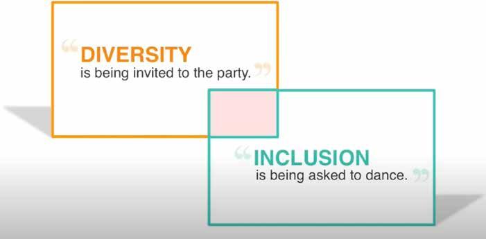
Applicants responded to the following prompt: Diversity, equity, and inclusion (DEI) programmes, however well-intentioned, stir a variety of responses in the corporate and political worlds and in the scientific and medical spheres. What DEI and unconscious bias issues are present in the current culture of ophthalmology training, practice, and clinical research? What are the potential benefits of addressing these issues for patients and ophthalmologists? What kind of meaningful changes need to happen to move beyond ‘talking the talk’ to ‘walking the walk’?
Indeed, diversity, equity, and inclusion (DEI) concepts have gained momentum across various fields. However, in the medical field, particularly in ophthalmology, they remain sporadically applied, poorly understood, and often seen as peripheral to clinical brilliance. Although well-intentioned, DEI programmes evoke varying responses within the medical community. Nonetheless, beneath the surface of this debate, a fundamental truth remains: unconscious bias and inequality continue to influence ophthalmology training, practice, and research, often to the disadvantage of clinicians and patients.
Diversity, equity, and inclusion in ophthalmology today
Traditionally, ophthalmology has been regarded as an exceptionally exclusive and competitive speciality. Securing a place in training remains heavily biased, with many individuals from lower socioeconomic backgrounds, underrepresented ethnic minorities, or without academic or family ties in medicine facing significant obstacles. When investigated, gender disparities among mentorship programmes in ophthalmology have shown that female trainees, compared to their male counterparts, reported significantly lower satisfaction. Furthermore, lower income, decreased rates of goal achievement, and less support in achieving these goals were among the concerns raised. Unconscious bias continues to affect recruitment decisions, assessments, interactions with patients, and even who gets access to prestigious cases. Research indicates that in surgical specialities, women experience less autonomy in surgeries compared to their male counterparts, even though their levels of expertise are similar. Similar disparities are also seen in academic authorship and research funding allocations. Racial and ethnic minorities are underrepresented as lead or senior authors in
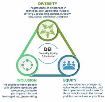
Adopting a DEI approach is not just a moral duty for ophthalmologists. It’s crucial for the long-term sustainability and quality of the field. Research has consistently shown that diverse teams make better clinical decisions, drive more innovative research, and achieve higher patient satisfaction. Figure 1 underscores how diversity, equity, and inclusion comprise a comprehensive institutional culture approach. Making trainees feel valued, supported, and mentored increases the likelihood of staying in the field, flourishing, and making a meaningful contribution. DEI promotes a sense of belonging, which helps reduce burnout—a growing problem in ophthalmology—and improves mental well-being.
Equity in care should not be a luxury for patients; it should be a given. Tackling unconscious bias can result in increased trust, earlier diagnoses, and improved treatment adherence. Additionally, involving more diverse patients in clinical trials ensures that healthcare innovations are safe and effective for all populations.
From talk to action: What must change?
While raising awareness is crucial, the real priority should be taking concrete, measurable steps to drive meaningful change.
1. Transparency in recruitment and promotion
Implement a blind review process that can highlight the true qualities of training applicants in hiring and promotion decisions. This should be supported by accurate diversity data reporting and equity audits conducted by institutions to ensure oversight of the process.
2. Programmes for mentorship and sponsorship
Engage senior ophthalmologists with expertise in identifying and addressing unconscious bias to set up targeted mentorship for minority groups. Additionally, sponsorship programmes should be created to motivate mentors to champion their trainees.
3. Research practices with a focus on inclusivity
Develop practical routes for publishing and co-authoring with clinicians from underrepresented minority backgrounds. Meanwhile, promote the use of DEI metrics in research designs for clinical trials to recruit diverse participants.
4. Compulsory DEI and bias training
Incorporating DEI education into curricula leads to meaningful, long-lasting change. This is necessary to tackle the deeply ingrained unconscious bias in our field. Regular assessment intervals are crucial for consistency and should involve clinical mentors and educators.
5. Environmental and policy reform
Institutional involvement is pivotal in facilitating flexible work arrangements, supporting caregivers, and employing proactive strategies to dismantle exclusive networks that restrict opportunities for minority groups. Invest in
physical and cultural initiatives that promote inclusivity and implement anonymised and accessible feedback mechanisms. Biases and inequities in ophthalmology can be mitigated through DEI. This concept should not be dismissed as merely a quota that institutions must fulfil. Its implementation should stem from our collective desire as medical professionals to provide our patients with the best care. It goes beyond fostering a more diverse field and has clinical, educational, and ethical implications. Let us remove the barriers that prevent talent and compassion from reaching their full potential. It is high time we stop simply ‘talking the talk’ and begin ‘walking the walk’ with commitment, courage, and transparency.
The complete version of the essay with citations and diagrams will appear in EuroTimes.
We’re willing to bet most eye care professionals don’t realize just how prevalent Demodex blepharitis is.
In fact, ~54% of eye care patients in Europe may have Demodex blepharitis (DB).1
Reference: 1. Data on File. Tarsus Pharmaceuticals, Inc.
LEARN HOW DB CAN FLY UNDER THE RADAR AT


Ophthalmologists and researchers (MD and/or PhD) as well as experienced ophthalmic nurses are encouraged to apply for the new ESCRS Sustainability Research (SURE) Award, which will fund projects that investigate meaningful, practical ways to promote environmental responsibility in ophthalmic care.
Two awards will be granted; each award will provide up to €10,000 per project. Applicants must be active ESCRS members at the time of application and must hold a current full- or part-time clinical or research position at a clinical or academic institution. Early-career researchers and young ophthalmologists are especially encouraged to apply.
The application period opens 28 July and closes 01 November. Award recipients must submit an article to a peer-reviewed journal within six months after the research period concludes. The article should be made open access if accepted and submitted to the Journal of Cataract & Refractive Surgery in the first instance.
ANDREW SWEENEY REPORTS
While the ESCRS may be a society dedicated to cataract and refractive surgery, the Annual Congress highlights many other areas of ophthalmology. Cornea Day, organised jointly with EuCornea, has become a favourite event preceding the main meeting, and more than lived up to expectations.
José Güell MD, PhD, head of the Cornea, Cataract, and Refractive Surgery Department at IMO Grupo Miranza in Spain and former president of both the ESCRS and EuCornea, said he was excited to be returning to another day of ‘cornea-copia.’
“I’m 65 years old, so as you can imagine, I’ve been to many meetings. Cornea Day at ESCRS is always a major attraction for me. If it’s such an attraction for me, it should be fascinating for anyone interested in cornea. If you’re interested in the cornea field, then this is the most exciting and fruitful part of ESCRS 2025 for you. It’s six hours of brainstorming on the latest cornea news,” said Professor Güell.
“We selected several topics based on the strong experience of cornea surgery specialists. No topics were repeated from the previous year; even if, say, keratoconus was covered the year before, new information would be provided.”
Prof Güell chaired “New Developments and Updates in Keratoplasty” with Prof Claus Cursiefen. The two-hour session provided valuable insights from several cornea specialists on topics including paediatric corneal transplantation, corneal regeneration (involving stem cells, hydrogels, and 3D bioprinting), deep anterior lamellar keratoplasty (DALK) techniques and indications, and more. Three separate discussions were also part of the symposium.
The session is just one of six symposia on Cornea Day, covering topics such as cataract patients with cornea pathology, infection of the ocular surface (including paediatrics), keratoconus and cross-linking updates, and one session dedicated to ‘the eternal battle between glaucoma and the cornea.’
“There’s something for everyone, whether they’re a cornea specialist or not,” he said.
Get funding for projects to promote environmental responsibility in ophthalmic care.
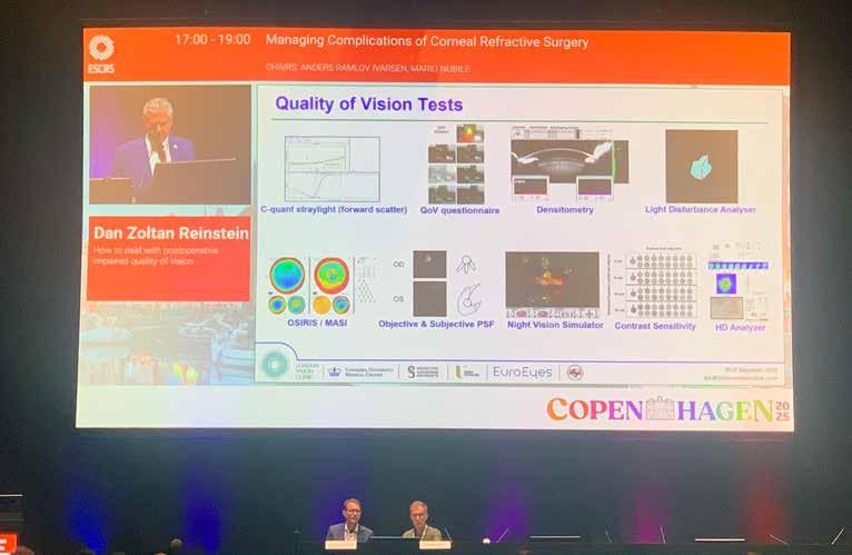
Prof Güell not only presided over the Cornea Day symposium—he’s participated in several of them. His presentation, “Corneal Surgery for Refractive Surgery Complications”, came during the “Managing Complications of Corneal Refractive Surgery” symposium.
In the presentation, he noted corneal complications in refractive surgery today are rare, but “when present, are quite challenging to deal with,” adding that he draws on his considerable experience when highlighting what to do in these situations.
“In cases involving opacity that is not significant and centrally located, many (with different degrees of irregularities) can be managed with rigid corneal or scleral contact lenses and corneal cross-linking. In other cases, an interventional procedure must be planned on a case-by-case basis,” Prof Güell said.
“Topo-guided transepithelial photorefractive keratectomy with or without corneal collagen cross-linking (TE-PRK +/- CXL) might be considered in selected cases. In the most difficult situations, DALK or even penetrating keratoplasty (PK) are the only options.”
Best practices and close cooperation
Cornea Day is made possible by the strong cooperation between ESCRS, EuCornea, and doctors like Prof Güell who are closely involved with both societies. He believes that, while ESCRS specialises in cataract and refractive surgery and has a broader scope than the more specialised EuCornea, the possibilities for working together are considerable.
Collaboration is especially important as new and increasing threats emerge in the cornea sphere, including changing profiles, pathologies, and characteristics of diseases that have emerged in regions they previously were not present. Challenges like these require closer cooperation.
“Pathogens change—they become resistant to treatment—and it’s never been easier to move around the world and take them from, say, India to Spain. Fungal infections are becoming much more common too, particularly in East Asia,” Prof Güell said.
“The ESCRS is closer to more general cornea subjects, like transplants and cross-linking, while EuCornea is more specialised. I’ve been involved in bringing these two societies together for years; the cooperation between us and our activity together is great.”


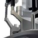
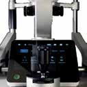
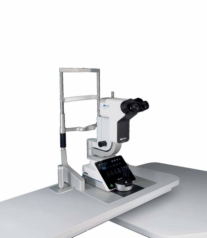
Haag-Streit invites ESCRS delegates to join its distinguished faculty for a one-hour Satellite Symposium on how innovations in slit lamp microscopy can address emerging & future trends in eyecare health. The Symposium will also address the benefits of the new Elara 900, including its superior clinical observation, improved efficiency & optimized ergonomics.
Heritage Lecture traces the origins of ophthalmic video.
Ophthalmic video pioneer Robert H Osher MD will present the 2025 Heritage Lecture, entitled “Video – Can’t Live Without It!”
Dr Osher is well known to ESCRS, having organised the annual Video Symposium since the early days of the European Intraocular Implant Council (EIIC), the predecessor of the ESCRS. He has received nearly a dozen awards in the ESCRS Video Competition, including Overall Winner twice. His courses on complication management have earned the Educators Award and the Kelman Award from ESCRS.
Noting his lecture is “guaranteed to entertain with minimal educational value,” Dr Osher traces the origin of ophthalmic video with priceless clips of the pioneers and groundbreaking changes in cataract surgery. He has also promised to present some humorous clips “to keep the audience awake!”
Dr Osher traces his love for video back to when he introduced the first video symposium in the early 1980s. He has served as the editor of the Video Journal of Cataract, Refractive, & Glaucoma Surgery (vcrgs.com) for 41 years, offering every issue free of charge to all members of ESCRS. Dr Osher said he watches every video from ASCRS, ESCRS, AAO, and the other major organisations to develop content for the VJCRGS. Surgical videos from ophthalmologists around the world demonstrating excellent skill and sophisticated technique are hand-picked by a distinguished editorial board that includes Drs Graham Barrett, Michael Snyder, George Waring IV, and Ike Ahmed.
I’m so lucky to be a member of this great Society. I’ve learned so much and have made so many lifelong friends.
“We feature an international cast of surgeons who show innovative techniques, challenging cases, and complication management, as well as award-winning videos and named lectures,” he said. “We avoid any advertising or promotional content [that interferes] with the viewer’s education.”
Each quarterly issue of the VCRGS offers at least an hour of high-quality education for the anterior segment surgeon. For example, outstanding corneal surgeons Kavitha Sivaraman and Jack Parker moderate the “Surgical Cornea” issue (Vol 40, Issue 2), now available on the journal website.
Dr Osher credits many of his ideas and awards, including the Lifetime Achievement Award from AAO and ESCRS, to his European colleagues.
“I’m so lucky to be a member of this great Society. I’ve learned so much and have made so many lifelong friends. I just hope a few will remain after this unconventional lecture!” he joked.
Dr Osher served his residency at the Bascom Palmer Eye Institute in Miami, Florida, US, and completed three fellowships in Miami and at the Wills Eye Hospital in Philadelphia, Pennsylvania, US. He is Professor of Ophthalmology at the College of Medicine of the University of Cincinnati and Medical Director Emeritus of the Cincinnati Eye Institute, Ohio, US. His practice has been limited to cataract and implant surgery by referral for 38 years.
The Heritage Lecture is scheduled for Sunday at 10:30 in Hall

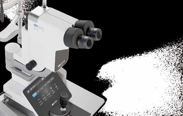
Combining the proven power of Scheimpflug imaging with the precision of ultra high-resolution OCT enables the detection of previously unseen corneal pathologies with unprecedented clarity.
Diagnose earlier. Treat smarter. Care deeper.
EPITHELIUM
BOWMAN’S LAYER
STROMAL LAYER
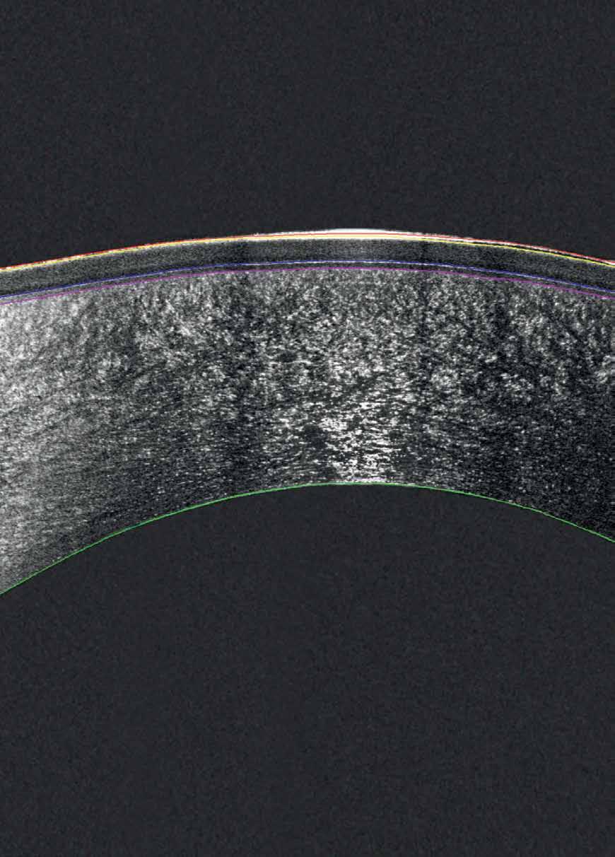

The Pentacam® Cornea OCT can increase the confidence that your diagnosis is correct.
Lunch Symposium I Room A3 Sat, 13 September 2025, 13-14 h
Integrating key modalities for a complete refractive work-up ESCRS 2025 | Booth C2.017 The availability of products and features may vary by country.
Cash reserves and a need to innovate drive changing landscape.
HOWARD LARKIN REPORTS
After subdued growth since 2022, investments in ophthalmology-related businesses rebounded in the first half of 2025, Citi investment banker Scott Bardo told ESCRS 2025 iNovation Day attendees. Private equity mergers and acquisitions for all sectors were up 47% over 2024, while overall M&A investments including strategic investments were up 29%, totalling more than $2 trillion for the period.
Many large ophthalmology deals were concluded in 2021 and 2022, but higher interest rates put a damper on deals in 2023 and 2024. Initial public offerings showed a similar pattern, slacking off after a flurry of activity in 2021, suggesting capital grew tighter over the period.
The environment is now shifting thanks to growing cash reserves and a need to innovate. Improved efficiency in manufacturing, distribution, digital technology, and integrated delivery systems may drive future financial growth.
“Strategics, or public companies acquiring ophthalmic businesses, were very, very low during the pandemic,” although a lot of investment went into improving ongoing operations, Bardo said. “What we are seeing now are very robust balance sheets poised to grow,” which is yielding more M&A investment this year, a trend likely to continue.
Since COVID, market prices for ophthalmology-related businesses, which historically have traded higher than other medical technology firms, have dropped relative to med-tech overall and the markets generally. “Top-line growth has been
harder to come by, and CEOs have done what they can on innovation, really driving operational efficiency, optimising portfolios, and reinvigorating growth with M&A,” Bardo said.
One result has been a growth in vertically integrated ophthalmology companies. These include companies from the optometry industry moving into diagnostics, dry eye treatment, and even buying surgical clinics, Bardo said. He sees further convergence of products, devices and services as ways to create end-to-end value chains. Integration of digital technologies will be central to this industry transformation.
Bardo also foresees portfolio optimisation, in which firms divest products and services not related to their core focus. An example would be selling off endoscopic surgery devices or contact lens businesses to concentrate on prescription drugs or service delivery.
“There will be a lot more deal activity in this area,” he said. “Growing cash piles, dwindling growth, and valuation dislocation create a fertile environment for M&A. Companies that are able to integrate operations and digital technology are likely to succeed.”


Take control of your patients’ ocular surface with Lacrifill - the hyaluronic acid canalicular gel that maintains natural lubricating tears for six months with a single treatment.
✔ FDA-cleared
✔ CE-marked
✔ One syringe, one patient


From VHS to digital, video continues to support ophthalmic education.
SEAN HENAHAN REPORTS
Agood coach can help turn an average athlete into a star. The same is no doubt true for ophthalmic surgery. Perhaps more than in any other specialty, ophthalmologists hone their skills with the help of videos. One of the best known of these is Cataract Coach at cataractcoach.com.
Uday Devgan MD is the founder and producer of the site, which releases a new five-minute instructional video online every day and has been doing so for more than seven years.
“We never miss a day,” he said. “The format is basically five minutes of video since people don’t have a lot of time. The US is the biggest audience, followed by India, Brazil, and the UK.”
In addition to the website, the videos appear on all the main social media platforms—YouTube, LinkedIn, Facebook, Instagram, etc.—and viewers can sign up for email updates.
“We have 2.5 million views per month, with half on mobile devices,” Dr Devgan explained. “We also do the leading podcast in ophthalmology, covering cataract and refractive topics along with cornea and glaucoma—the entire anterior segment.”
The video cases come from many sources, including Dr Devgan himself. He uses templates for videos, conducts the voiceovers, and employs a team to help with the uploads.
“At first, it was all my videos,” he said. “Now I get 50–60 videos [each] week. I go through all of them and choose the best. There is amazing stuff from around the world.”
The site has no sponsors and avoids commercialism, he noted.
“I don’t want to risk our credibility. I’m not giving you an infomercial plugging a product. Viewers are smarter than that,” he noted. “I want viewers to learn something. The trick in the video is not which lens to use, but the technique itself.”
Cataract Coach came to Glaucoma Day on Friday in the video-based “Legends of the Lens: Navigating Cataract and Glaucoma Together”. Dr Devgan’s Cataract Coach Live session reviewed “Winning Strategies for Tough Cases”, with videos covering complicated cases involving subjects such as cataract and glaucoma, pseudoexfoliation, and MIGS.
Dr Devgan is also hosting “Best of Cataract Coach” today at 11:00 in Auditorium 10.
“There is so much to learn, and we all have to share our knowledge,” he said. “This was designed to provide fast-paced, interesting, challenging cases. It is like a sports highlight reel, only the good stuff.”
Kudos to Dr Osher
Dr Devgan cited Dr Robert Osher as a primary inspiration.
“He is the best: the godfather of ophthalmic surgery and a number one podcast guest. He has had the most impact on me and my career,” Dr Devgan reflected. “I first learned surgery as a resident by watching his videos. In the 1990s, there was no one to teach you phaco chop, so I had to learn by watching his videos on VHS tapes.”
Dr Devgan stressed that his favourite part of coming to the ESCRS Congress is interacting with fellow ophthalmologists.
“It’s all about meeting people in person. You can’t do that online. You want to shake hands, break bread, take some selfies. Also, since Europe isn’t regulated by the FDA, we often give a preview of what’s coming to the US.”
Green and clean
Innovative eco-design saves resources
Less waste and lower disposal costs
Swiss made Medicel quality


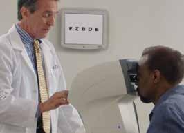
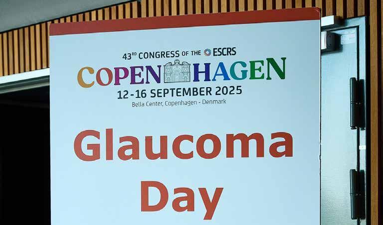
Glaucoma Day attracts more than just glaucoma specialists.
With the advent of minimally invasive glaucoma surgery (MIGS), some cataract surgeons are expanding their practices to include glaucoma care. This year’s Congress accommodates this increasing interest with several learning opportunities.
On Friday, Glaucoma Day started with an introduction from Ingeborg Stalmans MD (President of the European Glaucoma Society) and Ziad Khoueir MD (consultant ophthalmologist at Beirut Eye Hospital, Lebanon), highlighting the positive collaboration between the European Glaucoma Society (EGS) and the ESCRS.
With a nod to Denmark’s Shakespearean connection, “Through Hamlet’s Lens” explored many of the controversies in glaucoma today through a look at clinical cases in glaucoma management. A subsequent session addressed innovations in laser, surgery, medication, and AI, polling attendees on what they think glaucoma management will involve in 25 years. Another session reviewed the best uses of MIGS, where Ike Ahmed MD presented on the perils and pitfalls of integrating MIGS into ophthalmic practice. A subsequent session covered the corneal implications of glaucoma surgery, with an emphasis on protecting the endothelium and optimising the ocular surface. The final session (“Legends of the Lens: Navigating Cataract and Glaucoma Together”) lived up to its billing with video presentations from two legends: Uday Devgan MD, founder of cataractcoach.com, and MIGS pioneer Dr Ahmed.
“Glaucoma Day also addressed a lot of controversies about surgical innovations,” said Luis Abegão Pinto MD, Head of the Glaucoma Clinic, Department of Ophthalmology, Lisbon University, Portugal. “We had cornea people, die-hard glaucoma surgeons who rarely
[perform] cataract, and cataract surgeons who rarely [perform] glaucoma surgery. We want to show that we can work as a team, complementing, not competing. We have to realise we are working towards a common goal of providing optimal care—this could mean a trabeculectomy for some, for others, it might be phaco MIGS, sometimes it might mean not doing anything.”
He also noted 2025 has been very good for glaucoma as a whole. A comprehensive update of the EGS Terminology and Guidelines for Glaucoma was launched at a recent meeting in Thessaloniki, Greece. This followed the release of an EGS book on surgical innovation, which examines the evidence base for many new devices and techniques in development. Meanwhile, the 11th World Glaucoma Congress (WGC) in Honolulu announced a consensus on angle closure, redefining the terminology based on all available evidence.
“If you weren’t in Thessaloniki for the EGS Members’ Meeting or Hawaii for the WGC, you can be sure these two big landmark studies will be discussed in Copenhagen,” Dr Pinto said. “Glaucoma Day offers attendees a crash course on everything happening in our field.”
Outside of Glaucoma Day, Congress attendees can participate in relevant surgical instructional courses during the meeting. In addition, free paper sessions on Sunday (Environmentally Responsible Eye Care, Hall B3, Podium 4; Advances in Glaucoma Surgery, 14:00, Hall B3, Podium 4) and Monday (Contemporary Surgical Interventions in Glaucoma, 16:30, Hall B3, Podium 2) cover a wide range of glaucoma-related topics.

Leadership, Business, and Innovation session seeks to enhance presentation skills.
Even for the most skilled and brilliant physician, building influence and finding opportunities requires effective communication. And polishing presentation skills can help, according to Artemis Matsou MD.
“Some of the most meaningful opportunities in my career—whether speaking at an international conference, joining a research collaboration, or being invited to teach—have come from moments when I stood up and shared my work effectively. Ophthalmology is a fast-moving specialty, and presenting well isn’t just about showcasing results. It’s about telling a story that connects with your audience,” said Dr Matsou.
“Good presentation skills help you make your science memorable, inspire confidence in your expertise, and leave the kind of impression that leads to new roles, partnerships, and recognition in the field,” she added. Dr Matsou will share her insights and tips on effectively presenting at the ESCRS Leadership, Business, and Innovation Bootcamp on Sunday, 14 September from 09:00 to 16:00.
“I’ve seen (and made!) my fair share of presentation mistakes over the years, the most common being overcrowded slides, reading every word on the screen, and forgetting to tailor the talk to the audience in front of you,” Dr Matsou told EuroTimes
“Another pitfall is diving straight into the data without setting the scene, which makes it harder for the audience to understand why the work matters. The best way to avoid these is to think of your talk as a journey: start with a clear purpose,

build a logical flow, use visuals that support rather than duplicate your words, and rehearse enough so you can focus on connecting with your audience rather than surviving the clock.”
David Lockington MBBCh, PhD, the current president of UKISCRS, will also present at the bootcamp. “Great leadership involves effective transfer of knowledge to empower action. Doctors are regularly trained to improve communication with patients, but never with their peers, so the LBI committee wanted to ensure ESCRS delegates address this training gap, whether young or established.
“Effective presentation skills ensure you are competent and credible, leading to greater confidence on the podium. If you can be memorable—and brief—the impact you leave on your audience will open opportunities for collaboration and career advancement,” Professor Lockington said.
“In our symposium, we will share techniques to clarify your talks, use audiovisuals appropriately, and [handle] Q&A effectively. Using body language and humour to elevate your talk takes practice, and we will demonstrate how to [translate] this into prize-winning performances. Practice makes perfect, and as I always say—my best ad-libs are my most practised.”
Effective presentation doesn’t end at the podium. Prof Lockington said communicating both before and after a presentation on social media can amplify its impact. “Before the event, share the good news [about] getting this opportunity, and include others involved in your session. During the presentation, bribe some friends to sit at the front and take some photos for you during your talk or beside your poster—and return the favour for them. Then post the picture with a short summary of your main learning points on LinkedIn or Instagram. Doing this promptly may even lead to future contacts with delegates at the event or in [its] immediate aftermath.”
Başak Bostancı MD will also present.
Artemis Matsou MD, FEBO, MRCP(UK) is a consultant ophthalmic surgeon at Queen Victoria Hospital, East Grinstead, UK.
David Lockington MBBCh, BAO (Hons), FRCOphth, PhD is a consultant ophthalmologist at Tennent Institute of Ophthalmology, Glasgow, UK.
Başak Bostancı MD is associate professor of ophthalmology at Bahçeşehir University School of Medicine and surgeon in Dunya Goz Hospital, Istanbul, Turkey.


Swept Source OCT biometer with high-speed acquisition and excellent penetration in dense cataracts. Fully automated bilateral measurement, including Placido ring-based topography – completed in under 60 seconds with no manual input required.

Biometry. Fast. Automatic. Complete.
Capture axial length, keratometry, ACD, lens thickness and topography in one scan. Fully automated – no manual alignment needed. Consistent, reproducible results for both eyes in under 60 seconds.
Smart IOL choices. Consistent outcomes.
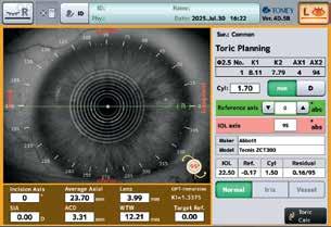
Comprehensive IOL calculation with multiple formulae and toric planning – including post-refractive cases. Compare up to eight lenses for confident decisions in routine and complex cataract surgery.
Using AI for a safer and more effective refractive surgery.
TIMOTHY NORRIS REPORTS
Per the proverb popularised by Benjamin Franklin, an ounce of prevention is worth a pound of cure. During the “Management of Complications in Corneal Refractive Surgery” symposium held on Cornea Day, Renato Ambrósio Jr, MD, PhD shared his expertise in the prevention and management of post-LASIK corneal ectasia.
“The purpose is to define Enhanced Ectasia Risk Assessment (E²RA),” Professor Ambrósio explained. “This concept goes beyond, not over, topography for detecting mild or subclinical cases, moving instead toward an integrated characterization of ectasia susceptibility, considering multimodal diagnostics.”
Multimodal imaging includes 3D Scheimpflug tomography, OCT-based epithelial and stromal mapping, and in vivo biomechanical assessment.
Thanks to the improved diagnostics and surgical technology now available, cases of postoperative corneal ectasia have dropped dramatically from 0.660% to 0.033% of operated eyes. However, it is still one of the most feared complications of refractive surgery.
As a biomechanical decompensation, ectasia arises from both intrinsic susceptibility and extrinsic impacts. The intrinsic first hit reflects the cornea’s genetic, structural, and biomechanical susceptibility, he said. In contrast, the second hit involves extrinsic impacts such as flap creation, ablation depth, or environmental stressors like chronic eye rubbing.
Thanks to a multimodal imaging approach, it is possible to identify these markers before surgery, providing the surgeon with all the tools and knowledge to fine-tune surgery and the patient with valuable information to lower the risk of postoperative onset of corneal ectasia.
According to Prof Ambrósio, this can be obtained by combining multimodal diagnostics with patient-specific factors such as age, family history, and behavioural habits, with a careful consideration of flap and ablation related to corneal thickness and patient age.
All the data can be integrated and assessed case by case thanks to the AI program BEES, a software that uses both the BrAIN (Brazilian Artificial Intelligence Network in Medicine) technology and the Relational Tissue Altered metric. Given the wealth of diagnostic data generated, Prof Ambrósio said applied artificial intelligence has become indispensable.
Postoperative ectasia management can be individualized and tailored to each patient by using corneal
cross-linking, intracorneal ring segments, phakic IOLs, and, in selected cases, custom therapeutic surface ablation enhancements. In this strategy, black box thinking becomes essential, said Prof Ambrósio, explaining how every complication provides an opportunity to learn and refine predictive models, integrating multimodal data with the AI for increased precision.
Prof Ambrósio’s session highlighted how an understanding of corneal ectasia has evolved beyond standard diagnosis and imaging. Attendees were able to leave with practical, evidence-based tools to refine screening, reduce risk, and improve outcomes, not only for laser vision correction procedures, but also for phakic IOLs and refractive cataract surgery.
“This journey—from the BAD-D display to BAD-Dv4, the Tomographic Biomechanical Index (TBI), the Relational Tissue Altered (RTA), and now BEES—represents a paradigm shift in patient safety,” he said.
Ambrósio Jr, MD, PhD is Adjunct Professor of Ophthalmology at the Federal University of the State of Rio de Janeiro, Brazil.


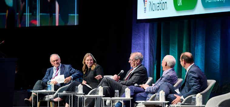
Demographics, AI, reimbursement, and research among top trends driving ophthalmology.
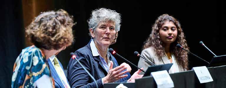
European ophthalmology is being reshaped by shifting demographics, rising patient expectations, and rapid advances in AI and digital health, according to Filomena Ribeiro MD, PhD, ESCRS president, who participated in the Global Ophthalmic Trends panel discussion at iNovation Day.
“Over the past few years, the field has moved from incremental innovation to a more transformative approach, with technology, sustainability, and equity of access now at the centre of the conversation,” she said.
Engaging representatives from regional and national ophthalmology societies, the iNovation programme focused on taking a proactive approach, Professor Ribeiro added. “We are bringing together the voices that can turn innovation into impact—defining how ophthalmology will look in the next ten years.”
Exciting though these transformations are, the field also faces significant challenges—such as declining reimbursement by both government and private insurers—that are affecting how and which services are delivered, Stephen McLeod MD said.
“Practice consolidation has been a significant factor over the last few years, largely driven by economic pressures related to increased practice costs in the face of stagnant or declining physician and practice reimbursement,” Dr McLeod explained. “The post-COVID inflation surge has never been fully absorbed into any meaningful adjustment to practice reimbursement, and costs continue to rise. Inevitably, this means that physicians are increasingly strained to meet their obligations to patients and are forced into difficult decisions regarding the services they can sustainably offer and the
range of insurances they can absorb.”
Recent cuts in US government funding for ophthalmic research, as well as a proposal to merge the National Eye Institute (NEI) with other neurology-related fields, are major concerns, Dr McLeod added. “Eliminating NEI as an independent institute threatens to dismantle the progress made since its establishment by Congress in 1968. The Academy has urged Congress to continue its longstanding history of bipartisan support of NEI.”
Global ophthalmology must step up to meet these challenges, Prof Ribeiro said. “While reimbursement pressures and reduced research funding present ongoing challenges— particularly as cuts in the US reverberate globally—Europe’s collaborative research networks, strong academic-industry partnerships, and cross-border investment offer a pathway to mitigate these constraints and maintain momentum in innovation. The aim is to identify practical, scalable solutions that will shape the next decade of eye care.”
Filomena Ribeiro MD, PhD, FEBO is ESCRS president.
Stephen McLeod MD is CEO of the American Academy of Ophthalmology.


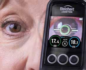


Experience the all-new Reichert® Tono-Vera® Tonometer featuring the patented, auto-measuring ActiView™ Positioning System, for fast, e ortless, objective, and repeatable rebound tonometry measurements. For nearly two centuries, Reichert has empowered a more e cient exam and shared in your passion to deliver exceptional patient outcomes.


Pioneer discusses the great achievements in refractive surgery and the future ahead.
LAURA GASPARI REPORTS
More than thirty years after the first excimer laser procedure, refractive surgery is now safer and more effective, while continuing to develop and improve, according to Marguerite McDonald MD.
Professor McDonald is a pioneer in the field, being the first eye surgeon to perform an excimer laser procedure in the late 1980s, and she describes modern refractive surgery as very exciting. “We are now both corneal and intraocular. There is something for virtually every patient,” she said.
“The development of the femtosecond laser was also a landmark in ophthalmic history, and it has changed the way we practice medicine.”
In the earliest days of refractive surgery—with keratomileusis, keratophakia, and radial keratotomy— surgeons were happy to get most of their patients to see 20/40 or better uncorrected. However, Prof McDonald said things have changed a lot. Published data now report
The development of the femtosecond laser was also a landmark in ophthalmic history, and it has changed the way we practice medicine.
20/15 and even 20/10 uncorrected results, with safer and more accurate procedures each year. The femtosecond laser improved the safety of LASIK and made KLEx possible. Even if EVO ICL procedures are more invasive, their safety record is excellent, with the advantage of being reversible.
The future of refractive surgery is bright, Prof McDonald added. It will still be a mix of corneal and intraocular procedures with substantial improvements in femtosecond laser-created flaps, tracking, wavefront- and topography-guided ablations, as well as refinements in EVO ICL implantation. The latter may be more appealing to younger patients because of its reversibility, even if their parents have already undergone successful LASIK surgeries.
Prof McDonald moderated a session with a panel of experts on long-term efficacy and safety outcomes on Friday morning during the Global Refractive Summit symposium. In it, she said patients are surgeons’ north star, and long-term safety and efficacy cannot be compromised.
“We have learned that short-term results do not necessarily predict long-term safety and efficacy. Synthetic intracorneal inlays are a good example, as well as radial keratotomy. Our minds should be open to anything that makes refractive surgery safer and more effective, with long-term follow-up as an essential element of success,” she concluded.
Marguerite McDonald MD, FACS is Clinical Professor, Department of Ophthalmology at NYU Grossman School of Medicine, New York, New York, US, and Tulane University Health Sciences Center, New Orleans, Louisiana, US.

OASIS® manufactures a wide variety of high quality ophthalmic microsurgical knives from PremierEdge® for use in cataract, refractive, retinal and glaucoma surgery.
PremierEdge® Microsurgical Knives provide trustworthy performance during surgical procedures with enhanced precision on the cutting tip and ergonomic handle design. Also available: PremierEdge® has safety scalpels designed for one-handed use.
Glaucoma and cornea surgeons find common ground in Copenhagen.
ANDREW SWEENEY REPORTS
Awar rages in the ophthalmology community—a conflict over a fundamental issue. At the 2025 ESCRS Congress in Copenhagen, the situation finally came to a head as attendees took sides during the Glaucoma Day session, “The Eternal Battle: Cornea vs Glaucoma”.
Thankfully, this battle is one for minds, especially for those corneal specialists attending the Congress to update themselves about the latest in glaucoma treatment. And who better to chair it than a glaucoma expert living amidst a very real war?
“The eternal battle between cornea and glaucoma is about which procedure to perform first. Do we first focus on glaucoma treatment and surgery or on cornea treatment?” said Volodymyr Melnyk MD.
“Glaucoma doctors must think about corneal protection. If a patient loses their sight due to glaucoma, we can’t salvage it, but we can if they’ve lost their vision due to corneal complications.”
From current conflict to an eternal battle
Dr Melnyk, who will also present the poster “Combined Glaucoma/Cataract Surgery” on Sunday, is one of Ukraine’s leading glaucoma specialists. He brings considerable experience to the symposium, reporting that he performs
The eternal battle between cornea and glaucoma is about which procedure to perform first.
around 15 glaucoma procedures per week—a busy schedule complicated by the kind of issues from which most ophthalmologists thankfully are spared.
Many glaucoma patients in Ukraine are unable to access proper treatment as they either reside in territory occupied by Russia or in settlements near the front line. By the time they’re finally able to receive treatment, the glaucoma has often progressed, requiring considerable ingenuity on the part of surgeons like Dr Melnyk.
“Our approach towards glaucoma surgery is different from you might expect in the United States and European countries; We are limited in what we can do. However, due to this situation, we [must] also become innovative: for example, I have developed my own technique for glaucoma surgery,” Dr Melnyk said.
“I use the patient’s own tissue so that I can carry out microinvasive glaucoma surgery or microinvasive bleb surgery
without artificial devices, and I’ve had very good results. This is what ophthalmology is in Ukraine, and it’s very important to share our results with surgeons in other countries.”
The contribution of these experiences provided valuable context as he presided over “The Eternal Battle: Cornea vs Glaucoma.” The session was packed with interesting topics, including optimising the ocular surface to achieve success in glaucoma surgery and protecting the endothelium to minimise trauma in glaucoma management.
Several sessions were also dedicated to real-world cases of glaucoma meeting refractive surgery. Each of these included live polling for attendees.
Dr Melnyk emphasised that sharing these cases at the ESCRS conference is also an opportunity to give back. The ESCRS has provided considerable assistance to Ukrainian ophthalmologists since Russia launched its full-scale invasion, and he is keen to highlight some of the organisation’s leading figures for their support.
“I’d like to express many thanks to Tom Ogilvie-Graham, Oliver Findl, Filomena Ribeiro, and Burkhard Dick for their deep involvement in all humanitarian projects of our country,” Dr Melnyk said. “The support of the ESCRS is really important to Ukrainian ophthalmologists, as many can’t travel to Copenhagen.”
If attendees want to meet those Ukrainian ophthalmologists who are unable to attend the Copenhagen session, then they might consider attending the upcoming Society of Ukrainian Ophthalmologists congress. Due to be held on 21–22 November in the country’s capital, Kyiv, the event is titled “Ophthalmic Light: Ophthalmic Surgery in Ukraine in 2025”.
Dr Melnyk said the society (which he chairs) is offering open invitations for all European ophthalmologists who wish to come and participate. Those who brave Ukraine’s dangerous skies will likely learn a great deal from the country’s clinicians.
In the meantime, plenty of pearls of wisdom are on offer at “The Eternal Battle: Cornea vs Glaucoma”. The expertise of those involved is considerable, and Dr Melnyk believes the symposium will be valuable to all attendees.
“Sometimes, glaucoma surgeons don’t have much experience in treating combined pathology, such as glaucoma and corneal disease,” he said. “The symposium featured clinicians who have faced this issue, and their experience is valuable.”
Inspired by the Natural Wonder of the Northern Lights: A New Dawn in Ophthalmic Surgery Innovation
Saturday 13th September 2025
13.00 to 14.00


Welcome & Introduction
Dr. Cathleen McCabe (USA)
Illumination on the Latest Teneo M2 Refractive Surgery Procedures: Octavius & TransEpi PRK
Prof. Suphi Taneri (Germany)
Elevating the Everyday with the enVista Aspire™ IOL Assoc. Prof. Christina Leydolt (Austria)
Connecting with a Natural Vision Experience with the LuxLife™ IOL
Dr. Francesco Carones (Italy)
Shining a Light on Glaucoma with Phaco+ ELIOS™
Dr. Ana Miguel (France)
Q&A & Closing Remarks
Dr. Cathleen McCabe (USA)
Location: Auditorium 10 @BauschSurgical

Session outlines methods to obtain a successful KLEx procedure.
TIMOTHY NORRIS REPORTS
Kerato-lenticule extraction (KLEx) is an innovative procedure and a welcome addition to the armamentarium of a refractive surgeon. While it is a safe and effective procedure, it still presents pre- and postoperative complications that can be dealt with and avoided through the correct combination of knowledge and experience, Leonardo Mastropasqua MD told the “Managing Complications of Corneal Refractive Surgery” session.
“Amongst skilled refractive surgeons, KLEx comes with an exceptionally low incidence of complications,” Professor Mastropasqua said. “Some may differ sensibly from femto-LASIK’s complications and require different strategic approaches.”
During the symposium, Prof Mastropasqua shared his recommendations for achieving an uneventful KLEx surgery and strategies for managing the unexpected.
Loss of suction, black spots, and incomplete removal of the lenticule are some of the crucial issues that a surgeon can encounter during a KLEx procedure. On the other hand, postoperative complications are more like those seen with femto-LASIK, but with an overall incidence that is far lower,
Prof Mastropasqua said. This is due to the lack of a flap that avoids cutting the sub-Bowman nervous plexus, as well as a different type of laser. He further explained KLEx protects the corneal architecture with a minimal risk of ectasia and dry eye, as well as a zero risk of regression in the years after the procedure.
Amongst skilled refractive surgeons, KLEx comes with an exceptionally low incidence of complications.
To avoid intraoperative complications, Prof Mastropasqua suggested careful preparation. A thorough preoperative evaluation of the patient is crucial to identify risk factors well before surgery. Moreover, surgical expertise can be a pivotal factor in this kind of surgery. KLEx is much
more of a true corneal surgery than other refractive techniques, he said, adding that this is fundamental for understanding how a broader know-how of the refractive surgeon can be key to avoiding even the more insidious and severe complications.
Prof Mastropasqua therefore stressed targeted training as essential to steepen the learning curve and improve the odds of an uneventful surgical procedure. He recommended empowering the use of wet labs, doing the first cases alongside a skilled surgeon with a postoperative video analysis of the procedure, and especially slowly increasing the difficulty, with surgeons only accepting patients with anxiety issues, high myopia, thin lenticules, irregular topography, excessive lacrimation, and deep-set eyes when confident enough with the procedure.
Exclusion criteria are the same as those for other refractive techniques: instability of the ametropia, corneal ectasias such as keratoconus, corneal or lenticular opacities, systemic diseases, and glaucoma. Pregnancy and children aged younger than 18 years are also not eligible for now, but with new data, this could change, he added.
“The future of KLEx is brilliant and encouraging, with many fields of application in both refractive and corneal surgery,” Prof Mastropasqua concluded.
Leonardo Mastropasqua MD is Full Professor in Ophthalmology and Head of the Ophthalmology Department at the University “G. d’Annunzio” of Chieti-Pescara, Italy.


Defined standards for keratoconus care receive a new and long-awaited update.
TIMOTHY NORRIS REPORTS
Ten years after the landmark publication of the First Global Consensus on Keratoconus and Ectatic Disease, a Cornea Day session discussed the recently released and updated Second Global Consensus.
Organised by José Álvaro Gomes MD, PhD; Renato Ambrósio Jr, MD, PhD; and Farhad Hafezi MD, PhD, the new document brings together experts from 12 international societies across 6 continents. The project aims to distil the collective judgment of the world’s leading cornea specialists.
As Professor Hafezi explained, the project is a momentary snapshot of the current opinion of international leaders in the field. Using a modified Delphi methodology, only statements reaching at least two-thirds (66%) agreement were accepted. “That threshold ensures the conclusions represent a broad consensus rather than individual preferences,” he observed.
And its impact extends well beyond academic debate. “The Second Global Consensus on Keratoconus is [essentially] a guidelines document that provides clinicians with a practical framework for diagnosis and management, as well as helping societies and health systems to harmonise their standards of care,” said Prof Gomes. “By consolidating expertise into clear, evidence-based recommendations, the Consensus influences not only clinical practice but also training and even policy decisions around corneal health.”
The organisers shared several important updates during the Cornea Day session on new developments in keratoconus and cross-linking. Immediate treatment of children upon diagnosis, adoption of the Belin ABCD for staging and monitoring, new protocols such as ELZA-sub400 for thin corneas, and new techniques and technology such as ELZA-PACE, SLAK, and CAIRS were all presented and discussed.
The Second Global Consensus on Keratoconus is [essentially] a guidelines document that provides clinicians with a practical framework for diagnosis and management.
“These updates reflect the continuous evolution and paradigm shift in keratoconus care,” Prof Renato Ambrósio noted. “The Second Consensus expanded beyond stabilisation, integrating multimodal diagnostics for individualised management, ectasia prevention, vision rehabilitation, and guidance in therapeutic, elective, and refractive cataract surgery.”
The Cornea Day programme featured a structured overview of the Consensus. Prof Hafezi introduced the initiative, followed by Michael Belin MD presenting “Highlights of the Consensus, part 1,” which focused on diagnostic definitions and staging. Cosimo Mazzotta MD then delivered “part 2,” covering CXL protocols and treatment strategies.
Prof Hafezi noted that behind these highlights lies the work of seven expert panels, each addressing a major theme: definition and staging, non-invasive treatment, cross-linking, invasive visual rehabilitation, keratoplasty, refractive surgery, and cataract in keratoconus. The forthcoming publication will detail both the agreements and the open debates that remain, he said.
If the First Consensus in 2015 helped establish cross-linking as the standard of care, the 2025 update reflects a broader ambition. “We can now move beyond stabilising the disease,” Prof Hafezi said. “With techniques such as PRK combined with CXL, PACE, CAIRS, and SLAK, we can offer patients meaningful visual rehabilitation. For many, that means not just stopping progression but regaining quality of life.”
As a result, the Second Global Consensus is expected to serve as both a practical roadmap for clinicians and a benchmark for future innovation in corneal surgery—an internationally representative document to guide the field until the next update.
Farhad Hafezi MD, PhD, FARVO is Medical Director at the ELZA Institute of Dietikon, Switzerland.
Renato Ambrósio Jr, MD, PhD is Adjunct Professor of Ophthalmology at the Federal University of the State of Rio de Janeiro, Brazil.
José Álvaro Pereira Gomes MD, PhD is Adjunct Professor at the Federal University of São Paulo, Brazil.
Optimizing the corneal surface before cataract surgery can provide a host of beneficial patient outcomes.
ANDREW SWEENEY REPORTS
Lumps and bumps on the corneal surface were discussed during one of the first presentations at ESCRS 2025; with the presenter noting that she has been so successful in addressing these irregularities that some of her patients have elected to delay cataract surgery.
Dr Nandini Venkateswaran kicked off Cornea Day by providing a comprehensive overview of things that can go bump on the cornea. Describing case studies on patients affected by Salzmann’s nodules, pterygia and epithelial basement membrane dystrophy (EBMD), Dr Venkateswaran’s enthusiasm for the topic was apparent.
“The question is, how do we proceed? Do we do cataract surgery; do we address the Salzmann’s nodule?” Dr Venkateswaran asked. “Corneal irregularities, which I like to refer to as lumps and bumps, determine how we proceed with surgery, which is why it’s important to optimise the corneal surface before proceeding.”
To that point, Dr Venkateswaran described a 68-yearold female patient presenting for cataract surgery with Salzmann’s nodules and nuclear sclerosis lasting for more than two years. After a superficial keratectomy was performed, she experienced a drop of 2 dioptres in corneal astigmatism; after the nodule was removed, she was able to enjoy BCVA of 20/20.
“I’m almost putting myself out of business,” Dr Venkateswaran said. “For the last five years, she's been so happy with her vision after the removal of the nodule that she continues to defer cataract surgery.
“Considerations for Salzmann’s nodules are so underrated. I think it’s really important to address them,” she said. “You can remove them with a superficial keratectomy and a nodulectomy. Consider a diamond burr or phototherapeutic keratectomy (PTK) to reduce scarring.”
When it comes to pterygia, Dr Venkateswaran pointed to her treatment of a 60-year-old female patient with a combined cataract and significant pterygium lasting over two years. She “took biometric measurements,” the results of which recommended an 18 dioptre monofocal lens. Topographic images found irregular astigmatism at nearly six dioptres with marked flattening of the nasal cornea.
“In a scenario like this, I would advocate for removal of the pterygium, given how much of an effect it's having on the corneal surface,” Dr Venkateswaran said. “I like to remove pterygia in the operating room, spending time to do a nice, clean dissection. I spend time at the head of the pterygium to ensure that I can really improve the corneal architecture and surface.”
Finally, regarding EBMD, Dr Venkateswaran recommends performing a superficial keratectomy if the disease is present in the central 3 mm to 4 mm of the cornea, especially if it’s causing vision fluctuation and irregular astigmatism. She also recommends diamond burr polishing and PTK if there is “a recurrent erosion component or subepithelial ridges.”
“EBMD can induce a great degree of irregular astigmatism,” she said. “Similarly to my Salzmann's nodule cases, I'd like to wait 8 to 12 weeks to allow for re-epithelialization and normalization of the cornea.”
Nandini Venkateswaran MD is an associate professor of ophthalmology at Harvard University Medical School, Massachusetts, US.
Study reveals similar safety and efficacy profile over time.
Aten-year follow-up comparative study showed high safety levels with no significant late complications, confirming a minimal decrease in efficacy over time and revealing no clear advantage of one laser procedure over the others, according to Prof Suphi Taneri, MD, PhD, FEBOSCR, FWCRS.
“Regression occurred with higher corrections, which are mostly not recommended today,” Prof Taneri said. “In contrast, regression was almost negligible in the current indication range.”
The follow-up collected data from 15 studies in literature, comparing data on safety and efficacy in all of the main laser refractive surgery methods. Starting in 2015, the study showed some early misses with SMILE, but after 12 months the enhancement rate was 1.17%, Prof Taneri said. The 12-month femto-LASIK enhancement value was 0% and PRK was 0.48%. One of the contralateral studies showed no difference in efficacy and predictability between SMILE and femto-LASIK, Prof Taneri said.
Regarding PRK, a very safe track record was observed over 20 years of data collection. A significant decrease in corrections above -6 Dioptres of myopia was noted, a value that is not recommended today for correction in Germany.
No significant vision complications such as ectasia or haze were recorded even in the earliest cases of PRK. Twenty years after PRK, the morphology is preserved, he added.
Sub-basal plexus was not fully recovered 13 years after LASIK, but the other morphology of the nerves was equal to preoperative status, Prof Taneri observed. He noted a puz-
zling decrease after 12 years that appeared to be higher in high-myopic eyes, probably due to regression and steepening but not thinning of the cornea.
The study revealed that 16 years after LASIK hyperopia correction, manifest hyperopia increased, especially in corrections over 3 Dioptres, and there was a decrease in efficacy. Keratometry did not change, however, and no vision-threatening complications occurred. According to Prof Taneri, the study comparing LASIK to PRK showed early regression at 6 months that indicated a stronger regression at 10 years. If the eyes did not regress at 6 months, they remained stable after 10 years. Initially, LASIK was better, but in the long term PRK was more stable.
A follow-up on 29 eyes 13 years after KLEx showed no complications, and regression was also negligible. Significant post-op increases in HOA and vertical coma were note and remained stable over the follow-up period, Prof Taneri said.
“Between treatment methods being performed today, safety and predictability are very similar,” he said. “PRK is a little bit of an outlier because of the indication.”
Prof Suphi Taneri, MD, PhD, FEBOS-CR, FWCRS spoke at the Global Refractive Summit at the ESCRS 2025 Annual Meeting in Copenhagen.
Are you concerned about burning out early? Wondering what you can do to help promote equitable eye care for all patients? Looking for advice on building your career?
The ESCRS BoSS (Building Our Sustainable Society) initiative is addressing these questions by sponsoring a symposium and courses as well as a speed mentoring programme at the ESCRS Annual Congress in Copenhagen.
Check out the details below and make plans to attend.
BoSS Symposium:
Are you satisfied?
From burned out to burning bright
Date: 14 September
Time: 11:00–12:30
Location: Hall B2-M1 (300 seats)
(Held at the ESCRS membership booth)
BoSS Course: Implicit bias
Date: 14 September
Time: 16:45–18:15
Location: Hall D2 (450 seats)
Speaker: Amy Johnson

Speed mentoring is a dynamic and interactive session where mentees have the opportunity to engage with experienced mentors in short, focused conversations. This format allows for the exchange of knowledge, guidance, and networking in a time-efficient manner. It also offers an excellent opportunity to build your professional network by meeting mentors and fellow mentees, fostering connections that could benefit your career for years to come.

BoSS Course:
Combatting unconscious gender bias in ophthalmology, industry, and research
Date: 15 September
Time: 09:00–11:00
Location: Hall B1-M5 (300 seats)
Friday, 12 September, 14:00–15:00
Saturday, 13 September, 10:00–11:00
Saturday, 13 September, 14:00–15:00
Sunday, 14 September, 10:00–11:00
Sunday, 14 September, 14:00–15:00
Monday, 15 September, 10:00–11:00
Monday, 15 September, 14:00–15:00

The 2025 Myopia Consensus Statement from the World Society of Paediatric Ophthalmology & Strabismus (WSPOS) helped fulfil the need for clear guidance on what works and what doesn’t work when attempting to slow the progression of myopia. The statement summarises the current consensus on the essentials of myopia management and treatment.
Myopia has become more of a public health concern, with a rise in the prevalence of nearsightedness globally. Experts are talking about a myopia pandemic, and by 2050, half of the global population may be myopic, with more than 740 million children and adolescents affected.
The need to control myopia progression stems from the risks associated with high myopia, which can increase the chances of retinal detachment, glaucoma, cataracts, and other eye conditions.
“The longer your eyeball, the more nearsighted you are, and as the eyeball becomes longer, it starts to affect the function and the outcomes of eye health,” Ken K Nischal MD, founder of WSPOS explained. “When the eyeball gets beyond 26 millimetres, the retina does not function properly.”
Several solutions with at least two years of follow-up and solid scientific evidence are now available to control myopia progression, including defocus lenses and contact lenses, low-dose atropine eye drops, orthokeratology, low-level red light therapy, and combination treatments. The WSPOS Consensus Statement presents all these options in an accessible and easy-to-understand format, while also explaining their mechanisms of action and potential side effects.
“The consensus statement provides new data for practitioners, and it is very important to update it regularly, because it is essential to keep children away from high myopia,” Dominique Brémond-Gignac MD, PhD noted. “Every dioptre increases the risk of maculopathy, so every dioptre counts.”
However, as the statement highlights, prevention and behavioural interventions remain key in the fight against
myopia progression. Daily exposure to natural light, for example, has strong scientific support in protecting children from myopia. Good habits—such as managing screen time and near work, taking breaks every 30 minutes, and avoiding reading in dim light—are also essential. In this context, the role of parents is pivotal, especially in ensuring children adhere to the prescribed treatment to support progress and prevent worsening. It is also important to consider customised solutions according to what the child and family need, especially financial ones, which must be thoroughly discussed, according to Professor Brémond-Gignac.
Collaboration with public health institutions and schools should also be effective in educating the public about the risks of myopia, as Prof Nischal wrote in an editorial published in Eye. Some countries, such as France, dedicate an entire week each year to national myopia campaigns, featuring free screening programmes and advertisements on social media, TV, and newspapers.
The myopia epidemic was discussed during a myopia innovation session at the WSPOS specialty day conference yesterday. Other topics included paediatric cataract, ocular surface disease, uveitis, and retinitis of prematurity.
The WSPOS Consensus Statement is available online at https://wspos.org/myopia-consensus-statement-2025.
Dominique Brémond-Gignac MD, PhD, FEBO is professor and head of the ophthalmology department at University Hospital Necker-Enfants-malades and Paris University, France.
ESCRS is deepening its commitment to sustainable ophthalmology by launching a new award to drive high-quality translational research that can reduce the carbon footprint and enhance the circularity of global cataract, refractive, and corneal practices.
The Sustainability Research (SURE) Award will fund projects examining meaningful, practical ways to promote environmental responsibility in ophthalmic care. From reducing surgical waste to analysing life cycle efficiencies in clinical workflows, the award aims to support research that aligns with ESCRS’s commitment to long-term sustainability in ophthalmology.
Award details. Two awards of up to €10,000 each will be presented per research project. Applications can be submitted by 1 November. The project duration is 12 months.
Award recipients must submit an article to a peer-reviewed journal within 6 months after the research period concludes. The article should be made open access if accepted and submitted to the Journal of Cataract & Refractive Surgery in the first instance. Evidence of this submission should be shown upon request to the ESCRS Research Committee in order to release the final instalment of funding.
Evaluation criteria. Applications will be assessed on the following criteria:
• A well-defined research question that addresses a gap in sustainability literature and is appropriate for the proposed methodology
• A strong methodology design (e.g., RCTs, systematic reviews, and life cycle analyses)
• A realistic scope for a 12-month project
• Clear, justifiedm and feasible use of award funds
• A dissemination plan that includes publication, conference presentation, and novel dissemination (e.g., podcasts/webinars)
Eligibility. The award is open to ophthalmologists and researchers (MD and/or PhD) and experienced ophthalmic nurses who are active ESCRS members. Applicants must hold a current full- or part-time clinical or research position at a clinical or academic institution. Early-career researchers and young ophthalmologists are especially encouraged to apply.
Note that applications from current ESCRS Research Committee members as well as the Society’s executive leaders, trustees, council members, and co-opted council members will not be accepted. This abstention extends to submissions from their respective departments and/or clinics. Please check with the ESCRS Head Office (escrs@escrs.org) if you are unsure of your eligibility.
ESCRS proactively compensated for 80% of estimated greenhouse gas emissions from its 2025 Annual Congress by supporting certified offset projects in Burkina Faso and Vietnam.
ESCRS selected the projects through its partnership with South Pole, a climate solutions organisation dedicated to delivering measurable climate change. The projects are the following:
• Providing environmentally ‘clean’ cookstoves in Burkina Faso to improve community health while reducing deforestation; and
• Developing hydropower and wind energy projects in Vietnam to displace the use of coal, create more local employment, and further Vietnam’s transition to renewable energy sources.
ESCRS is committed to calculating its overall Congress emissions and offsetting the remaining balance after the event.
Comparing efficacy and safety over the long term
LAURA GASPARI REPORTS
When it comes to high myopia, scientific literature offers a good insight into what is best between laser refractive surgery and phakic IOLs to avoid complications in the long term, according to Jesper Hjortdal, MD, PhD.
“Just have this in mind, something long term to me is at least more than one year, possibly five years,” Prof Hjortdal clarified. “Efficacy means post-op uncorrected versus pre-op BCVA. Stability is the change of refraction over time, and safety is the post-op BCVA versus the pre-op BCVA.”
When comparing major laser refractive surgeries like surface ablation, LASIK and KLeX with phakic IOLs (mostly Iris-claw based IOLs and ICLs), one thing to bear in mind when talking about efficacy and stability over the long term is that changes in corneal curvature (mostly after corneal laser surgery) or changes in the lens can affect the outcomes, according to Prof Hjortdal.
Concerning laser refractive surgery, he suggested that in surface ablation there was 1 D of regression over 15 years, while in LASIK there has been 1 D of regression over 10 years. With KLeX, only 0.3 D of regression over 10 years was reported. As for phakic IOLs, Prof Hjortdal noted there are very few studies on the matter and none reported any regression over the long term. This suggests that phakic IOLs are better than laser refractive surgery in terms of efficacy over time as no corneal aberration is induced.
Regarding safety, the complications from laser refractive procedures are severe persistent haze in surface ablation, epithelial ingrowth, flap dislocation (for LASIK) and ectasia (for both LASIK and KLeX). However, Prof Hjortdal emphasised
Something long term to me is at least more than one year, possibly five years.
that the risk of losing two or more lines of BCVA in laser refractive surgery is less than 1% over the long term. Citing a study of Wagner and Sekundo , the risk of post-refractive ectasia in eyes without identifiable pre-op risk factors is 0.02% in PRK, 0.1% in LASIK and 0.01% in SMILE. The worst-case scenario is the need for keratoplasty due to severe ectasia, but Prof Hjortdal estimates very few cases, with 2 in 2,000 for mLASIK and 0 in 5,000 with SMILE.
Possible complications of phakic IOLs are cataract formation, corneal endothelial failure, and glaucoma. Prof Hjortdal found that cataract formation in phakic IOLs is 10% over 10 years, while endothelial failure is probably more than 1% over 10 years. The worst cases involve the need for keratoplasty due to endothelial decompensation. According to risk estimates , young high-myopic males can develop a pseudophakic retinal detachment in 5% of cases and cataract in 10% of cases over 10 years, with a total risk of 0.5%.
Safety risks are possibly similar in laser refractive surgery and phakic IOLs without rare complications involved. Prof Hjortdal recommended focusing on patient selection and assessing possible risk factors to minimise complications. “In laser refractive surgery, avoid ectasia and focus on pre-op risk factors with tomography and on the stability of refraction,” he said. “In phakic IOL implantation, focus on endothelial cell loss.”
Prof Jesper Hjortdal spoke at the Global Refractive Summit at the ESCRS Annual Meeting 2025 in Copenhagen
1 Wagner & Sekundo W. Klin Monbl Augenheilkd. 2023 Jun;240(6):783-794. doi: 10.1055/a-2073-8478.
2 Boberg-Ans et al. Acta Ophthalmol Scand. 2006 Oct; 84(5);613-8
Cilioscleral interpositioning device lowers IOP in new way.
Coinciding with the iNovation session yesterday, Ciliatech recently announced it received the CE Mark under the scope of the EU Medical Device Regulation (MDR) for Intercil, a uveal spacer offering a new approach to glaucoma treatment.
An ab externo device inserted between the sclera and ciliary body below the limbus, Intercil has been shown to provide reliable IOP reduction while lowering the risk of damaging the corneal endothelium, hyphaema, and postoperative inflammation, compared with procedures that penetrate the iris root.
“Ciliatech was created to develop an approach to glaucoma treatment that had never been considered previously: to lower IOP without entering the anterior chamber or creating subconjunctival filtration,” explained Ciliatech’s co-founder and medical director Philippe Sourdille MD. “This makes Ciliatech’s approach significantly different from existing surgical propositions. It provides procedural simplicity, with a fast learning curve. It does not inhibit future treatment options.”
The device is inserted through a penetrating 3.5 mm radial scleral incision 2.0 mm away from the limbus, which Dr Sourdille said offers the surgeon a precise view of the supraciliary body and allows creation of a space between the sclera and the muscle before inserting the device. It also leaves space for filtration surgery if that eventually is needed. The procedure takes about 15 minutes.
Intercil might be especially appropriate for phakic glaucoma patients, said Karsten Klabe MD, a member of Ciliatech’s key opinion leader advisory board. “In addition to reducing the risk to the corneal endothelial cells by not
opening the anterior chamber, there is less risk of cataract or damaging the iris. It might be safer [than angle-based or filtering glaucoma surgery] for such a group of patients.”
“Ciliatech is thrilled to obtain CE certification for our Intercil Uveal Spacer under the scope of MDR. This critical step is the culmination of years of hard work and significant investment,” said Olivier Benoit, CEO of Ciliatech in a company press release. His company was included in the emerging company section of the “Interventional Glaucoma in the Cataract Practice” session of iNovation Day.
“In our steadfast commitment to offering a genuine solution to patients who endure the impacts of glaucoma, and thanks to new funding, we can now accelerate the commercial availability of Intercil, roll out its distribution in select European countries, and plan for future registrations in key markets, notably the US and China. We anticipate that glaucoma surgeons and early adopters of innovation will see the value in our ‘no-bleb-no-cleft’ approach in achieving robust IOP lowering with minimal postoperative care.”
Intercil is the first in a brand-new class of glaucoma surgical procedure, ‘Cilioscleral Interpositioning Device’ (CID), designed to lower IOP by increasing uveoscleral outflow without penetrating the anterior chamber. This technique may preserve the integrity of the anterior chamber by using a ‘no-bleb-no-cleft’ approach, offering potential benefits in minimising the risk of corneal endothelial cell loss. CID transforms the way surgeons approach the supraciliary space of the eye, demonstrates a high safety profile, minimises complications, and provides a comfortable postoperative recovery for patients, the company reported.
“The Short-Sighted World”
Don’t miss today’s presentation by Professor Thomas Kohnen as he shares insights from his remarkable career as a leader and teacher in cataract and refractive surgery.
Prof Kohnen is chairman of the Department of Ophthalmology at Goethe-University in Frankfurt. He is also a visiting professor of ophthalmology at the Cullen Eye Institute at Baylor College of Medicine Houston (USA) and at the Medical University of Silesia in Katowice (Poland).
Prof Kohnen’s research focuses on IOLs, laser technology, and cataract, refractive and corneal surgery. He has authored and co-authored almost 630 publications and has given more than 500 invited lectures.
Time: 10:00 a.m. (part of opening ceremony) Place: Hall A1
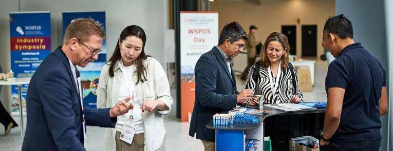
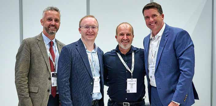
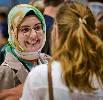
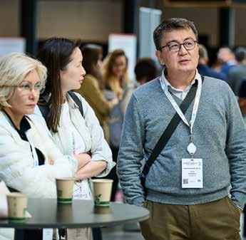
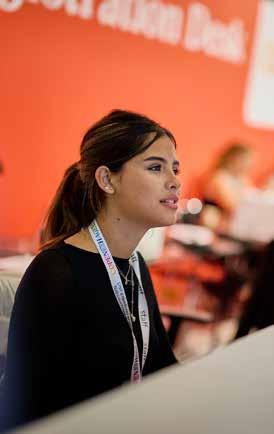
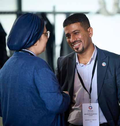
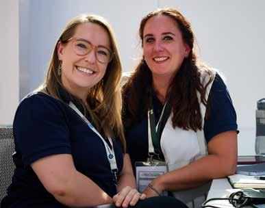
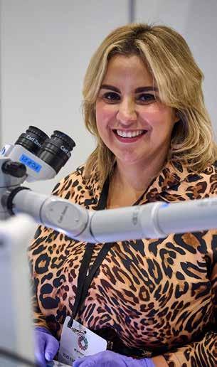
Memorable moments from the ESCRS 2025 Congress, capturing key connections and collaborations.

Don’t miss the ESCRS Video Awards
This session is a long-standing tradition that announces the best ophthalmic surgery videos of the year. The all-star list of chairpersons includes Zsolt Biro, Richard Packard, Boris Malyugin, and many more.
Sunday 14:15 – 16:15 A1
Don’t miss the JCRS Symposium
Moderated by Drs Thomas Kohnen and Liliana Werner, elite surgeons tackle major debates in ophthalmic surgery, including the future of lens removal, evidence-based IOL classification, and the future of training.
Sunday 14:30 – 16:30 D2
Surgery:
Don’t miss today’s IME Forum!
Join us today at the ESCRS Independent Medical Education Forum on Refractive Surgery. Gain insights into practical strategies to manage patient expectations, optimise the ocular surface, and learn how to address common patient objections with real-life case examples from the latest ESCRS Clinical Trends Survey.
Room D4, Hall D Saturday, 13 September 2025 16:45–17:45 CEST
Chaired by Professors Bruce Allan and Thomas Kohnen and accompanied by faculty Prof Béatrice Cochener-Lamard and Drs Andreia Rosa, Pooja Khamar, and Walter Sekundo, expect specialist insights, interactive live polling, and a closing panel discussion—all focused on improving surgical outcomes and boosting patient satisfaction.
Don’t miss this chance to learn directly from world-leading experts!


Learn about iStent inject® W and the new iStent infinite® trabecular micro-bypass, the only truly microinvasive technologies offering safe interventional glaucoma helping delay disease progression.
Saturday 13th and Sunday 14th
Saturday 13th
iStent infinite® Evolution or Revolution?
SCAN THE QR CODE TO ACCESS THE FULL PROGRAM
SCAN THE QR CODE TO ACCESS THE FULL PROGRAM
iStent infinite® IMPORTANT SAFETY INFORMATION: INDICATION FOR USE. The iStent infinite System is intended to reduce intraocular pressure safely and effectively in adult patients diagnosed with primary open-angle glaucoma, pseudo- exfoliative glaucoma or pigmentary glaucoma. The device is safe and effective when implanted in combination with or without cataract surgery in those subjects who require intraocular pressure reduction and/or would benefit from glaucoma medication reduction. The device may also be implanted in patients who continue to have elevated intraocular pressure despite prior treatment with glaucoma medications and/or conventional glaucoma surgery. CONTRAINDICATIONS: The iStent infinite System is contraindicated under the following circumstances or conditions: •In eyes with primary angle closure glaucoma, or secondary angle-closure glaucoma, including neovascular glaucoma, because the device would not be expected to work in such situations.• In patients with retrobulbar tumor, thyroid eye disease, Sturge-Weber Syndrome or any other type of condition that may cause elevated episcleral venous pressure WARNINGS/PRECAUTIONS:
• For prescription use only.
• Intended users are trained ophthalmologists only.
• This device has not been studied in patients with uveitic glaucoma.
• Do not use the device if the Tyvek® lid has been opened or the packaging appears damaged. In such cases, the sterility of the device may be compromised.
• Due to the sharpness of certain injector components (i.e., the insertion sleeve and trocar), care should be exercised to grasp the injector body. Dispose of device in a sharps container.
• iStent infinite is MR-Conditional
• Physician training is required prior to use of the iStent infinite System.
• The surgeon should monitor the patient postoperatively for proper maintenance of intraocular pressure. If intraocular pressure is not adequately maintained after surgery, the surgeon should consider an appropriate treatment regimen to reduce intraocular pressure.
• Do not re-use the stent(s) or injector, as this may result in infection and/or intraocular inflammation, as well as occurrence of potential postoperative adverse events • There are no known compatibility issues with theiStent infinite and other intraoperative devices (e.g., viscoelastics) or glaucoma medications. • Unused product & packaging may be disposed of in accordance with facility procedures. Implanted medical devices and contaminated products must be disposed of as medical waste.
• Patients should be informed that placement of the stents, without concomitant cataract surgery in phakic patients can enhance the formation or progression of cataract. ADVERSE EVENTS: The most common postoperative adverse events reported in the iStent infinite pivotal trial included IOP increase ≥ 10 mmHg vs. baseline IOP (8.2%), loss of BSCVA ≥ 2 lines (11.5%), ocular surface disease (11.5%), perioperative inflammation (6.6%) and visual field loss ≥ 2.5 dB (6.6%). CAUTION: Please see DFU for a complete list of contraindications, warnings, precautions, and adverse events. iStent inject® W IMPORTANT SAFETY INFORMATION: INDICATIONS FOR USE: The iStent inject® W, is intended to reduce intraocular pressure safely and effectively in patients diagnosed with primary openangle glaucoma, pseudo-exfoliative glaucoma or pigmentary glaucoma. The iStent inject® W, can deliver two (2) stents on a single pass, through a single incision. The implant is designed to stent open a passage through the trabecular meshwork to allow for an increase in the facility of outflow and a subsequent reduction in intraocular pressure. The device is safe and effective when implanted in combination with cataract surgery in those subjects who require intraocular pressure reduction and/or would benefit from glaucoma medication reduction. The device may also be implanted in patients who continue to have elevated intraocular pressure despite prior treatment with glaucoma medications and conventional glaucoma surgery. CONTRAINDICATIONS: The iStent inject® W System is contraindicated under the following circumstances or conditions: • In eyes with primary angle closure glaucoma, or secondary angleclosure glaucoma, including neovascular glaucoma, because the device would not be expected to work in such situations. • In patients with retrobulbar tumor, thyroid eye disease, Sturge-Weber Syndrome or any other type of condition that may cause elevated episcleral venous pressure. WARNINGS/PRECAUTIONS:• For prescription use only. • This device has not been studied in patients with uveitic glaucoma. • Do not use the device if the Tyvek® lid has been opened or the packaging appears damaged. In such cases, the sterility of the device may be compromised. • Due to the sharpness of certain injector components (i.e. the insertion sleeve and trocar), care should be exercised to grasp the injector body. Dispose of device in a sharps container. • iStent inject®