SWT/KWS Meru Veterinary Unit Report for May June 2025
7 Cases in May/June 2025
May/June Report by Dr.
Aminga Duncan
3 Poaching Cases 3 Elephant Cases
The month of June was characterized by dry weather conditions and moderate daytime temperatures. The gradual loss of green vegetation across the Meru ecosystem signalled the onset of the dry season. Despite this, rivers within the area continued to flow, ensuring the availability of water for wildlife in the coming months.
Notably, despite the varied outcomes of elephant cases attended to during the period, there was an encouraging increase in the sighting of baby elephants across the ecosystem. During the reporting period, the SWT/KWS Meru Mobile Veterinary Unit recorded an increase in the number of wildlife cases managed, attending to a total of 7 cases involving African elephants and Southern white rhinos.
Acknowledgement
We deeply appreciate the steadfast commitment of Sylvie Chantecaille and the Sheldrick Wildlife Trust, whose generous contributions continue to empower the Meru Mobile Veterinary Unit. Their vital support significantly strengthens wildlife interventions and enhances species conservation within the Meru ecosystem. We also commend the Kenya Wildlife Service for its strong institutional leadership. The consistent guidance from the KWS-Head of Veterinary Services and the Senior Assistant Director for the Eastern Conservation Area has been key in shaping effective conservation strategies and reinforcing wildlife protection initiatives throughout the region.
Case Details
April 2025
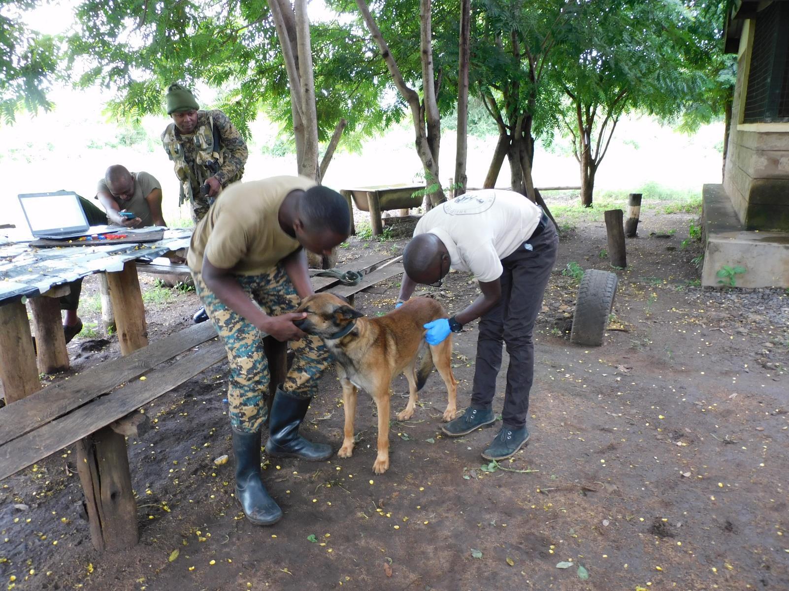
Security Dog Technical Cases
Meru National Park
The security dogs in Meru National Park play a crucial role in wildlife conservation by tracking poachers and detecting illegal activities within the Meru ecosystem. Despite their significant contributions to combating poaching, these dogs are at risk of contracting Trypanosomiasis, a highly fatal disease transmitted by Tsetse flies. To protect these valuable canines, the Meru Veterinary team administered a vaccination against Trypanosomiasis to Sep, a security dog at the Meru Canine Unit, ensuring he remains in optimal health for his important duties.
Examination and treatment
To ensure the dog remained calm during the examination, he was introduced to the veterinarian beforehand. During the physical examination, it was observed that he was in fair body condition (3/5), active, and alert. His body weight was recorded at 27.8 kg, and the appropriate drug dosages were calculated accordingly. He was manually restrained and given a subcutaneous injection of 0.8 ml of Triquin® (a combination of Quinapyramine sulphate and Quinapyramine chloride) on the dorsal side of his neck.
Prognosis
No adverse reactions were noted following the administration of the drugs, so a good prognosis is indicated.
Case 2 – 31st May 2025
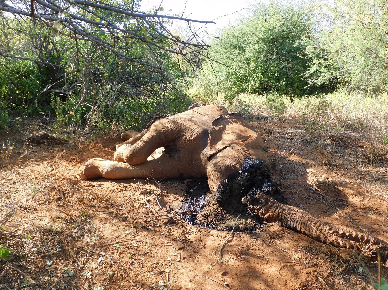
Elephant Poaching Postmortem Mwingi National Reserve
The Meru Mobile Veterinary Unit was notified about an elephant carcass sighted in the Kasalani area of Mwingi National Reserve, located in Kitui County.
Postmortem examination
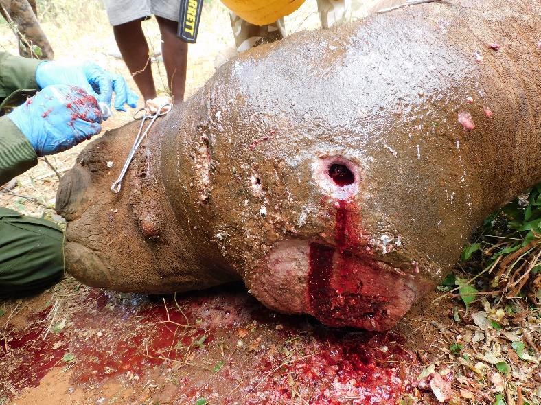
The carcass was found lying in left lateral recumbency, in the active decay stage of decomposition. The body condition score was estimated at 3/5, indicating a fair nutritional state prior to death. The elephant was identified as a mono-tusker, bearing only the right tusk, which had been forcibly removed. Tool marks on the maxillary bone and surrounding soft tissue suggested tusk extraction using a sharp instrument There was a large area of soft tissue excision on the right rump, with margins consistent with sharp-force trauma, likely inflicted using machetes. No external injuries were identified. A thorough examination with a handheld metal detector did not detect any metallic foreign objects. On internal examination, the liver was found to be enlarged, friable and diffusely pale. The kidneys were markedly congested. The lungs were oedematous, exuding copious frothy, blood-tinged fluid. The gastrointestinal tract revealed severe mucosal damage, with the intestinal mucosa appearing friable and easily sloughed upon minimal traction. The remaining internal organs showed rapid autolysis, complicating further detailed evaluation.
Prognosis
The gross pathological changes observed are consistent with a toxic substance. Given the condition of the carcass, the missing tusk, and evidence of human activity, it is suspected the elephant could have succumbed to being shot using a poisoned arrow. However, the mechanism of death remains inconclusive pending toxicological and histopathological evaluation.
Case 3 – 6th June 2025
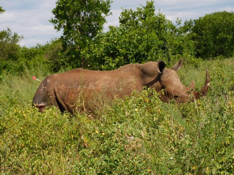
White Rhino Natural Causes
Meru National Park
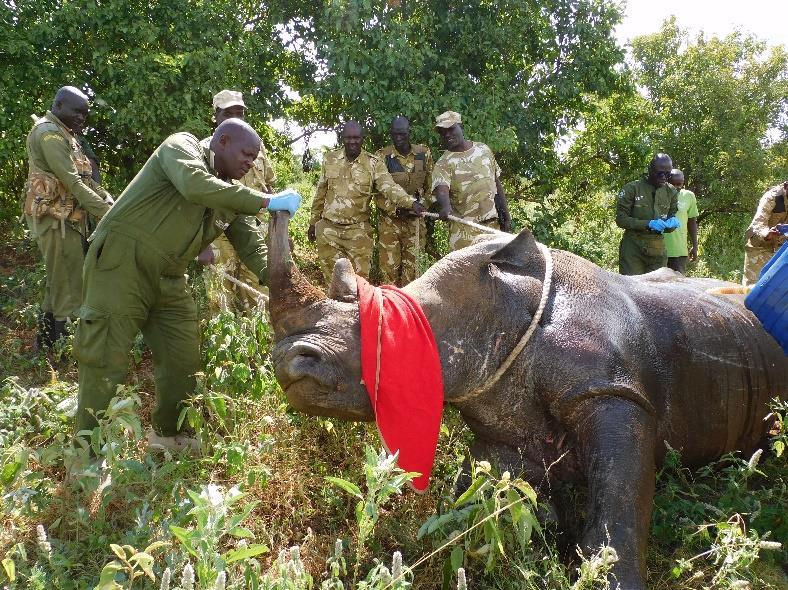
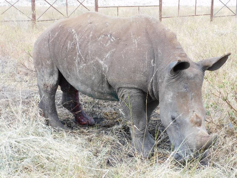
An adult male white rhino was exhibiting marked lameness of the right hind limb. Preliminary observations indicated the animal had sustained multiple injuries suspected of being from a fight with another bull.
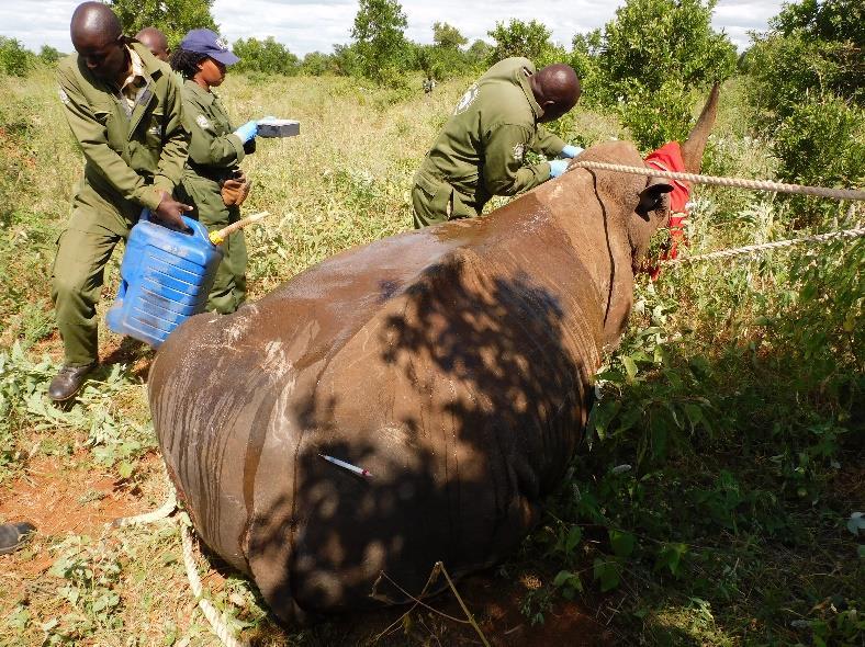
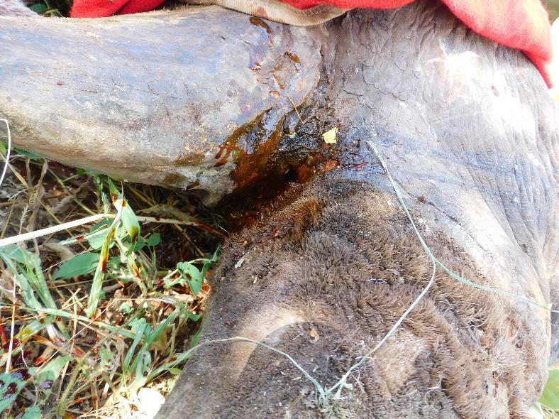
Immobilisation, examination and treatment
The white rhinoceros was darted from a helicopter with 5mgs Etorphine and 80mgs Azaperone. Prior to the commencement of the clinical examination, 50mgs of Butorphanol tartrate was administered intravenously to antagonize the undesirable side effects of etorphine respiratory depression and systemic hypertension.
The injured rhino was found to have sustained multiple traumatic injuries involving the head, thoracic region, and hind limbs, consistent with injuries, likely from a territorial fight with another adult male. Notably, a deep puncture wound was observed on the right hind limb leading to marked lameness. All the wounds were cleaned with water and debrided using Hydrogen peroxide to remove necrotic tissue. This was followed by flushing with Iodine for disinfection. The wounds were then packed with Oxytetracycline pessaries and covered with green clay, known for its wound-healing properties. An antibiotic aerosol spray was applied topically to deter fly infestation and minimize the risk of myiasis. The rhino was given 2,500 mgs of Flunixin, 18,000mgs of long-acting Amoxicillin as a broad-spectrum antimicrobial and 6,000mgs of Metabolic stimulants were administered to enhance physiological recovery and support systemic function.
Prognosis
Given the extent of the injuries, the prognosis for full recovery was assessed as poor to favourable
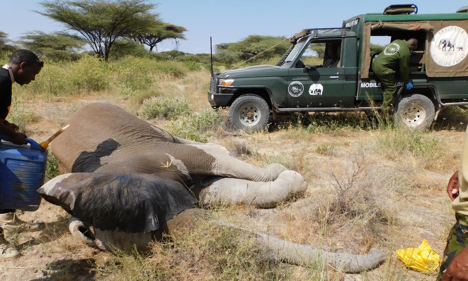
Case 4 – 7th June 2025
Elephant Bullet Wound
Wikithuki area- Kitui County
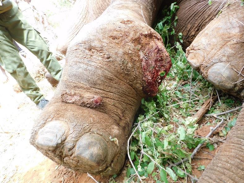
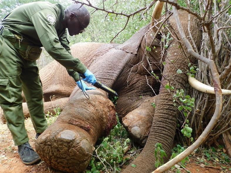
An adult male elephant was sighted in Wikithuki area, Kitui County exhibiting pronounced lameness in the right forelimb.
Immobilisation, examination and treatment
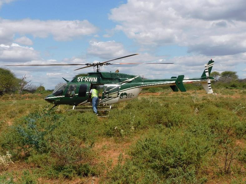
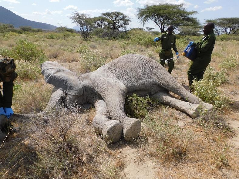
On the morning of 7th June 2025, ground teams continued tracking the injured elephant while a Kenya Wildlife Service helicopter airlifted the veterinary team to the site. The elephant was successfully immobilized via aerial darting using 18mgs Etorphine
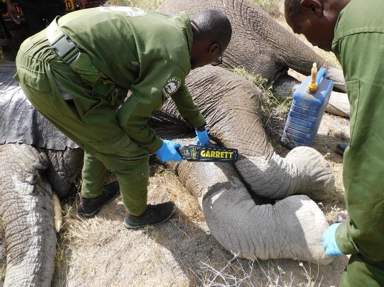
Clinical examination revealed a severely swollen carpal joint with a penetrating gunshot wound approximately 7 inches deep. Additionally, two old, infected arrow wounds were noted. A metal detector scan confirmed the presence of a metallic foreign body; however, the bullet could not be retrieved due to its deep location distal to the elbow joint, where extraction posed a significant risk of further tissue damage. The wounds were thoroughly cleaned with water, debrided using Hydrogen peroxide, and flushed with Iodine. Oxytetracycline pessaries were inserted into the wound cavities and subsequently packed with green clay, known for its antimicrobial and healing properties. The elephant was administered a high dose of anti-inflammatory agents, systemic antibiotics, and metabolic stimulants to support recovery.
Prognosis
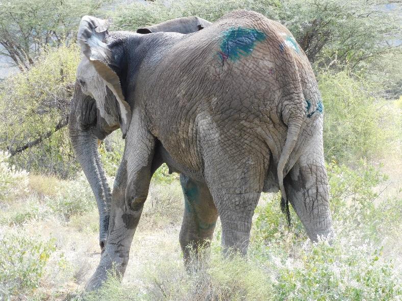
Anaesthetic reversal was uneventful, and the elephant regained full consciousness with a guarded to favourable prognosis for complete recovery. The monitoring teams were instructed to conduct regular follow-ups over the subsequent weeks and report any significant changes to facilitate potential follow-up intervention.
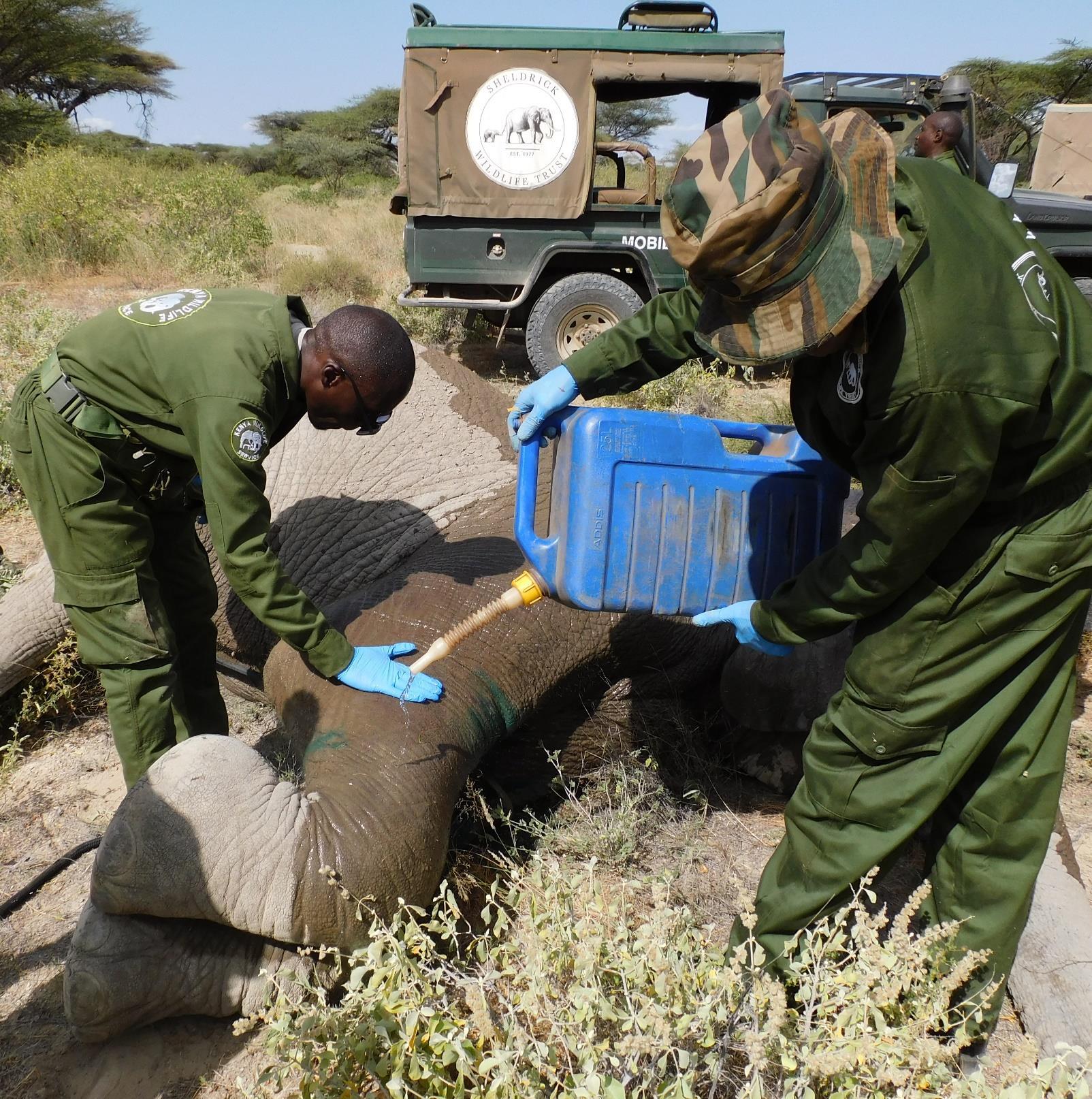
Case 5 – 11th June 2025
Elephant Bullet Wound
Shaba National Reserve
A juvenile female elephant was observed to be limping within Shaba National Reserve. The reporting team was requested to keep track of the elephant to aid in timely intervention upon the team's arrival.
Immobilisation, examination and treatment
She was located browsing in nearby vegetation and was cautiously approached using a vehicle. She was darted on the left rump with 13mgs of Etorphine hydrochloride. Following darting, the animal exhibited a flight response and moved away slowly before the drug took effect leading to recumbency on the right lateral side.
Clinical examination revealed a gunshot wound located on the elbow region of the right forelimb. The wound was approximately 7 inches deep; however, the elbow joint was fortunately not compromised. Use of a handheld metal detector confirmed the presence of a metallic foreign body within the limb. The bullet was lodged deeply distal to the elbow joint, making retrieval unfeasible without risking further soft tissue trauma or neurovascular injury. The wound was thoroughly cleaned with clean water, flushed with Hydrogen peroxide and Iodine, and subsequently packed with Oxytetracycline pessaries to allow for sustained antibiotic release. An antibiotic aerosol spray was applied topically to deter flies and minimize the risk of myiasis. Systemically, the animal was administered 15,000 mgs Amoxicillin intramuscularly for bacterial infection control, along with 100mgs Dexamethasone sodium to manage inflammation.
Prognosis
The animal stood up three minutes after reversal and walked away to re-join her herd. The prognosis for recovery is favourable
Case 6 – 17th June 2025
White Rhino Postmortem
Meru National Park
On 17th June, the Meru Veterinary Unit received a report regarding the sighting of a white rhinoceros’ carcass within Meru National Park. The animal was positively identified as an individual previously treated by the team a few days earlier for injuries sustained during a territorial altercation. The veterinary team was subsequently assigned to carry out a post-mortem examination to ascertain the cause of death.
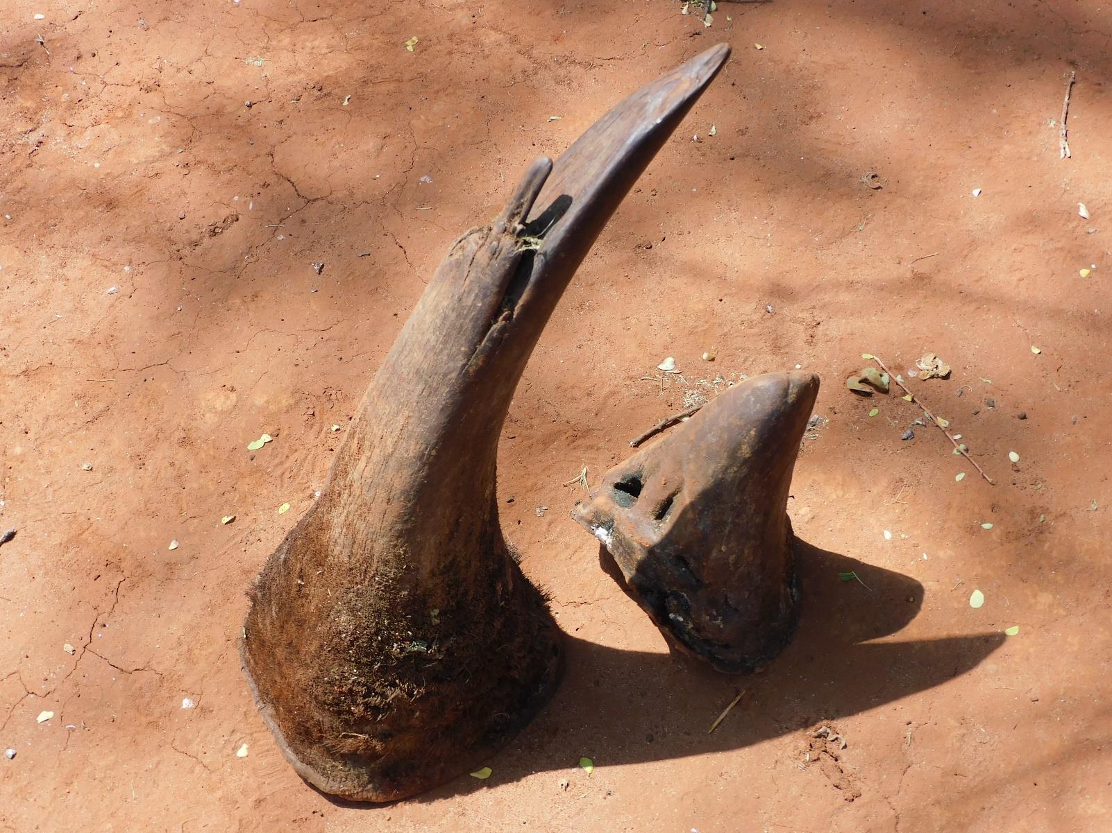
Postmortem examination
The carcass was found in left lateral recumbency and had been extensively scavenged, predominantly by hyenas. Most internal organs and significant muscle masses, including those over the neck, back, and limbs had been consumed, thereby limiting a thorough assessment of the internal injuries. Both horns were intact and were recovered by the KWS Meru Rhino Monitoring Team for secure custody. The remaining distal limb tissues exhibited marked oedema.
Prognosis
The gross pathological findings were indicative of a systemic inflammatory response. Considering that the rhino had been treated a few days earlier for severe traumatic injuries, and based on the observed lesions, the cause of death was attributed to septicaemia.
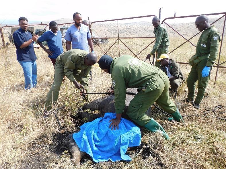
Case 7 – 19th June 2025
White Rhino Natural Causes Lewa Wildlife Conservancy
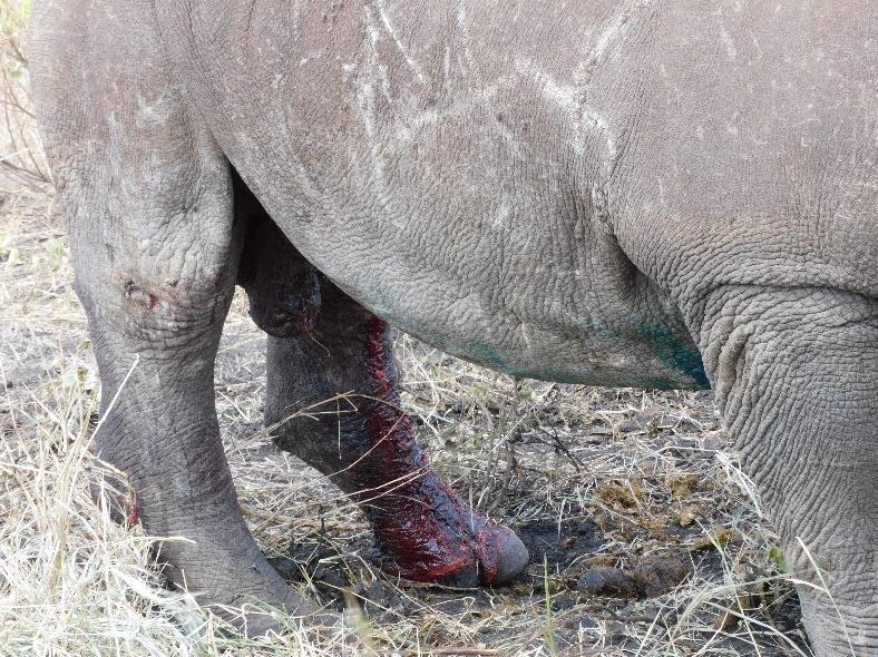
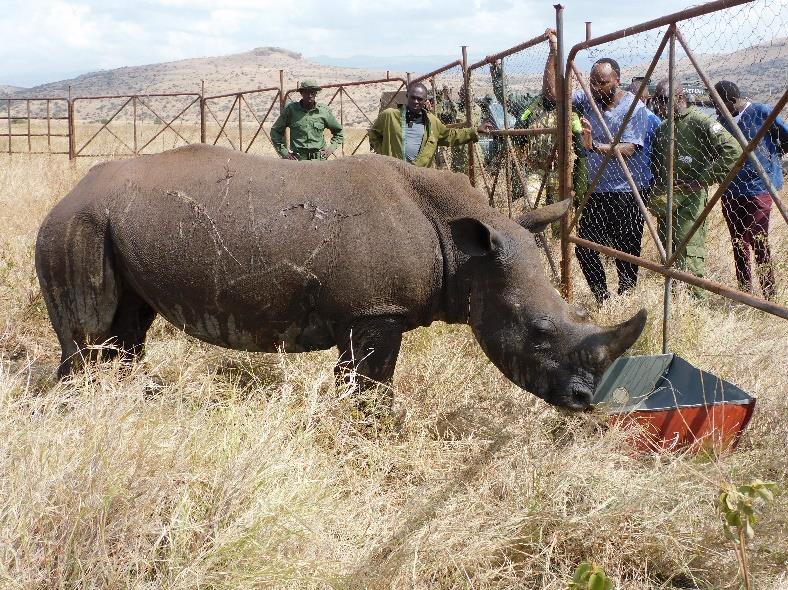
The Meru Mobile Veterinary Unit received a report indicating that a previously treated juvenile White rhino was not responding well to the initial intervention. The white rhino was initially treated on 13th June 2025 for a deep puncture wound involving the left inguinal region extending into the proximal aspect of the left thigh, which was causing marked difficulty in ambulation. The injuries were suspected to have been sustained during an aggressive encounter with an older bull. Following the treatment, the animal was placed in a holding enclosure to facilitate close monitoring and supportive care.
Immobilisation, examination and treatment
However, despite the initial medical intervention, the animal's condition progressively deteriorated. Notably, the animal had an active bleeding from the wound site during attempts to bear weight on the affected limb. The animal was reported to be inactive and had not been observed feeding for over 24 hours. Physical restraint was achieved using ropes to facilitate a thorough clinical examination. Palpation and manipulation of the affected hind limb revealed crepitation, suggestive of an underlying skeletal injury. Additionally, a bone fragment was retrieved from the wound, raising a strong suspicion of a femoral fracture. A portable X-ray machine was used; however, the location of the suspected injury limited the getting of conclusive radiographic evidence.
Prognosis
Unfortunately, the animal succumbed during the assessment. The cause of death is attributed to severe traumatic injury to the left hind limb, resulting in a complete mid-shaft femoral fracture with associated massive internal haemorrhage. The extensive blood loss, compounded by tissue damage and vascular disruption, likely led to hypovolemic shock and subsequent death.