

International, Interdisciplinary Conference of Innovative Technologies in Dentistry, June 2–3 2023 “ DENTISTRY
– VIENNA Österreichische Post AG MZ14Z039972M Universitätszahnklinik Wien, Sensengasse 2a, 1090 Wien Special Edition 2023 The Journal of the University Clinic of Dentistry Vienna Congress lectures on current topics in dental medicine – 13 Scientific workshops from the dentistry practice and for the dentistry practice – 26 Poster competitions for young scientists – 30 Mit Strahlenschutzkurs für Zahnärzt:innen und Assistent:innen!
4.0”
Preface
Dear Friends and Colleagues,
The digital revolution in dentistry is advancing rapidly. This brings advantages for dentists and patients, but such rapid changes can be disruptive if not carefully implemented. Hence, it is our pleasure to announce that the “Dentistry 4.0” – International, Interdisciplinary Conference of Innovative Technologies will be held at the University Clinic of Dentistry in Vienna, Austria.
Liebe Freunde und Kolleg:innen!
Die digitale Revolution in der Zahnheilkunde schreitet rasant voran, was Vorteile für Zahnärzt:innen und Patient:innen bringt. Solche schnellen Änderungen können jedoch verunsichern, wenn sie nicht sorgfältig begleitet werden. Daher ist es uns eine Freude, die „Dentistry 4.0 – Internationale, interdisziplinäre Konferenz innovativer Technologien“, anzukündigen, die in Wien Österreich, stattfnden wird. „Dentistry 4.0“ wird von der Universitätszahnklinik der Medizinischen Universität Wien in Kooperation mit verschiedenen nationalen und internationalen zahnmedizinischen Fachgesellschaften veranstaltet.
Zahnmedizin 4.0 stellt die Vision der Digitalisierung, deren praktische Umsetzungsansätze sowie Chancen, Fähigkeiten, aber auch weniger bekannte Seiten der künstlichen Intelligenz vor. Die Konferenz bietet ein reichhaltiges und vielfältiges wissenschaftliches Programm zu den neuesten Entwicklungen in allen Bereichen der Zahnmedizin. International und national renommierte Expert:innen stellen den aktuellen Forschungsstand vor, die Vorträge reichen von Praxisworkshops bis hin zu Networking-Events. Die Konferenzteilnehmer:innen erhalten zudem die Möglichkeit, Unternehmenspartner:innen und Sponsor:innen von „Dentistry 4.0“ im Rahmen unserer Ausstellung persönlich zu treffen.

„Dentistry 4.0“ ist „the place-to-be“!
Wir freuen uns darauf, Sie 2023 in Wien begrüßen zu dürfen!
“Dentistry 4.0” will present the vision of digitalization and practical implementation approaches, as well as the opportunities, abilities and dangers of artifcial intelligence. The conference features a rich and varied scientifc programme that highlights the latest developments in all areas of dentistry. Internationally and nationally renowned experts will present the current state of research. An exciting programme is available to all participants, ranging from lectures and practical workshops to networking events. Corporate partners and sponsors of “Dentistry 4.0” will be able to meet conference delegates face-to-face in our exhibition.
“Dentistry 4.0” is organised by the University Clinic of Dentistry, Medical University of Vienna, in cooperation with various national and international dental societies.
”Dentistry 4.0“ is “the place-to-be” and we are looking forward to seeing you in Vienna in 2023!
2 DentUnique 1/2023
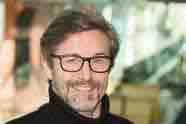
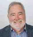
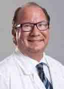
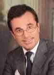
Univ.-Prof. DDr. Andreas Moritz, MD, DMD, Kongresspräsident
Preface
Sehr geehrte Konferenzteilnehmer:innen!
Wien ist weltweit eine der beliebtesten Städte für internationale Kongresse und Konferenzen. Das hängt nicht allein damit zusammen, dass Wien die lebenswerteste Stadt der Welt ist, wie renommierte internationale Studien regelmäßig belegen. Vielmehr ist Wien auch einer der führenden europäischen Forschungs- und Wissenschaftsstandorte. Gerade im medizinischen und naturwissenschaftlichen Bereich forschen und lehren viele hoch angesehene, innovative Wissenschaftler:innen an ausgezeichneten öffentlichen und privaten Hochschulen und Institutionen. Ich freue mich daher sehr, dass mit „Dentistry 4.0“ eine internationale und interdisziplinäre Konferenz zu innovativen Technologien in der Zahnmedizin in Wien stattfndet.
Digitalisierung und künstliche Intelligenz bieten auch in der Zahnmedizin neue Chancen und Hoffnungen im Bereich Diagnose und Behandlung. Diese Möglichkeiten künstlicher Intelligenz, aber auch die damit verbundenen Herausforderungen stellen ausgewiesene nationale und internationale Expert:innen in zahlreichen wissenschaftlichen Vorträgen und praktischen Workshops vor. „Dentistry 4.0“ präsentiert dabei den aktuellen Stand der Forschung in wichtigen medizinischen Zukunftsfeldern und rückt Wien einmal mehr in den Mittelpunkt des akademischen Diskurses.
Als Bürgermeister der Stadt Wien möchte ich mich bei der Universitätszahnklinik Wien und der Wiener Medizinischen Akademie GmbH für die ausgezeichnete Organisation von „Dentistry 4.0“ bedanken und wünsche allen Teilnehmer:innen eine spannende und informative Konferenz.
Dear Conference Participants,

Vienna is one of the most popular cities worldwide for international congresses and conferences. This is not only due to the fact that Vienna is the most liveable city in the world, as regularly proven by renowned international studies. Vienna is also one of the leading European research and science locations. Especially in the medical and natural sciences, many highly respected, innovative scientists conduct research and teach at excellent public and private universities and institutions. I am therefore very pleased that “Dentistry 4.0”, an international and interdisciplinary conference on innovative technologies in dentistry, is taking place in Vienna.
Digitalisation and artifcial intelligence offer new opportunities and hopes in the feld of diagnosis and treatment in dentistry. The possibilities artifcial intelligence offers, but also the challenges it creates, will be presented by renowned national and international experts in numerous scientifc lectures and practical workshops. “Dentistry 4.0” presents the current state of research in important medical felds of the future and once again places Vienna at the centre of academic discourse.
As Mayor of Vienna, I would like to thank the University Clinic of Dentistry Vienna and the Vienna Medical Academy for the excellent organisation of “Dentistry 4.0” and wish all participants an exciting and informative conference.
DentUnique 1/2023 3
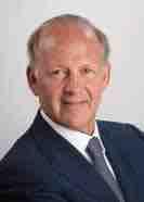
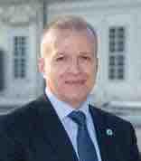
Dr. Michael Ludwig, Bürgermeister und Landeshauptmann von Wien
Foto: PID/Gregor Kuntscher
Impressum:
Medieninhaber: Universitätszahnklinik Wien GmbH, 1090 Wien, Sensengasse 2a, Tel.: +43 1/400 70, E-Mail: office-unizahnklinik@meduniwien.ac.at, www.unizahnklinik-wien.at

Herausgeber: Geschäftsführer Univ.-Prof. DDr. Andreas Moritz und Thomas Stock
Erscheinungsort: Wien
Auflage: 4.500
Verlag: Albatros Media, H. Wollner Straße 20, 2602 Blumau, office@ albatros-media.at, www.albatros-media.at, Redaktion: Mag.a Erika
Hofbauer (Leitung) Grafik & Produktion: Albatros Media, Designkonzept: Albatros Media, Verlagsleitung: Otto Koller
Lektorat (DE): Mag.a Eva Kainrad, Lektorat (EN): Dr.in Sara Crockett
Coverfoto: Universitätszahnklinik Wien
Fotos: Falls nicht anders angegeben: Universitätszahnklinik Wien
Druck: Druckerei Janetschek GmbH, 3860 Heidenreichstein
Offenlegung gemäß § 25 Mediengesetz
Die Universitätszahnklinik Wien GmbH ist eine 100-%-Tochtergesellschaft der Medizinischen Universität Wien, www.meduniwien.ac.at/ homepage/info/impressum. Grundlegende Richtung des Magazins: DentUnique informiert Zahnärzt:innen, Studierende und Mitarbeiter:innen der Universitätszahnklinik Wien über Forschung, Fallstudien, Weiterbildungsangebote und die Tätigkeiten der Institution.
4 DentUnique 1/2023 05 General Information 06 Networking 07 Sessions Overview 08 “Dentistry 4.0” Programme Details 12 Orientation Plan 13 Speakers and Abstracts 26 Workshop Details 27 Satellite Workshop Details 30 Posters 31 Sponsors and Partners 34 Registration Fees 35 Information Contents
Organiser
University Clinic of Dentistry
Medical University of Vienna
Sensengasse 2a
1090 Vienna, Austria
T: +43 1 40070
E-Mail: office-unizahnklinik@meduniwien.ac.at
Professional congress organiser
Vienna Medical Academy (WMA GmbH)
Alser Straße 4
1090 Vienna, Austria
T: +43 1 4051383-25
F: +43 1 4051383-925
E-Mail: dent2023@wma.co.at
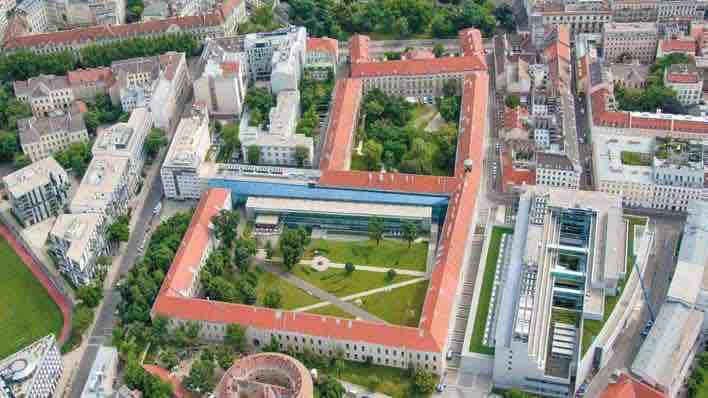
Venue University Clinic of Dentistry
Congress president and chairman of the scientific organising committee
Univ.-Prof. DDr. Andreas Moritz
Scientific organising committee at the University Clinic of Dentistry
Assoc. Prof. Priv.-Doz. Dr. Oleh Andrukhov
Univ.-Prof.in Dr.in med. dent. habil Katrin Bekes, MME
Dr.in Corinna Bruckmann, MSc
Univ.-Prof. Dipl.-Ing. Dr. Reinhard Gruber
Ao. Univ.-Prof. DDr. Erwin Jonke
DDr. Martin Krainhöfner
Assoc. Prof.in Priv.-Doz.in DDr.in Ulrike Kuchler
Dr. med. dent. Christoph Kurzmann
Univ.-Prof. DDr. Andreas Moritz
DDr. Michael Nemec
O. Univ.-Prof.in DDr.in Eva Piehslinger
Univ.-Prof.in Univ.-Doz.in Dr.in Xiaohui Rausch-Fan
Univ.-Prof. Dr. Harald Sitte
Ao. Univ.-Prof. DDr. Christian Ulm
Priv.-Doz. DDr. Christoph Vasak
Mst. Tom Vaskovich, MSc
Conference registration
Please note that Dentistry 4.0 will be held as a live event.
General Information
Travelinformation
Arrival is possible by plane or train. Please fnd detailed information here:
Vienna International Airport Vienna Main Train Station
Public transport
From Schottentor: Tram lines 37, 38, 40, 41 und 42
Stop: Schwarzspanierstraße
From Währinger Straße Volksoper: Tram lines 40, 41 und 42
Stop: Sensengasse
From Döbling: Tram lines 37 und 38
Stop: Sensengasse
Tram lines 5 und 33
Stop: Lazarettgasse
By car
There are very few parking spaces on Sensengasse. You can use the BOE underground parking at Sensengasse 3 (for a fee). Please note that the entire 9th district is a short-term parking zone.
Accessibility
The whole building provides accessibility to handicapped people. The parking area for disabled persons can be reached via the entrance Währinger Straße 25a.
Hotel
The organiser has reserved a small contingent of rooms in each hotel listed, which will be handed out first-come-first-serve basis.
Please find more information here:
CME/ZFP Credits
Dentistry 4.0 has applied for accreditation from the European Accreditation Council of Continuing Medical Education, also Dentistry 4.0 will be seeking accreditation from the Austrian Dental Association (ZFP-ÖZÄK) as part of the diploma training program.
The continuously updated information can be found here:
In order to obtain the credits, pertaining to each day, participants will be requested to scan their badges daily at the General Information Desk. After the congress, a survey will be sent out to all participants, and those seeking credits will have to complete the survey in order to download their certificate of attendance, including the credits obtained during the conference.
Please note that if you do not scan your badge on any given day, the conference will not be able to provide you with the credits of that day.
Should you have any question about CME credits, please do not hesitate to contact us at dent2023@wma.co.at.
DentUnique 1/2023 5
Networking
Dinner in the Vienna City Hall
Friday, June 2
The networking evening is a great opportunity to meet with friends and colleagues from around the world in a relaxed atmosphere and to enjoy the unmatched local charm of the host city. You are invited by the mayor of Vienna!
Start: 19:00 CEST, End: 00:00 CEST
Tickets: 50,00 EUR
Dinner & drinks are included in the price.
Dress code: Business Casual
Registration is mandatory
Networking in the Garden at the University Clinic of Dentistry
Saturday, June 3
Start: 16:15 CEST (after the Closing Ceremony at Van Swieten Saal)
End: open
Tickets: Included in the fee. Registration is mandatory. Snacks & drinks are included.
Dress code:
6
Fotos:
stock.adobe.com
Tryfonov/suronin
17:00-21:00
Thursday, June 1, 2023
Big Lecture Hall
Strahlenschutzkurs für Zahnärzt:innen
Fortbildung für Strahlenschutzbeauftragte (gemäß § 82 Allgemeine Strahlenschutzverordnung)
This course is only approved for Austrian dentists and is included in the registration fee. Registration is required in order to take part. First come, frst served! (in German)
Friday, June 2, 2023
Big
13:45-15:15 15:15-16:15
Saturday,
Problems of Digitalization
Closing Ceremony Awards Short Talks/Poster Prize
Programme changes reserved. The continuously updated programme can be found here:
DentUnique 1/2023 7
Sessions Overview
Van Swieten Hall
Lecture Hall
Basics of Digitalization 10:30-11:00 Break Break 09:00-10:30 Prosthodontics 11:00-12:30 Paediatric Dentistry Digital Dentistry 12:30-13:45 Lunch Break Lunch Break Orthodontics Oral Surgery 15:15-15:45 Break Break Digital Dentistry Periodontology 13:45-15:15 15:45-17:15 19:00-23:00 Networking Dinner in Vienna City Hall
Van Swieten Hall Periodontology 10:30-11:00 Break Break 09:00-10:30 Conservative Dentistry-Laser 11:00-12:30 Oral Surgery Orthodontics
Lunch Break Lunch Break
Paediatric
June 3, 2023 Big Lecture Hall
12:30-13:45
Digital Dentistry
Dentistry
16:15–open end
in the Garden
Networking
Friday, June 2
09:00-09:15
Plenary Speaker
Big Lecture Hall
Opening Ceremony
Basics of Digitalization
09:30-10:30
Wido Menhardt
09:30-10:30
Digitalization, artifcial intelligence, and the metaverse
Hands-on Workshop
Van Swieten Hall Seminar Room A
Prosthodontics
09:30-10:30
Nazzareno Bassetti
Integration between function and aesthetics in complex cases using VieSID concepts and OMRT Basetti
10:30-11:00 Break Break
Pediatric Dentistry
11:00-12:30
Norbert Krämer, Katrin Bekes
11:00-12:30
Molar incisor hypomineralization: Where do conventional and where do digital therapy approaches make sense?
Digital Dentistry
11:00-12:00
Falk Schwendicke
Digital dentistry and artifcial intelligence: A revolution ongoing?
12:00-12:30
André Gahleitner
Current cross-sectional imaging of the jaw
Orthodontics Hands-on Workshop
09:30-11:00 Part I
Heinz Winsauer
Newly emerging digital workfows in bone-borne devices
Palatal and buccal-shelf applications of mini screws in everyday practice
11:00-11:15 Break
11:15-12:25 Part II
Trend digitization - opportunities and risks for case planning and workfow with mini screws
12:25-12:40 Lunch Break
12:40-13:40 Part III
12:30-13:45
Lunch Break
Orthodontics
13:45-14:15
Erwin Jonke
Implementation of modern 3D imaging in orthodontic diagnostics and treatment
13:45-15:15
14:15-15:15
Alexander Schwärzler
CAD/CAM orthodontic appliances for complex cases: The fully digital workfow
Lunch Break
Oral Surgery
13:45-14:15
Alwin Sokolowski
Digital technologies in implant treatment: Surgical and restorative considerations
14:15-14:45
Werner Zechner
Digitally aided bone augmentation concepts in implant surgery
14:45-15:15
Georg Strbac
Novel clinical applications of computer-guided surgery
15:15-15:45 Break Break
Digital Dentistry
15:45-16:15
Andreas Moritz, Tom Vaskovich
Digital workfow at the University Clinic of Dentistry - an overview
15:45-17:15
16:15-16:45
Eva Piehslinger, Lana Zupančič Čepić Evolution of complete denture in the digital age – update on possibilities and boundaries in clinical practice
16:45-17:15
Xiaohui Rausch-Fan, Eva Piehslinger
Decision of therapeutic position in interdisciplinary treatment of temp-oromandibular joint disorders
Periodontology
15:45-16:30
Andreas Stavropoulos
Management of periimplantitis: A programatic approach
16:30-16:50
Gabor Fürst
Is there an advantage of digital processing of implant abutment in prevention of peri-implant disease?
16:50-17:10
Michael Müller
Ways to improve soft tissue around implants – in view of digital considerations
Testing and comparing different workfows with digital components in an everyday practice routine including Hands-on Workshop for Orthodontics
13:40-15:20
Afternoon Break
15:20-17:50 Part IV
Non-surgical maxillary suture opening in adults
19:00-23:00
Networking Dinner in the Vienna City Hall
8 DentUnique 1/2023
Satellite Workshops
Friday, June 2
Cadaver Course Scientifc Workshops
Seminar Room B Seminar Room C1 Institute of Anatomy Pre-Clinic
09:30-11:00
Amann Girrbach
Wibke Rosin, Markus Hares
DRS in teams –The intraoral scan: Let‘s go digital
Break
11:30-13:00
Amann Girrbach
Wibke Rosin, Markus Hares
09:30-12:30
Scanner course for assistants (theoretical introduction), followed by practical exercises in Unit 1 (in German)
09:30-13:00
CADAVER COURSE
Christian Ulm
Werner Zechner
Danijel Domic
Detailed programme can be found under Workshops
DRS in teams –The intraoral scan: Let‘s go digital Break Break
Seminar Room B
13:30-15:15
Oral-B
Katrin Bekes
Norbert Krämer
Current challenges in paediatric dentistry
Seminar Room C1
Seminar Room C2
13:45-15:15
Strahlenschutzkurs für Assistent:innen
13:45-16:00
Poster Session during the break
(This course is only approved for Austrian assistants, in German.)
Dr. Dino Imsirovic
Fortbildung für Strahlenschutzbeauftragte (gemäß § 82 Allgemeine Strahlenschutzverordnung)
PERIDONTOLOGY WORKSHOP
Michael Müller
Lukas Wolschner
Red-white Aesthetics
Management
Detailed programme can be found under Workshops Break Break Seminar Room B
15:45-17:30
Oral-B
Katrin Bekes
Norbert Krämer
Current challenges in paediatric dentistry
Poster Session during the break
Seminar Room C2
15:45-17:15
Strahlenschutzkurs für Assistent:innen
(This course is only approved for Austrian assistants, in German.)
Dr. Dino Imsirovic
Fortbildung für Strahlenschutzbeauftragte (gemäß § 82 Allgemeine Strahlenschutzverordnung)
Networking Dinner in the Vienna City Hall
1/2023 9
DentUnique
Saturday, June 3
Plenary Speaker
Big Lecture Hall
Periodontology
09:00-09:30
Niklaus Lang
Periodontal care using continuously the data of the patient for optimal treatment outcomes
09:30-10:00
09:00-10:30
Paul P. Lin
Soft tissue management around teeth and implant in the esthetic zone for thin periodontium
10:00-10:30
Christoph Ramseier tba
Satellite Workshops
Van Swieten Hall Seminar Room A
Conservative Dentistry-Laser
09:00-09:30
Roeland De Moor
Laser-activated irrigation. On how the “power of the bubble” does make the difference in endodontic irrigation.
09:30-10:00
Peter Verheyen
Insight in the photochemistry of laserand light-activated systems
10:00-10:30
Andreas Moritz
Latest scientifc results in dental hard tissue preparation
09:00-10:30
Dentsply Sirona Workshop #1
Roland Felber
Digital Impressions
10:30-11:00
Oral Surgery
11:00-12:00
Giorgio Tabanella
11:00-12:30
Digital soft-tissue mapping for peri-impl, soft tissue augmentation procedures
12:00-12:30
Robert Sader
The effects of digital dentistry workfow on the development of innovative technologies
Orthodontics
11:00-12:30
Christos Katsaros
Digital orthodontics: Is there added value for the patient?
11:00-12:30
Dentsply Sirona Workshop #2
Roland Felber Chairside Workfow
12:30-13:45
Digital Dentistry
13:45-14:15
Jan-Fredrik Güth Digital technologies and monolithic materials
14:15-14:45
Ottmar Kullmer
13:45-15:15
Occlusal fngerprint analysis (OFA)digital view of dental evolution and function
14:45-15:15
Benedikt Sagl
Computational modeling of temporomandibular joint biomechanics
15:15-16:15
16:15
Pediatric Dentistry
13:45-14:45
Richard Steffen
Dental craftsmanship meets high tech - stories from daily dental practice in which challenging situations must dealt with using innovative technologies or trend-setting solutions
14:45-15:15
Sarra Altner
Minimally invasive treatment using CAD/CAM technology in patients with structural anomalies
15:15-15:45
Problems with digitization
Florian Beuer
Digitization: Where are the real benefts?
13:45-15:15
OCMR
Automated diagnotics through artifcial intelligence (AI) in oral surgery: How useful are these new systems in daily practice?
10 DentUnique 1/2023
Break Break Break
Lunch Lunch Lunch
Networking in the Garden
Closing Ceremony Awards Short Talks/Poster Prize
Satellite Workshops
Seminar Room B Seminar Room C
09:00-10:30
REGEDENT GmbH
Anton Friedmann
Paradigm shift - successful nonsurgical treatment of deep periodontal and peri-implant infections
Saturday, June 3
Speakers and Chairs in alphabetical order
Sarra ALTNER
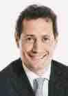
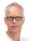
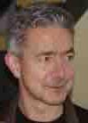
Nazzareno BASSETTI
Katrin BEKES
Florian BEUER
Corinna BRUCKMANN
Roeland DE MOOR
Danijel DOMIC
Gabor FÜRST
André GAHLEITNER
Jan-Frederik GÜTH
Erwin JONKE
Christos KATSAROS
Norbert KRÄMER
Ottmar KULLMER
Markus LAKY
Niklaus P. LANG
Eva PIEHSLINGER
Christoph RAMSEIER
Xiaohui RAUSCH-FAN
Robert SADER
Benedikt SAGL
Alexander SCHWÄRZLER
Falk SCHWENDICKE
Anton SCULEAN
Alwin SOKOLOWSKI
Andreas STAVROPOULOS
Richard STEFFEN
Georg STRBAC
Giorgio TABANELLA
Christian ULM
Tom VASKOVICH
Peter VERHEYEN
11:00-12:30
3Shape
Andreas Keßler
New possibilities of digital implant planning and immediate loading
Lunch
13:45-15:15
3Shape
Andreas Keßler
New possibilities of digital implant planning and immediate loading
Poster Session during the break
Paul P. LIN
Martin LORENZONI
Wido MENHARDT
Andreas MORITZ
Michael MÜLLER
Heinz WINSAUER
Lukas WOLSCHNER
Werner ZECHNER
Lana ZUPAN
13:45-15:15
ÖGI Next Gen
Networking in the Garden
DentUnique 1/2023 11
ČIČ ČEPIĆ
Break Break
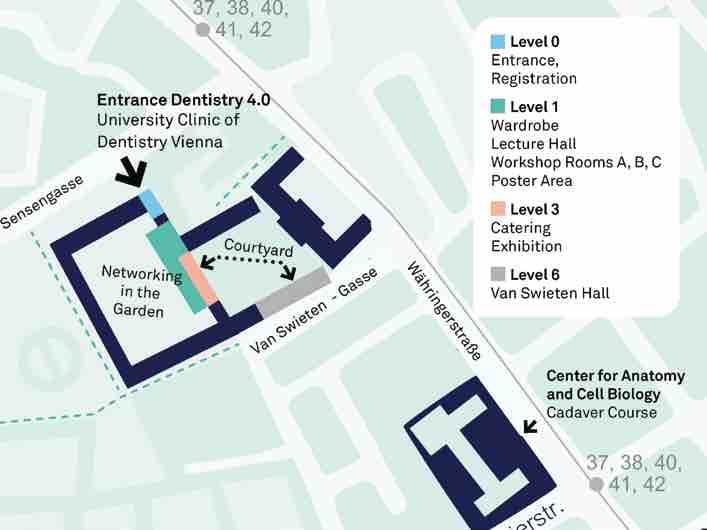
Orientation Plan
12 DentUnique 1/2023
Abstract
Speakers in alphabetical order and their topics
Minimally invasive treatment using CAD/CAM technology in patients with structural anomalies
Dr.in Sarra Altner University Clinic of Dentistry, Clinical Division of Paediatric Dentistry, Vienna, Austria

Complete dental rehabilitation in children and adolescents with abnormalities of tooth structure (amelogenesis imperfecta and molar incisor hypomineralisation) can present particular challenges in the dental practice. Due to chronic hypersensitivity and enamel loss, these teeth often require restorative therapy as early as possible. Especially the task of restoring the vertical dimension of occlusion due to defects in the enamel structure presents a considerable therapeutic challenge. This restoration requires
precise treatment planning and analysis. The aesthetic and functional aspects must be considered as a relevant prerequisite in order to ensure the success of the treatment.
The conventional methods used to create artificial teeth in pediatric dentistry are associated with many challenges. The conventional procedures followed to obtain a dental impression in children are considered to be quite awkward and difficult due to various clinical factors in dental medicine. In recent years, it has become possible to take digital impressi-
ons and successfully use them to create various types of prosthetics. This, in turn, has reduced the workload and increased patient comfort.
Various case studies of children and adolescents with anomalies of tooth structure are used to explain the digital workflow and the associated indications and advantages. The medium- and long-term options for restoring teeth with structural abnormalities, i.e. with an incursion in the enamel, will be discussed to provide insight into pediatric dentistry using digital tools.
Integration between function and aesthetics in complex cases using VieSID concepts and OMRT Bassetti
Dr. Nazzareno Bassetti Studio Dentistico, Ascoli Piceno, Italy
The plan for the treatment of complex dysfunctional and non-dysfunctional cases requires an interdisciplinary approach that involves the most common branches of dentistry and an orthodontic, prosthetic approach respecting a functional-occlusal concept according to the philosophy of Professor Slavicek and Professor Sato. The pre-therapy with an occlusal splint or with temporary prosthesis allows the algic symptomatology to be resolved and and the therapeutic positon to be tested. The subsequent orthodontic therapy using Sato‘s MEAW
philosopy, allows for the spaces of the missing teeth to be reopened, the vertical dimension to be increased, the inclination of the occlusal plane to be corrected, and the mandibular to be repositioned three-dimensionally. Implant therapy will allow you to have the back support, the control of the vertical dimension and to guide the mandible in a sagittal and traversal reposition (3D). You can proceed and verify the therapeutic position by reassembling the temporary crowns in the articulator and valuate the R.P. (reference position) and any corrections to be made on the resin so as to reach the Therapeu-
tic Position(TP). We can say that the position of the implant, the consequent bone tissue management and the subsequent prosthetic reconstructions are gnathologically guided. The control of the vertical dimension and the individual inclination of the occlusal plane allow the threedimensional repositioning of the mandible in the Therapeutic Position (TP). The BRUX CHECKER® highlighted an occlusion that was supported by the occlusal concept of Slavicek and was able to control the parafunction. This type of approach is shown to be important for the long-term stability of the complex rehabilitations.
DentUnique 1/2023 13
Speakers and Abstracts
Foto: © Studio Bassetti
Speakers and Abstracts
Molar incisory hypomineralization: Where do conventional and where do digital therapy approaches make sense?
Univ.-Prof.in Dr.in med. dent. habil. Katrin Bekes, MME1 Prof. DDr. Norbert Krämer2
1University Clinic of Dentistry, Head of Clinical Division of Paediatric Dentistry, Vienna, Austria


2Justus-Liebig-Universität Gießen, Director, Gießen, Germany
Abstract
The treatment of children with molar incisor hypomineralization (MIH) is playing an increasingly important role in dental practice. It is a frequently encountered dental condition worldwide, affecting around 13% of children. MIH is defined as the hypomineralization of systemic origin of one to four permanent first molars, which is frequently associated with affected incisors. Affected teeth are more
prone to caries and post-eruptive enamel breakdown and should be diagnosed and managed as soon as possible. Furthermore, tooth hypersensitivity is a common symptom in these patients. It is important to diagnose affected children at an early stage and to offer treatment that is appropriate with respect to the severity of the condition. The “Würzburg Concept” which was developed by an international working group with representatives from
German-speaking universities (Austria, Germany, Switzerland) can provide assistance here. The joint lecture by Prof. Bekes and Prof. Krämer will provide insights into the diagnostics and therapeutic possibilities of MIH, highlight conventional restoration techniques and discuss the potential to use the digital workflow.
Digitalization: Where are the real benefits?
Univ.-Prof. Dr. med. dent. Florian Beuer, MME Department of Prosthodontics, Geriatric Dentistry and Craniomandibular Disorders, Director, Charité Berlin, Germany
Abstract
The transition into the digital age has
are real benefits for our patients and our profession? Do our restorations last
our treatment? Some of these questions will definitely be answered with “no”.
Krämer/privat
Foto: ©
Foto: © Beuer/privat
Extraordinary Full Professor Dr. Roeland De Moor, DDS, PhD, MSc
Head of the Research Cluster of Endodontics, Department of Oral Health Sciences, Ghent University, Belgium Director of the three-year Master after Master programme in Endodontics, Ghent University, Belgium
Adjunct Professor at the Medical University of Vienna, Austria, Ghent Dental Laser Centre
Abstract
Laser-activated irrigation (LAI) with Erbium lasers was marketed in 2009. This approach is based on the creation of expanding and imploding cavitation bubbles in the irrigation solution, resulting in three-dimensional spreading and agitation of the fluid in the root canal system. Hence, it is a powerful approach to cleaning and disinfecting root canals. From activation with the fiber in the
root canal, we evolved to the use of the laser tip positioned in the pulp chamber. Originally, the fiber was positioned in the root canal and moved up and down. Together with the development of super short pulses (50 µsec), a new approach was launched in 2013. A double-pulse modality was introduced in 2018 with the goal to enhance the disinfecting and activating efficacy of SSP (super short pulse) laser-assisted activation (PIPS). This SWEEPS (Shock Wave Enhanced Emissi-
on Photoacoustic Streaming) approach consists of delivering a subsequent laser pulse into the liquid at an optimal time when the initial bubble is in the final phase of its collapse. In this presentation, the differences between the different approaches are clarified, the impact on and differences in their efficacy are demonstrated with high-speed imaging, and new insights on how LAI interacts with the biofilm are shared.
Is there an advantage of digital processing of implant abutment in prevention of peri-implant disease?
Ao. Univ.-Prof. DDr. Gabor Fürst University Clinic of Dentistry, Clinical Division of Periodontology, Vienna, Austria
Abstract
In recent years, the evidence of a relationship between restorative design factors and peri-implant health has increased and, especially, the structure of implantabutment junction and mucositis and periimplantitis. In this presentation,
the background and clinical aspects of the concept of one-abutment at one time will be highlighted. One special issue is the prevention of peri-implant disease. Factors that can influence the inflammatory reaction of the peri-implant tissue will be discussed, especially
focussing on biomarkers associated with inflammation and tissue destruction in peri-implant crevicular fluid (PICF) of implants. Finally, a case report will show the procedures used to scan, design and produce the individual abutment for dental implant.
Current cross-sectional imaging of the jaw
Ao. Univ.-Prof. Dr. André Gahleitner University Clinic of Dentistry, Clinical Division of Radiology, Vienna, Austria
Abstract
By ending analog imaging techniques and and shifting towards digital imaging, Dental Radiology has made huge advances without sacrificing well-established examination methods. With the advent of computed tomography in dental radiology, additional cross-sectional imaging techniques have recently become more and more available in dentistry as well.
Hence, it is helpful to introduce the most important ones and provide an overview. More specifically, you will learn about the diagnostic possibilities of:
• Selective-Slice Panoramic X-ray
• Dental Computed Tomography (dCT)
• Cone Beam Computed Tomography (CBCT)
• Sonography of the jaw
• Dental Magnetic Resonance Imaging (dMRI)
None of these techniques is ”better“ or ”worse“ than the other, but rather has its own strengths or weaknesses for answering a specific question or for using in an application. This presentation will give an overview of the benefits, risks and accompanying caveats when using them in the jaw region.
DentUnique 1/2023 15 Speakers and Abstracts
Laser-activated irrigation. On how the “power of the bubble” does make the difference in endodontic irrigation.
Speakers and Abstracts
Digital technologies and monolithic materials – an ideal synergy?
Prof. Dr. med. dent. Jan-Frederik Güth Director and Chair, Department of Prosthetic Dentistry, Department of Prosthodontics, Center for Dentistry and Oral Health, Goethe University Frankfurt am Main, Germany
Abstract
In the dental world, two trends are currently emerging more frequently. One is the progression of digital technology, and the other one is the minimally invasive “close-to-nature-dentistry”. The current digital development goes far beyond the use of intraoral scanners or the fabrication of dental restorations and is strongly supported by new innovative materials. Especially in the field of monolithic restorations, dentists, dental technicians
and patients have access to a wide range of new treatment strategies and potential. However, whenever new materials and workflows are introduced, we need to understand the specific potential, but even more the associated limitations to estimate what is possible, what makes sense and where there might be room for improvement.
The presentation discusses the synergy that occurs when up-to-date technology, like intraoral scanners, 3D face-scanners,
milling technology and 3D printers are combined with innovative polymer and ceramic materials. Based on evidence, clinical experience and common sense, the lecture also gives you sound background knowledge as well as clinical information combined with specific practical tips and tricks. In short, it gives you an introduction to new concepts and their integration into our daily work.
Implementation of modern 3D imaging in orthodontic diagnostics and treatment
Ao. Univ.-Prof. DDr. Erwin Jonke University Clinic of Dentistry, Medical University Vienna, Austria, Head of Clinical Division of Orthodontics Deputy Head of the University Clinic of Dentistry, Head of Special Clinic for Aligner-Therapy
Abstract
As in many other fields, digitalisation has also arrived in the field of orthodontics. New digital processes make treatment faster, more precise, and more comfortable. However, the technical innovations are developing at a rapid pace, making it increasingly difficult to keep up with them. This presentation of current promising developments provides an overview of the state of the art. Digital orthodontic workflows and new diagnostic tools include CBCT/CT imaging and its combination with digital intraoral scans. In this way, highly predictable outcomes can
be achieved when performing vital tooth transplants, because both the trauma to the root element and the extra-corporeal time needed for the autotransplantation can be minimised by careful digital planning and by fabricating 3D-printed replacement teeth. Modern dental magnetic resonance imaging (MRI) is used to achieve a high contrast between soft and hard tissue without exposure to radiation. Teeth, bone, and gums can be visualised by acquiring a single image, avoiding the need for further examinations. Since 2018, our department has been 100% digital: Digi-
tal CT, digital intraoral scans, digital photos, and virtual treatment planning have been part of our digital daily routine for years. Since 2022, dental MRI has been used as a radiation-free diagnostic tool to address specific clinical issues. These include displaced teeth, the evaluation of inadequate bone or soft tissue supply, and the differential diagnostic evaluation of inflammatory processes in the bone. Therefore, our current research focuses on dental MRI, as modern orthodontics requires both high-quality diagnostic tools for planning treatment and minimal radiation exposure for the patient.
16 DentUnique 1/2023
Foto: © Sedan7/Frank Hanewacker
Speakers and Abstracts
Digital orthodontics: Is there added value for the patient?
Prof. Dr. Christos Katsaros, PhD Director Clinic of Orthodontics, University of Bern, Switzerland
Despite the initial hesitation by the majority of the orthodontic clinicians, digitalization in orthodontics has developed rapidly during the last 20 years and has now become an integral part of the everyday clinical orthodontic practice. Digital 3D/4D-documentation, diagnosis and treatment planning, CAD/
CAM appliances, and remote monitoring of treatment have been adopted by a sizable percentage of practitioners. A digital hype is occurring with the help of intense advertisement, while artificial intelligence is considered to be the next innovation that will change the way orthodontics is practiced. Despite the fact that digital technology has helped in
solving some of previous challenges, the question still remains: Is there added value for the patient from digital orthodontics? This lecture will critically discuss the influence of digital orthodontics in diagnostics, treatment planning and treatment result quality in our way towards personalized precision medicine.
Occlusal Fingerprint Analysis (OFA) − digital view on dental evolution and function
Apl. Prof. Dr. Ottmar Kullmer Senckenberg Research Institute and Natural History Museum Frankfurt, Germany
Abstract
The evolutionary history and dynamics of the relationship of upper and lower tooth crowns in mammals provides the essential biological fundament for an interpretation of dental function and occlusion in modern humans. In our ancestors, we can only hypothesise occlusal dynamics based on dental architecture, morphology, tooth wear patterns and the reconstruction of jaw mechanics, because we are not able to test and comparably render in vivo the physiology of chewing and para-masticatory behaviour. Those facts inspired us in the Sencken-
berg Research Institute and Natural History Museum in Frankfurt am Main to develop a digital method, the Occlusal Fingerprint Analysis (OFA), for taking a quantitative approach and a detailed visualization of antagonistic occlusal kinematics derived from virtual analysis of dental wear facets and the simulation of power stroke antagonistic dental contacts.
The OFA provides a spatial map of and quantitative data for crown macrowear areas and occlusal kinematics. It records the spatial position and size of complementary dental contacts and wear facet
pairs, supporting an individual reconstruction of occlusion and of antagonistic crown surface morphology for a proper contact situation.
The presentation introduces the biological and historical background of quantitative wear facet analyses and two principal OFA components, i.e. the static macrowear mapping and the sequential analysis of occlusal contacts when applying the OFA software. Furthermore, a potential application of the virtual OFA for dental functional analysis in modern dentistry will be discussed.
DentUnique 1/2023 17
Foto: © Sven Tränkner,
Senckenberg
Foto: © Universität Bern
Speakers and Abstracts
Periodontal care using continuously the data of the patient for optimal treatment outcomes
Prof. Dr. med. dent. Dr. odont. h. c. mult. Niklaus P. Lang University of Bern, Switzerland
Abstract
Periodontal diseases represent opportunistic infections and, hence, periodontal therapy has to address an anti-infective treatment approach.
A careful history and examination of the patients provides the necessary information for the decision making process, i.e.
the diagnosis and treatment plan. The clinical parameters will subsequently be used throughout the entire treatment to evaluate treatment results and assess the quality of the treatment steps rendered. Depending on the standards reached, treatment may be stopped, and the patient may enter a practice of
life-long supportive therapy, or additional therapy may have to be provided to reach optimal outcomes. The standard of care provided will be reflected in quality standards to be reached for optimal outcomes. Quality management allows the clinician to steadily improve their performance.
Soft tissue management around teeth and implant in the esthetic zone for thin periodontium
Paul P. Lin, DDS, MS Diplomate, The American Board of Peridontology, Clinical Assistant Professor, The Ohio State University, Columbus, USA
Soft tissue is the major periodontal tissue found either around natural teeth or dental implants, contributing to gingival (so-called pink) esthetics. It takes part in all stages of the dentogingival junction with white esthetic, namely, periodontal, pontic, implant (when reconstructing) and peri-implant (when treating). Thin periodontium occurs in high percentage of Asians and makes it difficult for them to achieve comparable results as other ethnic groups. Management of such tissue requires particular attention in both non-surgical curettage and surgical
flap design, incision and flap closure. It is the purpose of my presentation to elaborate on the concept and techniques for obtaining pleasant and esthetic results for managing thin periodontium, whether in disease control or in the correction of deficiency-affecting esthetics. Definition and classification of various tissue types in terms of their health and deficiencies will be re-visited in order to establish a simple and effective treatment plan to tackle bone loss, recession, papilla loss and poor angulation around teeth and implants. Key concepts in managing thin tissues rely on the maintaining the stability
and integrity of periosteum, mucogingival junction and gingival margin. Proper surgical techniques will be demonstrated by using 3D illustration and animation to provide more clarity regarding the surgical details. Management of thin periodontium can be predictable when treating disease in the esthetic zone without causing recession and papilla loss; meanwhile, the correction of soft tissue deficiency with marginal and papillary discrepancy even with implant poor angle can be esthetically enhanced.
Digitalization, aritificial intelligence and the metaverse
Dr. Wido Menhardt
Independent advisor and consultant to digital health companies, USA
Abstract
Digitalization is revolutionizing our personal and professional lives in many ways: customer interactions, workflows, business models, access to informati-
on are all changing. In healthcare, the benefits are many, as are the risks. In this talk, we will discuss some of these aspects of digitalization before we review the impact of the next wave of digitaliza-
tion, artificial intelligence, on healthcare in particular. Finally, we will shed some light on the metaverse and its promise for healthcare applications.
18 DentUnique 1/2023
Foto:
Foto: © Lang/privat
© Lin/privat Foto: © Menhardt/privat
Latest scientific results in dental hard tissue preparation
Univ.-Prof. DDr. Andreas Moritz
Head of the University Clinic of Dentistry, Head of the Clinical Division of Conservative Dentistry & Periodontology, Vienna, Austria
Among a broad variety of indications for the use of lasers in dentistry, there is a field of application where the laser has brought major advances and specific advantages: dental hard tissue preparation. Using the Erbium laser, enamel and dentin can be ablated without thermal side effects due to the photo ablative impact of this wavelength: The laser light is absorbed by water within the hydroxy-apatite, the water is heated, and it evaporates instantly. Due to the volume change, particles of the dental hard tissues are blasted out of the tissue, and
a cavity is created. The whole procedure is almost painless and, therefore, well accepted by the patients. Composite restorations can be placed without acid etching because of the retentive surface formed by the laser. To find the right power settings and how to optimally handle the laser fiber, a comprehensive SEM study has been performed. In addition, different bonding systems have been compared, and an optimal quality of bonding has been evaluated. Investigations show that through the optimization of different influence factors, results far superior to those achieved by the use of
traditional preparation techniques can be achieved. Furthermore, you will learn the latest results of the influence of lasers on collagen fibers.
CO2, diode and erbium lasers are widely used in dentistry, showing very satisfying results. Another field of application for these wavelengths in conservative dentistry is the treatment of hypersensitive dental necks. The laser is applied at rather low energy settings in combination with fluoride gel. In most cases, one appointment is sufficient to achieve permanent freedom from pain, even in patients who suffer from severe symptoms.
Digital workflow at the University Clinic of Dentistry –an overview
Univ.-Prof. DDr. Andreas Moritz1 Mst. Tom Vaskovich, MSc2
1Head of the University Clinic of Dentistry, Head of the Clinical Division of Conservative Dentistry & Periodontology, Vienna, Austria
2University Clinic of Dentistry, Dental Technology Laboratory, Vienna, Austria
Ways to improve soft tissue around implants –in view of digital considerations
Dr. Michael Müller
University Clinic of Dentistry, Clinical Division of Periodontology, Vienna, Austria
Abstract
To avoid periimplantitis, the presence of soft tissue around implants is very important. Not only for prevention of periimplantitits but also for esthetic reasons, it is essential to have enough soft tissue of good quality. Treatment
planning in collaboration with digital planning and implant solutions can save, stabilize and improve soft tissue around implants. Interdisciplinary cooperation is very important, especially in the highly aesthetic field. The hand-in-hand work with the dental laboratory with regard to
material-technical conditions and biological factors is also important. Planning, key factors and patient management in light of the digital world in dentistry will be discussed.
DentUnique 1/2023 19
Speakers and Abstracts
Speakers and Abstracts
Periodontology – title and abstract will be announced soon

PD Dr. med. dent. Christoph Ramseier, MAS
University of Bern
Department of Periodontology, Switzerland
Decision of therapeutic position in interdisciplinary treatment of temporomandibular joint disorders
Univ.-Prof.in Univ.-Doz.in DDr.in Xiaohui Rausch-Fan1
O. Univ.-Prof.in DDr.in Eva Piehslinger2
1University Clinic of Dentistry, Deputy Head of the Clinical Division of Periodontology, Vienna, Austria
2University Clinic of Dentistry, Head of the Clinical Division of Prosthodontics, Vienna, Austria
Abstract
In the rehabilitation and reconstruction of functional occlusion, in particular for patients with temporomandibular joint disorders (TMD), big challenges are still associated with digital technology for defining therapeutic position of temporomandibular joint (TMJ). Diagnostic procedures like anamnestic inquiry, clinical functional analysis, occlusal
of the sagittal x-rays, are important for case planning. In complex cases where we have to define a joint-related jaw position, the pretreatment with occlusal splints is mandatory. In patients with internal derangements, we try to position the condyle three-dimensionally in the Mandibular Position Variator (MPV), according to axiographic data. This lecture will present a functional diagnostic system, a therapeutic method of controlled
interdisciplinary treatment approaches for rehabilitation of therapeutic position of TMJ. Namely, for TMD cases resulting from occlusal discrepancy, the therapeutic position is defined by mandibular repositioning splint therapy, further supported by orthodontic and prosthodontic treatments. The innovation and limitations of applying digital technology for establishing the functional occlusion of TMD cases will be discussed.
Foto: © Universität Bern
Speakers and Abstracts
The effects of digital dentistry workflow on the development of innovative technologies
Univ.-Prof. Prof. h. c. mult. Dr. mult. Robert Sader Department of Oral, Cranio-Maxillofacial and Facial Plastic Surgery, University Hospital Frankfurt, Frankfurt, Germany
The development of a digital workflow based on new medical 3D devices has changed the world of dental treatment. Optimizing the treatment result adds to the increasing complexity of the treatment`s workflow; increasing costs are an additional side effect. Innovations have created not only new possibilities, but also new problems and hazards. This increases the demand not only to develop new other technologies, but also to include more fundamental biological principles in reconstructive oral surgery. The presentation should clearly show
that the goal should be to imitate nature more and more and not to replace biological approaches by technical ones. As a consequence, old techniques like osseofixation could experience a revival and could be an alternative to osseointegration. But if it becomes possible to use new digital technologies and combine these with physiologic principles, then new application fields could be established for the benefit of the patients. To prove these new possibilities of 3D operation, planning and CAD/CAMbased manufacturing of patient-specific implants in dental implantology will be
presented. It will be shown that using new digital tools lead directly to surgical innovations, such as the development of new surgical devices like laser osteotomy or the production of 3D-printed dental implants to achieve further improvements of the esthetic and functional result in oral reconstructions. In this way, implantbased dental restorations will be made possible even in complex patients who could not be treated up until now. The next step will be the inclusion of artificial intelligence.
Computational modeling of temporomandibular joint biomechanics
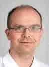
Benedikt Sagl, PhD Research Associate at the University Clinic of Dentistry, Vienna, Austria
Abstract
Functional impairment of the masticatory region can have a significant impact, ranging from a loss of quality of life to severe health issues. Commonly, increased temporomandibular joint (TMJ) loading is connected with temporomandibular disorders (TMD). Due to the small size and complex structure of the masticatory region, in vivo investigations of TMJ biomechanics are difficult to perform. Hence, biomechanical computer simulation remains an important tool for the investigation of the function of this complex system. This talk will present current computational modeling projects
performed by our group at the University Clinic of Dentistry, focusing on TMJ stress during chewing and bruxing. Our investigations use a novel biomechanical computer model that enables dynamic, muscle-driven simulations of mandibular function. The computed model was used to thoroughly investigate the effect of tooth morphology and position on TMJ biomechanics during laterotrusive tooth grinding. Additionally, we investigated the effect of food bolus parameters on TMJ biomechanics during a unilateral chewing cycle. Our bruxing simulations suggest that steeper teeth have a protective effect on TMJ loading, which might suggest a benefit to a lateral
canine guidance. Moreover, our chewing simulations show decreased loading of the TMJ when chewing softer and smaller food pieces, which supports the current TMD self-management guidelines.
Overall, the aim of this talk is to highlight various projects that use computational modeling approaches to study research questions that are virtually impossible to perform in vivo without the serious risk of harming patients. These computational investigations can be used to improve our understanding of the masticatory system in a safe and controlled manner and are hence a highly relevant tool in modern dental research.
DentUnique 1/2023 21
Foto: © Sader/privat
Speakers and Abstracts
CAD/CAM orthodontic appliances for complex cases: The fully digital workflow
Dr. Alexander Schwärzler University Clinic of Dentistry, Clinical Division of Orthodontics, Vienna, Austria
Abstract
The digital revolution in dentistry opens up various possibilities in orthodontics and interdisciplinary communication. Treatment planning based on digital records is considered to be standard today. To further expand the opportunities for treating complex orthodontic cases, a CAD/CAM protocol was established at the Orthodontic Department at the University Clinic of Vienna. Softwarebased appliance design and laser
melting technology are the main tools the speech focuses on. Starting with the evolution of customized CAD/CAM appliances, the “Vienna Palate Expander” and the “Vienna Palate Guide” will be presented to the dental audience. To connect the interdisciplinary dots between various dental fields, the “Virtual Orthodontic Setup” creates links in terms of communication and provides visualization of comprehensive treatment plans; examples will be
shown. Furthermore, the function and accuracy of 3D-printed Indirect Bonding Trays and the importance of the virtually placed orthodontic Temporary Anchorage Devices (TAD) will be discussed, accompanied by evidence from today‘s literature. Finally, the speech will be underlined with comprehensive clinical cases having one thing in common: the fully digital workflow.
Digital dentistry and artificial intelligence: A revolution ongoing?
Univ-Prof. Dr. med. dent. Falk Schwendicke, MDPH Head of the Department of Oral Diagnostics, Digital Health and Health Service Research at Charité – Universitätsmedizin Berlin
Abstract
At the dawn of the data era, medicine and dentistry are seeing change all over the place: Artificial intelligence (AI) revolutionizes diagnostics and treatment planning, medical apps place the patient into the center of his or her healthcare, augmented and virtual reality increasingly support treatment conduct, and
with the coronavirus pandemic as fuel, telemedicine and dentistry are finally taking off. The lecture will discuss why we see this change right now, i.e. what drivers are behind it, and will systematically lay out the various domains doctors and dentists need to consider: Data-driven, patient-centred, automated and increasingly remote and digital care.
For dentists, the message will be clear: Having access to new digital technologies, knowing their chances and risks, and being able to integrate them into their workflow and their practice will be a must if you plan to stay in healthcare in the next decade and beyond.
22 DentUnique 1/2023
Foto: © Schwendicke/privat
Abstract
Digital technologies in implant treatment: Surgical and restorative considerations
Dr. med. dent. Dr. med. univ. Dr. scient. med. Alwin Sokolowski1 Martin Lorenzoni, MD, DDS, PhD2
1Division of Restorative Dentistry, Periodontology and Prosthodontics, Medical University of Graz
2Department of Prosthodontics (Prosthetics, Periodontology, Restorative Dentistry and Implantology) University Dental Clinic, Medical University Graz, Austria
How can modern technologies help in diagnostics, treatment and follow-up?
Patients with previous illnesses, conditions following chronic inflammation or tooth trauma, and sites with alveolar bone deficiencies as well as high esthetic
demands represent a particular challenge for the restorative dentist. Modern technologies have expanded the horizon for new therapy options, and existing approaches have been simplified, accelerated and improved. Using highly developed implant systems in
combination with digital techniques enables treatment that meets high standards and leads to predictable and precise results. These approaches in implant surgery and prosthodontics should be discussed.
Management of peri-implantitis: A pragmatic approach
Prof. Dr. Andreas Stavropoulos University of Malmö, Sweden
Peri-implantitis is a major burden in everyday praxis, with a large and increasing number of patients suffering from it. Management of peri-implantitis
is often complex, and current approaches consist first of anti-infective, non-surgical measures, followed in the majority of cases by surgical intervention. The lecture presents the decision-making
process for a pragmatic management of peri-implantitis, and an outlook for the future in the Dentistry 4.0 era.
Dental craftsmanship meets high-tech stories from daily dental practice in which challenging situations must dealt with innovative technologies or trend-setting solutions
Dr. Richard Steffen Clinic for Pediatric Dentistry and Orthodontics, University of Zurich, Zurich, Switzerland
Working as a dentist means a continuous demand to keep up to date. Nowadays, it is necessary to keep up with the latest developments in our field of expertise. This is in particular true for paediatric dentistry, because our patients are the most vulnerable and, as professionals, we have an even greater responsibility to take care of them. In addition, the profile of requirements for the dental treatment
of paediatric patients is very complex and involves almost all fields of dentistry. However, there is no question that, even in rare cases, treatment should be given with the best possible therapy option. Such challenging situations must also be able to be mastered in everyday life by “normal” practitioners. This requires a constant search for new treatment methods, improvements to existing treatment recommendations and personalised
treatment plans adapted to the paediatric patient. Examples are used to show the diversity of these medical challenges. An algorithm shows how the best possible treatment option can be selected in a scientifically correct way and in the best interest of the child. With a bit of luck, this might even turn into a successful treatment trend.
DentUnique 1/2023 23
Speakers and Abstracts
Foto: © Sokolowski/privat Foto: © Steffen/privat Foto: © EFP Foto: © Lorenzoni/privat
Speakers and Abstracts
Novel clinical applications of computer-guided surgery
Assoc. Prof. Priv.-Doz. Dr. med. dent. et scient. med. Georg Strbac University Clinic of Dentistry, Clinical Division of Oral Surgery, Vienna, Austria
Abstract
In current dental implant treatment, accurate clinical and radiological examination can enhance treatment planning, ensuring an atraumatic and minimally invasive surgical procedure. Modern diagnostic techniques, such as 3D imaging, not only allow imaging of anatomic structures for accurate diagnosis and treatment planning but can also be used for the fabrication of surgical models and for the construction of surgical templates in guided implant
surgery. Today, the latest implant planning software programs can simplify the previously used template production techniques by shortening the fabrication process and by avoiding additional radiographic exposures. This can be achieved directly after the initial 3D radiographic assessment by merging the uploaded radiographic data with the STL files of scanned dental models or intraoral scans of the jaws to fabricate accurate surgical templates prior to the intervention. In addition to the
virtual planning of dental implants and prosthodontics reconstructions, these modified computer-supported planning and guided surgical approaches can also be used for sinus grafting procedures. Additionally, new surgical methods could be developed to ensure precise and safe guided osteotomies for future treatment methods. Moreover, recently introduced innovative techniques such as Guided Autotransplantation of Teeth and Guided Modern Endodontic Surgery have become reality.
Digital soft tissue mapping for peri-implant soft tissue augmentation procedures
Dr. Giorgio Tabanella
DDS, MS Diplomate of the American Board of Periodontology, O.R.E.C.-Oral Reconstruction and Education Center, Rome, Italy
Abstract
As long as the implants are inserted in a 3D prosthetic-driven direction and surrounded by a minimum volume of bone matrix, the implant treatment can be safely considered as long-lasting. However, the fact that an implant is properly functioning is not necessarily associated with success. In fact, many dental implants can present esthetic or functional alterations in the soft tissue morphology and texture that may jeopardize the success rate. Soft tissue defects around dental implants, such as papilla or volume loss, peri-implant recession and alterations of the ridge color and/or texture, lead to critical esthetic and functional critical issues. Treatments of these defects around implants are more demanding than in teeth because peri-implant tissue exhibits different anatomical and histological characteristics. Recent studies have shown that the peri-implant mucosa
can play a major role in the sealing of the implant platform, thus preventing bacterial ingrowth. A good quality and quantity of the mucosa is also critical for achieving biomimetics and pink esthetics. It has been reported that about one-third of the inserted implants may then require a connective tissue graft. However, this approach could be considered to be too invasive when multiple implants need to be treated with mucogingival plastic procedures. The “Buccal Pedicle Flap” and its modifications will be presented as a novel technique to boost the peri-implant mucosa without the need for invasive procedures, thus allowing the dentist to obtain the desiderable mucosa volume and quality. This approach would lead to more stable tissues, no peri-implant bone loss or peri-implantitis and ultimately a higher success rate in modern implant dentistry.Finally, recent studies will be presented to describe digital soft tissue
mapping to monitor the stability of the augmented soft tissue collar but also to properly plan the reconstruction of the pink esthetics.
OBJECTIVE:
• understand the importance of the peri-implant mucosa
• be able to detect the critical numbers for mucosa health
• evaluate the surgical alternatives to boost the peri-implant mucosa
• understand in details the critical steps to perform the “Buccal Pedicle Flap”
• obtain a full understanding of the concept of mucosa integration and soft tissue sealing
• understand how to prevent or treat complications and failures in dental medicine
• learn how to use digital technology for soft tissue mapping and monitoring
24 DentUnique 1/2023
Foto: ©
Tabanella/privat
Speakers and Abstracts
Insight in the photochemistry of laser- and light-activated systems
Peter Verheyen, DDS Private Practice, Gruitrode, Belgium
Abstract
In 1917, Albert Einstein published a paper ‘On the Quantum Theory of Radiation’, which resulted many years later in the development of the first laser devices. However, already in 1905 he described ‘The Photoelectric Effect’, for which he received a Nobel Prize in 1921. In this article, Einstein explained how photo-
nenergy can interact with matter. This formed the basis for the study of photochemistry where photonenergy induces chemical reactions. The most commonly known of these is the photochemistry in the chlorophyll of plant cells, which creates life on Earth. But also in our daily work in dentistry, we use photochemistry all the time. It is the intention in this
lecture to summarize our actual insights on how laser and light interactions with matter and tissues induces the effects we experience and use in our patient treatments. And, as an example of this, we show how photochemistry may form the basis on how dental bleaching without the use of hydrogen peroxide may be possible.
Digitally aided bone augmentation concepts in implant surgery
Univ.-Prof. DDr. Werner Zechner University Clinic of Dentistry, Clinical Division of Oral Surgery, Vienna, Austria
Abstract
Bone atrophy and defects due to inflammatory or traumatic lesions still increase the complexity of implant treatments. For the patients and for the surgeons, the efforts needed regarding time and technical skills as well as the incidence of complications can increase substantially, also depending on the
chosen treatment concept. Furthermore, this can aggravate the decision-making related to the choice of an appropriate bone augmentation concept. Digital planning and computer-aided manufacturing has become an important part also in oral surgery: In this lecture, various surgical concepts are introduced which are implemented at the University Clinic
of Dentistry Vienna for the regeneration of vertico-lateral bone defects before implant placement, including CAD/CAMallogenic block bone augmentation and 3D-printed titanium meshes. Surgical challenges, limits and advantages of these techniques and additional surgical measurements will be presented.
Evolution of complete dentures in the digital age –update on possibilities and boundaries in clinical practice
Dr.in Lana Zupančič Čepić University Clinic of Dentistry, Clinical Division of Prosthodontics, Vienna, Austria
Abstract
Digital denture technology has considerably changed the manufacturing of complete and implant-supported dentures with the aim of accelerating and facilitating everyday clinical practice. The fundamental principles, however, are still the same - an accurate impression of edentulous jaws and recording of the
jaw relationship with a well-thoughtout occlusal concept are the key to successful prosthodontic treatment.
The Viennese Occlusal Concept works in natural teeth as well as in fixed and removeable prosthodontic rehabilitations, including implant-supported restorations. This lecture will provide a brief overview of the fundamentals and an
update on digital concepts for complete dentures by focusing on advantages and limitations reported so far. Furthermore, the treatment efficacy of digital versus conventional dentures will be discussed based on clinical and patient-based outcomes from a clinical study conducted at the University Clinic of Dentistry Vienna.
DentUnique 1/2023 25
Foto: © Verheyen/privat
Workshops
Strahlenschutzkurs für Zahnärzt:innen
Ort: Universitätszahnklinik Wien, Sensengasse 2a, 1090 Wien
Großer Hörsaal
Datum: Donnerstag, 1. Juni 2023
Zeit: 17:00-21:00
Referent:
MR Dr. Franz Hastermann
(Präsident des ZIV, Präsident des ÖGHZ)
Kosten: Die Gebühr ist in der Registrierungsgebühr inkludiert (Anmeldung erforderlich!)
Programm:
Seit 1.1.2011 besteht die Verpflichtung für Strahlenschutzbeauftragte, alle 5 Jahre eine geeignete Fortbildung von mindestens 4 Stunden (sofern die Tätigkeit auf eine Zahnarztordination beschränkt) ist, nachzuweisen. (§ 82 (1) Allgemeine Strahlenschutzverordnung
v. 1.8.2020) Das Bundesministerium für Soziales, Gesundheit, Pflege und Konsumentenschutz hat schriftlich bescheinigt (2021-0.690.478), dass
die Veranstaltung den Forderungen des § 82 Abs. 1 Z 1 der Allgemeinen Strahlenschutzverordnung 2020, BGB. Il Nr. 339/2020, entspricht. Rechtliche Grundlagen und deren praktischen Auswirkungen für Bewilligung, Überprüfung und Betrieb werden ebenso besprochen wie bauliche Konsequenzen. Nutzen und Grenzen neuer Verfahren und Aufnahmetechniken (z.B.: DVT) mit Fallbeispielen runden die Veranstaltung ab.
Newly emerging digital workflows in bone-borne devices – Friday, June 2
Venue: University Clinic of Dentistry, Sensengasse 2a, 1090 Vienna
Seminar Room A
Date: Friday, June 2, 2023
Time: 09:30-17:50
Speaker:
Dr. Heinz Winsauer
Registration Fee: ¤ 290 (Due to limited space, registration is on a first-come-firstserve basis.)
Programme:
09:30-11:00 Part I
Palatal and buccal-shelf applications of mini screws in everyday practice
11:00-11:15 Morning Break
11:15-12:25 Part II
Trend digitization - opportunities and risks for case planning and workflow with mini screws
12:25-12:40 Lunch break
12:40-13:40 Part III
Scannerkurs für Assistent:innen –Freitag, 2. Juni
Ort: Universitätszahnklinik Wien,
Sensengasse 2a, 1090 Wien, Seminarraum C1
Datum: Freitag, 2. Juni 2023
Zeit: 09:30-12:30
Kosten: Die Gebühr ist in der Registrierungsgebühr inkludiert (Anmeldung erforderlich!)
Programm:
Theoretische Einführung mit anschließenden praktischen Übungen
Cadaver course – Friday, June 2
Venue: Institute of Anatomy, Währinger

Straße 13, 1090 Vienna
Date: Friday, June 2, 2023
Time: 09:30-13:00
Speakers:
Ao. Univ.-Prof. DDr. Christian Ulm
Univ.-Prof. DDr. Werner Zechner
Dr. Danijel Domic
Programme:
Testing and comparing different workflows with digital components in an everyday practice routine including Hands-on Workshop in Orthodontics
13:40-15:20 Afternoon Break
15:20-17:50 Part IIII
Non-surgical maxillary suture opening in adults
Scanner course for dental assistants (German language) – Friday, June 2
Venue: University Clinic of Dentistry, Sensengasse 2a, 1090 Vienna, Seminar Room C1
Date: Friday, June 2, 2023
Time: 09:30-12:30
Course Fee is included in the registration fee. Registration is required in order to take part. First come, frst served!
Programme:
Theoretical introduction followed by practical exercises
Anatomical structures and their variants relevant to oral implantology (atrophic alveolar ridge, maxillary sinus, mandibular canal, local bone quality) Sinus floor elevation techniques (lateral and transcrestal sinus lift technique)
26 DentUnique 1/2023
Fotos: © carmenbobo stock.adobe.com
(only approved for Austrian dentists, German language) – Donnerstag, 1. Juni
Strahlenschutzkurs
Ort: Universitätszahnklinik Wien, Sensengasse 2a, 1090 Wien Seminar Room C2
Datum: Freitag, 2. Juni 2023
Zeit: 13:45-17:15
Referent: Dr. Dino Imsirovic (Vizepräsident des ZIV)
Kosten: Die Gebühr ist in der Registrierungsgebühr inkludiert (Anmeldung erforderlich!)
Programm:
Seit 1.1.2011 besteht die Verpflichtung für Strahlenschutzbeauftragte, alle 5 Jahre eine geeignete Fortbildung von mindestens 4 Stunden (sofern die Tätigkeit auf eine Zahnarztordination beschränkt) ist, nachzuweisen. (§ 82 (1) Allgemeine Strahlenschutzverordnung
v. 1.8.2020) Das Bundesministerium für Soziales, Gesundheit, Pflege und Konsumentenschutz hat schriftlich bescheinigt (2021-0.690.478), dass
Red-white Aesthetics Management – Friday, June 2
Venue: University Clinic of Dentistry, Sensengasse 2a, 1090 Vienna
Pre-Clinic
Date: Friday, June 2, 2023
Time: 13:45-16:00
Speakers:
Dr. Michael Müller
Dr. Lukas Wolschner
Registration Fee: ¤ 200 (Due to limited space, registration is on a first-come-firstserve basis.)
Programme: Pathological impact of gingival recession on dental healthy and its aetiology has been discussed over the years. Neverthe-
less it can lead to functional limitations and be one of reasons for orthodontic, prosthodontic and implantological complications. However, the trend seen today of increasing numbers of such patients in plastic surgery treatment is mostly due to aesthetic demand. Plastic surgery for treatment gingival recession is generally considered to be a challenge in surgical technique, however, relevant indications and periodontal preand post-operative care are essential for clinical successful outcome. Therefore, the morphological alteration of marginal gingiva for esthetical and functional demands precise the surgical design and technique.
Amann Girrbach – Friday, June 2
DRS in teams – the intraoral scan “let’s go digital”
Venue: University Clinic of Dentistry, Sensengasse 2a, 1090 Vienna
Seminar Room B
Date:
Friday, June 2, 2023
Time: 09:30-10:30 / break / 11:00-12:30
Speakers:
Wibke Rosin, Markus Hares
Registration Fee: ¤ 100 (Due to limited space, registration is on a first-come-firstserve basis.)
Programme:
In this workshop, the product specialists Wibke Rosin and Markus Hares illustrate the path from digital scanning to manufacturing crowns using the new, innovative system solution Ceramill DRS from Amann Girrbach.
The digital workfow is presented as a whole and the 3 kits with intraoral scanner, milling unit and sintering oven, as well as the digital platform AG.Live, are discussed in detail. The lecture also deals with the integration of digital
die Veranstaltung den Forderungen des § 82 Abs. 1 Z 1 der Allgemeinen Strahlenschutzverordnung 2020, BGB. Il Nr. 339/2020, entspricht. Rechtliche Grundlagen und deren praktischen Auswirkungen für Bewilligung, Überprüfung und Betrieb werden ebenso besprochen wie bauliche Konsequenzen. Nutzen und Grenzen neuer Verfahren und Aufnahmetechniken (z.B.: DVT) mit Fallbeispielen runden die Veranstaltung ab.
This theoretical lecture accompanied by hands-on training with pig jaws focuses on the following topics:
1. Aetiology of gingival recession and indication for different mucogingival surgeries
2. Microsurgery concepts in periodontal plastic surgery
3. Surgical procedures with emphasis on:
a. Vestibular extension procedures
b. Root and recession covering procedures: Gingival grafts, connective tissue grafts and alternatives with collagen-based biomaterials.
Satellite Workshops
temporomandibular joint measurement with Zebris for Ceramill. The fully digital approach makes it possible to produce small restorations with a high degree of patient comfort in just one appointment. The cooperation in the interdisciplinary team works quickly and effciently – even with larger restorations
DentUnique 1/2023 27
Workshops
für Assistent:innen (only approved for Austrian assistants, German language) – Freitag, 2. Juni
Satellite Workshops
Oral-B – Friday, June 2
Current Challenges in Paediatric Dentistry
Venue: University Clinic of Dentistry, Sensengasse 2a, 1090 Vienna Seminar Room B
Date:
Friday, June 2, 2023
Time:
13:30-15:15 / break / 15:45-17:30
Speakers:
Prof. Dr.in Katrin Bekes
Prof. Dr. Norbert Krämer
Registration Fee: ¤ 100 (Due to limited space, registration is on a first-come-firstserve basis.)
Programme:
Early childhood caries (ECC) is still a highly prevalent disease affecting millions of preschool children worldwide. Currently, 13.7% of 3-year-olds already have early caries in the primary denti-
Dentsply Sirona – Saturday, June 3, Workshop 1
Digital Impression
Venue: University Clinic of Dentistry, Sensengasse 2a, 1090 Vienna
Seminar Room A
Date:
Saturday, June 3, 2023
Time: 09:00-10:30
Speaker:
Dipl.-Ing. Roland Felber
Registration Fee: ¤ 100 (Due to limited space, registration is on a first-come-firstserve basis.)
Programmme:
Digitization is also progressing unstoppably in dentistry. This is how digital impressions with an intraoral camera fnd their way into the dental practice. Today, around 25% of dental practices have a scanner for digitizing the patient‘s dentures. But how should an intraoral camera be handled? What possibilities does this open up for the dentist and what should be considered when using it?
The “Digital Impression” workshop starts with the preparation and hygiene measures in the treatment room and on the 3D scanner. After the patient case has been created, scanning and handling of
Dentsply Sirona – Saturday, June 3, Workshop 2 Chairside Workflow
Venue: University Clinic of Dentistry, Sensengasse 2a, 1090 Vienna Seminar Room A
Date:
Saturday, June 3, 2023
Time: 11:00-12:30
Speaker: Dipl.-Ing. Roland Felber
Registration Fee: ¤ 100 (Due to limited space, registration is on a first-come-firstserve basis.)
Programme:
Optimized workfows and the resulting time and cost savings are the challenge
of new developments and future-oriented technologies. With the “Chairside Workfow” of dental CAD/CAM systems, the dentures produced on the treatment unit not only save the patient a second visit to the dentist. The practice can also allow the saved follow-up appointment to another patient and thus use the treatment time effciently.
The digital data of the impression enable the restoration to be fabricated in just a few minutes. In the workshop, the participants go through the production of a single-tooth restoration. The digital impression and the design of the dentures are carried out step by step. The optimized parameter settings as well as the initial
tion. Although it is not life-threatening, ECC negatively impacts the quality of life as it may lead to infections, swelling, pain, and other symptoms. Notably, ECC is also a global public health burden, medically, socially and economically. The aims of the workshop are to present current preventive approaches and restorative options for the treatment of carious primary teeth.
the IO camera for the digital impression begins. The participants create several scans of different patient situations (quadrant scan, full jaw scan and implant situation). The options for post-processing the scan data are presented, as well as for quality control of the impression. Finally, the advantages of digital impressions are shown and applied. These are the storage of the scan data in different formats, fast transmission via the Internet and the multiple design and analysis options on the computer.
To ensure an intense , only 2 participants will work on one scanner.
proposal for the restoration and the manual design tools are also discussed. Subtractive and additive manufacturing processes offer a wide range of esthetic materials and at the same time guarantee consistent quality of the restorations. Getting to know the range of materials available for the chairside workfow is on the agenda. The applications for zirconium, lithium disilicate up to PMMA are presented here. Whether milling, grinding or printing, the workshop presents the individual manufacturing processes with their requirements and advantages.
To ensure an intense training, only 2 participants will work on one scanner.
28 DentUnique 1/2023
REGEDENT GmbH – Saturday, June 3
Paradigm shift – successful nonsurgical treatment of deep periodontal and peri-implant infections
Venue: University Clinic of Dentistry, Sensengasse 2a, 1090 Vienna Seminar Room C
Date:
Saturday, June 3, 2023
Time: 09:00-10:30
Speaker:
Univ.-Prof. Dr. Anton Friedmann
Practice-relevant treatment options for dentists, oral surgeons or maxillofacial surgeons working in periodontology and implantology, as well as committed dental hygenists.
Registration Fee: ¤ 100 (Due to limited space, registration is on a first-come-firstserve basis.)
Programmme:
Despite careful post-surgical oral hygiene protocols and regular SPT intervals, deep pockets and infammatory reactions can persist around teeth or implants, leading to resorption of the surrounding
3Shape – Saturday, June 3
tissue and increasing the risk of tooth or implant failure.
Usually, an invasive, surgical treatment is performed which is undesirable for the patient. Due to the complexity, this treatment is usually only carried out by experienced practitioners. Therefore, practitioners are longing for effective, less-invasive non-surgical protocols for subgingival treatment.
Previously established non-surgical protocols for subgingival treatment often do not lead to a lasting clinical improvements in the probing depths and attachment level.
The latest publications impressively demonstrate that the combination of a cleaning gel based on sodium hypochlorite and cross-linked hyaluronic acid (xHyA) represents a promising and superior material combination for the non-surgical treatment of periodontal and peri-implant infammation. In this workshop, it will be fundamentally explained on which fndings this so-called Clean & Seal® concept is based
and what differs to established treatment methods. The possibilities and limitations of this new, minimally invasive method are described based on various clinical case studies.
CV Uni.-Prof. Dr. Anton Friedmann: University professor and Head of the Periodontology Department, Department of ZMK, Faculty of Health, University Witten/Herdecke (GER)
Specialist Periodontology (DGP) Specialized Dentist for Periodontology (LZÄKWL)
ITI Speaker
ITI Fellow
Director of ITI Study Club Witten
Research focus and national and international speaker activities on bone augmentation, periodontal regeneration, implantology in patients with periodontal harm, soft tissue design and prosthetic concepts in periodontology. Numerous publications in international peer-reviewed journals.
New possibilities of digital implant planning and immediate loading
Venue: University Clinic of Dentistry, Sensengasse 2a, 1090 Vienna Seminar Room B
Date:
Saturday, June 3, 2023
Time: 11:00-12:30 / break / 13:45-15:15
Speaker: PD Dr. Andreas Keßler, MSc
Registration Fee: ¤ 100 (Due to limited space, registration is on a first-come-firstserve basis.)
Programme:
The focus of this workshop will be on the hands-on part. After a short theoretical introduction, you will learn how to scan an implant case with the 3Shape TRIOS.
OCMR – Saturday, June 3 – a hands-on workshop
Based on the scan, you will proceed with a complete digital implant backward planning with immediate loading using 3Shape studio apps. To ensure an intense training, only 2 participants will work on one PC, and the planned case will be a real implant case from our speaker PD Dr. Andreas Keßler.
Automated diagnostics through AI (artificial intelligence) in oral surgery:
How useful are these new systems in daily practice?
Venue: University Clinic of Dentistry, Sensengasse 2a, 1090 Vienna
Seminar Room A
Date:
Saturday, June 3, 2023
Time: 13:45-15:15
Moderator:
Assoc. Prof.in Priv.-Doz.in DDr.in Ulrike Kuchler
Speaker: tba
Registration Fee: ¤ 100
DentUnique 1/2023 29
Satellite Workshops
Posters
Dentistry 4.0 – Posters
Present your work at Dentistry 4.0
The Scientific Organising Committee is pleased to invite you to submit a poster or oral presentation abstract for consideration for Dentistry 4.0.
Abstract submission guidelines
Please read the instructions carefully.
Abstract format
• The text must not exceed 250 words, excluding title and authors.
• Grant references must be included at the end of the abstract text.
• A maximum of 50 authors with 3 institutions per author is allowed. However, the size of the author block must not exceed 3,000 characters.
• Tables may be included.
• No graphics are allowed.
• Remember that you may be presenting author of only one paper, but co-author of any number of abstracts.
• Download a sample abstract with instructions
Before you get started…
Type your abstract using a word processing program (e.g. MS Word). Please save
Submitted pre-clinical research abstracts as well as clinical research abstracts can be considered for oral presentations (as ‘Short Talks’) and for poster presentations.
a copy of the file with the abstract text only (no title or authors). This file can then be used for a “file upload” (most convenient way of submission). The upload feature supports and converts tables within the document. Special characters are widely supported.
When entering the submission portal… You will have to create a new user account by clicking on “Create new account”. Fill in all required fields and save your personal information by clicking “Save & Continue” in the online form.
Important notes:
The abstract submission system used by Dentistry 4.0 (OASIS by CTI) is based in the United States. Hence, by submitting an abstract to our meeting, you agree to submit personal data (name, institution address, email address) to a ‘data processor’ located outside of the EU. It is CTI’s policy to be compliant with the European
Research Forum & Poster Session
Presentation of best clinical research posters: Short Talks Seminar Room C
MODERATORS:
Prof. Ulrike Kuchler
Prof. Xiaohui Rausch-Fan
Presentation of best pre-clinical research posters: Short Talks Seminar Room C
MODERATORS:
Prof. Reinhard Gruber
Prof. Oleh Andrukhov
Jury Members: International Scientific Committee
General Data Protection Regulation (GDPR), see here: https://www.ctimeetingtech.com/privacy-policy.
How to start the abstract submission… Once you have logged into the submission portal, you will be directed to the abstract submission site for Dentistry 4.0. Click on the “+New” button to start with your abstract submission. If you cannot see this button, please make sure you are located in the tab labelled “submissions”. When clicking the “+New” button, a new tab will open in your browser. Follow the step-by-step instructions for submission.
At the end of the process, you will see a summary of your submission in the last step (“Review my work”), which you may print. Your abstract should now show as complete in the submission site.
Dentistry 4.0 Research Prize
June 3 at closing ceremony
The three best presentations presented in two categories during the Research Forum will be awarded a prize.
30 DentUnique 1/2023
Silver Sponsoring
Sponsors and Partners
Platin Sponsoring
Satellite Workshops
Bronze Sponsoring


DentUnique 1/2023 31
Sponsors and Partners

Exhibitors as of March 2023
Dentistry 4.0 is organised in cooperation with following dental societies
österreichische gesellschaft esthetic dentistry

32 DentUnique 1/2023
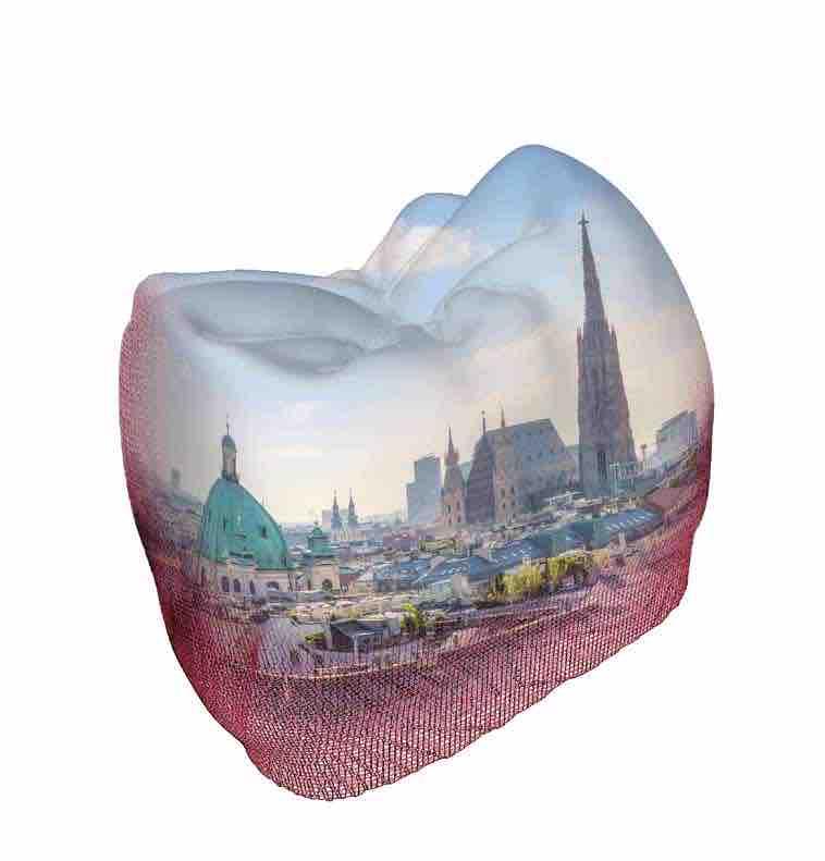
DentUnique 1/2023 33 Register now! www.dentistryvienna.com Mit Strahlenschutzkurs für Zahnärzt:innen und Assistent:innen!
Registration Fees
Payment received
Early Bird (until April 30, 2023)
All registration fees include 20% Austrian VAT.
1Applies to undergraduate & graduate students (MSc/PhD) up to the age of 27. You will need to upload a copy of a Student ID or a confrmation signed by the head of department when you register.
ÖGZMK members have a 10% reduction on the applicable registration fee. Proof of membership is required. When registering for the conference, you will have to agree to the General Terms & Conditions for participants.
What is covered by the registration fee?
During the conference:
• Access to all scientific plenary sessions, company exhibition and poster area
• Electronic Abstract Book and Programme
• Coffee/Tea, drinks, snacks during coffee breaks and lunch buffet during the lunch breaks from June 2–3, 2023
Conference registration
By registering for the meeting, you agree to complete an independent health check prior to entering the event side. (Do you have cough? Do you have shortness of breath? Did you have contact to a person who tested COVID-19 positive?)
In case of symptoms of disease, suspected or confirmed COVID-19 infection, do NOT enter the venue under any circumstances.
This responsibility is subject to the requirements of the Federal Ministry’s COVID-19 guidelines, applicable at the time. In case of non-compliance with these or any regulations set by the authorities, the organiser is free to ask the person concerned to leave the venue. In this case, a refund of the registration fee is excluded.
Registration to the event should be made online prior to the event.
Only fully completed online registrations will be accepted. An invoice will be sent after the receipt of your registration, and a confirmation of payment will be sent upon receipt of payment in full. If the full payment has not been received before the deadline indicated, the registration will remain valid, but the due fee will be increased according to the payment period (early/regular/onsite).
To be able to register as a student, individuals must present proof of full-time enrolment at a recognised university or college or an according programme (i.e. student ID, confirmation of head of department, etc.) by uploading their student ID during registration or by sending a photo of the ID by email immediately after completing the registration.
The registration fee for registered participants includes entry to all scientific plenary sessions, the company exhibition and poster area, the opening and the closing session, provided that sufficient seating space is available. Please note that the on-site security personnel for the staff are responsible for granting or denying access to conference halls in case of overfilling, according to room capacities and national legislation. The conference organiser cannot guarantee available space in every session.
The conference organisers cannot guarantee that a conference bag and other conference materials will be available for those who register late. All conference materials will be distributed on-site.
If the maximum delegate capacity is reached, the conference organisers reserve the right to refuse additional registrations. The registration fees include 20% of Austrian VAT.
Payment modalities
Payment of registration fees must be made in EUR by:
• Credit Card: Mastercard and Visa
• Bank transfer to the conference account until May 31, 2023.
Payments need to be made without charges to the beneficiary. Clearly state the registration number, invoice number and participant’s name when processing the transaction.
IMPORTANT
The reduced registration fee only applies if it has also been paid to the congress account by the listed deadlines. Registering without performing an actual payment will automatically mean that the conference fee must be paid in full on site. Please note that it is only possible to pay on site in euros.
Register now:
34 DentUnique 1/2023
Regular Fee
-
ON-SITE Regular** 590 EUR 690 EUR 720 EUR Student1**, Assistant Doctor**, Nurse**, Dental Technician** 190 EUR 290 EUR 320 EUR Networking Dinner at the Vienna City Hall – 50 EUR 50 EUR
(May 1
May 31, 2023)
Good to know
University Clinic of Dentistry, Sensengasse 2a, 1090 Vienna
ARRIVAL:
Public transportation
• 37/38/40/41/42 from Schottentor –Stop: Schwarzspanierstraße
• 40/41/42 from Währinger Straße –Volksoper – Stop: Sensengasse
• 5/33 Stop: Lazarettgasse
• 43/44 Stop: Lange Gasse (short walk through the Alte AKH)
By Car
You can use the BOE underground parking at Sensengasse 3 (for a fee).
Please note that the entire 9th district is a short-term parking zone.
CONTACTS:
Central Admission
Phone: +43 1/400 70-2000
E-Mail: aufnahme-unizahnklinik@ meduniwien.ac.at
Emergency Clinic with Trauma Care
Phone: +43 1/400 70-2023
E-Mail: notfallambulanz-unizahnklinik@ meduniwien.ac.at
CLINICAL DIVISIONS:
• Units and restorative dentistry
• Unit 1: Phone: +43 1/400 70, DD: 2111, 2112, 2113
• Unit 2: Phone: +43 1/400 70, DD: 2114, 2115, 2116

• CCUnit: Phone: +43 1/400 70-2117

• Orthodontics
Phone: +43 1/400 70-4321
Phone: +43 1/400 70-4301
E-Mail: kfo-unizahnklinik@ meduniwien.ac.at
• Paediatric Dentistry
Phone: +43 1/400 70-2820
Phone: +43 1/400 70-2825
E-Mail: kinder-unizahnklinik@ meduniwien.ac.at
• Oral Surgery
Phone: +43 1/400 70-4121
Phone: +43 1/400 70-4101
E-Mail: oralechirurgie-unizahnklinik@ meduniwien.ac.at
• Periodontology
Phone.: +43 1/400 70-4720
E-Mail: parodontologie-unizahnklinik@ meduniwien.ac.at
• Prophylaxis Center
Phone: +43 1/400 70-4720
E-Mail: prophylaxecenterunizahnklinik@meduniwien.ac.at
• Prosthodontics
Phone: +43 1/400 70-4930
E-Mail: prothetik-unizahnklinik@ meduniwien.ac.at
• Radiology
Phone: +43 1/400 70-2420
E-Mail: radiologie-unizahnklinik@ meduniwien.ac.at
• Postgraduate Master
Phone: +43 1/400 70-5401
Phone: +43 1/400 70-5402
E-Mail: dentalmaster@ meduniwien.ac.at
OPENING HOURS:
• All departments: Mo.–Fr., from 8:00 am by appointment
• Central Admission: Mo.–So., from 7:30 am
• Emergency Clinic with Trauma Care: Mo.–So., 8:00-14:00
• Prophylaxis Center: Mo.–Fr., from 8:00 am by appointment
DentUnique 1/2023 35
Besuchen Sie uns auch auf www.unizahnklinik-wien.at Besuchen Sie uns auch auf www.unizahnklinik-wien.at Information
Jetzt wird geheiratet!
Gemeinsam haben wir, Pluradent Austria und dental bauer uns das Ja-Wort gegeben und folgendes Ehegelübde abgelegt:
„In guten wie in schlechten Zeiten, bei Rabattaktionen, aber auch bei Reklamationen schwören wir unseren Kunden Treue, beste Beratung und als verlässlicher Partner immer an ihrer Seite zu sein. Wir nehmen den Namen austro dent hiermit dentalamtlich an.“
Unsere Standorte:
Salzburg/ Eugendorf
Mit vereinten Kräften bieten wir gemeinsam unter unserem neuen Namen austro dent gleichbleibend hohe Qualität und verlässliche Beratung rund um die Ausstattung Ihrer Dental-Praxis.
Weitere Informationen unter: www.austrodent.at

Innsbruck
austro dent GmbH Goldschlagstraße 172/1, 1140 Wien Tel 01 5441594, wien@austrodent.at
Save the Date: 03.06.2023
Existenzgründungstag in Wien
Klagenfurt
Mehr Infos und Anmeldung
Weitere Termine in Graz und Tirol folgen: www.austrodent.at/seminare
36 DentUnique 1/2023
Wien
Graz/ Seiersberg
