ISSUE 74 JUNE 2024
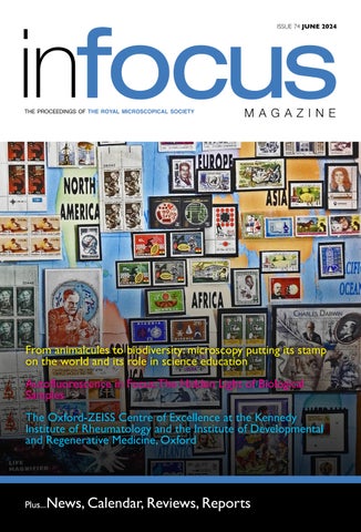
From animalcules to biodiversity: microscopy putting its stamp on the world and its role in science education Autofluorescence in Focus: The Hidden Light of Biological Samples The Oxford-ZEISS Centre of Excellence at the Kennedy Institute of Rheumatology and the Institute of Developmental and Regenerative Medicine, Oxford
Plus...News, Calendar, Reviews, Reports 1
ISSUE 74 JUNE 2024