
17 minute read
The Oxford-ZEISS Centre of Excellence at the Kennedy Institute of Rheumatology and the Institute of Developmental and Regenerative Medicine, Oxford
Josie Eade, NDORMS, University of Oxford
The Oxford-ZEISS Centre of Excellence (CoE) at the University of Oxford is an imaging centre with an eye on the future. It’s been built on a concept that successfully brings together academia and industry partners to not only provide state-of-the-art technology, but also give researchers access to the unique expertise of ZEISS engineers in their research and development team. Creating a melting pot for new imaging ideas, the centre’s ground-breaking technologies allow researchers to observe scientific processes in increasingly remarkable ways to address novel biological research questions. The centre celebrated its official opening in February 2024.
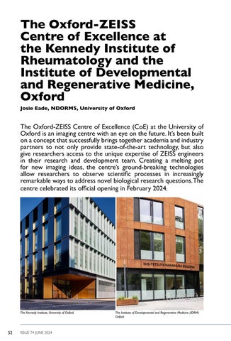


The Oxford-ZEISS CoE is a strategic partnership between the Kennedy Institute of Rheumatology (KIR), the Institute of Developmental and Regenerative Medicine (IDRM), and Carl ZEISS AG (ZEISS), a leader in the field of optics and optoelectronics. The KIR and IDRM sit together on the Old Road Campus at the heart of the University of Oxford’s biomedical research. They have broad research interests across immunology, neurology, embryology, cardiology, and oncology. Their goal is to study the key biological processes that underpin normal healthy development, and where these may go wrong in diseases such as cancer, cardiovascular disease, neuro-degenerative diseases, or conditions related to inflammation or tissue repair.

Although it was officially opened by the University of Oxford’s Vice-Chancellor Professor Irene Tracey on 21 February 2024, the centre has been building up its microscopy capabilities for the last three years. In 2021, it became the first place in the UK or Europe to install a ZEISS Lattice Lightsheet 7, two years before anywhere else. The microscope was later equipped with two high-speed cameras for simultaneous two-colour high-speed acquisition. These sit alongside a multi-photon microscope for powerful deep imaging, Airyscan confocals for imaging across the scales from sub-cellular structures to the large tissue tile regions, and a high throughput slide scanner, to ensure that the Centre provides a whole suite of light microscopy. It offers its service to researchers around the University for a diverse range of applications from mitochondrial morphology at a sub-cellular scale, to the development of whole organisms, and is actively looking for additional partners to come into the fold. A team of experts has been appointed to support the widest range of applications for microscopy and analysis.

The Oxford-ZEISS CoE is the brainchild of Marco Fritzsche, Professor of Biophysical Immunology at the KIR, and Scientific Director of the centre. Having studied physics and mathematics in Germany, he went on to study Experimental Physics and Biology at University College London. While conducting postdoctoral research at the University of Oxford, Marco met physicist and Nobel Laureate Eric Betzig who developed the Lattice Lightsheet microscope at the Janelia Farm Research Campus in the USA. Marco became fascinated by the Lattice Lightsheet microscope technology and began a long-term collaboration with the Janelia Research Campus. Speaking about the technology, Marco said: “Because of the minimally invasive Lattice Lightsheet we can study cells for long periods of time, gaining an understanding of their history, studying individual interactions of the cell within the biologically designed environment, and we can change the microenvironment to analyse the cells’ response. I like to call it the ZEISS Lattice Lightsheet revolution and I think that the lattice will replace confocal microscopes one day.”
Development leaps from disruptive technology
While many imaging centres provide cutting-edge technology, the Oxford-ZEISS CoE has taken a powerful step forward by bringing together Oxford scientists with the ZEISS research and development team. This partnership creates powerful new technologies developed alongside the research questions they are designed to solve.
“What this delivers”, says Marco, “is the freedom to raise questions relating to our areas of biological study that may challenge current microscopy capabilities. The idea is to create leap changes in the biomedical field by disruptive technology and close the gap between scientific needs and technological developments.”
Dr Bernhard Zimmermann, Head of Life Sciences at ZEISS Research Microscopy Solutions explained: “Developing our solutions in line with the rapidly changing demands of the scientific community continues to be vital for our success. Strong collaborations with leading-edge academic partners are key for reaching this goal. Thus, extending and reinforcing our interactions with the world class research teams at Oxford through the OxfordZEISS CoE is putting us in an excellent position to develop new technologies that are tailored to support new and upcoming application demands.”
Breaking the boundaries (imaging in four dimensions)
The Lattice Lightsheet microscope was developed to overcome one of the greatest challenges in imaging live cells: to observe them without light affecting their behaviour or damaging the sample.
Lightsheet microscopy uses a plane of light to illuminate a very thin slice of a sample. In comparison to standard confocal laser-scanning microscopy, lightsheet microscopy causes less photodamage to cells while gathering the same amount of data. Multiple planes can be used to render an image in four dimensions: three physical dimensions and time. Lightsheet microscopy can be used to image whole structures like organoids and whole organisms.
Lattice Lightsheet microscopy goes a step further, using successive planes of light in a ‘lattice’ shape to generate a series of images rendered in three dimensions and over time. It benefits from the same gentle illumination of lightsheet microscopy which minimises photobleaching. The technology enables researchers to watch biology live, as it unfolds over time by capturing previously unseen dynamic biological phenomena in three dimensions, in multiple colours, and for multiple hours per experiment.
As an example, Professor Paul Riley, Director of the IDRM, together with Professors Shankar Srinivas and Professor Georg Hollaender have used the ZEISS Lattice Lightsheet to study the cellular dynamics of embryos during the development of their cardiovascular and immune systems, allowing unprecedented insights under physiological conditions.
If the Lattice Lightsheet is the star of the OxfordZEISS CoE, the confocal microscopes are what Dr Kseniya Korobchevskaya and Dr Helena Coker, the Advanced Facility Managers, call the ‘pillars’. Confocal microscopy is a widely used ‘workhorse gold standard’ technique, and the Centre is equipped with two of the latest fast ZEISS LSM 980 confocal microscopes. These systems offer a range of capabilities including enhanced-resolution imaging using Airyscan 2, as well as spectroscopy and life-time measurements for the F-techniques; FCS, FCCS, FLIM, FRET and FRAP. This allows users not only to take images with confocal optical sectioning but probe the dynamics of molecules within cells and tissues.

Fluorescence correlation spectroscopy (FCS) is a statistical microscopy method which uses a laser beam to study precisely how proteins diffuse and interact in cells. However, FCS typically uses a strong, narrow laser beam to focus on a single spot which can bleach fluorescent molecules. Dr Narain Karedla, a postdoctoral researcher at the KIR and a specialist in advanced microscopy, combined the ZEISS LSM 980 with scanning FCS to develop a technique which scans in a laser line across the cell surface rather than in a single spot. This technique reduces photobleaching while increasing the amount of data collected. Narain used this technique to study the interaction of cell surface and cytosolic proteins in a remarkably precise way.
“We can now estimate these molecular spatiotemporal organisation with previously unachieved sensitivity and accuracy”, said Narain.
Where dynamic imaging is not the goal, the centre also houses a ZEISS LSM 880 Non-Linear Optical (NLO) confocal, a multi-photon microscope for powerful deep tissue and intravital imaging.
The centre has a ZEISS Axioscan 7 automated slide scanner which can be used for static rather than live-cell imaging. It takes 100 different slides at a time and can rapidly digitise samples in bright field and up to seven fluorescent channels.
“We leverage the ZEISS microscopy platform, especially the Axioscan, to acquire high-resolution whole-slide chromogenic and fluorescent images for tissue biology research”, said Ray Shih-Hsuan Mao, M.D., DPhil Student in the Nanchahal Group, KIR. “This enables comprehensive analysis of the tissue microenvironment, guiding therapeutic development. The platform facilitates unbiased image analysis and efficient protocol optimisation for advanced spatial technologies, such as spatial transcriptomics and multiplexed imaging.”
Most recently, the KIR won a UKRI Medical Research Council (MRC) grant in partnership with the Centre for Human Genetics to install a ZEISS ELYRA 7 with Lattice Structured Illumination Microscopy (SIM). Thanks to its sub-diffractionlimited spatial resolution, this fast, super-resolution microscope adds to the highly versatile imaging technology employed to answer the kind of research questions that are active in the health and disease space.
For DPhil student Robert Mitchell, the ELYRA 7 opened up new possibilities in his immunometabolism studies. “The ZEISS ELYRA 7 with Lattice SIM has enabled me to image mitochondrial networks in 3D with high resolution, making it possible to distinguish separate mitochondria which are packed tightly together in the small cytoplasmic space in B cells -something that had been a barrier for me even with Airyscan technology. It’s helping me to characterise how mitochondria in B cells change their morphology in response to different stimuli.”
Dr Jacky Ko, a Bioimage Specialist at the CoE added: “Complex biological processes are a puzzle for microscopes to unfold across multiple scales. Through the orchestration of multimodal machines at the ZEISS Centre, tiny details are gathered in one place to bring the full scientific picture out of the mist.”
Expertise and vision
Another strength of the centre is the quality of the team that supports researchers with a complete end-to-end service – providing training and support from imaging to data management and analysis.
“This core team is small, so the group’s approach to bringing new technologies to researchers and their diverse biological applications requires a certain element of adaptability”, said Marco. But the breadth of expertise and passions within the team are reinforced by teamwork and collaboration to help each other deliver excellence across disciplines.This approach works well for the researchers who find that the advice and guidance from the team provide quick and optimal solutions for their studies. If their requirements sit beyond the immediate capabilities of the centre, active collaboration between the Oxford and ZEISS R&D team, and technical modifications to ZEISS instruments, ensure needs are met that offer exciting possibilities for new scientific discovery.
KIR DPhil student Kaitlyn Purdie said: “The help that I’ve had from Kseniya, Helena and Jacky has helped me understand more intricately how the microscopes actually work, and why specific methods of analysis are better than others, which has deepened my knowledge beyond just understanding what buttons to press. The expertise they offer exponentially improves the quality of the data I can collect and present.”
The Oxford-ZEISS CoE supports researchers from interdisciplinary centres where people have different levels of expertise. Some have never even seen a microscope while others are expert users. Bringing them all up to the same quality of expertise is seen as a challenge but it is a core goal of the centre that the visualisation of microscopy data is never a barrier to important scientific discoveries. It has connected to the Oxford microscopy community to deliver courses, and in the autumn of 2023, the centre ran its first Oxford-ZEISS Deep Tissue Imaging course.
David Grainger was one of 12 participants on the highly specialised, week-long course. Aimed at researchers with experience in microscopy but looking to accept more challenging tasks for their research, the group explored five imaging platforms, accumulating a grand total of 48 hours of imaging and 12 hours of image analysis. “The major benefit was being taught how different microscopes acquire their images enabling me to make an informed decision on the most appropriate technology for my tissue research question. It’s given me the confidence to do experiments that I previously would have deemed too challenging”, said David.
A huge success, and a learning curve also for the Oxford-ZEISS team, the course will be replicated again this year.
Staff profiles

Name: Marco Fritzsche
Position: Scientific Director
Qualifications: DPhil. Biophysics, MSci Theoretical Physics, BSc Math, BSc Physics
Background: Prof Fritzsche is the Scientific Director of the Oxford-ZEISS Centre of Excellence and leads the Biophysical Immunology Laboratory (www.bpi-oxford.com) between the Rosalind Franklin Institute and the Kennedy Institute of Rheumatology at the University of Oxford, UK. The BPI Laboratory aims to unravel the impact of biophysics and mechanobiology on the human immune response in health and disease. For this mission, the BPI lab develop custombuilt microscopy technology at the interface of biophysics and immunology. Prof Fritzsche holds a MSc in theoretical physics and conducted his PhD in experimental biophysics and cell-biology at the London Centre for Nanotechnology at the University College London, UK. He performed his Postdoctoral work at the University of Oxford in close collaboration with the Howard Hughes Medical Institute Janelia Farm, USA.
Favourite microscope: I think that Lattice Lightsheet will change the microscope landscape.
How do you relax in your spare time? I love walking cities, mostly London.

Name: Kseniya Korobchevskaya
Position: Advanced Microscopy Facility Manager
Qualifications: PhD
Background: I am an optical physicist by training. I did my PhD in the field of ultra-fast spectroscopy. However, I was always fascinated by biology and this motivated me to move to the field of fluorescent microscopy, where my optical skills can be applied answering biological questions. I have led several development projects, which resulted in fully functioning bespoke imaging systems: Large Field of View Light Sheet, TIRF-SIM and Near IR Label Free Transient Absorption microscopes. I enjoy working together with biomedical researchers on exciting imaging projects, where my experience and expertise in fluorescent and super-resolution microscopy can contribute best.
Favourite microscope: ZEISS Airyscan Confocal
How do you relax in your spare time? I love hiking, potholing, canoeing and all other outdoor activities. But as those are quite demanding on my time, on most days I have to limit myself to less fancy reading and knitting.

Name: Helena Coker
Position: Advanced Microscopy Manager
Qualifications: DPhil. Physical and Theoretical Chemistry, MSci. Natural Sciences
Background: My background is quite broad as I studied Natural Sciences (mostly chemistry and pharmacology) for my MSci, then completed a DPhil in Physical and Theoretical Chemistry. During my DPhil., I built and optimised an interferometric scattering (iSCAT) microscope for very fast tracking of diffusing particles - this is where my love for optics began to drive my career. In 2018 I joined the University of Warwick as an Imaging Specialist in Lattice Lightsheet microscopy (LLSM). I moved back to Oxford in 2021 to manage the TIRF-SIM microscope and in 2023 I joined the microscopy facility full time.
Favourite microscope: ZEISS LLS7
How do you relax in your spare time? I am a ‘do-er’ inside and out of work so in my spare time I do a lot of art and crafts including dressmaking (I made my Inauguration dress!), cycling including racing on Zwift, and once I’ve tired myself out ‘doing’, I sit with my cat.

Name: Jacky Ka Long Ko
Position: Advanced Bioimage Analyst
Qualifications: MPhil/PhD in Imaging and Interventional Radiology
Background: I trained as a physicist, then redirected my research focus towards leveraging computational methods for biomedical imaging applications. Throughout my career, I have been deeply involved in medical image analysis, particularly in integrating artificial intelligence into the field. In 2017, I spearheaded the development of image-based surgical navigation systems with respiratory-gated robotic controls. Building upon this experience, my doctoral studies delved deeper into cluster and GPU computing for neurovascular investigations, specifically exploring atherosclerosis treatments through hemodynamic simulations with AI-assisted modelling. In 2022, I brought my expertise to the Oxford-ZEISS CoE, where I introduced advanced computer vision techniques in bioimage research.
Favourite microscope: ZEISS Axioscan 7 Slide Scanner
How do you relax in your spare time? In my spare time, I indulge in my passion for photography, particularly exploring landscapes as I travel across cities and fields. Recently, I’ve delved into aerial photography using drones, which offers a thrilling opportunity to capture unseen perspectives of the world around me.
Case study
Case study – the building blocks of life itself
Every human body begins as a clump of cells. The fertilized egg undergoes dramatic changes in shape over the course of its development in the womb, during which we are transformed into something more recognisable as a developing foetus. The process is called morphogenesis and it’s incredibly dynamic. A myriad of distinct cell types have to be created and positioned correctly as the basic plan of the body is laid down to produce a healthy embryo. An understanding of embryonic morphogenesis at the cellular and molecular level would advance our understanding of the fundamental process of tissue remodelling, and how this might go wrong in disease. Knowing the normal fate of cells is important not only for an understanding of development but also has implications for therapy in humans.
Shankar Srinivas, Professor of Developmental Biology at the IDRM, is an expert in morphogenesis. Lattice Lightsheet technology at the ZEISS CoE has transformed his research. ‘Embryos are really very sensitive to imaging and so the Lattice Lightsheet is transformative to our research, as it preserves their viability during our experiments,’ he said. ‘It enables us to image at much higher spatial and temporal resolution than any other microscope that we’ve used before, allowing us to watch the cells in the embryos as they migrate and shape various features.’
Recent work from his group has characterised the migration of a group of cells in the embryo responsible for setting up the head-tail axis. Lattice Lightsheet imaging enabled Dr Shifaan Thowfeequ in Shankar’s team to characterise a mutant with a defect in this important migratory process (now accepted for publication in Developmental Cell). This advances our understanding not only of how the body forms, but also more broadly of how cells in the body migrate, a normal process that can be subverted in diseases such as cancer.
Shifaan has been able to observe cells during gastrulation, a very early process in embryonic development where cells diversify from a single layer into different cell types which migrate and begin to form the structures of the body. ‘This imaging technology has enabled us to understand how individual cells leave one cell layer, emerge into another layer, and start moving in a coordinated manner,’ explained Shankar.

While it’s still early days for the CoE, the centre has already enabled Shankar to do more ambitious experiments and has helped his team to upskill through the centre’s commitment to training and development.
