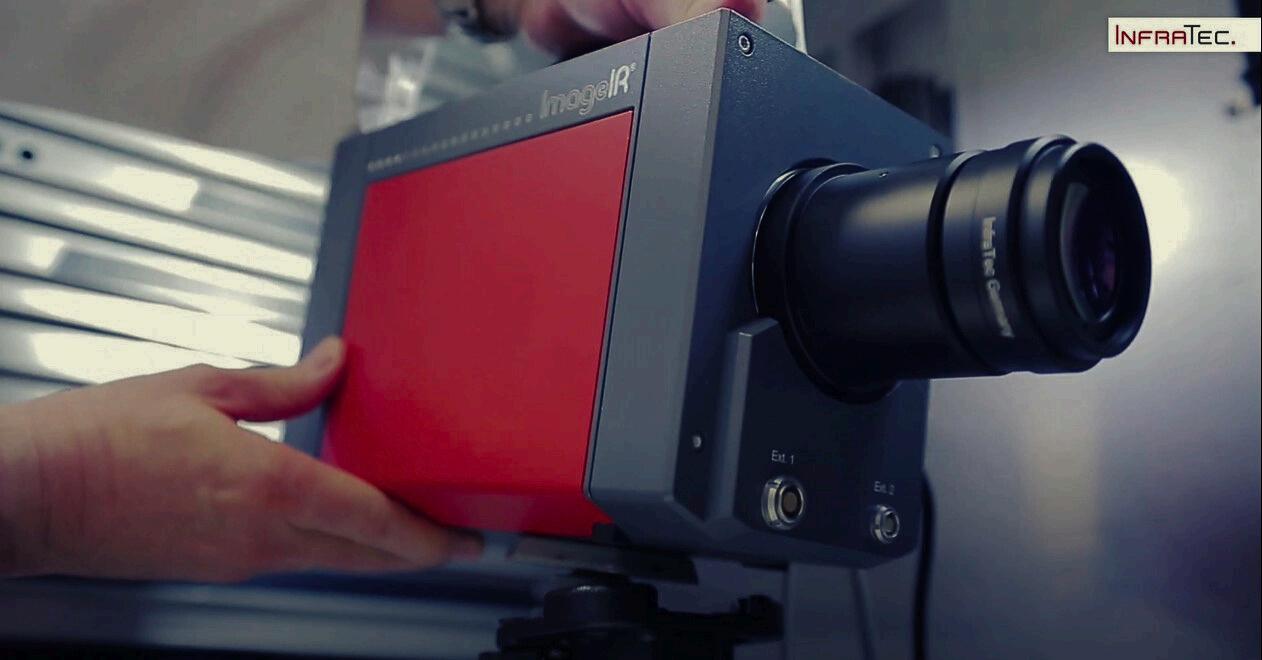
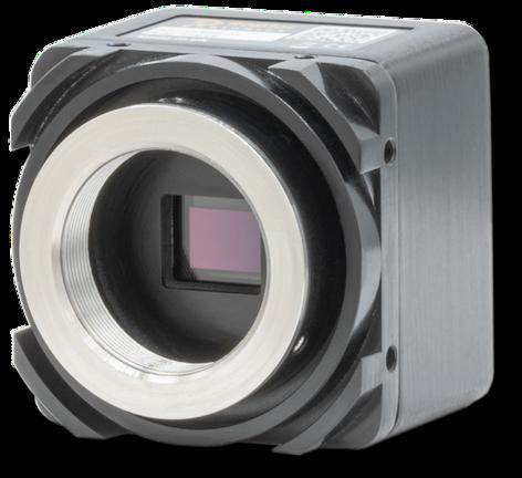
Raptor Launches RCMOS – Cameras Ruggedised for Any Environment
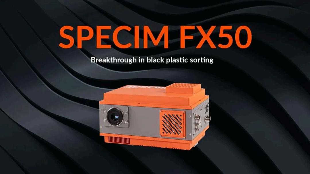
BREAKTHROUGH IN BLACK PLASTIC SORTING
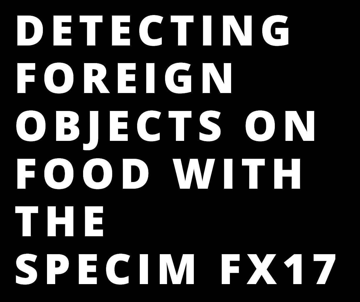
Detecting Foreign Objects On Food With Hyperspectral
The study of circadian rhythms and suprachiasmatic nucleus (SCN) neurons
Rental Cameras Available For Your Next Project
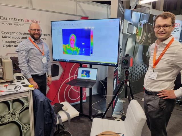
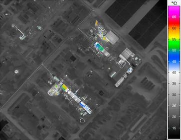
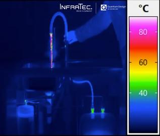
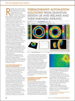
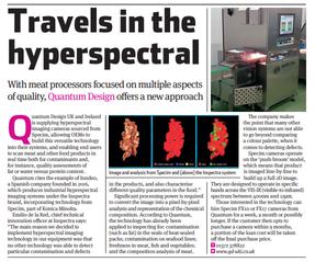
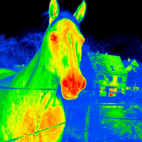
Thermography on the Trail of the Fault
WITH CONTENT CONTRIBUTIONS FROM OUR PARTNERS:
Raptor Photonics
Raptor Photonics aims to provide world class low light level camera solutions to industrial, research and governmental organisations around the globe. Raptor Photonics Limited is a high-tech company based in Northern Ireland, which was established in September 2006 Its main focus is to design, manufacture and sell the next generation of high performance, cutting edge, low light level digital cameras.
Specim
Specim is a globally leading supplier in hyperspectral imaging. As a true pioneer and forerunner in this field, we celebrated our 25th anniversary in 2020. Our international team of 70+ professionals, with expertise in optics, electronics, software, and machine vision, serves the market with the broadest range of hyperspectral cameras, imaging spectrographs, systems, and accessories. We are known as a trusted partner with products and support of superb quality and cost-efficiency
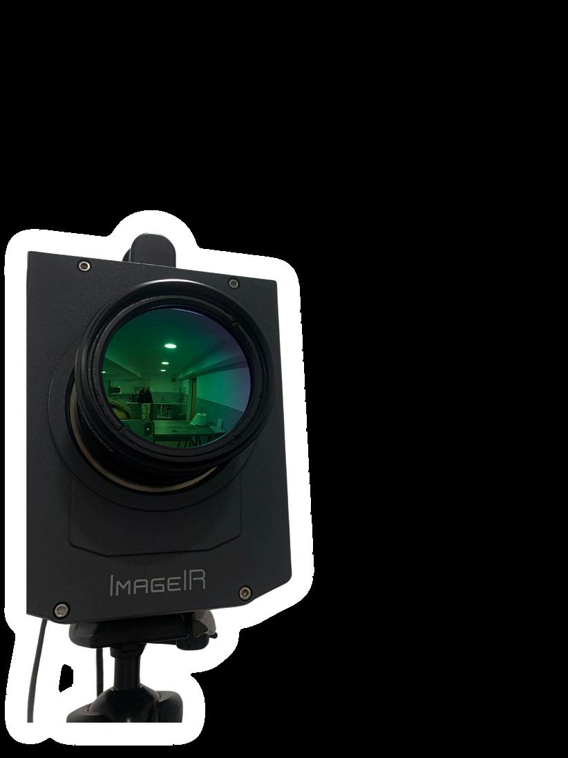
InfraTec
The Dresden-based company InfraTec GmbH is a specialist for products and services in the field of infrared technology. InfraTec offers solutions for every kind of thermographic measurement task Discover the new generation of stationary and handheld infrared cameras with megapixel formats and automated thermography solutions.
Drone-Based Daylight Electroluminescence Imaging of PV Modules using SWIR Cameras A
RAPTOR LAUNCHES
RCMOS – CAMERAS RUGGEDISED FOR ANY ENVIRONMENT
Detecting Foreign Objects On Food With Hyperspectral
BREAKTHROUGH IN BLACK PLASTIC SORTING
RENTAL CAMERAS AVAILABLE FOR YOU
Hyperspectral Imaging to Revolutionise Textile Sorting
Trade association memberships
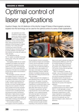
Technical Sales Manager
Luke joined Quantum Design a few years ago, following a Postdoc in the properties of complex beams at King’s College London in the Photonics & Nanotechnology group, headed by Prof. Anatoly Zayats.
Luke Nicholls started as our Camera Sales Engineer and is now our Technical Sales Manager
He completed his PhD in Physics, also at King’s College London, with a thesis entitled “Controlling light with light: exploiting fast free-electron nonlinearities in plasmonic metamaterials for the control of light polarisation”
His research experience in Photonics and Nanotechnology has helped customers across the Scientific and R&D market find camera solutions throughout the EM spectrum, from X-ray, through to visible and infrared, for their applications
Luke has brought his wide ranging knowledge of optics to assist customers in providing solutions to their applications. He looks forward to hearing from you about your next research project
luke@qd-uki co uk
SEE MORE FOR GREATER FLEXIBILITY
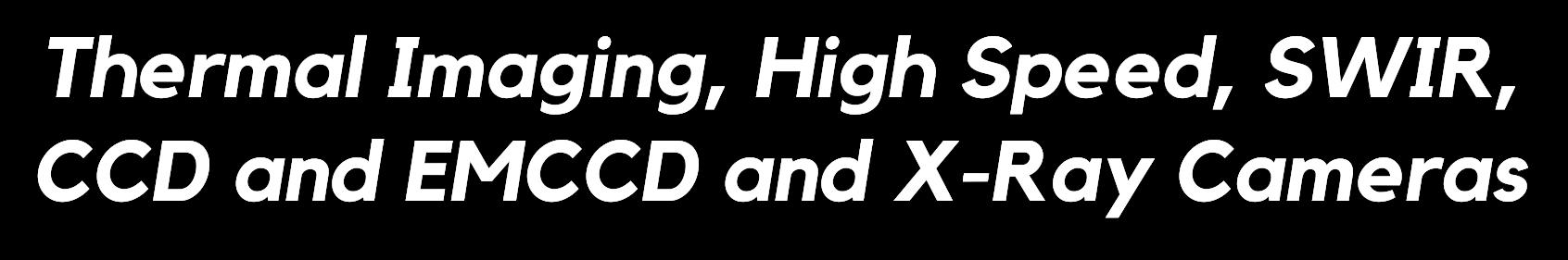
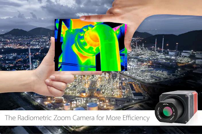
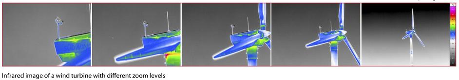
The new ImageIR® 6300 Z expands the portfolio of powerful cameras with zoom lens
InfraTec has been developing infrared cameras for various industries and applications for years The product range includes amongst others the ImageIR® 8300 Z and ImageIR® 9300 Z cooled zoom infrared cameras with 30× high-performance zoom lens and autofocus function Both infrared cameras are ideally suited to defense research and surveillance activities.
Both infrared cameras are ideally suited to defense research and surveillance activities The VarioCAM® HD Z complements these two models as an uncooled zoom thermal imaging camera in the long-wave infrared range and is used primarily for 24/7 operation in the industrial security sector
InfraTec's latest zoom camera, which measures in the midinfrared range like its counterparts in the ImageIR® series, is now much smaller and easier to handle.
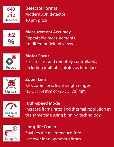
Efficiency has a New Name – ImageIR® 6300 Z
The radiometrically calibrated ImageIR® 6300 Z uses a compact, robust SWaP (Size, Weight and Power) detector, which has been combined with a powerful zoom lens Capable of thermographic temperature measurement in various fields, this camera is designed for any user wanting to cover a broad, universal range of applications
NO EXTENSIVE LENS CHANGE
The consistent use of the latest technologies in optics, detector and electronics is the basis for the remarkable user-friendliness of the camera series The thermal imaging ImageIR® 6300 Z has a 7.5× zoom lens integrated as standard, which allows, in combination with the motorised focus, a fast and flexible adjustment to different object distances and sizes while maintaining stable image quality and high measurement accuracy.
POWERFUL IMAGE PROCESSING ELECTRONICS
The ImageIR® 6300 Z has high-performance image processing electronics and can output the IR image data in real time to several video and data interfaces as well as record and evaluate it autonomously. This camera can also be operated via smartphone or tablet using its web interface With these features and the possibility to power it from an external battery, this camera is ready for mobile outdoor use. The zoom camera can be easily integrated into existing system environments in a space-saving way. It is suitable for universal use in research and development, but also for integration into GIMBAL systems in the field of flight thermography
DETECT SMALLEST TEMPERATURE DIFFERENCES USING HIGH THERMAL RESOLUTION OF 30 MK
The thermal resolution of the ImageIR® infrared camera series allows you to measure smallest temperature differences of better than 0.03 K (at 30 °C)
Thermal images with a narrow temperature span display details absolutely sharp You will find interesting signatures even there where other infrared camera systems cannot detect any temperature differences any longer.
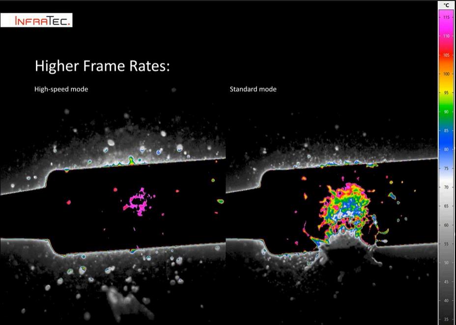
HIGH-SPEED MODE TO INCREASE FRAME RATES AND THERMAL RESOLUTION AT THE SAME TIME
If you want to analyse the thermal behavior of objects and processes from a wide variety of perspectives, you can use the high-speed mode for maximum flexibility. This function allows the camera to be used in two different operating modes. The standard mode suits best for depicting the smallest geometrical details This allows users to record images with the native number of pixel of the camera detector. Switching to high-speed mode opens up the option of more than triple the frame rate, while the field of view (FOV) remains identical. This enables for most accurate time monitoring of fast processes At the same time, the thermal resolution increases by a factor of 2 in high-speed mode, which additionally improves the analysis of temperature differences.
Broad Range of Applications
The ImageIR® 6300 Z is designed specifically for challenging applications and can be easily integrated into existing system environments in order to save space. This zoom camera's extreme flexibility – even for close-range applications – makes it the ideal solution for numerous measuring and inspection tasks
Fields of application:
Research and development
Aerial thermography: inspection and monitoring tasks
Quality assurance
Materials testing
Integration solutions
find out more
Here at Quantum Design UK and Ireland, we only want to send you the information that you would like to see When filling out the form, please tick the particular fields and product suppliers that interest you, and we will make sure you are kept up to date with ONLY the most relevant information
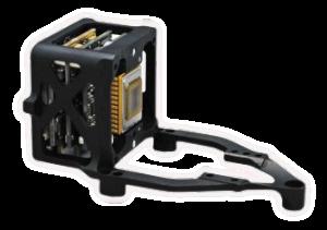
FLIGHT-READY HAWK 247 EMCCD OEM CAMERA
Raptor has launched a family of ruggedised CMOS cameras The Hawk series of ultrasensitive RCMOS OEM cameras cover UV, VIS and NIR wavelengths and are designed to reliably perform in the harshest conditions from the seabed to space, withstanding temperature, pressure, moisture, radiation, shock and vibration extremes.
Available in a range of resolutions, the Hawk cameras are perfect for the most demanding conditions for airborne, space, surveillance, industrial and marine applications
find out more
Applications
Surveillance
Low-light Surveillance
Airborne Surveillance
Driver Vision Enhancement (DVE)
Digital Night Vision
Ground Based Surveillance
Embedded Vision
Plastics / PET Semiconductor Inspection
Space
Docking
Navigation
Space Situational Awareness
Inspection
Hyperspectral Imaging
Earth Observation
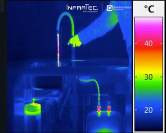
Firstly, cold tap Then hot tap And then the steamy Quooker! then...
Smaller, lightweight, without tedious lens changes –efficiency has a new name: The ImageIR®6300Z with a powerful zoom lens and a SWaP detector
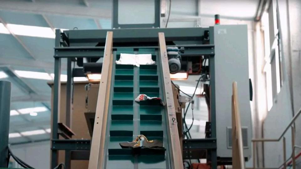
Black plastics are everywhere; cars, vehicles, electronics, packaging, and plastic bags. Instead of recycling, they are burned for energy or dumped in a landfill since efficient and reliable technology has not been available to sort black plastics – until now.
Hyperspectral imaging is the only technology capable of sorting different black plastic types when used on the MWIR spectral range.
Specim announces the upgraded Specim FX50 middle-wave infrared (MWIR) hyperspectral camera The Specim FX50 is the first and only push-broom hyperspectral camera on the market that covers the full MWIR spectral range of 2.7 – 5.3 μm.
Specim FX50 allows fast and reliable sorting of:
Black plastics such as PS, PE, PP, ABS and PVC
Rubbers
Non-black plastics and rubbers
This breakthrough product with enhanced features and capabilities expands the possibilities in industrial sorting, quality control, process optimisation, and research by detecting materials that are not detectable with any other wavelength or imaging method, such as hydrocarbons, minerals, oil, and contamination on metal surfaces.
“The launch of the improved Specim FX50 marks a significant milestone for Specim and our customers. The first
version of the camera
was
released in
2019 and its demand exceeded our expectations, and we’re excited about the untapped possibilities this camera offers.”
Tapio Kallonen, CEO at Specim
The new Specim FX50 drives cost efficiency and sustainability by enabling efficient raw material detection, manufacturing, and identifying valuable materials worth recovering for recycling and reuse. In particular, it revolutionises the field of black plastic sorting, as it is the only product with the required wavelength range capable of sorting difficult black plastics.
“One remarkable benefit of the Specim FX50 is its game-changing impact on black plastic sorting. The FX50 camera enables the identification and separation of black plastics effectively, helping to boost the efficiency, accuracy, and profitability of the crucial process,”
Tapio Kallonen, CEO at Specim
find out more
THE NEW SPECIM FX50 OPENS DOORS TO NEW OPPORTUNITIES FOR SCIENTIFIC EXPLORATION.
Accurate Inspection and Robust Industrial Design
With a high spatial resolution, image speed, and enhanced signal-to-noise ratio, the Specim FX50 enables fast and accurate inspection and classification of materials with very similar spectral features.
Beyond its impressive spectral capabilities, the Specim FX50 boasts new optimised thermal management that maximises sensor lifespan and minimises downtime, leading to increased operational efficiency and cost savings
In addition, the Specim FX50 offers several notable benefits, including temperaturestabilised optics, built-in image correction capabilities, unified spectral calibration between units, and a standard GigE Vision interface. These features improve the performance and reliability of the camera and ensure easy integration into industrial environments
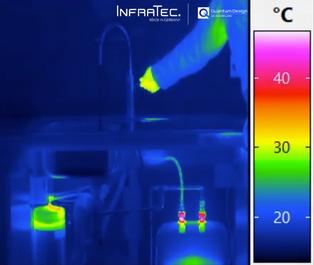
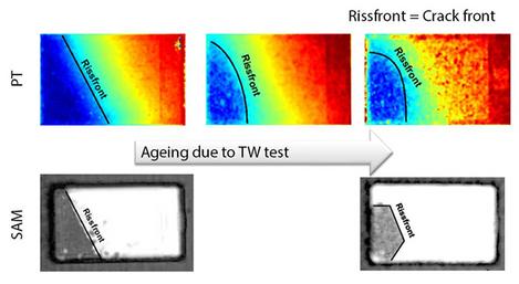
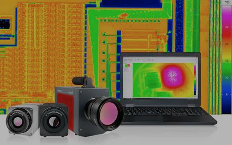
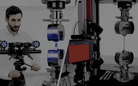
This method is also used for research purposes at the Institute for Electrical Systems and Energy Logistics at BTU Cottbus-Senftenberg In this context, Prof Dr Ralph Schacht is intensively involved with the material and system characterisation as well as the non-destructive failure analysis of printed circuit boards, electronic components, microelectronics as well as composite systems of packaging and interconnection technology.
Non-destructive testing (NDT) – in contrast to destructive component testing – implies that the test application must not influence or reduce the usability of the test object in any way Thermal imaging is a non-destructive, very efficient method It enables the imaging detection of thermal radiation as well as the interpretation of the observed surface temperatures
At the Institute of Electrical Systems and Energy Logistics at the BTU CottbusSenftenberg, non-destructive component testing is carried out using various thermography methods: passive IR thermography, active IR pulse thermography and active IR lock-in thermography.
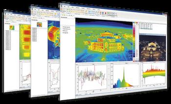
The infrared camera used for this purpose is the ImageIR® 8300 from InfraTec with a M=3x magnification microscope lens or a macro attachment. The measurements are evaluated with the IRBIS® 3 thermography software belonging to the camera and the additional module IRBIS® 3 active
Passive IR Thermography
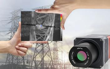
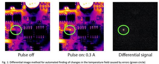
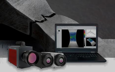
With this method, electrical "short-circuit currents" are selectively injected into the assemblies to be tested This can lead to increased current flows in faulty areas, which in some cases trigger the slightest temperature changes there and can be detected using thermography Short-circuit tests of electrical through-hole platings in printed circuit boards serve as an example here. Due to the possibility of evaluating the differential image within the IRBIS® 3 thermography software, the contact point can be precisely localised in real time (Fig. 1).
The utilisation of, for example, power MOSFETs in 3-phase inverter operation can also be displayed and monitored very well using this method (Fig. 2). With the aid of the high-resolution M=3x microscope lenses used on the ImageIR® 8300 infrared camera, it is possible to characterise integrated passive components at wafer level The thermal behaviour of nickel-chromium resistors as a function of their power dissipation was analysed here (Fig. 3).
Active IR Pulse Thermography
This form of non-destructive testing is a frequently used method to detect hidden damage in components Heat is pulsed into a sample while the temperature field of the stimulated surface is observed in parallel
This enables the evaluation of both heating and cooling curves. The advantage of impulse thermography is its high speed and, as a result, the high potential for a 100 percent inspection during a production process
Using this method, it is possible to take advantage of the fact that locally different cooling processes occur after the excitation impulse The time of the maximum temperature difference and the difference as such are determined.
This method also allows conclusions to be drawn about structures that are not directly located on the component surface (Fig 4)
Practical examples of this measurement method can be seen in connection with the assembly of power semiconductors Delaminations in a solder layer after cyclic loading or the propagation of crack fronts in sintered layers can, for example, be detected.
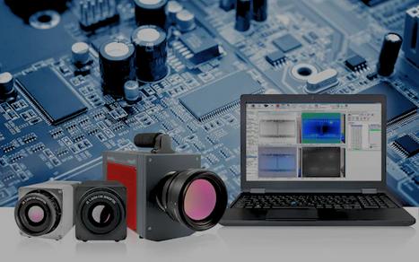
Active IR Lock-In Thermography
The lock-in principle or lock-in amplification is employed when a useful signal is to be found in a statistically noisy overall signal This must be periodically amplitudemodulated in a targeted manner.
In the simplest case, this is done by switching the supply voltage of an electronic component on and off or by sinusoidal modulation.
In addition, this method allows very small and weak heat signals to be detected Taking, for example, a test structure with a 50 µm small diode and exciting it with 10 mW at 0 1 Hz (duty cycle 50 %), the sought-after measurement signal in the pure thermal image is below the detection limit. However, the power dissipation source can be localised very clearly via a generated amplitude image – even in real time using the IRBIS® 3 active thermography software
The BTU Cottbus-Senftenberg has been using the ImageIR® 8300 for the measurements presented here for several years This camera is equipped with a 25 mm standard lens as well as M=1x and M=3x magnification microscope lenses. Internally, two filteraperture wheels have also been installed, a unique feature of the camera series, with which both signalattenuating filters and spectral filters can be swivelled in remotely in parallel In addition to the high spatial resolution of this system, excellent measurement accuracies are also achievable with each lens, so that exact and repeatable results are obtained.
The associated IRBIS® 3 active software allows, among other things, convenient data evaluation options of image sequences, the calculation of phase and amplitude images as well as the analysis with various methods of active thermography These include, for example, the quotient method, pulse phase and lock-in thermography.
The ImageIR® 8300, including its software tools, has become an indispensable aid in scientific training as well as in research work at the institute
FOREIGN OBJECTS IN FOOD POSE SIGNIFICANT RISKS TO CONSUMER SAFETY. THE DISCOVERY OF FOREIGN OBJECTS OFTEN RESULTS IN PRODUCT RECALLS, WHICH CAN BE COSTLY FOR BUSINESSES AND DAMAGE THE BRAND’S REPUTATION.
To ensure the quality and safety of food products, manufacturers must implement robust quality assurance throughout the production process, where hyperspectral imaging offers a solution.
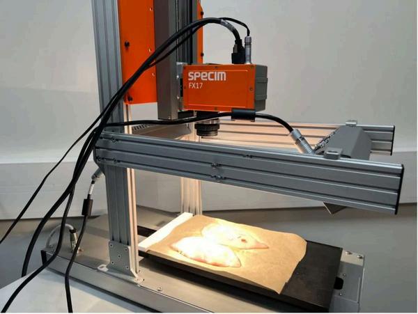
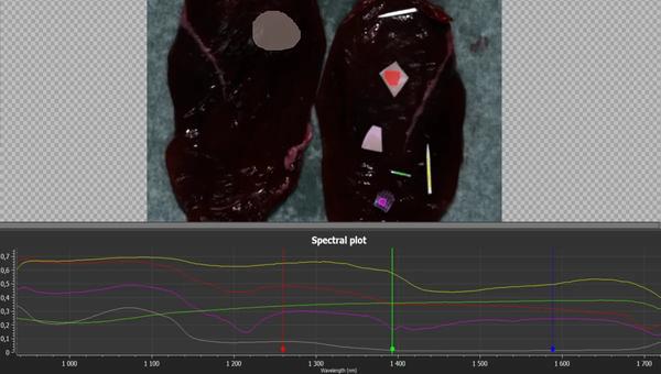
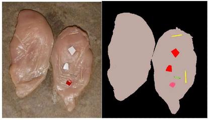
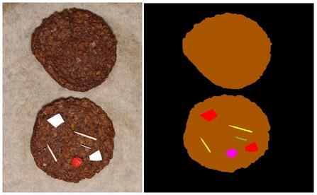
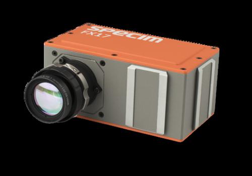
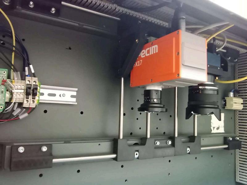
In this study, the Specim FX17 hyperspectral camera (900 – 1700 nm) and Specim Lab Scanner 40*20 were used to measure three food products: chicken fillet, veggie patties, and goat cheese containing contaminants Foreign objects were placed on each food product with the aim of creating a classification model to detect those.
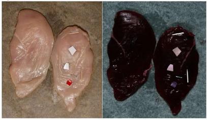
First up, the chicken fillet, a valuable food product. Contaminants used were wood, metal, and two kinds of plastics (PE and PS). The food was placed on baking paper.
Figure 2 shows the measured chicken fillet with and without the contaminants.
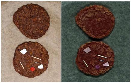
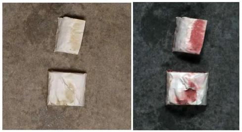
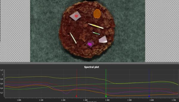
After this, they examined the veggie patties using the same contaminants as those used for the chicken fillet (Fig 3)
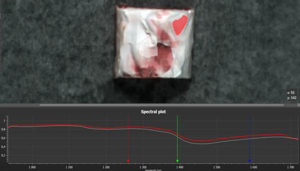
Finally, the goat cheese was measured. Specim used a piece of packaging material, i.e., a small piece of thin white plastic wrapping, as a contaminant Figure 4 shows that the contaminant looks very similar to the cheese and is hardly visible with an RGB camera or the naked eye.
SPECTRAL COMPARISON BETWEEN FOOD AND FOREIGN OBJECTS
Each data was normalised with respect to white and dark references We processed the resulting reflectance data with the SpecimINSIGHT analysis software. Area selection from the chicken fillet and contaminants were taken, and a mean spectrum from each selection was drawn to the spectral plot for comparison (Fig 5).
The colour of each spectra matches the colour of the corresponding selection on the image. The spectral plot shows that the spectral signatures of the chicken fillet and all the contaminants are clearly different.
The spectral signature of the veggie patties also differs from the contaminants, as shown in Figure 6
The goat cheese packaging material is slightly transparent, which causes the spectrum of the goat cheese to mix with the spectrum of the packaging material (Figure 7). Therefore, the spectral signatures of the goat cheese and the contaminant don’t differ as significantly as they do with the chicken fillet and veggie patties and the contaminants
CLASSIFICATION
CONCLUSION
This study used a Specim FX17 hyperspectral camera (900 – 1700 nm) to detect foreign objects on food products. Based on the measurement and analysis, we can conclude: Spectral signatures of food products and contaminants are distinguishable. Based on data captured with the Specim FX17 camera, creating a classification model to detect contaminants is possible.
The Specim FX17 hyperspectral camera can detect contaminants that are invisible to the human eye and undetectable with a standard RGB camera
Rent the Specim IQ
Hyperspectral camera for a week or a month at a time for your next research project at competitive rates.
Lightweight | Portable | Compact
luke@qd-uki co uk
At Quantum Design UK & Ireland, we try to attend all the top industry events, conferences, and symposiums. That way we can be available for discussions on the products we offer.
If you’re there, pop by to see demo models and chat about your upcoming research projects and applications
find out more
I M A G I N G : A P I V O T A L
P L A Y E R I N T
Food waste is a global challenge that demands immediate attention. Shockingly, approximately one-third of all food produced for human consumption is wasted annually, according to the United Nations.
This staggering waste represents a colossal economic loss and carries severe environmental and social consequences. In the ongoing battle against food waste, technology has emerged as a potent ally, and one of its most promising champions is hyperspectral imaging.
Revealing the Invisible
Hyperspectral imaging takes us beyond the limits of human perception. It grants us access to the unseen, unveiling the molecular composition of food. This innovative technology collects data across the electromagnetic spectrum, exposing hidden intricacies that remain invisible to the naked eye.
At its core, hyperspectral imaging allows us to capture and analyse the unique spectral signatures of various materials. This ability is particularly useful in the agricultural sector, where it can help identify and sort fruits and vegetables based on their ripeness, quality, and potential for spoilage.
F I G H T A G A I N S T
F O O D W A S T E
By doing so, farmers and distributors can make more informed decisions on managing their produce and reducing unnecessary waste.
Early Spoilage Detection
One of the standout advantages of hyperspectral imaging is its capacity to spot spoilage at its earliest stages. It can discern minute alterations in the spectral signatures of food items, allowing for the identification of spoilage or contamination before visible signs manifest. This early detection is instrumental in averting the distribution and consumption of unsafe or substandard food products.
A Catalyst for Research and Innovation
Hyperspectral imaging continues beyond waste reduction. It catalyses ongoing research, driving innovation and offering new perspectives on tackling the global challenge of food waste.
In conclusion, hyperspectral imaging is a powerful weapon in the crusade against food waste Its ability to elevate food quality, avert spoilage, and enhance the efficiency of the food supply chain positions it as a transformative force in the pursuit of a more sustainable and wastefree future.
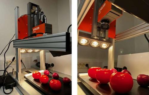
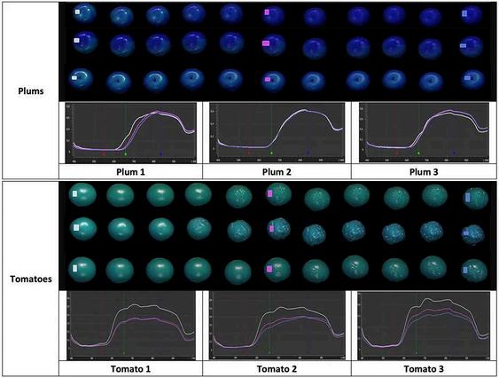
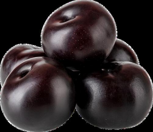
MEASURING THE AGEING OF PLUMS AND TOMATOES WITH SPECIM FX10 HYPERSPECTRAL CAMERA
Food ageing is an important parameter to be quantified when evaluating freshness.
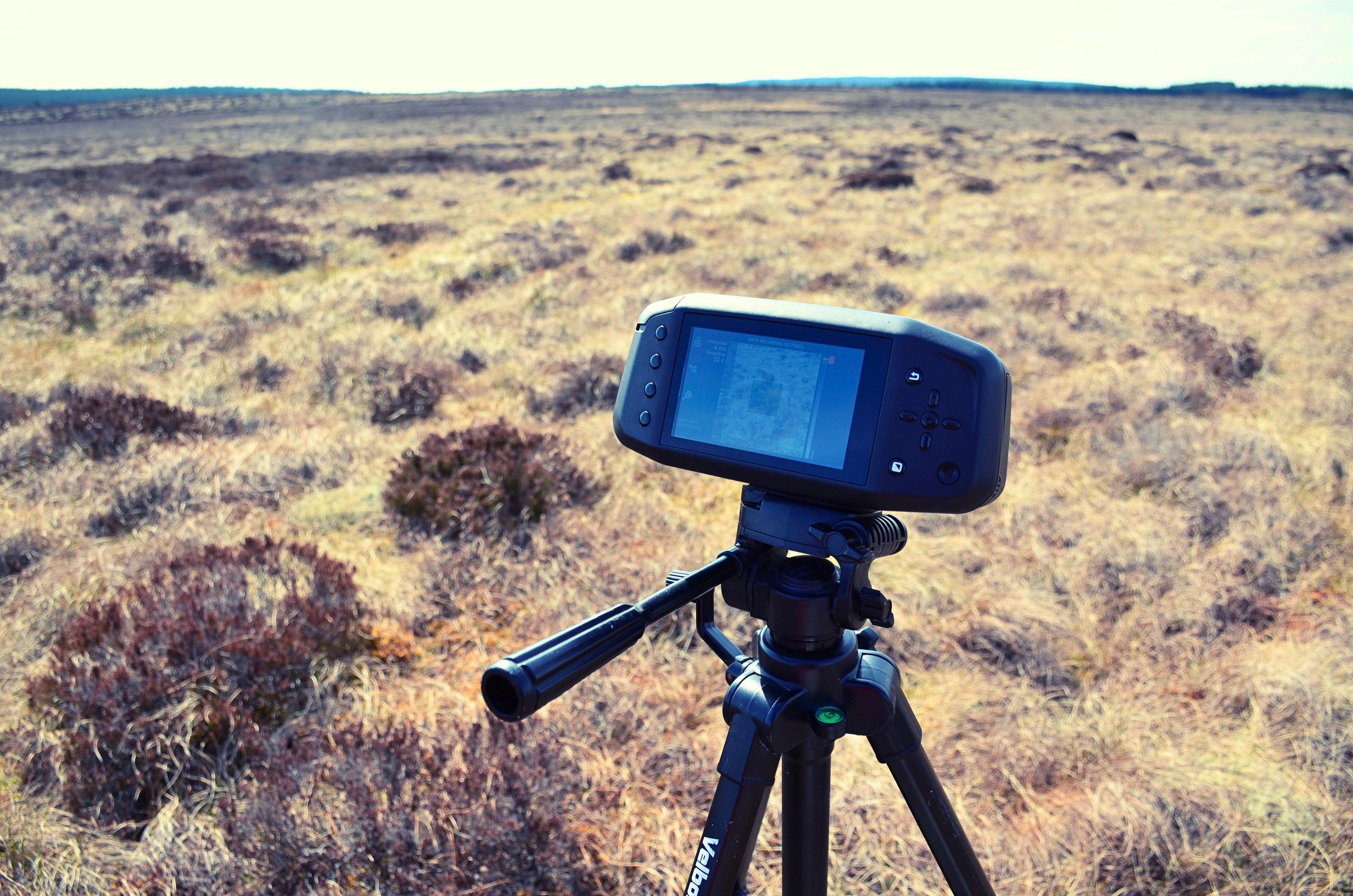
In this study, we used the Specim FX10 hyperspectral camera and a lab scanner to inspect plums and tomatoes for 20 days to assess the aging process (Fig 1). The Specim FX10 is a visible-near infrared (VNIR) camera that covers the spectral range from 400 to 1000 nanometers. The first part of the analysis focuses on the spectral features of the samples over time. Then, a regression model of tomatoes’ and plums’ ageing is presented.
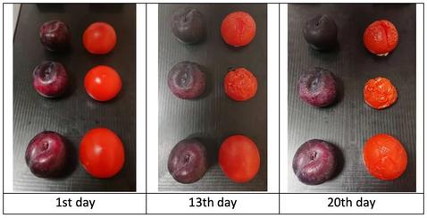
Photos of the samples were taken, along with the hyperspectral data. The pictures show that the freshness of the plums, especially the tomatoes, degraded firmly over time (Fig 2). A small cut was made in the middle of one tomato and plum. It seemed to have a substantial impact on accelerating the ageing of the tomato but not on the plum.
Spectral reflectance reveal chemical changes
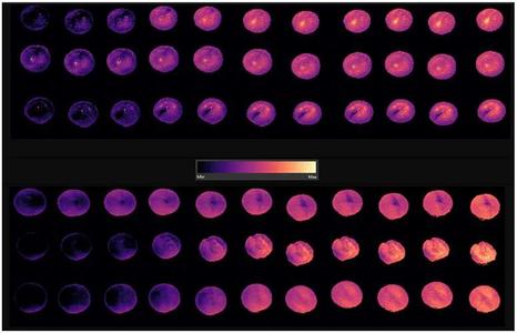
A rectangular selection was made on each plum and tomato each day when the spectral measurements were made (1st, 2nd, 3rd, 6th, 9th, 13th, 14th, 16th, 17th, and 20th day). Only the spectra obtained on the 1st, 13th, and 20th day are presented in Figure 3 to ease the reading of the results. Spectra are averaged over the selection.
The spectral differences are more significant for the tomatoes than for the plums. This is already visible in the photos taken on the 1st, 13th, and 20th days (Fig 2).
The spectra reveal chemical changes which happen over time within the fruits and vegetables. Plums and tomatoes are green at early growing stages due to the chlorophyll they contain. But when ripening, the chlorophyll breaks down into another chemical. For tomatoes, chlorophyll breaks down into lycopene, which explains the red colour. This chemical change explains the spectral variation of the plums and tomatoes over time between 550 and 750 nanometers. The ripening process of the fruits and vegetables also affects the moisture level or structure, impacting their spectra at 970 nanometers. Other properties (e.g., sugar content) also change over time, shaping the spectral reflectance.
Regression model to quantify the ageing
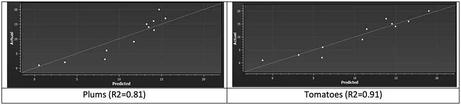
A regression model was built to quantify the ageing of the plums and tomatoes (Fig 3). The imaging day was the actual regression variable.
With the plums, the R2 was 0.81, whereas, for the tomatoes, it was 0.91. Those were computed on other selections than those used to train the model. The regression graph of Actual value vs. Predictions is presented in Fig 4.
For the plums, the model was based on the reduced spectral range from 588 to 976 nanometers. For the tomatoes, the model was based on the spectral bands between 445 and 993 nanometers.
Conclusion
The Specim FX10 camera is suitable for measuring fruits and vegetables’ ripeness and ageing as it is sensitive to traits related to freshness for agri-food products. When building a typical regression model, laboratory measurements should be used as a reference value to develop and validate the model. However, those are not needed for accessing fruits and vegetables’ ageing.
Hyperspectral cameras operating in visible-near infrared (VNIR) provide an efficient tool for monitoring the product quality of fresh food products. Hyperspectral imaging is an especially suitable method for food grading, sorting, and classification compared to conventional pointbased methods due to its non-destructive nature.
Electroluminescence (EL) imaging of photovoltaic (PV) solar panels provides high accuracy in detecting defects and faults, such as cracks, broken cells, interconnections, shunts, among many others; furthermore, the EL technique is used extensively due to a high level of detail and direct relationship to injected carrier density.
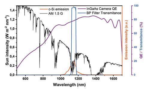
This technique is commonly practiced only indoors - or outdoors from dusk to dawnbecause the crystalline silicon luminescence signal is several orders of magnitude lower than sunlight. This limits the potential of such a powerful technique to be used in utility scale inspections, and therefore, the interest in the development of electrical biasing tools to make outdoor EL imaging truly fast and efficient.
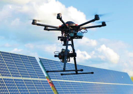
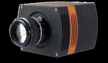
A team at Technical University of Denmark (DTU) lead by Gisele Alves dos Reis Benatto and Peter Behrensdorff Poulsen have used a drone-based system capable of acquiring EL images using an Owl 640 SWIR camera, running at a frame rate of 120 frames per second and imaging in the 1125-1175nm range. In a single second during high irradiance conditions, this system
can capture enough EL and background image pairs to create an EL PV module image that has sufficient diagnostic information to identify faults associated with power loss See Figure 1 Figure 2 highlights the Signal and sensor involved in daylight EL imaging.
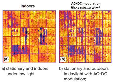
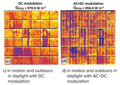
Figure 3i highlights drone based EL images, acquired with global horizontal solar irradiance close to one sun in the plane of the array, where one sun equals 1000W m2.
When the drone starts to fly overhead, you can see further EL images shown in figure 3ii. It shows images with DC modulation (c) and AC & DC modulation (d). It presents lower quality compared to those obtained indoors and stationary in daylight in figure 3i, but still having sufficient quality to identify the main features related to the module power loss.
With further work on the algorithms, this technique shows much promise. It is an obvious advantage to use a drone to inspect PV modules on a solar farm during daylight hours.
find out more
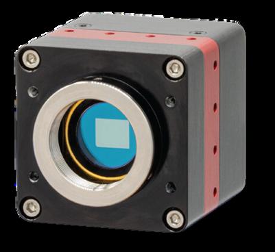
VIS-SWIR technology
Enables high sensitivity imaging from 0.9μm to 1.7μm
High quality sensors 99.5% operability, 640x512, 15μm pixel pitch
15μm x 15μm pixel pitch Enables highest resolution VIS-SWIR image
Ultra high intrascene dynamic range
Enables simultaneous capture of bright & dark portions of a scene
On-board Automated Gain Control (AGC)
Enables clear video in all light conditions
Ultra compact, Low power Ideal for handheld, mobile or airborne systems
More Magazines
Space
QuantumDesignUKandIrelandbringsyou the latest in space and aerospace flight qualified testing and components Our suppliers are responsible for supporting the development of iconic inventions such as the James Webb Space Telescope and theMarsPerseveranceRover
READEDITION1NOW READEDITION2NOW
02
01 03
Semiconductors
Quantum Design has been a leader in high-tech instrumentation for over 40 years with systems such as the MPMS and VersaLab. As distributor for other market leaders who provide solutions for semiconductors, including J. A. Woollam, InfraTec, Sigray, 4D Technology and Lake Shore Cryotronics, we can provide our customers with the right solutions for theirsemiconductorapplications.
READEDITION1NOW
Cryogenics
Featuring updates on new products, publications, and white papers related to our offerings. Quantum Design is heavilyinvolvedineducationandinthis edition we explore various global initiativesfromQD.
READEDITION2 NOW
Textile waste is a growing global concern with far-reaching environmental, social, and economic implications. The global textile industry is one of the largest and fastest-growing industries, producing an enormous quantity of textile products each year.
Estimates suggest that less than 1% of textiles are currently recycled. A significant portion of textile waste ends up in landfills where natural fibers, such as cotton, take years to decompose, while synthetic fibres, like polyester, persist for much longer, polluting the environment.
CASE STUDY
We chose Specim for their exceptional product and service. It’s a safe bet, as they offer an industrial-grade solution with fantastic capabilities” “
Daniel Carrero, Technical Director PICVISA
To boost textile recycling, the EU requires all its member countries to implement systems to manage 100 percent of their textile waste, more than 16 million tons per year, by January 1st, 2025. Despite efforts to regulate and promote recycling, textile recycling faces challenges due to the complexity of sorting textile materials, including blends of different fibers, presenting a pressing need for efficient textile sorting technologies.
PICVISA, a Spanish company specialising in optical sorting, robotics, AI, and deep learning solutions, had a growing number of clients with a significant interest in advanced textile separation solutions, which led to their decision to invest in textile sorting.
In pursuit of innovative textile sorting technology, PICVISA successfully developed a fully automated machine that can classify and automatically separate several types of textile waste by composition (cotton, polyester, viscose, and other fibres), colour, and shape. Thanks to the technological solution implemented by PICVISA, Coleo Recycling in A Coruña, Galicia, classifies and traces some 5,000 tons of textile waste annually.
HYPERSPECTRAL IMAGING ENABLES ACCURATE TEXTILE IDENTIFICATION AND SORTING
The key component of PICVISA’s fully automated textile sorting machine is the Specim FX17 hyperspectral camera. The Specim FX17 operates in a line-scan mode, collecting hyperspectral data in the nearinfrared (NIR) region spanning from 900 to 1700 nm. NIR hyperspectral imaging allows the identification of the composition of textile products since different textile fibres (natural, artificial, and synthetic) have unique spectral characteristics that can be used for classification.
Using the Specim FX17 hyperspectral camera, PICVISA can capture images and analyse the spectral responses of different materials with exceptional precision.
SENSOR-FUSION FOR ENHANCED SORTING ACCURACY
In addition to the Specim FX17 camera, PICVISA integrates various complementary technologies into their textile sorting machine to ensure unparalleled accuracy. These technologies encompass colour separation, defect identification, and contaminant extraction. RGB systems are employed for colour classification, while inductive sensors are used to identify metals in garments.
Artificial intelligence (AI) also plays a critical role in PICVISA’s sorting process. By utilising AI algorithms, the company achieves the classification of materials that may be identical in colour and composition but differ in appearance or shape, guaranteeing precise sorting outcomes.
RESPONDING TO CUSTOMERS’ NEEDS IS THE PATH TO SUCCESS
PICVISA’s diligent efforts have resulted in an extensive classification library comprising over 20 compositions. However, customer demand for an even wider range of classifications remains steadfastly increasing. Notably, tackling the identification of elastane has proven challenging for PICVISA, as its
Specim is a partner with exceptional products and service
its recognition is contingent upon particular compositions and proportions. To overcome the obstacle and further develop their solution, PICVISA is actively exploring regression-based classification besides classbased as a potential solution.
REGULATIONS DRIVE INNOVATION AND GROWTH OF TEXTILE RECYCLING
Lluís Seguí, Managing Director of PICVISA, states:
For
PICVISA, the textile sorting market represents one of the fastest-growing sectors of the future. The development of chemical recycling for mixedfibre garments and finding a solution for already-sorted pure garments will be crucial.”
PICVISA already had experience working with Specim, as the company has previously implemented Specim’s hyperspectral cameras in their automated sorting machines for other segments, such as plastic recycling.
PICVISA’s collaboration with Specim has been highly satisfactory, setting the benchmark for successful partnerships. Regarding their decision to work with Specim, Daniel Carrero emphasises, “We chose Specim for their exceptional product and service. It’s a safe bet, as they offer an industrial-grade solution with fantastic capabilities.”
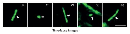
THE STUDY OF CIRCADIAN RHYTHMS AND SUPRACHIASMATIC NUCLEUS (SCN) NEURONS
How does studying jet lag in mice improve our health? A recent study led by Huiyan Li, from the National Center of Biomedical Analysis in Beijing, China is looking to understand how Chronobiology affects our wellbeing.
Targeting cilia-mediated SHH signaling might be a potential therapeutic strategy for the treatment of human diseases related to circadian disruptions
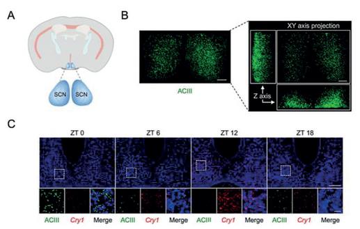
The culture dish was sealed and placed on the stage of an inverted microscope with 10x objective lens in a dark room See Fig 3 Images were acquired with three deep-cooled CCD cameras: Andor iKon-M 934, Raptor Eagle 47-10 and Raptor Eagle CCD 42-40 Images of 60 min exposure duration were collected continuously The Eagle CCD42-40 camera came out as the winner with the optimum performance
To test whether primary cilia-mediated SHH signaling is required for intercellular coupling among SCN neurons, the researchers used realtime luciferase luminescence imaging of SCN slices isolated from Per2::Luciferase (Per2::Luc) transgenic reporter mice to track Per2 rhythmic expression in single cells ex vivo. In the up bioluminescence imaging
Raptor has been developing deep cooled CCD cameras for years The Eagle family of cooled cameras enables longer integration times Both 4MP and 1MP models are available offering -70°C and -90°C absolute cooling
entical culture conditions for a higher concentration of hich was added for imaging.
find out more
7-year vacuum guarantee – Protection and integrity of the sensor
Extremely low dark current – Deep cooled to greater than -110°C delta enables long exposure times
Back illuminated 4MP sensors from e2v Enables large field of view imaging
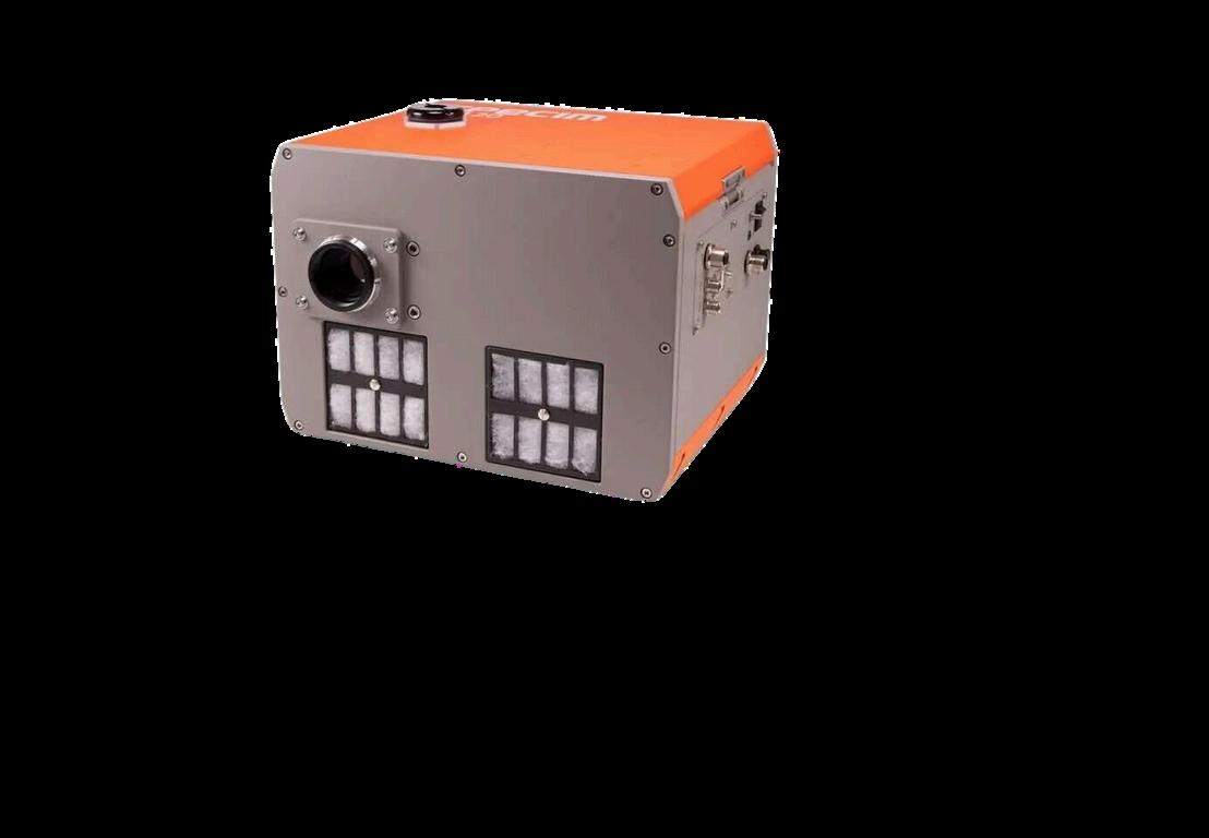
Compared to its predecessor, the Specim OWL, the new Specim FX120 boasts more spatial and spectral pixels, increased imaging speed, GigE Vision compliant interface, and modern detector technology. This translates into improved uniformity and a greater full well capacity, allowing you to capture more detailed and accurate information.”
AN ADVANCED THERMAL PUSH-BROOM HYPERSPECTRAL CAMERA
Specim announces its latest innovation, the Specim FX120, an advanced long-wave infrared hyperspectral camera with a full LWIR spectral range of 7 7 to 12 3 µm This fast push-broom thermal hyperspectral camera is set to redefine chemical imaging capabilities in challenging environments, day and night.
With its excellent spectral and spatial imaging performance, the Specim FX120 allows for the
Jere Hartikainen, Chief Technical Officer at
Specim
simultaneous capture of all 160 swath-width spectral bands at a high image speed of 240 frames per second.
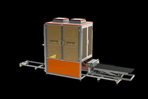
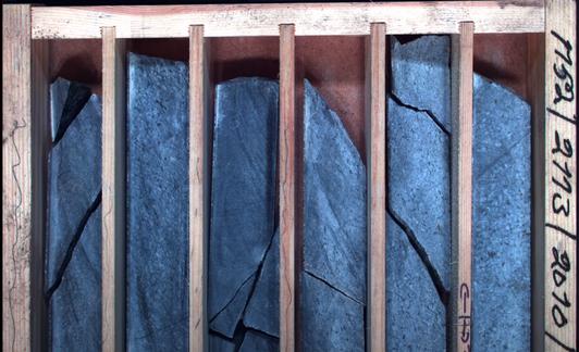
In conjunction with the SisuRock workstation, the Specim FX120 revolutionises drill-core scanning Specim FX120 can detect minerals undetectable by other wavelengths, and its speed and accuracy in capturing all sizes of core enhance productivity in drillcore analysis.
HAVE YOUR SAY
Show off your best visuals
What have you been using imaging cameras for recently? SEND IN YOUR