5 minute read
QDUKI Imaging Cameras Magazine Ed. 2


THE STUDY OF CIRCADIAN RHYTHMS AND SUPRACHIASMATIC NUCLEUS (SCN) NEURONS
How does studying jet lag in mice improve our health? A recent study led by Huiyan Li, from the National Center of Biomedical Analysis in Beijing, China is looking to understand how Chronobiology affects our wellbeing.
Circadian rhythms are physical, mental, and behavioural changes that follow a 24-hour cycle. This rhythm is governed by the master circadian pacemaker, suprachiasmatic nucleus (SCN) which can orchestrate the peripheral clocks in multiple tissues throughout the body. They have established primary cilia–mediated Sonic Hedgehog (SHH) signaling in the SCN as a novel inter neuronal coupling mechanism and may lead to novel therapy of circadian disruption linked diseases like high blood pressure, obesity and other metabolic disorders.
In vertebrate animals, the master clock is a group of about 20,000 nerve cells (neurons) that form a structure called SCN, which is in a part of the brain called the hypothalamus and receives direct input from the eyes. It is the central pacemaker of the circadian timing system and regulates most circadian rhythms in the body Your body tries to align your sleep-wake cycle to cues from the environment, such as when it gets light or dark outside, when you eat, and when you are physically active
Environmental circadian disruptions, such as acute jet-lag and long-term shift work, cause temporal unsynchronisation between the internal circadian clock and external time cues, leading to physiological stress Circadian disruption has been implicated in tumorigenesis and various psychiatric, neurological and metabolic diseases, including depression and diabetes. The study of circadian rhythms and suprachiasmatic nucleus (SCN) neurons is an active area of research, and new discoveries continue to expand our understanding of these complex processes.

Primary cilia, specialised hair-like structures are found on the surface of many cells, play crucial role in mammalian embryonic development Tu et al revealed that primary cilia in a subset of SCN neurons cilia exhibit pronounced circadian rhythmicity in abundance and length, and further identify primary cilia as a critical device for intercellular coupling to maintain the circadian clock in the SCN
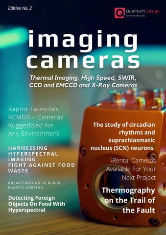
Isolated live SCN neurons from postnatal mice were transduced with modified baculovirus encoding mCherry tagged, constitutively ciliarylocalised protein, 5-hydroxytryptamine receptor 6 (5-HT6). The numbers on the images indicate the time in hours Arrows indicate primary cilia Scale bar, 5 μm (J) Quantitative analysis of the cilium length in (I). Each dot represents one cell.

In vertebrates, primary cilia are required for transactivation of the Sonic Hedgehog (SHH) signalling. SHH pathways play a significant role in embryonic development and tissue patterning, primarily during early development
Targeting cilia-mediated SHH signaling might be a potential therapeutic strategy for the treatment of human diseases related to circadian disruptions
The research highlighted that cilia-mediated SHH signalling in the SCN is essential to control the central clock function in mice. They also found that a clinical drug, Vismodegib, blocking SHH signaling, could be used to regulate circadian rhythm, and render mice susceptible to rapid resetting by light. Thus, targeting cilia-mediated SHH signaling might be a potential therapeutic strategy for the treatment of human diseases related to circadian disruptions
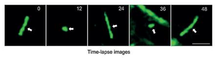
The culture dish was sealed and placed on the stage of an inverted microscope with 10x objective lens in a dark room See Fig 3 Images were acquired with three deep-cooled CCD cameras: Andor iKon-M 934, Raptor Eagle 47-10 and Raptor Eagle CCD 42-40 Images of 60 min exposure duration were collected continuously. The Eagle CCD42-40 camera came out as the winner with the optimum performance
To test whether primary cilia-mediated SHH signaling is required for intercellular coupling among SCN neurons, the researchers used realtime luciferase luminescence imaging of SCN slices isolated from Per2::Luciferase (Per2::Luc) transgenic reporter mice to track Per2 rhythmic expression in single cells ex vivo. In the experimental set-up bioluminescence imaging was used with identical culture conditions for SCN slices with a higher concentration of luciferin (1mM), which was added for imaging
The culture dish was sealed and placed on the stage of an inverted microscope with 10x objective lens in a dark room See Fig 3 Images were acquired with three deep-cooled CCD cameras: Andor iKon-M 934, Raptor Eagle 47-10 and Raptor Eagle CCD 42-40 Images of 60 min exposure duration were collected continuously. The Eagle CCD42-40 camera came out as the winner with the optimum performance
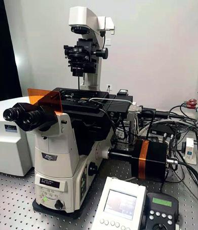
Raptor has been developing deep cooled CCD cameras for years. The Eagle family of cooled cameras enables longer integration times Both 4MP and 1MP models are available offering -70°C and -90°C absolute cooling
Ref 1 - Rhythmic cilia changes support SCN neuron coherence in circadian clock- Hai-Qing Tu et al - National Center of Biomedical Analysis in Beijing, China. doi: https:// DOI: 10.1126/science.abm1962
---------------------------
Eagle Scientific CCD
-90°C 1MP and 4MP Scientific CCDs
Features
7-year vacuum guarantee – Protection and integrity of the sensor
Extremely low dark current – Deep cooled to greater than -110°C delta enables long exposure times
Back illuminated 4MP sensors from e2v Enables large field of view imaging
13.5μm x 13.5μm pixels (4MP) – Enables ultra sharp image resolution
High QE: >90% @ 525nm and 50% @ 380nm & 720nm –Optimum photon collection
find out more
-----------------------
To discuss your application, please get in touch with our Technical Sales Manager, Dr. Luke Nicholls by email (luke@qd-uki.co.uk) or call (01372) 378822.