BioTools Magazine from QDUKI

Page 1


NEXT-GEN BIOTOOLS

High-Tech Instrumentation for Bio Applications

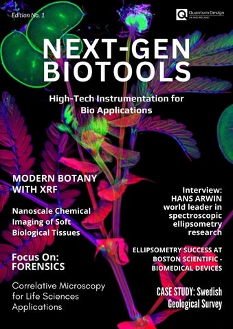
MODERN BOTANY

WITH XRF

Nanoscale Chemical Imaging of Soft Biological Tissues

Focus On: FORENSICS

Correlative Microscopy for Life Sciences Applications

ELLIPSOMETRY SUCCESS AT BOSTON SCIENTIFICBIOMEDICAL DEVICES

CASE STUDY: Swedish Geological Survey

Welcome to the first issue of NextGen BioTools, the new magazine from Quantum Design UK and Ireland dedicated to showcasing the latest advances in high-tech scientific instrumentation for life science and biomedical research Our mission is to highlight the tools, techniques, and success stories that are shaping the future of biological discovery, from fundamental plant science to cutting-edge medical applications.

In this issue, we explore the unexpected intersections of biology and technology. Discover how XRF analysis is revolutionising modern botany, offering fresh perspectives on plant physiology and adaptation. We are proud to feature an exclusive interview with Hans Arwin, a world leader in spectroscopic ellipsometry, who shares insights from decades of pioneering research

We also examine correlative microscopy for life sciences, where advanced imaging techniques are enabling scientists to uncover biological complexity with unprecedented detail A case study from the Swedish Geological Survey demonstrates how instrumentation designed for earth sciences can unlock biological knowledge, while Boston Scientific shares its journey using ellipsometry to drive success in biomedical device development Finally, we highlight how the Raptor SWIR camera has been applied in a unique project exploring the science of happiness.

NextGen BioTools celebrates innovation, collaboration, and the transformative role of instrumentation in bioscience.

Foreword

Correlative Microscopy for Life Sciences Applications

Correlative in-situ Analysis of Bone Collagen

Nanoscale Chemical Imaging of Soft Biological Tissues

Focus On: FORENSICS

Ellipsometry for Biomedical Devices

Ellipsometry Success at Boston Scientific, Clonmel

Raptor SWIR Camera Used in the “Pursuit of Happiness”

INTERVIEW: HANS ARWIN

in spectroscopic ellipsometry research in biological material applications

ILLUMINATING THE Process of Biomineralisation in Baby Teeth

In-Vivo NIR-II / SWIR Imaging

Correlative Microscopy for Life Sciences Applications

Easy to use Correlative AFM with SEM Microscopy Platform

Combine the complementary strengths of AFM and SEM like never before! The FusionScope fully integrates a wide range of advanced AFM measurement techniques with the benefits of SEM imaging. Seamlessly image your sample, identify areas of interest, measure your sample, and combine your imaging data in real time

Discover the FusionScope

APP NOTES AND ARTICLES ON LIFE SCIENCES

Using FusionScope in Life Science applications allows you to acquire the nanoscale morphology of biological samples accurately and easily. Especially for hard-to-reach sample areas or very small features, FusionScope allows you to characterise physical properties such as 3D topography, stiffness, and adhesion with the highest precision.

DISCOVERPROPERTIESOF HARD-TO-REACHSAMPLE AREASUSINGATOMICFORCE MICROSCOPY

CHARACTERISESUB-WAVELENGTH PARTICLESUSINGAFMANDENERGY DISPERSIVESPECTROSCOPY(EDS) ALONGWITHPROFILEVIEW

Mode: AFM Topography

Sample: Bone

FusionScope offers a fast and easy identification and imaging of the lacunae structures

Read the Article

EASILYLOCATEANDIMAGE DIATOMSONASEASHELL SURFACE

Mode: AFM Topography, SEM

Sample: Seashell

Modes: SEM, AFM Topography and Phase, EDS

Sample: Superparamagneti c Particles

For this example, we use carboxyl (COOH) group functionalised magnetic particles as a model system to demonstrate FusionScope’s imaging capability. The FusionScope allows us to image the particle’s surface with picometer resolution The wide field of view of the SEM is employed to localise particles for AFM imaging

The topography and phase data from the AFM permit high-resolution inspection of the particle surfaces. We then use the correlated coordinate system and correlative in-situ measurement modes to capture relational AFM and SEM data Higher-resolution AFM images from a single particle are shown in Figure 1 Finding particular particles of this size to take AFM data would be extremely difficult without FusionScope's correlated coordinate system and Profile View

Read the Article

Diatoms are fascinating unicellular organisms that make up a significant amount of the Earth's biomass. In this application we used the power of correlative microscopy to locate diatoms on the surface of a seashell using the high-resolution of the FusionScope SEM With Profile view you can easily position the AFM cantilever tip on a diatom structure of your choice and perform a 3D topography analysis

F S E M ® :

Bonetissueiscontinuouslyremodeledthrough the concerted actions of bone cells Whereas osteoblasts synthesise the bone matrix and are responsible for its mineralisation, osteoclasts breakdownbonematrix.

The third cells, osteocytes, are inactive osteoblasts that have become trapped within thebonetheyhaveformed.

The small space containing the osteocyte is called lacuna When the tissue is dead, the lacuna topography gives information about the osteocytethatoccupieditbefore

This is particularly advantageous when the sample is extremely difficult to reach with the AFM cantilever as in the case of bovine vertebra.

®

AFSEM combines the strengths of both SEM and AFM for a quantitative analysis of bone structure

SEM’s large field of view enables identifying lacunae in bone tissue and the precision of the SEM is used for positioning the cantilever directlyontolacunae

Correlative AFM/SEM microscopy allows for complementary SEM and AFM topography information of lacunae and collagen fibres Real 3D representation of the topography can be used for analysing collagen fibres with highresolution

Quantitative analysis of the AFSEM® data reveals characteristic 67 nm periodic banding pattern on collagen fibres with sub-nm resolution The cross-section of the banding pattern shows a corrugation heightof3nm.

In summary, AFSEM is the solution of choice to obtain correlative AFM/SEM microscopy analysis. It accepts any sample that fits the SEM/FIB. SEMguidance makes the identification and cantilever positioning even on very complicated samples very easy. AFSEM enables quantitative 3D analysis of features with sub-nmresolution.

AFSEM ensures SEM-guided easy identification and cantilever positioning on lacunae in bone structure and enables complementary SEM and AFSEM 3D topography information of lacunae and collagen fibres. sample courtesy : Prof. G. E. Fantner, (EPFL)

Interactivecorrelativein-situ

SEMandAFManalysis

Identifyyourregionof interestfastandeasily

Quantitative3Dtopography withsub-nmresolution

AFSEM® enables quantitative 3D analysis of collagen fibres with sub-nm resolution.

AFSEM positioned above the bone sample mounted on the SEM stage

NANOSCALE CHEMICAL IMAGING OF SOFT BIOLOGICAL TISSUES

Exploring the capability of PiFM on fixed human skin samples that were penetrated by topically applied dexamethasone and show that there is similarly a good correlation between XAS microscopy and PiFM, with PiFM offering much higher spatial resolution.

one and red and bined to ponents; s shown d to the elow) at sorption From mission drug is ard the orneum, angular previous in slices posited r XAS as then PiFM um of PiFM 08 cm 1 ighlight and band), d was e has a d 1660

FTIR spectrum of dexamethasone; (b) combined PiFM images (top) acquired at 1708 cm 1 1 for corneocytes (green) and X-ray transmission image of the same region. Region A is further investigated by PiFM in Figure 2.

Credit: The samples were provided by E. Rühl, Freie Universität Berlin (Germany).

Figure 2 shows PiFM images at higher magnification along with the topography. The top images (12 mm × 12 mm) show the stratified structure of the stratum corneum clearly in all imaging modes; the bottom portion of these images coincide with the region A defined in Fig 1b Fig 2c shows clearly how the drug is concentrated in the uppermost layer of the stratum corneum as seen in Fig 1 The bottom row of images (3 mm × 3 mm) in Figure 2 zoom into the region defined by white boxes in the top images

Figure 2b’ shows that no amide II signals are seen in between the corneocytes, most likely due to the thin lipid lamellae that are known to occur between the corneocytes. However, Fig 2c’ shows that the topically applied drug penetrates into these lipid lamellae between the corneocytes and fills them partially; this is even more clearly visible in Fig 2d’ (combined PiFM image, red for dexamethasone and green for corneocytes), which is magnified by 2X for easier observation.

To discuss your application or upcoming research project, get in touch with our Technical Director, Dr. Shayz Ikram.

Call (01372) 378822

Email shayz@qd-ukicouk

Fig 2. Detailed view on the stratum corneum of fixed human skin: (a) AFM topography visualising the structure of corneocytes; (b) chemical PiFM contrast related to amides probed at 1558 cm 1; (c) PiFM contrast related to dexamethasone at 1708 cm 1; (d) two PiFM images combined to map chemical components.

Vista One makes nanoscale chemical maps and point spectra with more detail than FTIR or nanoFTIR. Characterise at the highest resolution.

Learn more at qd-uki.co.uk

Focus on Forensics

X-ray Fluorescence for Forensics

Not only is XRF a non-destructive technique, it requires essentially no sample preparation The ATLAS from IXRF Systems is a small spot µ-XRF unit which is capable of analysing extremely small samples down to 10 microns or scanning a small area of interest. µ-XRF is especially useful for analysing trace levels of elements in the ppm range, which can mean differentiation where other techniques falter or it could add to the weight of the evidence by showing samplescannotbedifferentiated

ATLAS can be used to analyse a wide variety of sample types, including but not limited to: glass chips, rubber, paint, soil/geological material, metals, money/coins,glitter,andmore.

ATLAS is the only µ-XRF unit that has software that automatically processes and presents data according to the specifications in the ASTM method: “ASTM E2926-13 Forensic Comparison of Glass using Micro-XRF Spectrometry”. The use of the software eliminates potential sources of human introduced errorwithinthenumericaldata.

Discover the ATLAS

IXRF ATLAS M : micro XRF Spectrometer

The 5 micron advantage

With the smallest X-ray spot in the industry at 5 μm, this unique XRF microscope is optimised for analysis speed without compromising accuracy

SpectrumofNIST610Glass Standard200-300ppmlevels

Paintinganalysis

ATLAS is a very powerful tool capable of single or multi-spot analysis as well as elemental mapping to identify location of elements within a sample This is extremely useful for paint chip/layeranalysis.

Metalcomparison

XRF can help distinguish between two very closely related steel standards by comparing trace levels of elements. Shrapnel from a pipe bomb scene could be compared to metal shavings foundinasuspect’sgarage

Tyrerubber

Identify trace elemental similarities or differences between tyre mark and tire Scrapings from a tyre mark at a hit and run scene can be compared to asuspectvehicle

Soilanalysis

XRF can easily identify differences between common potting soil and contaminated soil Soil found from a shoe print at a crime scene can potentially indicate the suspect has beentoorisfromaspecificregion

Due to the micron-sized spot of ATLAS different layers of paint chips are easily resolved not only elementally but also by comparative concentrations from one sample to the next This can help differentiate closely related samples by the coating, paint, or base coat in any combination. ATLAS can also be used as a bulk spot size XRF for homogenous evidence materials such as soil, metals, or otherunidentifiedmaterials

Cracking Cases with Light: How Hyperspectral Imaging Could Revolutionise

Forensics

Analyse forensic evidence either in laboratory conditions or directly at the crime scene.

Hyperspectral imaging is a noncontact, non-destructive method that requires no sample preparation, ensuring the integrity of evidence

A crime scene can hold a myriad of evidence – stains, marks, or residue. It is for the forensic investigators to identify, gather and analyse the samples to help find out what happened.

Crime Investigation Goes Mobile

While forensic science has made leaps and bounds in the past, could it still benefit further from the use of hyperspectral imaging?

Several independent studies indicate that it could There is room for improvement in the current crime scene investigation methods: while some of them use chemicals and therefore risk contaminating the sample, others require sending the samples for laboratory analysis, which takes time and money.

What is missing is a non-contact, non-destructive way to identify materials quickly, reliably, and on location

Here are just a few examples of how hyperspectral imaging can improve forensic investigation methods.

Identifying Blood Stains

A stain is found at the possible crime scene Is it blood, or something else? Is it from the nosebleed that happened last week or from a more recent event? Hyperspectral imaging finds blood stains in crime scenes quickly and easily What is more, it can even help determine how old the blood stain is. No chemicals are involved, therefore there is no risk of diluting or altering the blood spatter.

“Hyperspectral imaging for the age estimation of blood stains at the crime scene”

Examining Gunshot Residue

Gunshot residue is delicate evidence. When a forensic investigator applies chemicals for visualisation, they can alter the pattern that is used for reconstruction and determining shooting distances and angles As a result, they may destroy valuable information. Hyperspectral imaging can provide a quick, non-contact tool for determining the presence of the gunshot residue at a scene and visualising the patterns.

“Visible and near-infrared chemical imaging methods for the analysis of selected forensic samples”

Analysing Paint Marks in Hit-andRun Cases

Paint traces left on a scene of a hitand-run case can give valuable information on the car that escaped the scene Paint analysis, however, requires microscopes and sample preparation. With hyperspectral imaging, a quick test performed at the scene could narrow down the possible makes of the car based on the paint spectra.

“Evaluation of Hyperspectral Imaging Visible/Near Infrared Spectroscopy as a forensic tool for automotive paint distinction”

THE FUTURE OF HYPERSPECTRAL IMAGING IN FORENSICS

Forensics is more than just crime scene investigation. The possible applications of hyperspectral imaging do not end there, either. Hyperspectral imaging could, for instance, help doctors determine the age of a bruise – objectively and accurately. It could also be used in a laboratory to identify fraudulent documents without having to destroy the sample. As the technology develops and becomes more portable and easier to use, it is quite possible that a hyperspectral camera will be a regular part of forensic investigators’ toolbox in the future.

EXAMINE AND ANALYSE EVIDENCE WITH SPECIM IQ

Blood stains: detection, identification, age

Bruises: ageing and detection

Paint analysis in hit and run cases

Document examination: altered documents, forged signatures

Gunshot residue

SPECIM IQ

Mobile, compact and autonomous

Integrated processing engine for obtaining instant results

Highest sensitivity and data quality on the market

Ellipsometryfor Biomedical Devices

Medical de to the coatings to ensure biocompatibility, improve functionality, and increase the lifespan of the device within the body. Coatings are engineered to reduce inflammation caused by the device and therefore reduce the associatedriskofinfection.

Thin-film coatings on these devices have also been designed to improve corrosion resistance caused by interactions with bodily fluids. Many different methods of deposition have been developed to optimise the coatings Ellipsometry is one form of metrology used in development and quality control of thin-film deposition on bio-implanted or adhered devices our technology is used to monitor the consistency of the coatin as well as ensureitspresenceonthedevice.

Boston Scientific’s Clonmel site includes a Centre of Excellence for metal additives used in a range of electronic components for medical technology. Boston Scientific employees based in Clonmel manufacture medical devices that support the treatment of more than 400,000 patients globally each year

Products created in Clonmel help patients suffering from conditions such as heart disease, neurological disorders, kidney stones, and diseases of the pancreas, bile ducts and esophagus.

“Here at Boston Scientific Clonmel, we have advanced capabilities to deposit thin film and have wide range of characterisation techniques,” says Zahra Ghaferi PhD, a Senior Scientist in Process Development at Boston Scientific.

“In this regard, Ellipsometry helped us to develop a recipe for ALD film deposition on our implantable devices. We have a J.A. Woollam alpha-SE ellipsometer on site which enables us to verify film thickness and identify layers by optical features.”

“To me, J.A. Woollam and Quantum Design UK and Ireland have the most reliable and user-friendly Ellipsometers. Not only equipment diversity but also robust technical knowledge and wonderful customer care make them unique in this field. I was working on a very complex project and was struggling to model film thickness. I was in touch with vendor and they kindly advised me how to sort it over phone call and email.”

J.A. Woollam alpha 2.0

ELLIPSOMETER FOR SPECTRAL RANGE OF 400-1000 NM

The alpha 2.0 is a budget-friendly option for routine measurements of thin film thickness and refractive index A compact footprint and simple design make the alpha 2.0 easy to use while harnessing the power of spectroscopic ellipsometry. It was designed for easeof-use: simply place the sample on the stage, choose the model that matches your film, click “measure”, and you will have results within seconds.

Zahra working with the J. A. Woollam alphaSE at Boston Scientific

Raptor SWIR Camera Used in the “Pursuit of Happiness”

Researchers at Janelia Research Campus, Howard Hughes Medical Institute, are investigating the vast and complex network necessary for an organism to sense and react to the world. To do this, researchers must study neurons, which are the building blocks of the nervous system. Neurons connect to create complex networks at highly specialised junctions Individual cells communicate at these ‘synapses’ by releasing chemical signals (or neurotransmitters) such as dopamine.

Despite the central role that synapses play in the brain, it remains challenging to measure exactly where neurotransmitters are released and how far they travel from their release site. Currently, most tools available to scientists only allow bulk measurements of neurotransmitter release. Using a Raptor Ninox II Vis-SWIR camera has enabled researchers to study how dopamine is released from subcellular compartments that have previously not been well characterised

To tackle this limitation, the team at Janelia developed a new way to measure neurotransmitter release from neurons, harnessing a technique which uses fluorescent nanosensors that glow brighter when exposed to dopamine. These sensors form a very thin film upon which neurons can grow; when the cells release dopamine, the sensors ‘light up’ as they encounter the molecule. Dubbed DopaFilm, the technology reveals exactly where the neurotransmitter comes from and how it spreads between cells in real time

Figure 1: Schematic of DopaFilm imaging protocol (see Ref [1]) (A) Schematic of DopaFilm (B) Workflow for preparing dopamine neuron primary cultures from the rat mid brain regions highlighted in blue Neurons are grown on dishes with an engineered, chemisensitive, and fluorescent surface (DopaFilm) at the bottom

In particular, the approach showed that dopamine emerges from ‘hot spots’ at specific sites in cells; it also helped the team study how dopamine is released from subcellular compartments that have previously not been well characterised. Figure 1 highlights the schematic and workflow of the experiments

Figure 2: DopaFilm enables interrogation of the molecular correlates of dendritic release

Activity from a dendritic process and its corresponding THGFP, MAP2, and Bassoon images. The NIR ΔF/F image contrast was set at 0–30% ΔF/F

DopaFilm requires use of detectors with InGaAs sensors for photon detection and additional optimisation of optical components to facilitate transmission of NIR and SWIR photons. Its nonphotobleaching fluorescence properties in the NIR to SWIR regions of the electromagnetic spectrum (0.85–1.35 μm) is spectrally compatible with existing optical technologies, as highlighted in figure 2.

For broad-spectrum (visible to SWIR, 400–1400 nm) imaging, the team developed a custom microscope, see figure 3, equipped with two fibrecoupled NIR lasers to excite far red, NIR and SWIR fluorophores:a 671 nm and a 785 nm laser. The microscope is equipped with an Ninox 640-II camera with optimised sensitivity in the SWIR range.

Raptor has been developing InGaAs cameras for years The Ninox family of cooled InGaAs cameras enable longer integration times. These cameras are available in various resolutions and pixel sizes As well as being compact and low power, they provide high sensitivity, ultra-low noise images in visible and SWIR wavelengths.

Figure 3: Custom Microscope Set-up with Ninox 640 II VIS-SWIR Imaging Camera.

What

made you choose physics?

What was your PhD dissertation topic? When did you become interested and involved with optics and materials?

I studied electrical engineering at Chalmers Uiversity in Gothenurg (19691974, with a one-year break for military service as a Radar Technician) My master’s thesis dealt with measuring the spectral dielectric function at kHz/MHz frequencies of cerebrospinal fluid from old people with dementia. The idea was toseeifproteinshaddifferentresonance frequencies due to different amount of bound water caused by Alzheimer’s and otherdiseases.

HANS ARWIN

HansArwinatLinköpingUniversityisa worldleaderinspectroscopic ellipsometryresearch Especiallyin biologicalmaterialapplications Prof Arwin’sarsenalofellipsometric capabilitiesincludedynamic(in-situ), spectroscopic,generalised (anisotropic),imaging,andtotal internalreflectionellipsometry.

After my master’s degree in electrical engineering (1974), I worked on a PhD in applied physics My dissertation in 1980 in Linköping was about a capacitance method to determine enzyme activity in blood. It includes some in-situ (solidliquid) null ellipsometry (mass determination) in combination with capacitance measurements (surface coverage) allowing determination of molecularsurfacearea.

How did you get interested and involved with biological applications of ellipsometry?

My biology approach is due to my PhD supervisor, Ingemar Lundström, who encouraged this research. It turned out to be very fruitful as the competition was small and it was easy to present new results obtained by ellipsometry techniques.

Ithasbeenanicejourneyincluding: single-wavelength thickness determination of protein layers in-situdynamicsofproteinadsorption in-situproteinlayerspectroscopy proteinsinporoussilicon TIREforbiosensing analysisofcellulosenanocrystallayers transparentwood andmuchmore!

An interest in beetles started in 2004 when I visited Mathias Schubert in Leipzig I had a beetle in my bag, and we did a measurement with his Woollam M-2000 system. I did not understand the data even though Mathias encouraged me to use models developed for liquid crystals Many years later, when we had the RC2 system, we could do some serious studies of the chrial circular Bragg structures in exoskeletons of the narrowband (green) Cetonia aurata beetle

Our first paper was in 2012, but [almost exactly] 100 years after Michelson, who first observed these effects in beetles We were lucky to get support during many years to perform curiosity-driven research with no demands on applications It became a fascinatingjourney

Tell us about the impact and usefulness of J. A. Woollam ellipsometers on your research...

J A Woollam ellipsometers have never disappointed me They are in most cases not limiting our progress. It is the modeling which is the bottlenec as samples are never ideal However, WVASE and CompleteEASE are excellent, and we skipped development of our own program in the 90s. We were lucky to have one of the first RC2s and J. A. Woollam was very generous and updateditseveraltimesforfree Thanks to the RC2 we could move to Mueller matrices very early, which has been a very good advantage. This made it possible to study depolarisation, perform differentual as well as sum decompositions, and also has given us more confidence in electromagnetic modelingofchiralstructures.

What do you think is your greatest contribution to modern ellipsometry? Your proudest work?

If you had asked me long ago, I [would have] said resolving spectral dielectric functions of protein monolayer first at air-solid interfaces (at Bell Labs), then at liquid/solid interfaces (with our homebuilt RAE) and finally in IR with the IRSE system. I had wished that our more applied research on ellipsometric memories, drug delivery using porous structures, optical nose, or TIRE had reached industry, but I am a bad marketing person and not at all an entrepreneur

I now consider my work on understanding structure and determining properties of chiral bioreflectors to be my greatest contribution We have some 25 publications on the subject, covering more than 10 different types of beetles. During this project we have developed themethodology

Initially we presented Mueller matrices only. The we performed electromagnetic modeling of twisted layered structures, sum decomposition for classification of bioreflectors, differential decomposition to resolve effective chiral parameters, resolved pitchgradientstoexplainnarrowversus broad, band chiral features (circular Bragg) to understand differences between silvery and green/red beetles, and more. Recently we have done transmission ellipsometry with ambition to generalise ellipsometry to include circular birefringence (CB) and circular dichroism (CD) It is pretty extensive, and it was very nice that it was acceptedasis.

Imaging ellipsometry on beetles at École Polytechnique in Paris when visiting Enrique GarciaCaurel and Razvigor Ossisovski (2010)

I am now very happy that our Mexican colleague Arturo Mendoza-Galván has a program on synthesis of chiral structures in cellulose He has participated in our beetle research and hascontributedconsiderably.

He now has protocols for fabricating thin films of nanocrystalline cellulose with polarising properties

I am fully focused on chirality and working on a follow-up paper to my latest contribution [1]. I also have in the pipeline to make a review of the beetle studies performed over the years We havealsoacollectionofCetonia aurata beetles, and we will investigate if the Mueller-matrices exhibit some differences between male and female beetles It is still unclear why they have thesepolarisingfeatures

Tell us about what you are doing now and what you plan and want to do in the upcoming years...

Iwillcontinuedevelopmytextbook Ialso runmyconsultingcompanyandperform onaverage10smallprojectseachyear Ialsodevelopshortpresentations(20-30 minutes) on various aspects of the Stokes/Mueller formalism I present them at our group meetings (Mueller School) So far I have done 25 presentations over theyears.

The things I do above are for keeping my brain alert For my body I am doing sports activitiesatleastfourdaysper week(skiing,skating, rollerskies, floorball, jogging, gym, etc.).

ILLUMINATING THE PROCESS OF BIOMINERALISATION in Baby Teeth with MicroXRF

The journey to understanding human growth and development traverses unexpected but fascinatingscientificroutes.

The study of human baby teeth and bones, far from being purely dental or orthopedic research, offers a unique lens into a child’s overall health. A particularly innovativeanalyticalapproach for understanding growth and development involves highspatial-resolutionmicroXRF.This powerful tool provides an indepth analysis of the elemental composition of teeth and bone down to 5μm pixel diameter. This blog post by IXRF Systems delves into the application of microXRF in studying biomineralisationinteeth.

MICROXRF

MicroXRF (Micro X-ray Fluorescence) spectroscopy is an advanced analytical technique used for non-destructive elemental analysis with high spatial resolution. While it shares the same foundational principles as XRF, microXRF allows researchers to scrutinise materials at microscopic levels. By directing an ultrafine X-ray beam – as small as 5µm –onto the sample, the technique excites atoms, and as these atoms revert to their ground state, they emit X-rays at wavelengths that are distinctly unique to each element This feature allows users to identify and quantify the elements MicroXRFcandetect,quantifyandmapthe spatial distribution of elements in solid, tissue, liquid, and powdered samples with minimalpreparation.

INSIGHTS THROUGH ELEMENTAL COMPOSITION AND DISTRIBUTION

The elemental composition of baby teeth (also referred to as primary or milk teeth) and bones hold valuable information about a child’s environmental and nutritional history This research helps us understand the influence of factors such asdiet,environmentaltoxins,andparental habits like smoking on their growth and development. Certain elements, including lead,arsenic,andmercury,areparticularly important to monitor due to their significanthealthimpacts.

Researchers can identify potential associationsbetweenearlylifeexposureto certain elements – as determined by microXRF – and subsequent health issues by linking the elemental data with health and development outcomes. This information plays a critical role in formulating public health initiatives to protectchildren’shealth

MICROXRF FOR STUDYING BIOMINERALISATION IN TEETH AND BONES

MicroXRF allows for the nondestructive examination of the elemental composition and distribution in teeth and bones. Aschildrengrow,traceelements from their environment and diet are integrated into their teeth and bone structures. Just like tree rings, teeth provide a chronological record of exposure to these elements MicroXRF allows us to analyse tooth development, shedding light on a child’s early life exposures and potential health implications.

THE DENTIN-ENAMEL JUNCTION (DEJ) – A CRUCIAL INTERFACE

One portion of the tooth that can illuminatedevelopmentaldifferences is the crucial interface between odontoblastsandameloblastsatthe junction where dentin and enamel usually meet and become tightly bound to each other. This interface, also known as the Dentin-Enamel Junction(DEJ),playsacriticalrolein toothstrengthandresilience.TheDEJ is the interface between the two primary mineralised tissues of the tooth:

enamel, the hardest tissue in the human body, produced by ameloblasts, and dentin, a slightly softer material produced by odontoblasts. This junction is critical for the mechanical integrity of the tooth. Despite the stark difference in the properties of enamel and dentin, they form a seamless connection at the DEJ, giving the tooth its unique combination of hardness and resilience.

Figure 1: 2D elemental heat maps of a sliced baby incisor tooth ~0.8 mm thick. Maps were collected on the Atlas M microXRF at a pixel size of 5µm.

At the DEJ, several elements are of key interest. Calcium (Ca) and phosphorus (P), the primary constituents of hydroxyapatite, the mineral that gives teeth their hardness, are present in different

amounts in enamel versus dentin. Mapping these elements using microXRFcanhelpusunderstandthe transition between these two tissues atthemolecularlevel

In addition, trace elements like magnesium (Mg), zinc (Zn), strontium (Sr), and others are of great interest These elements can subtly alter the properties of hydroxyapatite and might play a role in the binding mechanism between dentin and enamel. Detailed mapping and quantification of these elements at the DEJ will provide more clarity on theirroleandfunction.

Knowledge about the elemental composition at the DEJ can potentially help develop new strategies for the prevention and treatment of dental diseases. For instance, a better understanding of the DEJ could lead to the development of more effective dental materials for restorations and prosthetics, which could mimic thenaturalstructureandfunctionof theDEJ

THE ROLE

OF IXRF

SYSTEMS IN THIS INNOVATIVE RESEARCH

IXRFSystemssta of this research supplying robu efficient microX systems offer

resolution, providing an X-ray spot size as small as 5µm, making it ideal for this analysis due to the necessity to see microscale featuresoftenpresent.

Moreover, this technology can configure up to four (4) silicon drift detectors (SDD), adding photon sensor area, directly translating to counts and mapping velocity in a linear relationship, providing highthroughputanalysis,andpavingthe wayforcomprehensivestudies

Figure 2: Atlas M benchtop microXRF spectrometer (above) and an inside view looking up at the polycapillary X-ray source with 4 SDDs incorporated (below).

The objective at IXRF Systems goes beyond providing cutting-edge analytical technology; we strive to propel research that contributes to a comprehensive understanding of human health. Collaborating with researchers and public health institutions,theyaspiretoenrichour knowledge of early-life elemental exposures’ impacts and aid in developing effective preventive measures.

CONCLUSION

The use of MicroXRF for studying biomineralisation in teeth and bones exemplifies the multidisciplinary and innovative nature of contemporary science. This approach empowers researchers to identify patterns of elemental exposure that could potentially influence a child’s health trajectory IXRF Systems remains committedtodeliveringtheessential tools for this critical work, as they continually refine their understandingoftheserelationships.

Intrigued by the intricate process of biomineralisation in teeth or bone? Your journey into this complex and critical biological phenomenon can start today with IXRF Systems. By utilising IXRF’s advanced microXRF technology, researchers, healthcare professionals, and innovators are makingstridesinunderstandinghow minerals are deposited in biological systems The implications span from enhanced dental care to groundbreaking treatments for bone-relateddisorders.

IXRF ATLAS M : micro XRF

Spectrometer

Micro X-ray Fluorescence (μXRF)

Hyperspectral Imaging Elemental Analyser

Benchtop / tabletop form factor

Spot size down to 5 microns with antihalo optic

SDD detectors w/ active area up to 150 mm2

Large chamber volume

50 kV / 50 W polycapillary optic X-ray source

Multi-point/multi-area automation & mapping

Air, vacuum, helium for solids, liquids, and powders

Iridium Ultra software running on Windows™ 10 OS

To discuss your application or area of research, please get in touch with our Sales Manager, Dr. Luke Nicholls. Call (01372) 378822

Email luke@qd-ukicouk

In-Vivo NIR-II / SWIR Imaging

Non-invasive fluorescence imaging of small animals has been a technique widely used for years as a method in preclinical research. Techniques have developed considerably in recent years, but still face a number of limitations.

Researchers detecting in the 500900 nm range suffer from shallow imaging depth and high background due to light scattering andtissueautofluorescence.

Over the last five years there has been a growing interest in NIRII/SWIR imaging detecting in the 1000-1700 nm range to greatly suppress these effects, affording single cell resolution at ~ 3 mm depth and useful resolving power at upto~1cmdepth.

This interest has been fuelled by the development of NIR-II markers / probes (molecules, carbon nanotubes, quantum dots and rareearth down-conversion nanoparticles) which provide a great combination of sensitivity, inherent biological safely, and relative ease of use. (see Nirmidas Biotech, Inc’s variety of dye materials.)

Images courtesy of a Project that developed the downconversion ErNP technology commercialised by Nirmidas Biotech, Inc

Introducing these probes into the systems of small animals enables researchers to see more in terms of pathophysiological processes. The goal is in evaluating the biodistribution, pharmacokinetics and biological activity of potentially therapeutics molecules that will identify and fight tumours. Deep tissue molecular imaging with diffraction limited resolution at the single cell level can also facilitate basic research in biology andmedicine.Buttodoso efficientlyrequiresaSWIR optimisedcamera.

Based on original inventions from Stanford University, a US based company, Nirmidas Biotech Inc. in Palo Alto California has developed and commercialised a series of near infra-red II (NIR-II) organic dye and inorganic probe products suitable forinvivo,exvivoandinvitroimagingapplications.

The DeepVision™ system is a high performance and cost effective NIR-II/SWIR fluorescence/ luminescence system with multiple, customisable lasers Itistheonlysmallimagingsystemdetectingin both NIR-I and NIR-II windows: wavelength of imaging spans 400 nm – 1700 nm with high detectionefficiencyinNIR-II1000–1700nmrange.

Raptor has been developing InGaAs cameras for years. The Ninox family of cooled InGaAs cameras enable longer integration times. These cameras are available in various resolutions and pixel sizes. As well as being compact and low power, they provide high sensitivity, ultra-low noise images in visible and SWIR wavelengths. Raptor has worked closely with Nirmidas to develop the right camera choice for DeepVision,theNinox640-II.

VIS-SWIR InGaAs technology

Enables imaging from 06μm to 17μm • Cooled to -15°C | Allows longer integration avoiding dark current build-up

Ultra-Low Noise Sensor: 18e- in High Gain

Enables ultimate low light VisSWIR image

15µm x 15µm pixel pitch

Enables the highest resolution SWIR image

Ultra high intrascene dynamic range

Enables simultaneous capture of bright & dark portions of a scene Onboard Automated Gain Control (AGC)

Enables clear video in all light conditions

Ultra compact, Low power Ideal for embedding into OEM systems

Discover the Raptor Ninox 640 II VIS-SWIR Imaging Camera

X-rays in Life Sciences

BIOLOGICAL SAMPLES

Biological and other low-density samples, can be extremely challenging to image using conventional x-ray techniques like microCT due to their low absorption and ultrahigh resolution requirements.

EclipseXRM provides both superior contrast and resolution, enabling the visualisation of sub-cellular details.

Shown in the examples here, is a mouse sciatic nerve sample, demonstrating how the system’s ultrahigh resolution clearly reveals the axons and myelin sheaths.

Mouse sciatic nerve samples at 0.25 µm
Cropped region of interest of mouse sciatic nerve cells (0.25 µm)

Modern botany is a broad, multidisciplinary subject with inputs from most other areas of science and technology. Research topics include: biochemistry and primary metabolism chemical products development the study of plant structure growth and differentiation evolutionary relationships systematics reproduction diseases plant taxonomy

Dominant themes in 21st century plant science are molecular genetics and epigenetics, which are the mechanisms and control of gene expression during differentiation of plant cells and tissues. Botanical research has diverse applications in providing staple foods, materials such as timber, oil, rubber, fibre and drugs, in modern horticulture, agriculture and forestry, plant propagation, breeding and genetic modification, in the synthesis of chemicals and raw materials for construction and energy production, in environmental management, and the maintenance of biodiversity.1

ATLAS X microEDXRF elemental maps of K, Ca, Se and sum of Xrays image of a hydrated youngest fully opened leaflet of Neptunia amplexicaulis

Reference Paper

Distribution and chemical form of selenium in Neptunia amplexicaulis from Central Queensland, Australia

ABSTRACT:

Selenium (Se), a trace element essential for human and animal biological processes, is deficient in many agricultural soils Some extremely rare plants can naturally accumulate extraordinarily high concentrations of Se. The native legume Neptunia amplexicaulis, endemic to a small area near Richmond and Hughenden in Central Queensland, Australia, is one of the strongest Se hyperaccumulators known on Earth, with foliar concentrations in excess of 4000 mg Se g-1 previously recorded Here, we report on the Se distribution at a whole plant level using laboratory micro X-ray Fluorescence Microscopy (µXRF) and scanning electron microscopy (SEM-EDS), as well as on chemical forms of Se in various tissues using liquid chromatographymass spectrometry (LC-MS) and synchrotron X-ray absorption spectroscopy (XAS).

ATLAS X microEDXRF elemental maps of K, Ca and Se of a hydrated infructescences of Neptunia amplexicaulis

The results show that Se occurs in the forms of methyl-selenocysteine and seleno-methionine in the foliar tissues, with up to 13600 mg Se g-1 total in young leaves. Selenium was found to accumulate primarily in the young leaves, flowers, pods and taproot, with lower concentrations present in the fineroots and stem and the lowest present in the oldest leaves Trichomes were not found to accumulate Se

We postulate that Se is (re)distributed in this plant via the phloem from older leaves to newer leaves, using the taproot as the main storage organ. High concentrations of Se in the nodes (pulvini) indicate this structure may play an important a role in Se (re)distribution The overall pattern of Se distribution was similar in a non-Se tolerant closely related species (Neptunia gracilis), although the prevailing Se concentrations were substantially lower than in N. amplexicaulis.

ATLAS X microEDXRF elemental maps of K, Ca and Se of a hydrated mature inflorescence of Neptunia amplexicaulis

Read the article

Agriculture has seen a large growth in investment for research and development in recent years and we have endeavoured to help our customers implement new techniques, such as multispectral, hyperspectral and thermal imaging to solve their agricultural challenges.

Precision farming, plant research, quality assurance, and animal husbandry are key topics featured in this magazine. QDUKI is proud to contribute to these developments, and look forward to hearing your challenges and finding suitable solutions.

A Z I N E

F R E E M A G

Quantum Design UK and Ireland (QDUKI) presents our first magazine dedicated to Agricultural applications Quantum Design (QD) has been a leader in high-tech instrumentation for over 40 years, with renowned systems like the MPMS and VersaLab. Features of the magazine include:

Remote sensing of Vegetation using SWIR cameras

Thermal Imaging in Agriculture

Infrared Cameras for Plant Research

Quality Assurance in Potato Storage

Precision Agriculture

CASE STUDY: Vertical Farming

Animal Husbandry and Thermal Imaging

SMALLSPOTMICROXRFEXPANDS GEOLOGISTS’ABILITIES

In studying the planetary processes and makeup of the Earth, as well as other bodies in our solar system, geologists routinely analyse the composition of a huge variety of materials including: rock, mineral, and meteorite samples Rapid elemental analyses for sodium through uranium at trace levels can be accomplished through X-ray Fluorescence (XRF) instrumentation without destruction, digestion or alteration of the sample. No sample preparation: highly polished samples are not required, and even rough samples can be analysed with excellent results

SPECIM SISUCHEMA CHEMICAL IMAGING ANALYSER

The SisuCHEMA is ideal for pharmaceutical, geological and agricultural applications where high spatial resolution is required and samples are small In the SisuCHEMA, the maximum sample size is 200 x 300 x 45 mm The system can image samples of 10 mm or smaller at a very high pixel resolution of 30 microns, and offers flexible settings to coarser resolutions.

HARNESSING MICROXRF FOR ENHANCED MINERAL EXPLORATION AND MINING

In the mining industry, there is a continual quest for innovative and efficient technologies that can provide deeper insights into the composition and structure of geological materials One such technology that has become a game-changer is Micro X-ray Fluorescence (microXRF microEDXRF µXRF, µEDXRF) Spectrometry This state-of-the-art technology is revolutionising mineral exploration, resource management, and quality control in mining

With sub-micron precision of the X/Y sample stage associated with MicroXRF instruments, exquisite elemental maps of thin section and core samples are obtained where each pixel, down to 5µm diameter, contains its own distinct elemental spectrum. This high resolution XRF mapping capability then allows further phase analysis, which is one of the most relevant mineralogy outcomes associated with MicroXRF

“It's actually part of what happened and started years ago..."

begins Kaj Lax, Head of the Mineral Resources Department of the Swedish Geological Survey (SGU). "The European Union realised that we had a problem with providing EU with metals and minerals so they started off a raw materials initiative The Swedish government set aside a fair amount of money for the survey to do more on Mineral Resources. The SGUscanningprojectisaresultofthat."

“The archive contains more than three million metres of drill core from more than 18,000 bore holes, some of them almost 100 years old. The Swedish Geological Survey made a decision to digitise a part of this little core archive, and publish theresultsforfreeontheinternet”saysKajLax

“When we started out, the Geological Survey looked around broadly at what was available on the market. We wanted a methodology that had been used before and that was already established. And that produced as much data as possible as quickly as possible for the money we had.Theresultwashyperspectralimaging.”

Specim Project Manager, Rainer Bärs, explains “SisuROCK is Specim’s hyperspectral core logging workstation. It contains three cameras for imaging the core Specim’s Phoenix hyperspectral imaging camera, Specim’s OWL hyperspectral imaging camera and a high-resolution RGB camera. The SisuROCK measures infrared light reflected from the surface of the drill core. The shape of the spectrum of the reflected light, the absorption in the spectrum tells us which mineral the light was reflected from. So, it tells us which minerals are in therock.”

Specim sisuROCK

Hyperspectral Core Imaging Station

Kaj Lax continues, “If we would have done this using a traditional core logging it would have taken us far, far, far too long! I don’t know how longitwouldhavetakenus,butyousimplywould never have done that, it would not produce what wewerelookingfor.”

TheSisuROCKcanmaintainascanningspeed ofmorethan200boxesinaneight-hourshift, meaning that more than 2,000 metres of drill core With the SisuROCK you can find minerals not visible to the naked eye, because they have a very small grain size, for example. You can also see and detect more gradual changes in mineral composition and mixing of several minerals You define more boundaries more accurately and make new interpretationsoftheoriginoftheformation.

“We’ve already seen that there are examples where there is a clear mismatch with what thehyperspectralimagingdatasaysandwhat the geologists on site had logged, and that is extremelyinteresting,”saysKajLax

In

just six months, SGU scanned 230,000 metres of drill core.

SisuROCK workstation is a fully automated, multi-camera spectral imaging instrument for easy and rapid scanning of drill cores and other geological samples SisuROCK is a scientifically proven and reliable spectral imaging instrument that significantly improves the efficiency and productivity of drill core analysis and mineral mapping

“TheobjectivefortheGeologicalSurveynowis to make sure that the SisuROCK available for exploration companies, mining companies, universities, anyone interested. The information in the data is probably going to leadtoamine,IwouldhopesoIthinkso.We’ve seen some pretty interesting stuff already In my view, the most valuable properties at this workstation used the SisuROCK are the reliability and speed. It’s a very good combinationwhenyouhaveboth!”

HAVE YOUR SAY

SEND IN YOUR IMAGES / RESULTS

Show off your best visuals

SEND IN YOUR RESEARCH OR WHITE PAPERS

We'd love to share your findings

SHARE YOUR APPLICATIONS

What have you been using QD products for recently?

Space:

Imaging Venus with SWIR

Quantum Technology for R&D How to Choose the Right Cryostat The Keys to Quantum Cryogenics Cryo for Quantum Networks M A G A Z I N E S

Temperature Sensors for Space Measurement of Space Optics/Structures

Optical Filters for Astronomy

Imaging Cameras: Advancing Geology with Hyperspectral Imaging Avoiding Contamination in the Food Industry SWIR Cameras for Telecomms Applications Streak Cameras in Action

Semiconductors:

Camera of PET Semiconductor Inspection Power Electronics Efficient Control of the Future Energy

Analyse Electronic Components Using AFM

Quantum 1 & 2:

Turn static files into dynamic content formats.

Create a flipbook
Issuu converts static files into: digital portfolios, online yearbooks, online catalogs, digital photo albums and more. Sign up and create your flipbook.