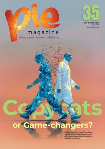
PIE Issue 35 - The Retina Issue

Page 1


Illustration:Diluck/Shutterstock com

Shifting Vision

Biosimilars, biomarkers and the brave new retina

Dear Readers,

Welcome to the latest edition of PIE magazine, where the retina takes center stage once more in a dynamic fusion of innovation, collaboration and clinical insight. If you are attending Euretina 2025 in Paris, France, make sure to drop by Media MICE’s booth to get a printed copy!

This issue showcases the vibrant evolution of retinal care—from bench to bedside breakthroughs that are redefining how we approach AMD and DME, to biosimilars quietly shifting the economic and therapeutic landscape. As we spotlight these “copycat” contenders, we also ask: Are they simply substitutes—or are they true game-changers?

Eight years ago when we launched PIE, anti-VEGF was all the buzz. Today, the emergence of biosimilars is making specialists assess costs and redefine treatment economics. Read this issue’s Cover Story (p18) to find out more.

Our exploration of the back of the eye continues beyond the macula, where retina specialists are stretching the boundaries of anti-VEGF durability and navigating the complex terrain of inherited retinal diseases (IRDs). Tools like next-generation imaging biomarkers and advanced genomic sequencing are no longer futuristic concepts but are presenting to be active players in decoding IRDs and personalizing care.

And then there’s geographic atrophy, always a hot topic! In this issue, guidance from Heidelberg Engineering’s clinical director, Christopher Mody, offers pragmatic insights into imaging and management of this challenging condition. Also, in ocular oncology and pathology, we celebrate team science and translational triumphs that bridge discovery and treatment—transforming lives through collaboration.

Beyond clinical science, this issue celebrates vision in every sense, especially outstanding women in the field. Our Enlightenment stories highlight the achievements and challenges faced by trailblazing women in ophthalmology. We also feature a compelling profile on Dr. Lee Shu Yen—surgeon, researcher and leader—whose remarkable career journey exemplifies dedication, excellence and resilience in retinal care.

From scientific rigor to human impact, we hope these pages inspire new ways of thinking, practicing and leading in the vitreoretinal field!

Sincerely,

Copycats or Game-changers?

A quiet revolution in ophthalmology is underway as biosimilars enter the retina space, promising to redefine practice economics

Front cover image created with assistance from Midjourney AI, ©Media MICE Pte Ltd, 2025.

Dr. Alay S. Banker

Banker’s Retina Clinic and Laser Centre Ahmedabad, India alay.banker@gmail.com

Dr. Arshad Khanani

Sierra Eye Associates; University of Nevada, Reno School of Medicine Nevada, USA arshad.khanani@gmail.com

Dr. Barbara Parolini

Eyecare Clinic Milan, Italy parolinibarbara@gmail.com

Prof. Gemmy Cheung

Singapore National Eye Centre (SNEC) Singapore gemmy.cheung.c.m@singhealth.com.sg

Dr. Hudson Nakamura

Bank of Goias Eye Foundation Goiânia, Brazil hudson.nakamura@gmail.com

Dr. Veeral Sheth

University Retina and Macula Associates; University of Illinois at Chicago, USA vsheth@gmail.com

Disposable

• Perfect view in every case

Single-use means no scratches or opacities

• Always st erile

Minimizes risk of infection and cross-contamination

• Always available on the spot

Increase your O.R. capacity utilization

Advancing the Management of Geographic Atrophy

As recommended by the Clinical Director at Heidelberg Engineering, Christopher Mody

Geographic atrophy (GA), the advanced non-neovascular form of age-related macular degeneration (AMD), represents a growing challenge in ophthalmic care, with an estimated global prevalence of 5 million cases worldwide.1 As life expectancy rises, so does the impact of GA on patient independence and healthcare systems, but with advancing treatment options and cutting-edge diagnostic technology, eye care professionals are better equipped than ever to manage the condition.2

Heidelberg Engineering aims to deliver imaging solutions that empower clinicians to diagnose, monitor and manage GA with clarity and confidence. The SPECTRALIS® platform, with its multimodal imaging capabilities, plays a central role in enabling high-precision diagnostics and guiding personalized treatment strategies.

From

observation to intervention: A new era in GA

care

Historically, clinical management of GA was limited to observation. However, two FDA-approved intravitreal therapies — pegcetacoplan (Syfovre) and avacincaptad pegol (Izervay) — have demonstrated efficacy in slowing lesion growth by 17-35%, depending on dosing regimen and treatment duration.3,4 These therapies do not reverse existing atrophy but represent a major step forward in slowing progression and preserving functional vision.

The power of multimodal imaging: Confident diagnostic decisions

Multimodal imaging is critical for identifying GA candidates that are likely to benefit from treatment. The SPECTRALIS imaging platform offers a comprehensive suite of imaging modalities essential for early detection, precise staging and

ongoing management of GA. Each imaging technique offers a different insight into a particular aspect of retinal pathophysiology, by reflecting the health of a certain component of the retinal tissue.5

Spectral-Domain OCT (SDOCT) technology provides high-resolution cross-sectional views of the retina. High-quality SPECTRALIS OCT images allow clinicians to identify pre-atrophic biomarkers such as ellipsoid zone (EZ) disruption, outer nuclear layer (ONL) thinning and choroidal hypertransmission. These features are key to applying CAM (Classification of Atrophy Meeting) definitions for incomplete (iRORA) and complete RPE and outer retinal atrophy (cRORA).6

Blue fundus autofluorescence (FAF) has been well-established for many years as a fast, easily repeatable and non-invasive measure for evaluating the pattern and distribution of lipofuscin in the retina.7,8 The SPECTRALIS BluePeak Module enables clinicians to visualize the size and potential for progression of GA lesions.

Near-infrared reflectance (NIR) and en-face OCT imaging offers additional insights, particularly in cases where FAF may be limited by media opacities or foveal pigmentation.9 The SPECTRALIS MultiColor Module and Widefield Imaging Module further expand the diagnostic perspective, enabling full visualization of atrophic changes across the macula and beyond.

A combined multimodal imaging strategy — using an imaging platform such as SPECTRALIS — is recommended for a comprehensive view and confident treatment planning.6

SPECTRALIS Multimodal Imaging Platform
SPECTRALIS infrared fundus image and OCT B-scan of eye with GA

SPECTRALIS BluePeak Module: GA progression overtime

Sign-up and stay tuned for GA: In Focus

Uncover more about GA classification, lesion patterns, risk factors for onset and progression, imaging protocols and the role of AI in GA management. Click here: www.he-newsletter.com

The role of artificial intelligence in GA analysis

Complementing the multimodal imaging strategy are a host of artificial intelligence (AI) tools that can be integrated into clinical practice that assist the clinician with insights such as automated layer segmentation, quantifying atrophy and photoreceptor loss, and stratifying patients as slow or fast progressors. However, effective use of AI requires high-quality OCT data, standardized scan patterns and compatible infrastructure. Solutions exist, such as HEYEX 2 with Heidelberg AppWay, which makes it easy to seamlessly integrate thirdparty AI-driven ophthalmic clinical and research applications securely into the existing clinical routine.

SPECTRALIS MultiColor Module: Widefield imaging of an eye with GA

HEYEX 2 with Heidelberg AppWay workflow

Preparing for the future: Clinical recommendations

Advancements, including the first FDA-approved therapies and the development of AI-driven diagnostic tools, offer new hope for managing GA. Eye care professionals can prepare to take advantage of these advancements right away:

• Implement routine imaging protocols for patients with intermediate AMD and GA

• Use fundus autofluorescence and OCT to document lesion extent, monitor progression and identify highrisk phenotypes

• Stay current with internationally accepted terminology and classifications

• Evaluate clinical systems for AI compatibility and standardized imaging outputs

• Engage patients in informed discussions about treatment expectations and disease trajectory

Shaping tomorrow’s vision care today

As therapeutic and technological advances converge, eye care professionals are uniquely positioned to deliver earlier, more targeted interventions that can meaningfully delay vision loss in patients with GA. Heidelberg Engineering, is here to support this evolution and unlock the full potential of retinal diagnostics

through the expandable SPECTRALIS multimodal imaging platform and AIenabled innovations via HEYEX 2 with Heidelberg AppWay.

Want to dive deeper into the future of GA diagnosis and management?

This summary highlights key insights from a comprehensive article to be released by Heidelberg Engineering later in 2025. Subscribe to the Heidelberg Engineering newsletter to be among the first to access the full article and stay informed about the latest advances in retinal imaging and AI-powered eye care.

Editor’s Note

A version of this article was first published on piemagazine.org.

References

1. Wong WL, Su X, Li X, et al. Global prevalence of age-related macular degeneration and disease burden projection for 2020 and 2040: a systematic review and meta-analysis. Lancet Glob Health. 2014;2(2):e106-16.

2. Patel PJ, Ziemssen F, Ng E, et al. Burden of Illness in Geographic Atrophy: A Study of Vision-Related Quality of Life and Health Care Resource Use. Clin Ophthalmol. 2020;14:15-28.

3. Heier JS, Lad EM, Holz FG, et al; OAKS and DERBY study investigators. Pegcetacoplan for the treatment of geographic atrophy secondary to age-related macular degeneration (OAKS and DERBY): two multicentre, randomised, doublemasked, sham-controlled, phase 3 trials. Lancet. 2023;402(10411):1434-1448.

4. Khanani AM, Patel SS, Staurenghi G, et al; GATHER2 trial investigators. Efficacy and safety of avacincaptad pegol in patients with geographic atrophy (GATHER2): 12-month results from a randomised, double-masked, phase 3 trial. Lancet. 2023;402(10411):1449-1458.

5. Crincoli E, De Rosa I, Miere A, Colantuono D, Mehanna CJ, Souied EH. Comparison of Multimodal Imaging for the Characterization of Geographic Atrophy. Transl Vis Sci Technol. 2022;11(11):21.

6. Sadda SR, Guymer R, Holz FG, et al. Consensus Definition for Atrophy Associated with AgeRelated Macular Degeneration on OCT: Classification of Atrophy Report 3. Ophthalmology. 2018;125(4):537-548. [Epub 2017 Nov 2. Erratum in: Ophthalmology. 2019;126(1):177].

7. Schmitz-Valckenberg S, Brinkmann CK, Alten F, et al. Semiautomated image processing method for identification and quantification of geographic atrophy in age-related macular degeneration. Invest Ophthalmol Vis Sci. 2011;52(10):7640-7646.

8. Hwang JC, Chan JW, Chang S, Smith RT. Predictive value of fundus autofluorescence for development of geographic atrophy in age-related macular degeneration. Invest Ophthalmol Vis Sci. 2006;47(6):2655-2661.

9. Corvi F, Corradetti G, Laiginhas R, Liu J, Gregori G, Rosenfeld PJ, Sadda SR. Comparison between B-Scan and En Face Images for Incomplete and Complete Retinal Pigment Epithelium and Outer Retinal Atrophy. Ophthalmol Retina. 2023;7(11):999-1009.

Bench to Bedside Breakthroughs

From imaging biomarkers to nextgen therapeutics, experts explore translational tools shaping the future of AMD and DME

We’re moving beyond just treating the symptoms of AMD and DME, thanks to dedicated scientists and doctors working together to understand these diseases on a deeper level. At a recent symposium, leading specialists shared how the latest research—from analyzing complex inflammation patterns to tracking microscopic changes—is already transforming diagnosis and treatment.

Illustration: Denayunebgt/Shutterstock.com

At the Translational Research in Age-Related Macular Degeneration and Diabetic Retinopathy symposium during the recent Congress of the Asia-Pacific Academy of Ophthalmology (APAO 2025), three leading retina specialists explored how the latest science is changing the landscape of posterior segment care.

Chaired by Dr. Cyrus Shroff (India) and Asst. Prof. Dr. Sruthi Arepalli (United States), the session covered

breakthrough findings in cytokine profiling, fundus autofluorescence imaging and gene therapy pathways—with each talk connecting the bench to bedside in novel ways.

The IMAGINE DME study

Asst. Prof. Dr. Sruthi Arepalli kicked off the session by challenging the conventional wisdom of a solely vascular endothelial growth factor (VEGF)-driven pathology in diabetic macular edema (DME). Presenting

findings from the IMAGINE DME Study,* she revealed a more complex interplay of inflammatory mediators.

By analyzing aqueous humor samples from DME patients, she classified responders into four categories: super responders (≥80% central subfield thickness reduction after one injection), early responders (≥50% reduction by three months), slow responders (<50% reduction by three months but achieving it later) and minimal responders (<50% reduction throughout) who stubbornly resist treatment.

Unsurprisingly, her team found super responders had significantly higher baseline VEGF levels but lower monocyte chemoattractant protein-1 (MCP-1) levels. Meanwhile, slow responders showed elevated angiopoietin-like protein 4 (ANGPTL4) levels.

Prof. Arepalli noted specific molecules that repeatedly surfaced as significant. “What stood out to us is that IL-6 came up quite a bit in diabetic macular edema and leakage throughout the fundus. ANGPTL4 was also prevalent in optical coherence tomography (OCT) macular edema and leakage, and also points to another area that we could experiment with later in the future.”

This suggests that while anti-VEGF treatments remain crucial, targeting pathways involving interleukin-6 (IL-6) and ANGPTL4 could offer therapeutic benefits, particularly for patients demonstrating suboptimal responses to current standards.

This multi-target perspective aligns with emerging treatments and makes a strong case for tailoring treatment to cytokine profiles, especially as newer therapies like faricimab (Vabysmo; Genentech, Inc.; California, USA) come online.

As Prof. Arepalli mentioned during the session’s Q&A, “We are seeing that in some patients, it has a better response than traditional anti-VEGF therapy.”

The secret life of photocoagulation lesions

Moving from molecular pathways to tissue effects, Dr. Kentaro Nishida (Taiwan) provided a fascinating

analysis of what happens to photocoagulation lesions over time. By classifying lesions into five groups based on fundus autofluorescence (FAF) patterns, he revealed that not all laser spots are created equal.

Most importantly, lesions exhibiting diffuse FAF patterns (Groups B and C) were associated with better retinal sensitivity compared to those lacking FAF or showing sharply demarcated hypo-autofluorescence (Groups A and D). OCT analysis confirmed the structural basis for this difference, revealing better preservation of the ellipsoid zone (EZ)—indicating viable photoreceptors—in areas corresponding to diffuse FAF.

As Dr. Nishida explained, “Remaining diffuse fundus autofluorescence implies the existence of photoreceptors,” linking their morphology and function directly to the FAF signal observed. Dr. Nishida proposed a photocoagulation index—a metric derived from the laser-treated area ratio—as a potential guide for optimizing PRP intensity, especially in neovascular glaucoma.

Applying this clinically, his research showed that patients with severe ischemic conditions like neovascular glaucoma (NVG) required significantly higher laser density for effective treatment compared to typical proliferative diabetic retinopathy (PDR) cases (index of 40.7% vs. 20.2%).

“The purpose of retinal photocoagulation is to destroy photoreceptors. The remaining diffuse fundus autofluorescence suggests the presence of photoreceptors. Some autocoagulation is needed to achieve autocoagulated lesions without underscored fluorescence for severe ischemic disease like neurovascular glaucoma,” noted Dr. Nishida, explaining the therapeutic goal.

The implication here is that FAF imaging shouldn’t be considered an afterthought, but actually as a potential roadmap. It could guide not only evaluation but real-time PRP planning, supporting a more refined and retina-preserving laser approach.

A three-front war

Dr. Alay Banker (India) closed the session with a tour de force of emerging age-related macular degeneration (AMD) therapies and the future landscape of intervention. With over 100 molecules in development, Dr. Banker focused on the most promising approaches.

“Exploring the future therapies for AMD, I think basically there are three main therapies which have been researched: gene therapy, stem cell therapy and novel pharmacological approaches,” he explained.

His talk reviewed gene therapies such as RGX-314 (REGENXBIO; Maryland, USA), ADVM-022 (Adverum Biotechnologies; California, USA), and 4D-150 (4D Molecular Therapeutics; California, USA)—each targeting different VEGF pathways for longer-lasting wet AMD control. In dry AMD, options ranged from complement inhibitors like JNJ-1887 to photobiomodulation and omega fatty acids engineered for retinal neuroprotection.

Dr. Banker also introduced pharmacologic options like DURAVYU™ (EyePoint Pharmaceuticals; Massachusetts, USA), a biodegradable tyrosine kinase inhibitor implant, which delivered VEGF suppression for up to six months—a potential gamechanger in reducing treatment burden.

“I feel that, ultimately, it is going to be a combination therapy,” he noted. “We have to integrate gene therapy, apply stem cells and also administer pharmacological agents to improve outcomes.”

Dr. Banker didn’t stop at the science. He closed with a playful nod to science fiction, imagining “retinal banks” where patients could receive custom implants and even teleportation to specialized treatment centers. The audience chuckled, but the underlying point was clear: Access and infrastructure remain critical hurdles in delivering advanced care globally.

The Q&A dove into real-world limitations, with panelists reflecting on the lack of access to dry AMD treatments in some regions and the need for better metrics, such as contrast sensitivity and reading speed, to capture patient-relevant outcomes.

Bridging bench to bedside in retinal disease

From deep molecular profiling to refined laser metrics and cutting-edge gene therapy, this symposium highlighted a clear shift in retinal research: from singletarget approaches to multi-modal treatments addressing the complex pathophysiology of retinal diseases.

Each presentation tackled different corners of AMD and DME, but together, they painted a picture of multifaceted management, increasingly defined by individual response profiles, imaging analytics and multi-modal therapeutics. Clearly, the future of retinal care is extremely bright and delightfully complex.

As speakers emphasized, innovation must go hand in hand with functional outcomes, accessibility and clinical common sense. In the coming years, integrating these diverse approaches may be less about finding a silver bullet and more about crafting the right cocktail for each patient.

Reference

* Kar SS, Abraham J, Wykoff CC, et al. Computational Imaging Biomarker Correlation with Intraocular Cytokine Expression in Diabetic Macular Edema: Radiomics Insights from the IMAGINE Study. Ophthalmol Sci. 2022;2(2):100123.

Editor’s Note

Reporting for this story took place during the 40th Congress of the AsiaPacific Academy of Ophthalmology (APAO 2025), held in conjunction with the 83rd Annual Conference of the All India Ophthalmological Society (AIOC 2025) from 3-6 April in New Delhi, India. A version of this article was first published on piemagazine.org

Could Cannabis Use Lower the Risk of PVR? A JAMA Study Offers a Surprising Clue

New real-world data suggests cannabis might play a protective role in retinal healing after retinal detachment surgery.

A newly published study in JAMA Network has revealed an unexpected association between long-term cannabis use and a lower risk of proliferative vitreoretinopathy (PVR) following primary retinal detachment (RD) repair.*

PVR remains one of the most common reasons RD surgery fails, often leading to complex repeat surgeries and poor visual outcomes. This new research raises a provocative question: Could cannabis be quietly offering protection where traditional approaches fall short?

The study: A two-decade deep dive

Drawing from electronic health records spanning 1 February 2005 to 1 February 2025, this retrospective cohort study analyzed outcomes for patients who underwent initial RD repair by pars plana vitrectomy (PPV), scleral buckle (SB) or pneumatic retinopexy.

Researchers compared patients with documented cannabis-related disorder—confirmed by urine or blood testing—against a control group with no recorded cannabis use. Using propensity score

matching, the final sample included 1,193 patients in each group.

Findings

At six months following surgery, patients with cannabis use had a notably lower rate of PVR, with only 2.1% developing the complication, compared to 4.36% in the control group. That translated to a relative risk reduction of over 50%.

Similarly, the rate of requiring a second, more complex retinal detachment surgery was 3.1% in cannabis users versus 5.03% in non-users—again, a significant reduction. These differences remained consistent at the oneyear follow-up mark, suggesting the protective association was not just short-lived.

While the study didn’t probe mechanisms, the authors speculate on cannabis’ anti-inflammatory and neuroprotective properties. Since PVR is driven by inflammation, retinal scarring and cellular proliferation, it’s plausible that cannabinoids may dampen these damaging pathways—though that remains unproven.

Of course, the protective association might reflect who uses cannabis, not what cannabis does. Lifestyle, comorbidities and genetic factors could also influence healing and inflammation, the study noted.

What it means for ophthalmology—and what it doesn’t

Let’s be clear: This doesn’t mean we’ll be prescribing cannabis after retinal surgery.

The difference in outcomes, while statistically significant, was modest in absolute terms—about 2%. The authors rightly warn that this was an observational study, not a clinical trial. Cannabis use is not a prescribed therapy, and the data alone can’t confirm a direct biological effect.

To move beyond correlation, we’ll need controlled, prospective trials that isolate the biological mechanisms and rule out external factors.

Still, the implications are intriguing. PVR remains one of the most stubborn complications in retinal surgery, and current prevention strategies are limited. This study offers an unexpected but promising angle: That systemic factors like cannabis exposure could influence retinal healing and inflammation in ways we’re only beginning to understand.

For now, it’s not a treatment—but it’s a conversation starter, and possibly a research pathway that could reshape how we think about inflammation modulation in surgical retina.

Reference

*Alshaikhsalama AM, Alsoudi AF, Mukhtar A, et al. Long-Term Cannabis Use and Risk of Postoperative Proliferative Vitreoretinopathy. JAMA Ophthalmol. 2025 Jul 3. [Epub ahead of print].

Photo: Anski21/Shutterstock.com

There’s Something for Everyone in Orlando

Join us for the latest clinical advancements and practical insights to help your practice thrive. Don’t miss:

• Subspecialty content from cornea to uveitis

• Cataract Spotlight, now on Sunday

• Courses for every level — residents to experts — and for the entire practice team

• AAO 2025’s must-attend social event, Illuminate, supporting ophthalmic education on Saturday

Plan your schedule with the Meeting Guide at aao.org/mtg-guide.

Register Early and Save

Secure your spot — and your Illuminate ticket — before they’re gone.

aao.org/registration Where All of Ophthalmology Meets®

Translational Triumphs

How collaborative team science can transform diagnosis and treatment in ocular oncology and pathology

Leading clinician-scientists at a recent major ophthalmic research meeting shared powerful new approaches to ocular oncology and pathology, showcasing how collaborative team science is rapidly transforming diagnosis and treatment for complex eye diseases.

Illustration: Roman Samborskyi/Shutterstock.com

The world of eye care is on the cusp of breakthroughs, with brilliant minds joining forces to tackle the most challenging diseases, even rare eye cancers. At the 2025 annual meeting of The Association for Research in Vision and Ophthalmology (ARVO) in Salt Lake City, Utah, experts shared how working together is uncovering new ways to diagnose and treat complex eye conditions.

In particular, during the Translational Scientist: Building Programs for a

Brighter Future mini-symposium, researchers and clinicians shared how they worked side by side, navigating complex paths to uncover innovative solutions for ocular oncology and pathology.

Tandem discovery in vitreoretinal lymphoma

Dr. Anita Chan and Ms. Mona Wang from the Singapore National Eye Centre (SNEC) delivered a tandem talk that brought translational science vividly to life. As a clinician-

researcher duo deeply immersed in the world of vitreoretinal lymphoma (VRL), they walked the audience through the nuanced journey of turning clinical puzzles into actionable lab solutions.

Ms. Wang opened with the realworld challenges of diagnosing VRL, a rare cancer that often hides behind the mask of chronic uveitis. She highlighted how fragile lymphoma cells often break down during sample processing, hence complicating cytologic diagnosis. But new approaches—like MYD88 mutation analysis—are changing the game. “MYD88 mutational testing is useful for the clinical diagnosis of vitreoretinal lymphoma, especially when you don’t have good cytology,” she emphasized.

Then Dr. Chan stepped in to describe how they brought that idea to the bench, developing a single-cell analysis pipeline that increases diagnostic sensitivity. “We tried to make it as simple as possible for clinical labs,” she said. “One of our research aims is to have these standardized test kits for widespread use... so that we can have multicenter collaborations and better understand tumor heterogeneity in VRL.”

“MYD88 mutational testing is useful for the clinical diagnosis of vitreoretinal lymphoma, especially when you don’t have good cytology.”

- Ms. Mona Wang

Together, Dr. Chan and Ms. Wang showed how close collaboration between clinic and lab can push the boundaries of what’s possible in rare disease diagnostics. Their talk felt less like a formal lecture and more like a dynamic conversation—one rooted in shared curiosity, practical problem-solving and a clear vision for better patient outcomes.

A mission to redefine uveal melanoma research

In a talk that blended scientific rigor with heartfelt urgency, Dr. Lauren

Dalvin (United States) of the Mayo Clinic shared her team’s ambitious journey to revolutionize uveal melanoma research, one patientderived organoid at a time.

“This is the most common thing I treat in my practice,” she began, “and my least favorite conversation is telling patients that no matter what I do for your eye, I can’t make your metastatic risk go to zero.” With limited treatment options and outdated lab models, Dr. Dalvin knew change was overdue.

So she built it—literally. With help from an unexpected crossdisciplinary network (“A pancreatic cancer researcher taught me how to make organoids!”), she and her team developed 3D tumor models that better reflect how this disease behaves in real patients. These organoids retain critical mutations like BAP1 loss and even mimic metastatic behavior when implanted in mice.

“They sort of take on their own personality,” Dr. Dalvin said with a smile. Even more exciting? These models respond differently to drugs based on their genetic profile, offering hope for more precise therapies in the future.

Dr. Dalvin’s story is one of persistence, collaboration and a fierce commitment to moving from “we can’t” to “what if?”—with science leading the way.

Two decades of translational research in action

Dr. Colleen Cebulla (USA) and Dr. Mohamed Abdel-Rahman (USA) offered a powerful look at how two decades of integrated clinical and molecular research have transformed our understanding of BAP1 tumor predisposition syndrome (TPDS) and uveal melanoma.

“What began as a simple clinical observation—that some uveal melanoma patients had strong family histories of cancer—evolved into a hypothesis-driven investigation of hereditary risk,” said Dr. AbdelRahman. Their work, grounded

in both cytogenetics and highresolution molecular profiling, was among the first to show that partial chromosome 3 loss behaves like monosomy 3, an insight now foundational in ocular oncology.

Together, they’ve built a biobank of patient samples that has not only validated key genetic markers but also informed national screening guidelines. “We’ve now identified germline mutations not just in BAP1, but also in other cancerassociated genes like CHEK2,” Dr. Abdel-Rahman noted, referencing prospective family history data that predicted a 10% to 15% germline mutation rate in selected uveal melanoma cases.

Their multi-specialty clinic at Ohio State follows rigorous protocols— fundus photography, ocular ultrasound and whole-body MRIs—to detect early manifestations of BAP1 TPDS across organ systems. “This is translational research in action,” Dr. Cebulla emphasized, pointing to their growing biorepository and collaboration with international consortia. “We’re not just studying mutations, we’re building systems to catch disease before it starts.”

The power of clinicopathologic collaboration

Prof. Dr. Martina Herwig-Carl (Germany) and Dr. Swathi Kaliki (India) took the stage to emphasize how pathology isn’t just a postscript to patient care; it’s central to it.

“Ophthalmic clinical pathology correlation is important not just for diagnosis, but for teaching, research and improving surgical outcomes,” said Prof. Herwig-Carl, whose presentation focused on nononcologic insights.

From differentiating nevi from melanoma by spotting tiny cystic spaces, to revealing keratocyte loss post-crosslinking that’s invisible clinically, she made one thing clear: histology tells the part of the story the eye can’t see. Her analysis of retinal implants in AMD patients even showed how histopathology

could validate long-term implant biocompatibility—something optical coherence tomography (OCT) alone could never confirm.

Dr. Kaliki picked up the thread with a series of jaw-dropping oncologic cases that underscored just how essential pathologists are in directing treatment. “This presentation also represents the teamwork that happens between the ocular oncologist and the pathology team,” she said. One case involved a child with aggressive eyelid swelling that turned out to be sebaceous gland hyperplasia associated with Muir-Torre syndrome—a rare but important finding.

“This presentation also represents the teamwork that happens between the ocular oncologist and the pathology team.”
- Dr. Swathi Kaliki

Another, a relentless conjunctival lesion, was ultimately diagnosed as IgG4-related orbital disease only after pathologic deep dives. Perhaps the most startling was an intraocular adenocarcinoma that stumped the team until pathology narrowed it down to a likely ciliary body origin. And her final case? A rare BCORassociated sarcoma of the orbit—a reminder that even in eye cancer, the frontier is always expanding.

Together, Prof. Herwig-Carl and Dr. Kaliki reminded the audience that real precision medicine lies in the details—and those are often microscopic.

Editor’s Note

Reporting for this story took place during the annual meeting of The Association for Research in Vision and Ophthalmology (ARVO 2025) held from 4-8 May in Salt Lake City, Utah, USA. A version of this article was first published on piemagazine.org

Nanoscope Kickstarts BLA Submission for Restorative RP Therapy, Adds to Advisory Board

BLA filed, big brains recruited— Nanoscope is charging ahead with its promising gene-agnostic therapy for retinitis pigmentosa.

Nanoscope Therapeutics (Texas, USA) is going full throttle with not one, but two major moves: initiating a rolling Biologics License Application (BLA) for its investigational, restorative, geneagnostic retinitis pigmentosa (RP) therapy, MCO-010, and assembling a powerhouse Visionary Advisory Committee to help steer its next chapter.

The BLA journey

Nanoscope has officially begun its rolling BLA submission to the U.S. Food and Drug Administration (FDA) for MCO-010, its gene-agnostic therapy for retinitis pigmentosa (RP). The first BLA modules are in, with full submission expected in early 2026. Thanks to MCO010’s fast track designation, the application qualifies for priority review, a potential fast lane to what could be a groundbreaking treatment.

“For the first time, patients who are considered to be on a path to permanent blindness may have a chance to regain sight,” said Sulagna Bhattacharya, CEO and co-founder of Nanoscope, in a news release. “We are deeply thankful to the FDA for their guidance as

we remain steadfast in our mission to restore vision and bring light back into the lives of those living in darkness.”

MCO-010 is the first gene-agnostic therapy for retinal disease to reach the BLA submission stage. But the hype goes beyond that: not only is MCO-010’s efficacy independent from a specific gene mutation, but it is also restorative, potentially bringing sight back to RP patients who have already lost vision.

Delivered via a single in-office intravitreal injection, MCO-010 activates dormant bipolar retinal cells, making them light sensitive and tapping into the eye’s remaining visual circuitry after photoreceptor loss.

In the pivotal RESTORE Phase IIb trial, MCO-010 met its primary endpoint for best corrected visual acuity (BCVA) at 52 weeks in both dose arms, compared to the shamcontrol. Patients experienced vision gains greater than 0.3 LogMAR, which is at least three lines on the eye chart. These improvements persisted through three years in ongoing follow-up.

Meet the visionary committee

Alongside the regulatory milestone, Nanoscope has launched its new Visionary Advisory Committee to guide its scientific, clinical and community outreach strategy.

Chaired by Dr. SriniVas Sadda (UCLA/Doheny Eye Institute), the committee brings together a startstudded cast of global leaders in retina, including:

• Dr. Allen Ho (Thomas Jefferson University)

• Dr. Peter Kaiser (Cleveland Clinic)

• Dr. Christine Kay (Vitreoretinal Associates)

• Dr. Arshad Khanani (University of Nevada, Reno)

• Dr. Vinit Mahajan (Stanford University)

• Dr. Jordi Monés (Institut de la Màcula, Barcelona)

• Dr. Mark Pennesi (Casey Eye Institute, Oregon Health & Science University).

“As retina specialists, we face a desperate need to change the conversation about blindness with our patients — from one of inevitability to one of possibility,” said Dr. Sadda. “I’m honored to be part of a team that believes the status quo is not set in stone, and that restoration of vision is not a dream—it’s a goal within reach.”

The committee’s formation marks a key moment in Nanoscope’s evolution as it prepares to bring a first-in-class, gene-agnostic therapy to market. With perspectives from both academic and clinical leaders, the group is expected to help shape future development, patient education and broader adoption strategies.

“One of the most challenging aspects of my entire career has been telling patients with RP there is no restorative treatment available as they experience progressive, irreversible, permanent vision loss,” said Dr. Ho, who also serves as Nanoscope’s chief medical advisor. “Based on the preclinical science and evidence in clinical trials, MCO-010 represents a potential, important paradigm shift for patients and retina specialists, providing hope for meaningful improvement in the quality of life for the neediest retina patients of all.” Editor’s

Beyond the Macula

From stretching anti-VEGF durability to decoding IRDs, specialists dissect new therapies and diagnostic challenges in retinal diseases

A pivotal symposium at APAO 2025 provided a comprehensive look into the rapidly evolving landscape of retinal disease diagnosis and treatment. Global retinal experts tackled pressing challenges across conditions like ROP, RVO and IRDs, alongside developments in AI diagnostics.

The recent Asia-Pacific Academy of Ophthalmology Congress (APAO 2025) featured a symposium on new approaches to retinal diseases, highlighting the evolving landscape of their diagnosis and treatment. Chaired by Dr. Raja Narayanan (India) and Prof. Dr. Hendrik Scholl (Switzerland), the session brought together global experts who tackled challenges in retinopathy of prematurity (ROP), retinal vein occlusion (RVO), artificial intelligence (AI) diagnostics, inherited retinal diseases (IRDs) and retinal toxicities from systemic therapy.

Presentations ranged from realworld data to pipeline innovations, highlighting how precision, access and vigilance are the triple threat keeping the retina world spinning (and retinal specialists on their toes) in today’s ever-evolving clinical landscape.

Stretching the injection timeline in RVO

Prof. Muna Bhende (India) opened the session with a focus on antiVEGF therapy for macular edema from retinal vein occlusion (RVO). Her analysis emphasized the value

of early and sustained treatment. Whether it was aflibercept 2 mg (Eylea; Regeneron Pharmaceuticals; New York, USA/Bayer; Leverkusen, Germany) , faricimab (Vabysmo; Genentech; California, USA) or the newer high-dose aflibercept 8 mg, the core message was clear: consistency counts.

“Anti-VEGF injections, regardless of the agent, when given monthly, show substantial improvements in visual acuity and central macular thickness reduction. However, with PRN , there is a loss of the initial benefit, which is more evident in central vein occlusion.”

- Prof. Muna Bhende

“Anti-VEGF injections, regardless of the agent, when given monthly, show substantial improvements in visual acuity and central macular thickness

(CMT) reduction. However, with PRN , there is a loss of the initial benefit, which is more evident in central vein occlusion,” Prof. Bhende cautioned.

Highlighting results from the BALATON and COMINO studies, she noted that more than half of branch RVO patients and just under half of central RVO (CRVO) patients could extend treatment intervals to 16 weeks with faricimab. Still, “about 50% of CRVO patients need treatment in year four,” she warned. “The newer injections may help us to increase the interval between injections.”

Masqueraders in ROP

Prof. Peiquan Zhao (China) presented diagnostic cases in retinopathy of prematurity (ROP) that revealed more than met the eye.

“The purpose of our work is to identify early lesions, particularly to prevent blindness caused by endstage ROP and other pediatric retinal disease,” he said, outlining several cases where presumed ROP turned out to be masquerading conditions such as retinoblastoma, Coats’ disease, incontinentia pigmenti or familial exudative vitreoretinopathy.

He concluded with a call for vigilance: clinicians must examine the entire retina—including the periphery—with an eye toward differential diagnosis.

“We need to identify the underlying disease masquerading as ROP, link heredity in every unexplained

peripheral retinopathy, and make the right choice for treatment or referral,” Prof. Zhao advised.

New guidelines in ROP management

ROP remains a leading cause of childhood blindness, especially in middle-income countries, explained Prof. Mangat Ram Dogra (India). In these settings, heavier and more mature infants remain at risk, requiring localized screening criteria. His central insight is that ROP is not a one-size-fits-all disease.

“Higher birth weight babies develop severe ROP in developing and middle-income countries. This led to a change in guidelines. We finally had guidelines [for babies weighing up to] 2 kgs [or born up to] 34 weeks ,” said Prof. Dogra.

“Higher birth weight babies develop severe ROP in developing and middle-income countries. This led to a change in guidelines. We finally had guidelines [for babies weighing up to] 2 kgs [or born up to] 34 weeks .”

- Prof. Mangat Ram Dogra

The talk emphasized that traditional 1,500 g/30-week cutoffs miss significant pathology in these settings. Prof. Dogra advocated for updated, locally relevant protocols that reflect real-world demographics and healthcare realities.

“Screening is very important to identify treatable ROP,” he reinforced. “Manage all treatable ROP promptly with laser or anti-VEGFs as per the standard of care.”

IRD therapies gain momentum

Switching gears to the future of inherited retinal diseases (IRDs), session co-chair Prof. Scholl highlighted two complementary approaches: pharmacologic modulation and gene editing.

“The pharmacological reduction of the availability of vitamin A to the retina with tinlarebant (Belite Bio; California, USA) has shown significant therapeutic effects with very good tolerability in Phase 2 studies,” he said, pointing to data from the Dragon 1 and Dragon 2 registration trials.

But the spotlight moment came when Prof. Scholl discussed base editing technology targeting the G961E mutation in Stargardt disease. “Base editing currently allows an editing rate of about 75% in cone photoreceptors in the primary retina with significantly lower editing in the fovea,” he said. While hurdles remain, these tools signal a new era in IRD precision medicine.

China’s blueprint for screening at scale

Prof. Haotian Lin (China) outlined how AI is being deployed to enhance both diagnosis and accessibility of retinal care in China. His team tackled AI’s weakest link—image quality— with an innovative three-dimensional quality control system for fundus photography.

“To achieve better performance of AI models in clinical practice, we collected representative fundus photographs from tertiary hospitals with abundant diseases,” he said. The model underwent validation in 35 medical institutions across 82% of China’s provinces, achieving an average area under the curve (AUC) greater than 0.95.

The initiative goes beyond the clinic with AI-equipped mobile screening vehicles now bringing diagnostics to rural areas. “The application of AI technology for retinal disease provides door-to-door eye disease screening service for residents in remote areas with scarce healthcare resources,” Prof. Lin concluded.

The hidden costs of cancer care

Closing the session with a dose of caution, Dr. Sruthi Arepalli (United States) shared insights into ocular complications from anti-cancer agents, particularly mitogenactivated protein kinase (MEK)

inhibitors, checkpoint inhibitors and tyrosine kinase inhibitors.

“MEK inhibitors are guilty of causing a self-limited serous neurosensory detachment, which is pathognomonic for MEK inhibitor-associated retinopathy. Almost 90% to 100% of patients will develop this at some point,” she said, emphasizing the importance of accurate differentiation from other retinal conditions.

Checkpoint inhibitors bring a broader spectrum of inflammatory complications, including VogtKoyanagi-Harada (VKH)-like syndromes and posterior uveitis.

Dr. Arepalli urged careful differential diagnosis: “While we learn about all of these new drugs and the sequelae that they can cause in the eye, including multiple types of uveitis, I always urge us to think about what else could be presenting even in a situation that seems like it’s a slam dunk.”

Eyes on the future

Presentations spanned early intervention in ROP, molecular innovations in inherited retinal diseases, and the systemic interface of oncology and ophthalmology. As the field advances, these discussions reflect a broader shift in retinal care—toward earlier diagnosis, more tailored interventions and a deeper awareness of intersecting systemic factors. The work presented offered not just solutions, but a framework for ongoing refinement in both clinical practice and global strategy.

Editor’s Note

Reporting for this story took place during the 40th Congress of the AsiaPacific Academy of Ophthalmology (APAO 2025), held in conjunction with the 83rd Annual Conference of the All India Ophthalmological Society (AIOC 2025) from 3-6 April in New Delhi, India. A version of this article was first published on piemagazine.org

A quiet revolution in ophthalmology is underway as biosimilars enter the retina space, promising to redefine practice economics

A quiet shift is gaining momentum in the field of ophthalmology. Biosimilars—near-identical copies of approved biologic drugs—are edging into the retina space, offering a fresh set of tools to tackle some of the most common and costly vision-threatening diseases.

Like a chef considering different brands of knives in their kitchen, retina specialists now face expanding choices in their treatment arsenal, each with potential implications for patient care, practice economics and the broader healthcare system.

“Biosimilars are an inevitable evolution of the lifecycle of a therapeutic,” explained Dr. Paul Hahn, a retina specialist at NJRetina (USA). “I don’t think they are likely to demonstrate any meaningful efficacy benefits compared to the reference therapy, but their potential cost savings may be meaningful drivers to promote utilization.”

A changing landscape

The biosimilar story in retina care began in 2021 when the FDA greenlit Byooviz (ranibizumabnuna; Samsung Bioepis; Incheon, South Korea). Since then, a few more ranibizumab biosimilars have entered the market, including Cimerli (ranibizumab-eqrn; Sandoz; Basel, Switzerland), which received an interchangeability designation from the FDA.

Now, aflibercept biosimilars are stepping into the spotlight. Most notably, Biocon Biologics (Bengaluru, India) recently reached a settlement with Regeneron (New York, USA), clearing the path to launch Yesafili (aflibercept-jbvf)—an interchangeable biosimilar to Eylea—in the U.S. by late 2026, or earlier under specific terms.

“This settlement clears the path for Biocon Biologics to be among the first to bring a reliable, highquality aflibercept biosimilar to patients and healthcare providers in the United States,” said Biocon’s CEO, Shreehas Tambe, in a news release.

It’s a bold step in a big territory. Eylea raked in roughly $9.4 billion in 2023, and the market is expected to grow to $12.73 billion in 2028—making it a tempting target for biosimilar competition.1

Understanding biosimilars

Unlike generic drugs—carbon copies of small-molecule medications— biosimilars are a different beast. They’re large, complex proteins brewed from living cells, which makes it impossible to create an exact replica of the original biologic. But although they can’t be identical, they’re very similar, close enough in structure, purity and potency to match the reference drug in performance.

“By definition, molecular and protein characteristics of a biosimilar must be similar to those of the reference product,” explained Bressler et al. in a review published in Survey of Ophthalmology. “Biologics have per se a certain inherent microheterogeneity.” Translation: Tiny differences are baked into the process. These arise from natural variations in the living cells used to make the drug, as well as minor shifts in manufacturing.2

This inherent variability means that even different batches of the same reference product aren’t absolutely identical to each other. As Bressler et al. noted, “batches of the reference ranibizumab product (Lucentis) from 2018 were likely not absolutely identical to batches of this product in 2022.”2

To get FDA approval, biosimilars must undergo rigorous testing to prove they’re clinically equivalent, though the development process is faster and less costly than for original biologics.2

“It’s frankly cheaper to develop a biosimilar because you don’t have an original pathway to go through,” explained Dr. Suber Huang, CEO of the Retina Center of Ohio (USA). “For instance, with Avastin, you would have to go through an entire new process to create a biosimilar, because Avastin (bevacizumab) was not a reference drug for the eye.”

The cost equation

One of the biggest motivators driving biosimilar development is cold, hard cash savings. The first ranibizumab biosimilar hit the market with a 40% price cut compared to its reference product, and the difference in

average sale price was approximately 31.3%. This represents significant potential savings, considering that patients with conditions like neovascular age-related macular degeneration (nAMD) or diabetic macular edema (DME) may receive an average of nine injections in the first year of treatment alone.2

A Canadian estimate projected that switching to ranibizumab biosimilars could save between 70 and 201 million CAD in 2023. With a similar market dynamic, the U.S. could be in line for major savings too, especially as more biosimilars enter the fray and start competing on cost.2

Of course, saving money is only a possibility if biosimilars are used. That’s where uptake comes in. “When you have a cheaper option… the patients, insurers, society and healthcare system in general will tend to favor those if there’s equal efficacy and equal safety,” said Dr. Huang.

Clinical experience and adoption challenges

Despite their potential to cut costs and expand access, biosimilars in ophthalmology have had something of a slow start. In 2023, they made up just 3% of usage, bumping up only modestly to around 10% in 2024.3

So, what’s behind the hesitation?

According to Dr. Hahn, it’s not that retina specialists doubt biosimilars work. “I think when biosimilars first became part of a consideration in the retinal armamentarium, there was a lot of hesitation around these,” he explained. “I think that exists because biosimilars are now here, not because of efficacy; they don’t work better than our reference products. But it’s a cost consideration, and many of us as doctors are used to not thinking about what costs less for the patient. Rather we’re used to thinking about what works best for our patient. It’s a different mindset.”3

That mindset shift is slowly happening, and among those who’ve already made the leap, the results have been promising. “We have incorporated ranibizumab biosimilars in our practice, both with new and existing treatment patients. The integration was generally smooth,” said Dr. Hahn. “Once our physicians

Dr. Huang, meanwhile, posed a thought-provoking question about priorities in drug development. “All the time and money spent in biomedical research…those resources are then used for something that is, in fact, not any better than something we already have,” he noted. “If it never improves, then we lose a very valuable part of what we do, which is to try to come up with something better.”

That said, Dr. Huang was quick to acknowledge the other side of the coin. “If a biosimilar costs less, more people can afford it. And if more people can access it—and it works just as well—then you’ve improved care in a different, but still meaningful, way,” he added.

Preparing for the Biosimilar Era

For retina specialists looking to integrate biosimilars into their practice, a little preparation can go a long way toward a smooth transition. Here are a few key things to keep in mind:

• Do your homework: Brush up on clinical trial data and realworld evidence to get a solid grasp of each biosimilar’s safety and efficacy.

• Know the fine print: Get familiar with regulatory terms like “interchangeability,” and more importantly, what those labels do (and don’t) mean for your day-today clinical decisions.

• Have your elevator pitch ready: A clear, no-nonsense way to explain biosimilars to patients will help put their minds at ease.

• Watch the insurance shuffle: As biosimilars become more common, be ready for shifting coverage policies and the possible maze of step therapy.

• Track your results: Monitor patient outcomes to build your own comfort and confidence in these agents.

Ultimately, trust is the secret sauce. "Patients trust their doctor," reminded Dr. Hahn. "If you are comfortable with the treatment plan that you are offering to the patient, the patient will often be comfortable as well."

A new set of tools in the kitchen

As we find our way in the new era of biosimilars in ophthalmology, perhaps Dr. Huang puts it best with a culinary metaphor: “If a knife is made by two different manufacturers, it’s still a knife, and you prepare dinner just the same.”

In other words, biosimilars don’t reinvent the recipe for treating retinal disease. But they do give retina specialists a wider array of tools to work with. Tools that, while functionally similar, come with different price tags. Just as you don’t need a Masamoto blade to chop an onion, you don’t always need a brand-name biologic to treat a patient effectively.

for physician choice rather than restricting options,” Dr. Hahn warned.

With smart education, thoughtful rollout, and a watchful eye on both safety and outcomes, biosimilars could play a meaningful role in expanding access to care while easing the financial strain of retinal disease treatment. In that way, they’re not just another set of knives. They’re a chance to cook up better outcomes for more patients.

The catch? Making sure these extra options empower physicians rather than tie their hands. “As a society, we must ensure that these additional modalities provide increased options

References

1. Eylea (Aflibercept) Market Research Report 2025: Strategic Partnerships and Biosimilars Enhance Accessibility, Diabetes-Related Retinal Issues and Myopia Surge Boost - Long-term Forecasts to 2033. Research and Markets. February 12, 2025. Available at: https://www.globenewswire.com/ news-release/2025/02/12/3024804/28124/en/ Eylea-Aflibercept-Market-Research-Report-2025Strategic-Partnerships-and-Biosimilars-EnhanceAccessibility-Diabetes-Related-Retinal-Issues-and-Myopia-Surge-Boost-Long-term-Forecast.html. Accessed on June 2, 2025.

2. Bressler NM, Kaiser PK, Do DV, et al. Biosimilars of anti-vascular endothelial growth factor for ophthalmic diseases: A review. Surv Ophthalmol. 2024;69(4):521-538.

3. Jeremias S. Retina Specialists’ Evolving View on Biosimilars in AMD Treatment. The Center for Biosimilars. March 16, 2025. Available at: https://www.centerforbiosimilars.com/view/retinaspecialists-evolving-view-on-biosimilars-in-amdtreatment. Accessed on June 2, 2025.

4. Freed M, Biniek JF, Damico A, et al. Medicare Advantage in 2024: Enrollment Update and Key Trends. August 8, 2024. Available at: https://www. kff.org/medicare/issue-brief/medicare-advantage-in2024-enrollment-update-and-key-trends/. Accessed on June 2, 2025.

5. Congress’ Physicians Prep Strong Message Opposing Step Therapy in Medicare. AAO. June 27, 2019. Available at: https://www.aao.org/advocacy/eye-onadvocacy-article/congress-physicians-prep-strongmessage-opposing. Accessed on June 2, 2025.

6. National Survey on Innovator Biologics and Biosimilars. Biologics Prescribers Collaborative. August 8, 2023. Available at: https://biologicsprescribers.org/wp-content/ uploads/2023/08/BPC_Patient-Choice_NationalSurvey-Report_August-2023.pdf Accessed on June 2, 2025.

Contributors

Dr. Paul Hahn, MD, PhD, FASRS, is a board-certified, fellowship-trained retina specialist in northern New Jersey. He graduated magna cum laude from Harvard University with a degree in Biochemical Sciences. He completed his combined MD-PhD education at the University of Pennsylvania School of Medicine, with a PhD dissertation in molecular pharmacology investigating novel treatment pathways for dry age-related macular degeneration and other retinal degenerative diseases. Dr. Hahn completed his ophthalmology residency at the Scheie Eye Institute at the University of Pennsylvania, followed by a two-year fellowship in vitreoretinal diseases and surgery at the Duke University Eye Center. After completion of his fellowship, Dr. Hahn remained at Duke University as an assistant professor of ophthalmology and retina specialist. He was the founder and director of the Duke Center for Artificial and Regenerative Vision, where he implanted the first Argus II “bionic eye” in the Southeast U.S., restoring vision to an individual with total blindness. Dr. Hahn moved back to the Northeast in 2016, where he joined NJRetina. He brought this “bionic eye” technology to New Jersey and was the first and only surgeon to perform this surgery in NJ. Dr. Hahn has published more than 100 peer-reviewed articles and book chapters. He has been invited to lecture at numerous national and international meetings on topics such as macular degeneration, diabetic retinopathy and retinal vascular disease, among others. He serves on the Board of Directors of the American Society of Retina Specialists (ASRS) and as an Oral Examiner for the American Board of Ophthalmology. He also serves on the editorial board of the Journal for VitreoRetinal Diseases, Retina Times and Retina Specialist. He has been the recipient of numerous prestigious honors and awards for excellence in ophthalmology research.

paulhahn@gmail.com

Dr. Suber S. Huang, MD, MBA, FASRS, is CEO of the Retina Center of Ohio; voluntary assistant clinical professor of ophthalmology at Bascom Palmer Eye Institute, University of Miami; and Hong Leong Visiting Professor of Ophthalmology, National University of Singapore. He is the founder/CEO/ executive producer of the Future Vision Foundation, and created the first-in-kind Future Vision Forum to bring visionary leaders in basic, translational and clinical research together to seek new directions that accelerate discovery and innovation in vision research. He received his undergraduate degree at Johns Hopkins University, a medical degree from the Albert Einstein College of Medicine, an ophthalmology residency at the Wilmer Eye Institute/ Johns Hopkins University and a fellowship in Vitreoretinal Diseases and Surgery from Bascom Palmer Eye Institute/University of Miami. He has graduate training at Harvard University, Wharton School of Business/University of Pennsylvania and received his Executive MBA from the Weatherhead School of Management/CWRU. Dr. Huang was formerly president of the American Society of Retina Specialists, chair of the Foundation of the ASRS, AAO Associate Secretariat of Federal Affairs and Chair of the Research, Regulatory, and External Scientific Affairs Committee. He founded the Retina Diseases Image Analysis Reading Center and was director of the Visual Sciences Research Center. Dr. Huang has participated in numerous clinical trials as PI and as director of the Retina Disease Image Analysis Reading Center at CWRU. He has published widely, given nearly 400 lectures and has served as Program Chair for retina surgery for the APAO and AAO Retina sub-specialty day. Dr. Huang founded the ASRS Retina Image Bank, now the world’s largest and most comprehensive open-access multimedia database of “all things retina.” He is the founding editor-inchief of the online ASRS Retina Atlas and the founding officer of Retina Global. Dr. Huang is an inductee to the Retina Hall of Fame and has received the “Top Doctors” and “Best Doctors in America” awards each year since 2003. He received the APAO Jose Rizal International Medal, APVRS International Award and the Chinese Vitreo-Retinal Society Senior Honor Award, among others.

DrHuang.RCO@gmail.com

A Story of Vision

From pioneering research and delicate surgeries to breaking barriers in leadership, Assoc. Prof.

Dr. Lee Shu Yen’s story is a true inspiration in the industry by Chow Ee-Tan

Assoc. Prof. Dr. Lee Shu Yen’s distinguished career in ophthalmology is a testament to both literal and metaphorical vision, shaped by her profound curiosity, commitment to research and steadfast dedication to patient care.

Dr. Lee Shu Yen’s story is one of vision. As a senior consultant at Advanced Eye Clinic and Surgery and a visiting senior consultant at the Singapore National Eye Centre (SNEC), she exemplifies leadership. Her journey, from discovering a passion for eye care during medical school to becoming a trailblazing surgeon and mentor, highlights a career built on excellence and a relentless pursuit of better patient outcomes.

From curiosity to calling

Her fascination with ophthalmology began during her medical school years at the National University of Singapore. While she entered medicine with a general interest in healthcare, it was her clinical exposure that drew her decisively toward eye care.

“I found ophthalmology to be a very balanced field,” she recalled. “It had the perfect mix of clinics, surgeries and lasers, offering very rewarding outcomes.”

Her medical officer rotation at SNEC cemented her passion for the field. There, she experienced an environment that blended excellent clinical and surgical training with dynamic teaching and emerging research. The newly established center was already making its mark, and its leaders—particularly the late Prof. Arthur Lim and his team—became her early sources of inspiration.

Mentorship that made a mark

If there is one thread that runs consistently through Dr. Lee’s

Her training later extended to the Lions Eye Institute in Perth, Western Australia, where she was guided by Prof. Ian Constable and Prof. Ian MacAllister, further shaping her expertise in complex retinal conditions.

Scientist, educator and role model

Dr. Lee’s early years as a vitreoretinal surgeon were also marked by robust research engagement. She immersed herself in the study of a wide range of topics, from retinal detachment outcomes and giant retinal tears to the complications of high myopia. She also led a prospective study on how scleral buckling surgery impacts eye biometry and participated in primate studies on gene therapy for choroidal neovascularization.

“I found ophthalmology to be a very balanced field. It had the perfect mix of clinics, surgeries and lasers, offering very rewarding outcomes.”

As her career matured, she moved from conducting research to supervising them. For several years, Dr. Lee chaired the Research and Innovation Grants Committee at SNEC-Singapore Eye Research Institute (SERI), which provided pilot funding for promising new ideas. This role allowed her to nurture the research culture that once nourished her own growth.

journey, it is the deep gratitude she holds for her mentors. She speaks with warmth and admiration for ophthalmic pioneers such as Prof. Arthur Lim, Dr. Peter Tseng and Dr. Chee Soon Phaik, who nurtured her clinical foundation.

But it was under the tutelage of Prof. Ang Chong Lye and Prof. Ong Sze Guan during her residency and fellowship at SNEC that she discovered her calling in vitreoretinal surgery.

“They performed elegant surgeries in quick and efficient ways,” she shared. “In those days, we trained in both surgical and medical retina. I enjoyed the variety of work—clinics, lasers, emergencies. But most of all, I loved the satisfaction of fixing a retina.”

Beyond her scientific rigour lies a devoted educator and mentor. For over a decade, she was a core faculty member in the SNEC residency program, mentoring numerous ophthalmology trainees.

“I enjoyed helping to develop the program and guiding young residents,” she said. “Watching them grow into capable and confident ophthalmologists has been very rewarding.”

She has also trained fellows in surgical retina and contributed significantly to courses and conferences in the region. She is a sought-after teacher and speaker in international ophthalmology circles.

A focus on pathologic myopia

Dr. Lee’s clinical passion lies in managing the complications of high

Image created with assistance from Midjourney AI, ©Media MICE Pte Ltd, 2025.

or pathologic myopia—a field that demands nuanced clinical judgment and refined surgical skills. She specializes in treating conditions such as myopic traction maculopathy, macula holes and associated retinal detachments.

“It’s a challenging area,” she acknowledged. “You need to be meticulous not only in surgery, but also in deciding whether and when to operate. The decisions are guided by careful clinical evaluation and imaging.”

This sub-specialty continues to evolve, and Dr. Lee remains at the forefront, actively collaborating with regional colleagues and maintaining a robust surgical practice in this niche.

Biosimilars in the treatment of retinal diseases

As a clinician-scientist who is aware of the evolving treatments and innovations in ophthalmology, she has been following with interest the growing use of biosimilars in the treatment of retinal diseases, particularly those requiring antivascular endothelial growth factor (anti-VEGF) therapy.

“Biosimilars have been shown to be cost-effective alternatives that can help reduce healthcare expenditure,” she noted. “However, the savings may not be huge when it comes to anti-VEGF antibody-based drugs because their production costs are still relatively high.”

In many parts of the world, biosimilars are beginning to gain traction as more affordable options to treat conditions like age-related macular degeneration and diabetic macular edema. Yet, the situation in Singapore remains tentative.

“At this point, biosimilars are not yet available in Singapore,” Dr. Lee said. She is keeping an open mind on this development, in which efficacy and safety are proven and the cost savings can benefit patients. Indeed, they could play an important role in our healthcare landscape.

A trailblazer in leadership

In 2013, adding another feather to her cap, Dr. Lee became the first woman to be elected president of the Singapore Society of Ophthalmology (SSO)—a position she held for 10 years. During her tenure, she championed many initiatives that expanded the reach and inclusivity of the society.

She helped establish the Young Ophthalmologists and Women in Ophthalmology sections and represented Singapore at international bodies such as the AsiaPacific Academy of Ophthalmology (APAO), International Council of Ophthalmology (ICO) and ASEAN Ophthalmology Society (AOS). Her leadership extended beyond the SSO, also serving as vice-president of the College of Ophthalmologists at the Academy of Medicine Singapore.

“We wanted to build a more connected and supportive ophthalmic community,” she shared. “That included outreach efforts like free eye screenings and cataract surgeries for the underprivileged, both locally and in the region.”

not denying the challenges she faces, feels her career and her life are paved with a deep sense of purpose.

“We all make our own choices in life, and with that comes the constant balancing act of personal and professional roles. Some days are better than others, but that’s life,” she enthused.

She believes that women in medicine don’t need to emulate men or feel disadvantaged. “I’ve never wished I were born male to make things easier,” she said. “We just aim to be the best doctors we can be—and everything else falls into place.”

What continues to drive her seems simple but also profound: the joy of seeing her patients regain their sight. “At the end of the day, restoring or saving someone’s vision—that’s the greatest job satisfaction there is,” she concluded.

She also played key roles in organizing major conferences, including the APAO Congress in 2017 and the Asia-Pacific Vitreo-Retina Society (APVRS) Congress in 2024, both hosted in Singapore.

“We all make our own choices in life, and with that comes the constant balancing act of personal and professional roles. Some days are better than others, but that’s life.”

Purpose-driven roles

As a successful professional who has multiple roles to juggle, Dr. Lee, while

Assoc. Prof. Dr. Lee Shu Yen , a vitreoretinal surgeon at Advanced Eye Clinic & Surgery and a visiting senior consultant at the Singapore National Eye Centre (SNEC), specializes in surgeries for retinal detachments, macula pathologies and retinal complications of high myopia. After residency and fellowship training at SNEC and Lions Eye Institute in Perth, Western Australia, she served as deputy head, then head, of SNEC’s surgical retina department from 2021 to 2024. She has been active in clinical education and was a core faculty member of the SNEC residency program for 10 years. Dr. Lee has trained numerous fellows in surgical retina and participated in various courses and international conferences over the years. She has been active in research with a special interest in the retinal complications of high myopia. Dr. Lee was also the director of operating theater and inpatient services at SNEC. She chaired the Research and Innovation Committee as well as the Grants Committee of the VisionSave (the philanthropy arm of SNEC and SERI). She was the president of the Singapore Society of Ophthalmology for 10 years and the first lady ophthalmologist to do so. She also served as the vice-president of the College of Ophthalmologists, Academy of Medicine Singapore. With these roles, she has represented Singapore on various platforms such as APAO and ICO.

drlee@aecs.sg

TAKE OUR SURVEY

Help Someone See.

We want to get to know you. What you love. What you read. What ma ers most.

Take our quick 2-minute survey, and for every completed response, Media MICE will donate $3 to the International Agency for the Prevention of Blindness (IAPB).

That’s your voice shaping our work, and helping someone else see the world.

Scan the QR code to take the survey.

Illustrations: LittleWhale/Shutterstock.com

Retina’s Leading Ladies

Trailblazing women in ophthalmology tackle real-world experiences in combating retinal diseases

The Women in Ophthalmology Symposium at APAO 2025 showcased leading female retina specialists delivering real-world insights—from evolving IRD genetics to practical infectious endophthalmitis management and multimodal imaging advancements.

At the 40th Congress of the Asia-Pacific Academy of Ophthalmology (APAO 2025), held in conjunction with the 83rd Annual Conference of the All India Ophthalmological Society (AIOC 2025), a dynamic Women in Ophthalmology Symposium took center stage, with leading female retina specialists sharing their realworld expertise in combating retinal diseases.

This stellar lineup of women leaders—including Dr. Choi Mun Chan (Singapore), Dr. Lia Zaini (Indonesia), Prof. Dr. Kyoko OhnoMatsui (Japan), Dr. Eli Pradhan (Nepal), Dr. Parveen Sen (India) and Dr. Suganeswari Ganesan (India)— covered a wide spectrum of topics from new perspectives in inherited retinal dystrophies (IRD) and managing infectious endophthalmitis to understanding complications of

pathologic myopia and leveraging multimodal imaging.

Each presentation offered practical, clinic-ready wisdom, highlighting critical advancements and real-world challenges in modern retina care.

New perspectives in IRDs

“Retinitis pigmentosa isn’t as simple as we once thought,” said Dr. Choi Mun Chan, opening her talk on New Perspectives in Inherited Retinal Dystrophies. She explained how evolving genetics are shifting IRD diagnosis and treatment.

While once viewed as monogenic, “there is a thinking about polygenic contributions in IRDs,” she explained, pointing to the role of modifier genes like RPGRIP1L, which can intensify disease severity in patients with RPGR gene mutations.

She emphasized the impact of noncoding variants—intronic, promoter and enhancer mutations—that don’t alter proteins directly but still disrupt gene regulation. “This can help us solve cases where we don’t find a causative mutation,” she said, advocating for whole genome and RNA sequencing over limited gene panels.

Dr. Chan also explored therapeutic frontiers, including CRISPRbased gene editing, antisense oligonucleotides and gene-agnostic strategies like Ocugen’s OCU400 (Pennsylvania, USA), targeting retinal transcription factors.

Management of infectious endophthalmitis

With her talk on Management of Infectious Endophthalmitis, Dr. Lia Zaini brought clinical reality to the stage. While the Endophthalmitis Vitrectomy Study (EVS) remains the gold standard, Dr. Zaini questioned its ongoing relevance, especially in low-resource settings.

“Surgery can reduce bacterial load, toxins and inflammatory mediators,” she said, adding that early vitrectomy should be considered even in patients with better than light perception—contrary to traditional EVS guidelines.

Drawing from her experience in Aceh, Indonesia, Dr. Zaini outlined the reallife challenges of managing infections in regions with limited access to retinal care, surgical equipment and follow-up. “Most of our patients come in with severe vision loss—no light perception or hand movements—and can’t return for regular care,” she said.

Her team’s novel protocol includes intravitreal antibiotics, systemic fluoroquinolones and corticosteroids for inflammation. In severe cases, they opt for vitrectomy, IOL extraction, and, when needed, silicone oil tamponade—though follow-up for oil removal remains a barrier. Her data showed improved outcomes, reducing severe vision loss from 88% to 50%.

“Most of our patients come in with severe vision loss—no light perception or hand movements—and can’t return for regular care.”
- Dr. Lia Zaini

“Infectious endophthalmitis is not one-size-fits-all,” Dr. Zaini concluded. “We need updated evidence that reflects the real-world conditions we treat every day.”

Complications of pathologic myopia

“Pathologic myopia remains a major cause of blindness,” shared Prof. Dr. Kyoko Ohno-Matsui. Ranked as the leading cause of blindness in several Asian populations, its complications often go unnoticed—until it’s too late.

Prof. Ohno-Matsui focused on three major culprits: myopic maculopathy (especially neovascularization), myopic traction maculopathy and glaucoma. Central to these issues is posterior staphyloma, a structural deformation of the eye.

She explained that myopic neovascularization, while initially treatable with anti-vascular endothelial growth factor (anti-VEGF) therapy, often leaves behind a more

insidious issue: “Macular atrophy develops even after successful treatment, leading to long-term vision decline.”

Another red flag? Myopic retinoschisis, which may progress to macular holes or retinal detachment. She emphasized newer surgical techniques like fovea-sparing internal limiting membrane (ILM) peeling to reduce damage to already fragile tissue.

Most alarming, though, was the silent role of glaucoma: “It is underdiagnosed, often until vision is already lost,” Prof. Ohno-Matsui warned. With distorted optic discs and misleading IOP readings, it hides in plain sight.

Her message was clear: Don’t just manage pathologic myopia—monitor it aggressively. The risk of permanent, preventable vision loss is too high to ignore.

Anti-VEGF for DME updates

“Diabetic macular edema (DME) is one of the most difficult challenges we face, especially when it doesn’t respond to treatment,” said Dr. Eli Pradhan during her session on the latest trends in anti-VEGF therapy for DME. Drawing from both international studies and local realities, she mapped out a rapidly shifting therapeutic landscape.

While anti-VEGF agents like bevacizumab (Avastin; Genentech;

California, USA), ranibizumab (Lucentis; Genentech; California, USA/Novartis; Basel, Switzerland) and aflibercept (Eylea; Regeneron Pharmaceuticals; New York, USA/ Bayer; Leverkusen, Germany) remain first-line treatments, Dr. Pradhan highlighted the role of newer agents like brolucizumab (Beovu; Novartis Pharmaceuticals; Basel, Switzerland) and faricimab (Vabysmo; Roche; Basel, Switzerland). “Brolucizumab showed promising results over 100 weeks, but we must be cautious due to risks of retinal vasculitis and inflammation,” she noted.

“Diabetic macular edema (DME) is one of the most difficult challenges we face, especially when it doesn’t respond to treatment.”
- Dr. Eli Pradhan

For refractory DME, she emphasized the need to consider inflammatory pathways and shift toward corticosteroids or combination regimens. “When monthly antiVEGF doesn’t work after six months, we need to think beyond VEGF inhibition,” she explained.

Cost and accessibility also play a role. “In Nepal, bevacizumab remains our go-to option due to affordability.” As biosimilars enter the market,

Dr. Pradhan called for more regionspecific evidence and strategies that reflect real-world settings.

How MMI is transforming AMD management

“Imaging in age-related macular degeneration (AMD) isn’t just diagnostic, it’s your best friend throughout the treatment journey,” said Dr. Parveen Sen in her dynamic talk on how multimodal imaging (MMI) has reshaped the way we understand and manage AMD.

Gone are the days of a simple dry versus wet AMD framework. Dr. Sen explained how we now recognize over 38 types of drusen, including subretinal drusenoid deposits (SDDs), which are linked to thinner choroids, type 3 neovascularization and a higher risk of geographic atrophy. “The terminology has evolved. What we once called choroidal neovascularization (CNV), we now call macular neovascularization (MNV),” she noted.

Using optical coherence tomography (OCT) and OCT-angiography (OCT-A), clinicians can now pinpoint lesion types, fluid compartments and disease activity with far more precision. “Type 3 MNV responds beautifully to anti-VEGF therapy,” she explained, “but Type 1 may need multiple injections.”

MMI also allows clinicians to safely observe non-exudative CNVs, assess treatment response and even determine when to pause therapy. “We’ve shifted from invasive to noninvasive tools, and OCT has become the new gold standard,” Dr. Sen emphasized. Still, she reminded the audience, “Technology is powerful, but clinical acumen remains irreplaceable.”

“Imaging in agerelated macular degeneration (AMD) isn’t just diagnostic, it’s your best friend throughout the treatment journey.”
- Dr. Parveen Sen

Unmasking masquerade syndromes

Dr. Suganeswari Ganesan took the audience on a diagnostic rollercoaster with her talk on Masquerade Syndromes—those tricky cases where dangerous pathology hides behind deceptively benign symptoms. “Not all pigmented lesions are melanomas, but a melanoma must never be missed,” she warned.

Dr. Ganesan shared a series of real-life cases that challenged even experienced clinicians. From a 17-year-old girl with fever and vitritis, later diagnosed with adolescent-onset retinoblastoma, to a supposed choroidal melanoma that turned out to be a leiomyoma, she showed how histopathology and immunohistochemistry can completely change the course of diagnosis and treatment.

One memorable case involved a melanocytoma misdiagnosed as melanoma, nearly leading to unnecessary enucleation. Another was a plasmacytoma that mimicked uveal melanoma but required a systemic workup for multiple myeloma. Even a history of renal cell carcinoma can mislead—what looked like a metastasis was just a benign lesion.

“The key message is that magnetic resonance imaging (MRI) is not diagnostic in pigmented uveal lesions; tissue biopsy is mandatory,” she said. Through storytelling, science and sharp clinical acumen, Dr. Ganesan reminded us why careful evaluation and a broad differential can make all the difference.

Delivering clinical clarity

The APAO Women in Ophthalmology Symposium didn’t just deliver academic insights, it brought realworld clarity to some of retina’s most challenging questions. Whether it was Dr. Choi Mun Chan reframing inherited retinal dystrophies through the lens of modifier genes, or Dr. Lia Zaini questioning long-held guidelines in endophthalmitis based on the lived realities of low-resource care, each speaker pushed the conversation forward.

Editor’s Note

Reporting for this story took place during the 40th Congress of the AsiaPacific Academy of Ophthalmology (APAO 2025), held in conjunction with the 83rd Annual Conference of the All India Ophthalmological Society (AIOC 2025) from 3-6 April in New Delhi, India. A version of this article was first published on piemagazine.org

That’s a First! Scientists Build Artificial Eye That Can See Color

From red to blue, this chip deciphers color like your retina— and it never needs charging.

A recent study published in Scientific Reports by researchers at Tokyo University of Science represents a major leap toward solving one of neuromorphic engineering’s biggest puzzles: replicating the human eye’s ability to perceive color.*

The team, led by Dr. Takashi Ikuno, has developed a self-powered optoelectronic artificial synapse that doesn’t just detect color—it mimics how our brains interpret it. This breakthrough brings intelligent, low-power machine vision closer to reality.

A color-sensing artificial synapse

Using dye-sensitized solar cells (DSCs), the researchers built a synaptic device that can recognize different wavelengths of light and respond with precision. Unlike previous artificial synapses, which focused on monochromatic detection, this one operates across the visible spectrum—something no other system has achieved.

At the heart of the system are two dye-sensitized solar cells (DSCs)— one tuned to short wavelengths (blue light) using a dye called D131, and another tuned to longer wavelengths (red light) with SQ2 dye. These DSCs convert light into electrical signals, but with a twist: depending on the wavelength, the output voltage shifts polarity. That means the device not only detects if light is present but also what color it is, using electrical cues.

How it works:

Multidimensional light interpretation

What sets this system apart isn’t just its sensitivity, but how it processes information. Rather than relying solely on light intensity, it

also reads polarity shifts and timing patterns—adding extra dimensions to the signal, similar to how biological neurons encode complex inputs.

The signal itself isn’t just a simple voltage blip. It encodes multiple layers of information:

• Polarity: Positive for blue, negative for red

• Temporal dynamics: How the signal changes over time

• Amplitude variation: Reflecting intensity and stimulus frequency

This rich data stream allows the device to interpret complex patterns in much the same way human neurons do—through a process called synaptic plasticity, which enables learning and adaptation.

And it’s self-powered. Like a solar panel with a built-in neural processor, the synapse collects light, analyzes its properties and transmits data—all without an external power source.

What it can do: Brain-like behavior in a chip

This prototype isn’t just a color sensor—it’s a learning system. Through a process similar to how the brain processes repeated stimuli (known as paired-pulse facilitation), the synapse adapts based on previous inputs.

Here’s what the system achieved:

• Color discrimination within 10 nanometers of precision with an accuracy of 82%

• Logic functions like AND, OR and XOR

• Pattern recognition, including classification of up to six-bit sequences

• Motion recognition in colorcoded video streams

• An exceptional PPF index (a measure of synaptic memory) ranging from –3776 to 8075

From prototype to practical use

While the study is a major breakthrough, several technical hurdles remain as with any other breakthrough. The current system uses a liquid electrolyte, which limits long-term stability. Future iterations will likely transition to solid-state designs for durability and scalability. There’s also a need to fine-tune voltage outputs for clearer digital signal reading.

But this study offers a clear proofof-concept: biological complexity can be emulated, and even exceeded by smart engineering.

In today’s world of smart machines, cameras and AI, energy consumption is a critical bottleneck—especially for systems that need to operate at the edge. Traditional machine vision systems are power-hungry, requiring multiple sensors and processors just to make sense of what they “see.”

This artificial synapse side steps that entirely. Inspired by biology, it offers a low-power, high-functioning model for next-gen vision systems— not just detecting shapes or movement, but understanding color, context and sequence in a compact, self-sufficient package.

As the researchers note, this isn’t just a sensor. It’s a model of cognition, opening the door to new possibilities in robotics, healthcare, wearable diagnostics and more.

Reference

*Komatsu H, Hosoda N, Ikuno T. Polarity-tunable dye-sensitized optoelectronic artificial synapses for physical reservoir computing-based machine vision. Sci Rep. 2025;15(1):16488.

Editor’s Note: A version of this article was first published on cakemagazine.org

Decoding Eye DNA

Next-gen genomic tools push beyond conventional sequencing to reveal the underlying truths behind IRDs

Genetic researchers revealed the most sophisticated tools for tackling major challenges in inherited eye diseases—some so advanced, you have to see them to believe them.

At the recent annual meeting of The Association for Research in Vision and Ophthalmology (ARVO 2025) in Salt Lake City, Utah, five global experts showcased cutting-edge genomic technologies aimed at addressing a range of diagnostic, treatment and analysis gaps in inherited eye diseases (IRDs).

His team’s validation studies showed OFFpeak outperformed existing tools for detecting CNVs, which constitute approximately 20% of pathogenic variants in human disease.

“Off-target reads that contaminate exome and panel next-generation sequencing (NGS) procedures do contain important information for CNV detection. They are not wanted, but they are useful,” Dr. Rivolta said in conclusion.

Long-read RNA sequencing revelations

Dr. Bin Guan (USA) followed with research on long-read RNA sequencing, a powerful tool that captures full-length transcripts rather than fragments. He explained how this approach is revealing numerous high-value, previously undocumented transcript variants in eye disease genes.

The Updated Toolkit for Genetic Analysis: New Methods for Heritable Eye Disease session demonstrated how researchers are pushing beyond the limitations of conventional sequencing to reveal the underlying truths of a wide variety of genetic eye diseases.

The hidden value in “junk” reads

The session opened with Dr. Carlo Rivolta (Switzerland) presenting OFFpeak, a tool that turns sequencing “contamination” into

valuable diagnostic information. His approach ingeniously leverages off-target reads—data typically discarded from targeted sequencing and exome analyses—to detect copy number variations (CNVs).

“Pathogenic variants in the human genome, retinal diseases, ocular diseases and any kind of genetic conditions are not represented by signal-to-signal sequence change, but a fairly large number of pathogenic variations are in fact CNVs,” Dr. Rivolta explained.

“The BEST1 gene was identified as the gene with the highest number of novel isoforms, which also has the highest number of reads,” Dr. Guan said, showing session attendees the surprising diversity in transcripts for this gene associated with Best disease.

The clinical relevance of such revelations could be massive, and already within reach of applicability. To illustrate, Dr. Guan described a mysterious patient initially lacking a genetic diagnosis. Long-read RNA sequencing identified a variant in an alternative exon of BEST1 not typically captured in clinical testing, leading to a definitive diagnosis where one would have otherwise been difficult or impossible.

“Long-read RNA sequencing can provide novel insights for gene function,” Dr. Guan said in conclusion. “The hope is that it perhaps could lead to better molecular diagnosis and potentially better therapeutic options.”

Photo: Freedomzaik/Shutterstock com

Is optical genome mapping the future?

Dr. Elfride De Baere (Belgium) shifted the session’s focus to an intriguing, (relatively) novel technique with profound implications for the field: optical genome mapping (OGM).

Unlike conventional sequencing, OGM creates high-resolution images of fluorescently labeled DNA to detect structural variations.

Dr. De Baere presented a case of Peters anomaly where conventional testing fell short when it identified a simple 2.5 MB deletion that didn’t fully explain the patient’s condition. When her team applied OGM, they uncovered a far more complex scenario.

“Using optical genome mapping, this allowed us to find complex structural variants that were previously missed by molecular karyotyping and to close the diagnostic gap in this family with inherited eye disease,” Dr. De Baere explained.

The technology revealed four separate events, including translocations affecting regulatory elements of the PITX2 gene—a finding that better explained the patient’s phenotype than the initially detected deletion.

“Using

optical genome mapping, this allowed us to find complex structural variants that were previously missed by molecular karyotyping and to close the diagnostic gap in this family with inherited eye disease.”

- Dr. Elfride De Baere

Dr. De Baere demonstrated how combining OGM with other approaches could provide gamechanging complementary insights. “The combination of optical genome mapping and long-read sequencing is important to advance the

understanding of difficult-to-interpret structural variants,” she said.

Ultra-long reads for phasing challenges

Session co-chair Dr. Gavin Arno (United Kingdom) presented work using Oxford Nanopore’s ultra-long read sequencing with adaptive sampling to target 309 retinal dystrophy genes. This approach generates reads exceeding 50 kilobases.

This technology could go a long way in overcoming pesky obstacles facing conventional methods.

“Even though this is common and it’s a clear explanatory factor for variability in females, it’s not taken into account systematically. It usually requires another test,” Dr. Gouil noted.

“A big issue for clinical labs is, of course, uncertain genotypes,” Dr. Arno explained. “Patients may go through clinical genetic testing and then have a positive result with two mutations. But without phasing, the report is often inconclusive because we don’t know if the variants are in cis or trans,” Dr. Arno explained.

His team successfully resolved molecular diagnoses in previously unsolved cases and accurately characterized the opsin array, a notoriously difficult genomic region containing nearly identical genes responsible for color vision.

“This enables us to phase distant variants in the absence of family members, so crucially being able to achieve a confirmed molecular diagnosis for patients in the absence of parents or family members,” Dr. Arno added.

Epigenetics explains female variability

Dr. Quentin Gouil (Australia) ended the session by addressing a longstanding mystery: Why do female carriers of X-linked eye diseases show such variable phenotypes? His approach uses nanopore sequencing to simultaneously measure genetic variants and DNA methylation patterns that indicate which X chromosome is active in a given cell.

By examining multiple tissues, his team found correlations between X-inactivation skew and disease severity in carriers of choroideremia and X-linked retinitis pigmentosa. In one case, a motherdaughter pair showed opposite skew patterns that explained why the younger patient had a more severe presentation despite her younger age.

“Adding this extra layer of epigenetic information really improves on linking the genotype and phenotype in female carriers,” Dr. Gouil concluded.

Complementary approaches

Together, these five technologies represent a formidable toolkit for tackling the heaps of remaining unsolved or misdiagnosed genetic eye disease cases. They address different challenges, from detecting elusive variants and interpreting their significance, to resolving complex structural rearrangements and explaining variable expressivity.

As these approaches mature and become more accessible, the field is inching closer to a highly sought-after comprehensive genetic diagnosis for all patients with IRDs—illuminating paths not just to diagnosis but potentially to life-changing treatments as well.

Editor’s Note

Reporting for this story took place during the annual meeting of The Association for Research in Vision and Ophthalmology (ARVO 2025) held from 4-8 May in Salt Lake City, Utah, United States. A version of this article was first published on piemagazine.org

Photo:Artacet/Shutterstockcom

Illustration: Roman Samborskyi/Shutterstock.com

Retina’s

Fresh View

AMD gets an upgrade as experts decode the future of imaging, treatments and take-home tech

Leading retina specialists explored the latest developments in AMD imaging and treatment at APAO 2025, examining the promise and pitfalls of new therapies and monitoring strategies.

Astandout symposium at the 40th Congress of the Asia-Pacific Academy of Ophthalmology (APAO 2025), held in conjunction with the 83rd Annual Conference of the All India Ophthalmological Society (AIOC 2025), brought together leading retina specialists to explore the latest developments in age-related macular degeneration (AMD) and related diseases.

The session offered comprehensive insights into cutting-edge advances, including imaging breakthroughs, home monitoring solutions, and sophisticated approaches to conditions like polypoidal choroidal vasculopathy (PCV), among others.

A new imaging era for AMD

Dr. SriniVas Sadda (USA) believes retinal imaging technologies

have evolved, leading to better visualizations and measurements of geographic atrophy (GA).

While flash color photography was historically the gold standard, newer modalities now offer superior visualization. “Atrophy can be assessed and measured with multimodal imaging,” noted Dr. Sadda. “I think color photography, or flash color, is the past. The present has been blue autofluorescence. But for my money, it’s these newer technologies that we’re going to be using going forward.”

Dr. Sadda also acknowledged the advantages that optical coherence tomography (OCT) and optical coherence tomography angiography (OCT-A) have displayed supplanted dye-based angiography for routine management, though indocyanine green angiography (ICG) remains

the gold standard for PCV diagnosis. He also discussed the importance of recognizing different types of neovascularization using modern tools, as they have distinct prognostic implications.

“I

think color photography, or flash color, is the past. The present has been blue autofluorescence. But for my money, it’s these newer technologies that we’re going to be using going forward.”

- Dr. SriniVas Sadda

The promise and limitations of GA treatments

Dr. Neil Bressler (USA) provided a balanced assessment of the recently approved treatments for geographic atrophy, one of the dominant debates in retina over the past year. While there’s excitement about having FDA-approved options, he outlined several key limitations of these newly approved complement inhibitors.

While these treatments slow GA growth by about 20% relative to placebo, said Dr. Bressler, the absolute difference is minimal— only about 0.4 mm² at one year. “The magnitude of the anatomic differences is very small,” he explained. “You can barely see it.”

More concerning, these anatomic benefits haven’t translated to functional improvements. “The anatomic benefits were not

associated with any clinically meaningful benefit across visual acuity, low luminance or reading speed,” Dr. Bressler stated. “With most macular diseases, including GA, vision outcomes should trump anatomic outcomes.”

Safety concerns include increased risk of neovascular AMD and rare but serious cases of occlusive retinal vasculitis. As a result, the European Medicines Agency (EMA) has repeatedly refused marketing authorization, citing unfavorable risk-benefit profiles. However, both Izervay (Astellas Pharma; Tokyo, Japan) and Syfovre (Apellis; Waltham, Massachusetts, USA) are approved for use in the United States.

Limited vitamin supplementation evidence for GA

In a separate presentation, Dr. Bressler addressed vitamin supplementation for GA and retinitis pigmentosa (RP). While the AREDS and AREDS2 supplements reduce the risk of progression to neovascular AMD, evidence does not support benefits for GA development or progression.

“Despite the ease of accessing vitamin A, and despite the potentially devastating effects of GA from AMD, or from RP, vitamin supplementation for either of these retinal diseases may not be warranted at this time,” Dr. Bressler said.

The potential and pitfalls of home monitoring

Is OCT monitoring coming home in 2025? It might be, but that doesn’t mean caution should be abandoned, argued Assoc. Prof. Gavin Tan (Singapore) as he discussed the evolving landscape of home monitoring for AMD.

As treatment intervals extend in exudative retinal disease, the hope is that home OCT can mitigate the increased risk of missing disease activity between clinic visits. But there are also drawbacks.

“Home monitoring holds potential,” said Dr. Tan. However, he also

cautioned that “hyperacuity-based tests have limitations in accuracy” and “may perform worse in real-world studies.”

While specialized devices like the ForeSeeHome monitoring device (Notal Vision; Virginia, USA) have shown efficacy in clinical trials, smartphone-based applications have demonstrated poor diagnostic accuracy in real-world settings. Patient compliance also remains a significant challenge, with usage declining over time.

Besides this, the practical realities of the device also put a damper on things—for now, at least. “Logistics, costs and compliance issues need to be addressed” before widespread adoption, Dr. Tan said.

Pachychoroid: Defining a complex condition

Dr. Xinyuan Zhang (China) discussed recent advances made by her team in understanding pachychoroid disease (PCD). Diagnostic criteria remain poorly defined, and her lab has developed objective criteria integrating demographic and clinical factors, including age, sex and refraction.

“Thick choroids can also exist in healthy individuals, requiring comprehensive imaging evidence for PCD diagnosis,” Dr. Zhang explained. Her research has focused on the role of lipid metabolism in pachychoroid pathogenesis, with AI-based tools showing potential for improving diagnosis.

Persistent challenges in PCV management

Assoc. Prof. Voraporn

Chaikitmongkol (Thailand) outlined ongoing challenges in managing PCV, despite therapeutic advances. While diagnosis has improved, treatment selection and long-term management remain difficult. “PCV is a disease with a highly recurrent nature, where long-term recurring fluid or massive hemorrhage could occur,” Dr. Chaikitmongkol cautioned.

She presented case examples demonstrating variable responses to

anti-vascular endothelial growth factor (anti-VEGF) monotherapy, with some patients requiring switching between agents or rescue photodynamic therapy (PDT). Complete polypoidal regression, a key predictor of reduced recurrence risk, is achieved in only 42% to 51% of patients with anti-VEGF monotherapy, compared to 80% with PDT.

But it’s not always just about the medical side of things, Dr. Chaikitmongkol argued, pointing to another area where disease outcomes can be improved. “Patient education is important to help better understand the chronicity and importance of follow-up,” she said.

Novel functional testing shows promise

Dr. Mark Pennesi (USA) presented encouraging data on splitspectrum amplitude decorrelation optoretinography (SSADOR), a new technique for objectively measuring photoreceptor function.

“This delivers an objective and reliable measure of photoreceptor function in both normal and patients with IRDs,” Dr. Pennesi explained. The technology correlates well with microperimetry in rod-cone dystrophies and may detect early cone dysfunction before other tests.

Over the course of the talk, Dr. Pennesi balanced both progress and persistent challenges in AMD management. While new imaging techniques, treatments and monitoring approaches show promise, questions remain about optimal patient selection, realworld effectiveness and long-term outcomes.

Editor’s Note

Reporting for this story took place during the 40th Congress of the AsiaPacific Academy of Ophthalmology (APAO 2025), held in conjunction with the 83rd Annual Conference of the All India Ophthalmological Society (AIOC 2025) from 3-6 April in New Delhi, India. A version of this article was first published on piemagazine.org

Mentorship in Motion

SOE 2025 highlights the power of mentorship and team science

The SOE 2025 provided a landmark gathering that reinforced the vital role of mentorship and global collaboration in eye care. This major international event offered a unique opportunity for professionals to share research, connect with leaders and foster the continuous engagement needed to nurture the future of ophthalmology.

The recent European Society of Ophthalmology Congress (SOE 2025) in Lisbon, Portugal, was a landmark event spanning all ophthalmic sub-specialties and featuring in-person sessions, debates, symposia and live surgery. The event emphasized the profound importance of fostering connections and global collaboration, highlighting the value of fostering connections among professionals

Bridging connections

The venue, the Lisbon Congress Center, provided a backdrop of the iconic bridge along the Tagus River connecting Lisbon and Alcantara. This setting was also symbolic, bridging or paving the way for ophthalmologists from different nations to come and attend the biennial meeting.

Dr. Pedro Meneres, president of the Portuguese Ophthalmological Society, and Dr. Wagih Aclimandos, president of the SOE, together with the energetic Dr. Angelina Meireles, vice president of the Portuguese Society of Ophthalmology (20222024), crafted a well-thought-out program for everyone.

The program featured topics carefully curated, spanning the areas of cornea, glaucoma, cataract, retina, uveitis and trauma; pediatric cases were also included.

The power of networking and mentorship

I organized a truly once-in-a-lifetime experience for the two residents, Dr. Arevella Ereno from Southern Philippines Medical Center and Dr. Mafel Montenegro from De La Salle University Medical Center. They had the invaluable opportunity to present their own cases, sharing their research on a global stage. This unique trip allowed them to connect with leading ophthalmologists and mentors, reinforcing the importance of international collaboration and mentorship in their professional development.

In particular, Dr. Montenegro delivered an impactful case report under the International Society of Ocular Trauma session, titled SelfEvisceration in a 92-year-old Female with Untreated Psychosis Due to

Uremic Encephalopathy: A Case Report. This opportunity allowed her to share her clinical insights on an international platform, highlighting her expertise in complex ophthalmic cases.

The residents were also treated to a lunch meet-and-greet with Dr. Anna Bauch, a German colleague I met during my stint in Pforzheim a decade ago. They also connected with Andrzej Grzybowski, the president and proponent of Case Reports in

Ophthalmology, with whom I have previously worked.

Connecting with global leaders

As the local co-organizer in the Philippines, we promote the importance of case reports for beginning residents as a way to hone their skills for research. This is the same platform where the two ophthalmologists first met me when they were both first-year residents.

Another exciting opportunity was the chance to visit the David J. Apple Laboratory, International Vision Research Center at Heidelberg University, a place where I am deeply connected as a guest researcher.

I brought Dr. Ereno and Dr. Montenegro to meet with my mentor, Prof. Gerd U. Auffarth. The residents also tried the EYESi simulator for the first time under my tutelage and guidance. Having a photograph taken in the Burgerstrasse with the plaque commemorating the hospital where our national hero, Dr. Jose Rizal, studied was indeed a treat and source of pride not only for me but even more so for the residents.

In the last leg of the trip, we attended the International SPECTRALIS Symposium (ISS) Congress at the Heidelberg Congress Center. Prominent international clinicians and researchers discussed the many uses of Heidelberg Engineering ophthalmic equipment, and the residents gladly noted these applications.

Two years ago in Berlin, Germany, I was the first Filipino ophthalmologist to attend the ISS. These two were the first residents from the Philippines to be invited to attend the exclusive, biannual congress, which is always sold out. There, they also met Dr. Martin Flores, a colleague and friend who visited me in Manila in 2024, and also happens to be the youngest Mexican Retina Society president.

The journey, marked by many firsts, began in Lisbon and concluded in Heidelberg. Ultimately, the trip highlighted the profound importance of international collaboration and professional relations in nurturing the future of ophthalmology.

Editor’s Note

Reporting for this story took place during the European Society of Ophthalmology Congress (SOE 2025) held from 7-9 June 2025, in Lisbon, Portugal.

Photos: Courtesy of Dr. Cagampang

Please refer to relevant products Instructions for Use for complete list of indications, contraindications and warnings. *When compared with Constellation and Centurion Vision Systems. Based on bench testing.

1. Alcon Data on File, 2024. [REF-24644];

2. Alcon Data on File, 2024. [REF-24379]

4.

Alcon Data on File, 2024. [REF-24576];

Data on File, 2024. [REF-24615]

This product is CE marked and commercially available in many markets including but not limited to Australia, Brazil, Europe, Japan, United States (FDA): however,regulatory approval is still pending in select regions. Eye care professionals should consult their local regulatory authority or company representative for availability and approved indications.

3.
Alcon

Turn static files into dynamic content formats.

Create a flipbook
Issuu converts static files into: digital portfolios, online yearbooks, online catalogs, digital photo albums and more. Sign up and create your flipbook.