Teenage girl with obesity amenorrhea and pcos- complete remission of the syndrome with weight loss.
Elpis-Athina Vlachopapadopoulou, Anatoli Fotiadou, Eirini Dikaiakou, Fani Athanasouli, Stefanos Michalacos
President A. Constantopoulos
Editorial board
Editor- in- Chief
S. Antoniadis
Members
S. Andronikou
E. Galanakis
A. Evangeliou
L. Thomaidou
M. Kanariou
A. Kapogiannis
S. Kitsiou-Tzeli
E. Mantadakis
P. Panagiotopoulou-Gartagani
A. Papadopoulou
V. Papaevagelou
A. Papathanassiou
A. Siamopoulou-Mavridou
A. Syrigou-Papavasiliou
Manuscript submission
e-mail: grammateia@e-child.gr
Instructions to authors: http://e-child.gr/publications/ instructions-to-authors
Owner Greek Paediatric Society 15, Mpakopoulou st. GR - 15451, Ν. Psychiko Tel.: +302107771140
e-mail: grammateia@e-child.gr
Annual subscription All foreign countries: US$50
204
A case report of an infantile hepatic haemangioma
D. Rallis, F. Balomenou, A. Drougia, V. Xydis, N. Giantzouli, I. Basoukas, A. Zellou, V. Giapros
210
UPDATE ARTICLE
Infantile haemangiomas
Alexis Alexopoulos
214 BETWEEN COLLEAGUES
Stelios Antoniadis
216
BOOK REVIEW
Stelios Antoniadis
220 INSTRUCTIONS TO AUTHORS
ΕΠΙΣΤΟΛΗ
(Random forest/ Support Vector Machine Radial-Linear/Generalized Linear Regression/Linear Discriminant Analysis)
F.
e-mail: agalli@auth.gr
Correspondence
Assimina Galli-Tsinopoulou
Stilponos Kyriakidi 1, 54636
Thessaloniki
T. +302310994801
F. +302310994803
e-mail: agalli@auth.gr
CpG methylation haplotypes of the
insulin gene promoter as a predictive biomarker in a cohort of children and
adolescents with type 1 diabetes
Eleni P. Kotanidou, Konstantina Mouzaki, Ioanna Chouvarda, Elisavet Koutsiana, Alexandra Kosvyra, Vasiliki Regina-Tsinopoulou, Styliani Giza, Anastasios Serbis, Assimina Galli-Tsinopoulou
Abstract
Eleni P. Kotanidou
Konstantina Mouzaki
Styliani Giza
Anastasios Serbis
Assimina Galli-Tsinopoulou 2nd Department of Paediatrics, School of Medicine, Faculty of Health Sciences, Aristotle University of Thessaloniki, AHEPA University Hospital, Thessaloniki, Greece.
Ioanna Chouvarda
Elisavet Koutsiana
Alexandra Kosvyra
Vasiliki Regina-Tsinopoulou
Laboratory of Computing, Medical Informatics and Biomedical Imaging Technologies School of Medicine, Faculty of Health Sciences, Aristotle University of Thessaloniki, Thessaloniki, Greece
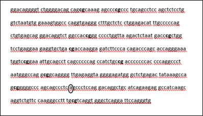
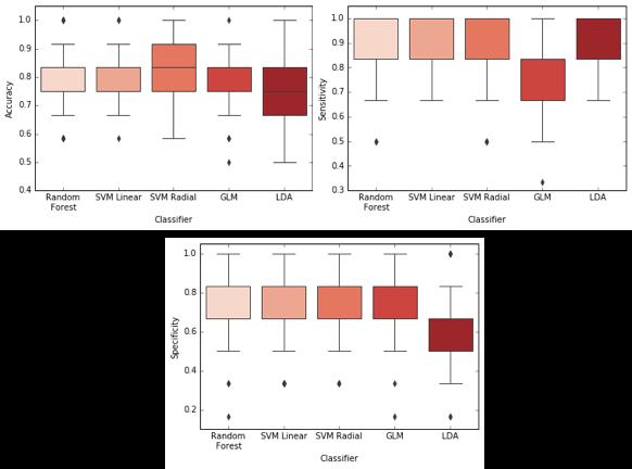
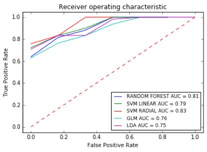
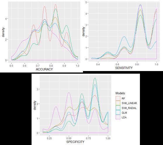
Background: Cytosine-guanine (CpGs) sites in genome can be identified as methylated or unmethylated; the combination of them in the genetic sequence of an individual includes a methylation haplotype (methyl-haplotype) for a specific locus. The insulin gene promoter (IGP) expression is highly regulated by methylation mechanisms. Our aim was to identify IGP methylhaplotypes among children/adolescents with type 1 diabetes (T1D) and to deploy a predictive model for the classification of cases and controls, using methyl-haplotypes as biomarkers. Methods: DNA from peripheral whole blood of 40 participants (20 T1D) was extracted and IGP region was sequenced by Next Generation Sequencing. A python-based pipeline for targeted deep bisulfite sequenced amplicons (ampliMethProfiler) was applied to estimate methylation status. Methylation profile at 10 CpG sites proximal to transcription start site of the IGP was recorded (site 1/-357, site-2/- 345, site 3/-234, site 4/-206, site 5/-180, site 6/-135, site 7/-102, site 8/69, site 9/-19, site 10/+60). Methylation of each site was coded as 0(zero) for unmethylation or 1(one) for methylation. A single read with the 10 CpG sites could result in “1111111111” methyl- haplotype (all methylated), in “000000000” methyl-haplotype (all unmethylated) or any other combination. The generated methyl-haplotypes were tested as predictive biomarkers in five different classifiers (Random forest/Support Vector Machine Radial and Linear/Generalized Linear Regression/Linear Discriminant Analysis). Predictive models were evaluated with the Receiver Operating Characteristics.
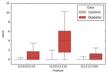
Results: 469 different methyl-haplotypes were recorded. After normalization of the features according to the number of readings, three distinct methyl-haplotypes: “1110101110”, “1110111110” and “1111111100” were more closely related to T1D compared to the controls (p-values: 0.00018,0.00032, 0.00095 respectively). The Support Vector Machine Radial presented the best accuracy (0.82±0.09) and a balanced performance between the two categories having sensitivity 0.86±0.12 and specificity 0.77±0.15.
Conclusions: Methylation based biomarkers, such as IGP methyl-haplotypes 1110101110, 1110111110 and 1111111100 could serve as a strategy to identify individuals at high risk for T1D.
Keywords: children, type 1 diabetes, DNA methylation, insulin gene, epigenetics
(cross-sectional)
ADA: American Diabetes Association
CpGs: Cytosine-Guanine sites
GLM: Generalized Linear Regression
IGP: Insulin Gene Promoter
ISPAD: International Society for Pediatric and Adolescence Diabetes
LDA: Linear Discriminant Analysis
NGS: Next Generation Sequencing
MWW: Mann-WhitneyWilcoxon test
RF: Random forest
SVM: Support Vector Machine
TSS: Transcription Start Site
ΣΔ1:
(International Society for Pediatric and Adolescence
(Nextera XT, DNA Library Preparation Kit, Research Illumina).
(pair–end reads)
NGS
Illumina.
(α) Random forest (RF), (β) Support Vector Machine Linear (SVM Linear), (γ) Support Vector Machine Radial (SVM Radial), (δ) Generalized Linear Regression (GLM), (ε) Linear Discriminant Analysis (LDA),
(Receiver Operating Characterisitc-
(16).
(MIS5033021),
Βιβλιογραφία
1. Krzewska A. Ben- Skowronek I. Effect of associated autoimmune diseases on type 1 diabetes mellitus incidence and metabolic control in children and adolescents. Biomed Res Int 2016;2016:6219730.
2. DIAMOND Project Group. Incidence and trends of childhood type 1 diabetes worldwide 1990-1999. Diabet Med 2006;23(8):857–866.
3. Allis CD, Jenuwein T. The molecular hallmarks of epigenetic control. Nat Rev Genet 2016;17:487–500.
4. Bird A. Perceptions of epigenetics. Nature 2007;447:396–398.
5. D’Angeli MA, Merzon E, Valbuena LF, Tirschwell D, Paris CA, Mueller BA. Environmental factors associated with childhood-onset type 1 diabetes mellitus: an exploration of the hygiene and overload hypotheses. Arch Pediatr Adolesc Med 2010;164:732–738.
6. Jerram ST, Dang MN, Leslie RD. The role of epigenetics in type 1 diabetes. Curr Diab Rep 2017;17:89.
7. Steck AK, Rewers MJ. Genetics of type 1 diabetes. Clin Chem 2011;57:176–185.
8. Fradin D, Le Fur S, Mille C, Naoui N, Groves C, Zelenika D, et al. Association of the CpG methylation pattern of the proximal insulin gene promoter with type 1 diabetes. PLoS One 2012;7:e36278.
9. American Diabetes Association. 2. Classification and Diagnosis of Diabetes: Standards of Medical Care in Diabetes-2018. Diabetes Care 2018;41:13–27.
10. Mayer-Davis EJ, Kahkoska AR, Jefferies C, Dabelea D, Balde N, Gong CX, et al. ISPAD Clinical Practice Consensus Guidelines 2018: Definition, epidemiology, and classification of diabetes in children and adolescents. Pediatr Diabetes 2018;19:7–19.
11. Scala G, Affinito O, Palumbo D, Florio E, Monticelli A, Miele G, et al. ampliMethProfiler: a pipeline for the analysis of CpG methylation profiles of targeted deep bisulfite sequenced amplicons. BMC Bioinformatics. 2016 ;17(1):484.
12. Kuhn M. Building predictive models in R using the caret package. Journal of Statistical Software 2008;28:1-26.
13. Jerram ST, Leslie RD. The genetic architecture of type 1 diabetes. Genes (Basel) 2017;8:209.
14. Nokoff N, Rewers M. Pathogenesis of type 1 diabetes: lessons from natural history studies of high-risk individuals. Ann N Y Acad Sci 2013;1281:1–15.
15. Morahan G. Insights into type 1 diabetes provided by genetic analyses. Curr Opin Endocrinol Diabetes Obes 2012;19:263–270.
16. Wang Z, Xie Z, Lu Q, Chang C, Zhou Z. Beyond genetics: What Causes Type 1 Diabetes. Clinic Rev Allerg Immunol 2017;52:273-286.
17. Cerna M. Epigenetic regulation in etiology of type 1 diabetes mellitus. Int J Mol Sci 2019;21:36.
18. Stefan M, Zhang W, Concepcion E, Yi Z, Tomer Y. DNA methylation profiles in type 1 diabetes twins point to strong epigenetic effects on etiology. J Autoimmun 2014;50:33–37.
19. Husseiny MI, Kaye A, Zebadua E, Kandeel F, Ferreri K. Tissue-specific methylation of human insulin gene and PCR assay for monitoring beta cell death. PLoS One 2014 ;9:e94591.
20. Neiman D, Moss J, Hecht M, Magenheim J, Piyanzin S, Shapiro AM, et al. Islet cells share promoter hypomethylation independently of expression, but exhibit cell-type-specific
methylation in enhancers. Proc Natl Acad Sci USA 2017;114:13525–13530.
21. Lehmann-Werman R, Neiman D, Zemmour H, Moss J, Magenheim J, Vaknin-Dembinsky A, et al. Identification of tissue-specific cell death using methylation patterns of circulating DNA. Proc Natl Acad Sci USA 2016 ;113:1826–1834.
22. Kuroda A, Rauch TA, Todorov I, Ku HT, Al-Abdullah IH, Kandeel F, et al. Insulin gene expression is regulated by DNA methylation. PLoS One 2009;4:e6953.
23. Herold KC, Usmani-Brown S, Ghazi T, Lebastchi J, Beam CA, Bellin MD, et al.; Type 1 Diabetes TrialNet Study Group. β cell death and dysfunction during type 1 diabetes development in at-risk individuals. J Clin Invest 2015;125:1163–1173.
24. Fisher MM, Watkins RA, Blum J, Evans-Molina C, Chalasani N, DiMeglio LA, et al. Elevations in Circulating Methylated and Unmethylated Preproinsulin DNA in New-Onset Type 1 Diabetes. Diabetes 2015;64:3867–3872.
25. Mouzaki K, Kotanidou EP, Fragou A, Kyrgios I, Giza S, Kleisarchaki A et al. Insulin gene promoter methylation status in Greek children and adolescents with type 1 diabetes. Biomed Reports 2020;13:31-36.
26. Mirabello L, Frimer M, Harari A, McAndrew T, Smith B, Chen Z, et al. HPV16 methylhaplotypes determined by a novel next-generation sequencing method are associated with cervical precancer. Int J Cancer 2015;136:146-153.
27. Clarke MA, Gradissimo A, Schiffman M, Lam J, Sollecito CC, Fetterman B, et al. Human papillomavirus DNA methylation as a biomarker for cervical precancer: consistency across 12 genotypes and potential impact on management of HPV-positive Women. Clin Cancer Res 2018;24:2194-2202.
28. Kavakiotis I, Tsave O, Salifoglou A, Maglaveras N, Vlahavas I, Chouvarda I. Machine learning and data mining methods in diabetes research. Comput Struct Biotechnol J 2017;15:104-116
29. Zhang H, Pollin TI. Epigenetics variation and pathogenesis in diabetes. Curr Diab Rep 2018;18:121.
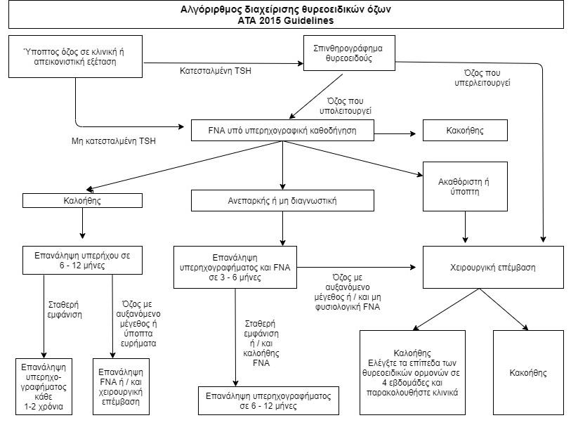
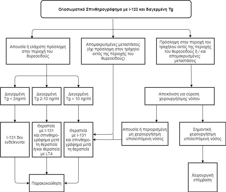
The incidence of thyroid cancer in the paediatric population is constantly rising during the last few decades. There are important differences in thyroid cancer between children and adults, as children present more frequently with advanced-stage disease in the time of diagnosis but have considerably lower mortality rates due to differences in molecular findings. Several risk factors, including iodine deficiency, previous radiation exposure, autoimmune thyroiditis and some genetic syndromes have been associated with the development of thyroid cancer. The greater risk of malignancy of thyroid nodules in children necessitates careful evaluation of a combination of clinical, imaging and histopathological findings aiming at early diagnosis and proper risk stratification, which will determine postoperative management and surveillance. Total thyroidectomy with or without adjuvant therapy with radioactive iodine is considered to be the gold standard in paediatric thyroid cancer and leads to high survival rates. The recent discoveries in the molecular mechanisms implicated in the various types of thyroid cancer and especially in the follicular subtype (the vast majority of cases) have provided new therapeutic opportunities through the use of targeted therapies for patients with disease not amenable to surgery and refractory to radioactive iodine. It is of the utmost importance that these patients are managed by a multidisciplinary team (paediatric endocrinologists, oncologists, radiologists, pathologists, endocrine surgeons and psychologists) in centers with experience in paediatric population.
Keywords: thyroid cancer, children, radioactive iodine, precision medicine
Eleni Tsotridou
Savvas Kolanis
Assimina Galli-Tsinopoulou
2nd Department of Paediatrics, School of Medicine, Faculty of Health Sciences, Aristotle University of Thessaloniki, University General Hospital
AHEPA
Skarpa, 2011 (16)
Corrias, 2008 (17)
Βασισμένο σε
• Francis GL, Waguespack SG, Bauer AJ, Angelos P, Benvenga S, Cerutti JM, et al. Management Guidelines for Children with Thyroid Nodules and Differentiated Thyroid Cancer. Thyroid. 2015;25(7):716–59.
1. Howlader N, Noone AM, Krapcho M, Miller D, Brest A, Yu M, et al (eds). SEER Cancer Statistics Review, 1975-2017, National Cancer Institute. Bethesda, MD, https://seer.cancer.
gov/csr/1975_2017/, based on November 2019 SEER data submission, posted to the SEER web site, April 2020.
2. Turcotte LM, Neglia JP. Subsequent malignant neoplasms in the survivor of childhood cancer: where we have been and where we are going? Future oncology.2017;13:23.
3. Francis GL, Waguespack SG, Bauer AJ, Angelos P, Benvenga S, Cerutti JM, et al. Management Guidelines for Children with Thyroid Nodules and Differentiated Thyroid Cancer. Thyroid. 2015;25(7):716–59.
4. Hogan AR, Zhuge Y, Perez EA, Koniaris LG, Lew JI, Sola JE. Pediatric thyroid carcinoma: incidence and outcomes in 1753 patients. J Surg Res. 2009;156(1):167–72.
5. Chan CM, Young J, Prager J, Travers S. Pediatric Thyroid Cancer. Adv Pediatr. 2017;64(1):171–90.
6. Paulson VA, Rudzinski ER, Hawkins DS. Thyroid cancer in the pediatric population. Genes (Basel). 2019;10(9).
7. Zimmermann MB, Galetti V. Iodine intake as a risk factor for thyroid cancer: a comprehensive review of animal and human studies. Thyroid Res. 2015;8:8.
8. Rose J, Wertheim BC, Guerrero MA. Radiation treatment of patients with primary pediatric malignancies: risk of developing thyroid cancer as a secondary malignancy. Am J Surg. 2012;204(6):881–7.
9. Veiga LHS, Lubin JH, Anderson H, de Vathaire F, Tucker M, Bhatti P, et al. A pooled analysis of thyroid cancer incidence following radiotherapy for childhood cancer. Radiat Res. 2012;178(4):365–76.
10. Lubin JH, Adams MJ, Shore R, Holmberg E, Schneider AB, Hawkins MM, et al. Thyroid Cancer Following Childhood Low-Dose Radiation Exposure: A Pooled Analysis of Nine Cohorts. J Clin Endocrinol Metab. 2017;102(7):2575–83.
11. Stefan AI, Piciu A, Mester A, Apostu D, Badan M, Badulescu CI. Pediatric thyroid cancer in Europe: An overdiagnosed condition? A literature review. Diagnostics. 2020;10(2):1–13.
12. Tuttle RM, Vaisman F, Tronko MD. Clinical presentation and clinical outcomes in Chernobyl-related paediatric thyroid cancers: what do we know now? What can we expect in the future? Clin Oncol (R Coll Radiol). 2011 ;23(4):268–75.
13. Radetti G, Loche S, D’Antonio V, Salerno M, Guzzetti C, Aversa T, et al. Influence of Hashimoto Thyroiditis on the Development of Thyroid Nodules and Cancer in Children and Adolescents. J Endocr Soc. 2019;3(3):607–16.
14. Won JH, Lee JY, Hong HS, Jeong SH. Thyroid nodules and cancer in children and adolescents affected by Hashimoto’s thyroiditis. Br J Radiol. 2018;91(1087):20180014.
15. Keskin M, Savas-Erdeve S, Aycan Z. Co-Existence of Thyroid Nodule and Thyroid Cancer in Children and Adolescents with Hashimoto Thyroiditis: A Single-Center Study. Horm Res Paediatr. 2016;85(3):181–7.
16. Skarpa V, Kousta E, Tertipi A, Anyfandakis K, Vakaki M, Dolianiti M, et al. Epidemiological characteristics of children with autoimmune thyroid disease. Hormones (Athens). 2011;10(3):207–14.
17. Corrias A, Cassio A, Weber G, Mussa A, Wasniewska M, Rapa A, et al. Thyroid nodules and cancer in children and adolescents affected by autoimmune thyroiditis. Arch Pediatr Adolesc Med. 2008;162(6):526–31.
18. Fiore E, Rago T, Latrofa F, Provenzale MA, Piaggi P, Delitala A, et al. Hashimoto’s thyroiditis is associated with papillary thyroid carcinoma: role of TSH and of treatment with L-thyroxine. Endocr Relat Cancer. 2011;18(4):429–37.
19. Mussa A, De Andrea M, Motta M, Mormile A, Palestini N, Corrias A. Predictors of Malignancy in Children with Thyroid Nodules. J Pediatr. 2015;167(4):886-892.
20. Zirilli G, Salzano G, Corica D, Pajno GB, Mignosa C, Pepe G, et al. Thyrotropin serum levels and coexistence with Hashimoto’s thyroiditis as predictors of malignancy in children with thyroid nodules. Italian journal of pediatrics. 2019;45(1):96.
21. Iliadou PK, Effraimidis G, Konstantinos M, Grigorios P, Mitsakis P, Patakiouta F, et al. Chronic lymphocytic thyroiditis is associated with invasive characteristics of differentiated thyroid carcinoma in children and adolescents. [published erratum in Eur J Endocrinol. 2016;174(2):X1] Eur J Endocrinol. 2015;173(6):827–33.
22. Hacıhamdioğlu B, Oçal G, Berberoğlu M, Savaş Erdeve S, Camtosun E, Kocaay P, et al. The evaluation of thyroid carcinoma in childhood and concomitance of autoimmune thyroid disorders. J Pediatr Endocrinol Metab. 2014 ;27(9–10):901–8.
23. Prasad PK, Mahajan P, Hawkins DS, Mostoufi-Moab S, Venkatramani R. Management of pediatric differentiated thyroid cancer: An overview for the pediatric oncologist. Pediatr Blood Cancer. 2020;67(6):1–10.
24. Cibas ES, Ali SZ. The 2017 Bethesda System for Reporting Thyroid Cytopathology. Thyroid. 2017;27(11):1341–6.
25. Wang H, Mehrad M, Ely KA, Liang J, Solórzano CC, Neblett WW 3rd, et al. Incidence and malignancy rates of indeterminate pediatric thyroid nodules. Cancer Cytopathol. 2019;127(4):231–9.
26. Arora S, Khoury J, Trout AT, Chuang J. Improving Malignancy Prediction in AUS/FLUS Pediatric Thyroid Nodules with the Aid of Ultrasound. Horm Res Paediatr. 2020;1–6.
27. Rago T, Vitti P. Role of thyroid ultrasound in the diagnostic evaluation of thyroid nodules. Best Pract Res Clin Endocrinol Metab. 2008;22(6):913–28.
28. Borysewicz-Sanczyk H, Dzieciol J, Sawicka B, Bossowski A. Practical Application of Elastography in the Diagnosis of Thyroid Nodules in Children and Adolescents. Horm Res Paediatr. 2016;86(1):39–44.
29. Cunha GB, Marino LCI, Yamaya A, Kochi C, Monte O, Longui CA, et al. Elastography for the evaluation of thyroid nodules in pediatric patients. Radiol Bras. 2019;52(3):141–7.
30. Hay ID, Gonzalez-Losada T, Reinalda MS, Honetschlager JA, Richards ML, Thompson GB. Long-term outcome in 215 children and adolescents with papillary thyroid cancer treated during 1940 through 2008. World J Surg. 2010;34(6):1192–202.
31. Chan CM, Young J, Prager J, Travers S. Pediatric Thyroid Cancer. Adv Pediatr. 2017;64(1):171–90.
32. Jeon MJ, Kim YN, Sung T-Y, Hong SJ, Cho YY, Kim TY, et al. Practical Initial Risk Stratification Based on Lymph Node Metastases in Pediatric and Adolescent Differentiated Thyroid Cancer. Thyroid. 2018;28(2):193–200.
33. Kim J, Sun Z, Adam MA, Adibe OO, Rice HE, Roman SA, et al. Predictors of nodal metastasis in pediatric differentiated thyroid cancer. J Pediatr Surg. 2017;52(1):120–3.
34. Spinelli C, Tognetti F, Strambi S, Morganti R, Massimino M, Collini P. Cervical Lymph Node Metastases of Papillary Thyroid Carcinoma, in the Central and Lateral Compartments, in Children and Adolescents: Predictive Factors. World J Surg. 2018;42(8):2444–53.
35. Jain NK, Mostoufi-Moab S, Hawkes CP, Nelson ND, Surrey LF, Jones ZS, et al. Extrathyroidal Extension is an Important Predictor of Regional Lymph Node Metastasis in Pediatric Differentiated Thyroid Cancer. Thyroid. 2020;30(7):1037–43.
36. Webb RC, Howard RS, Stojadinovic A, Gaitonde DY, Wallace MK, Ahmed J, et al. The utility of serum thyroglobulin measurement at the time of remnant ablation for predicting disease-free status in patients with differentiated thyroid cancer: a meta-analysis involving 3947 patients. J Clin Endocrinol Metab. 2012;97(8):2754–63.
37. Klain M, Zampella E, Manganelli M, Gaudieri V, Nappi C, D’Antonio A, et al. Risk of structural persistent disease in pediatric patients with low or intermediate risk differentiated thyroid cancer. Endocrine. 2020 Jun;
38. Karapanou O, Tzanela M, Rondogianni P, Dacou-Voutetakis C, Chiotis D, Vlassopoulou B, et al. Long-term outcome of differentiated thyroid cancer in children and young adults: risk stratification by ATA criteria and assessment of pre-ablation stimulated thyroglobulin as predictors of disease persistence. Endocrine. 2020
39. Liu L, Zhang X, Tian T, Huang R, Liu B. Prognostic Value of Pre-Ablation Stimulated Thyroglobulin in Children and Adolescents with Differentiated Thyroid Cancer. Thyroid. 2020;30(7):1017–24.
40. Pawelczak M, David R, Franklin B, Kessler M, Lam L, Shah B. Outcomes of children and adolescents with well-differentiated thyroid carcinoma and pulmonary metastases following 131I treatment: a systematic review. Thyroid. 2010;20(10):1095–101.
41. Fard-Esfahani A, Emami-Ardekani A, Fallahi B, Fard-Esfahani P, Beiki D, Hassanzadeh-Rad A, et al. Adverse effects of radioactive iodine-131 treatment for differentiated thyroid carcinoma.
Nucl Med Commun. 2014;35(8):808–17.
42. Albano D, Bertagna F, Panarotto MB, Giubbini R. Early and late adverse effects of radioiodine for pediatric differentiated thyroid cancer. Pediatr Blood Cancer. 2017 ;64(11).
43. Selvakumar T, Nies M, Klein Hesselink MS, Brouwers AH, van der Horst-Schrivers ANA, Klein Hesselink EN, et al. Long-term effects of radioiodine treatment on salivary gland function in adult survivors of pediatric differentiated thyroid carcinoma. J Nucl Med. 2018;60(2):172177.
44. Sharma E, Dahal S, Sharma P, Bhandari A, Gupta V, Dahal S. Secondary Salivary Gland Malignancy in Thyroid Cancer: A United States Population Based Study. J Clin Med Res. 2018;10(7):601–5.
45. Dong P, Wang L, Huang R, Li L. Bone marrow suppression in pediatric patients with differentiated thyroid cancer following empirical radioiodine therapy. Medicine (Baltimore). 2020;99(31):e21398.
46. Brown AP, Chen J, Hitchcock YJ, Szabo A, Shrieve DC, Tward JD. The risk of second primary malignancies up to three decades after the treatment of differentiated thyroid cancer. J Clin Endocrinol Metab. 2008;93(2):504–15.
47. Molenaar RJ, Sidana S, Radivoyevitch T, Advani AS, Gerds AT, Carraway HE, et al. Risk of Hematologic Malignancies After Radioiodine Treatment of Well-Differentiated Thyroid Cancer. J Clin Oncol. 2018;36(18):1831–9.
48. Nies M, Cantineau AEP, Arts EGJM, van den Berg MH, van Leeuwen FE, Muller Kobold AC, et al. Long-Term Effects of Radioiodine Treatment on Female Fertility in Survivors of Childhood Differentiated Thyroid Carcinoma. Thyroid. 2020;30(8):1169–76.
49. Waguespack SG, Sherman SI, Williams MD, Clayman GL, Herzog CE. The successful use of sorafenib to treat pediatric papillary thyroid carcinoma. Thyroid. 2009;19(4):407–12.
50. Aller S, Popescu A, Rao S, Morgan E, Gosiengfiao Y. Transient partial response to sorafenib treatment in an adolescent patient with MEN2B syndrome and end-stage medullary thyroid carcinoma. Pediatr Blood Cancer. 2012;58(1):98–100.
51. Higuchi Y, Motoki T, Ishida H, Kanamitsu K, Washio K, Oyama T, et al. Sorafenib treatment for papillary thyroid carcinoma with diffuse lung metastases in a child with autism spectrum disorder: a case report. BMC Cancer. 2017;17(1):775.
52. Iyer P, Mayer JLR, Ewig JM. Response to sorafenib in a pediatric patient with papillary thyroid carcinoma with diffuse nodular pulmonary disease requiring mechanical ventilation. Thyroid. 2014;24(1):169–74.
53. Mahajan P, Dawrant J, Kheradpour A, Quintanilla NM, Lopez ME, Orth RC, et al. Response to lenvatinib in children with papillary thyroid carcinoma. Thyroid. 2018;28(11):1450–4.
54. Milner TD, Ronghe M, Shaikh MG, MacGregor FB, Reed N. Vandetanib Tumor Shrinkage in Metastatic Medullary Thyroid Cancer Allowing Surgical Resection of the Primary Site: A Case Report. J Pediatr Hematol Oncol. 2019;41(5):e329–32.
55. Kraft IL, Akshintala S, Zhu Y, Lei H, Derse-Anthony C, Dombi E, et al. Outcomes of Children and Adolescents with Advanced Hereditary Medullary Thyroid Carcinoma Treated with Vandetanib. Clin Cancer Res. 2018;24(4):753–65.
56. Narayanan VK, Ronghe M, MacGregor FB, Bradshaw N, Davidson R, Welbury R, et al. Use of Vandetanib in Metastatic Medullary Carcinoma of Thyroid in a Pediatric Patient With Multiple Endocrine Neoplasia 2B. J Pediatr Hematol Oncol. 2016;38(2):155–7.
57. Chuk MK, Widemann BC, Minard CG, Liu X, Kim A, Bernhardt MB, et al. A phase 1 study of cabozantinib in children and adolescents with recurrent or refractory solid tumors, including CNS tumors: Trial ADVL1211, a report from the Children’s Oncology Group. Pediatr Blood Cancer. 2018;65(8):e27077.
58. Laetsch TW, DuBois SG, Mascarenhas L, Turpin B, Federman N, Albert CM, et al. Larotrectinib for paediatric solid tumours harbouring NTRK gene fusions: phase 1 results from a multicentre, open-label, phase 1/2 study. Lancet Oncol. 2018;19(5):705–14.
59. Hillier K, Hughes A, Shamberger RC, Shusterman S, Perez-Atayde AR, Wassner AJ, et al. A Novel ALK Fusion in Pediatric Medullary Thyroid Carcinoma. Thyroid. 2019;29(11):1704–7.
60. Subbiah V, Yang D, Velcheti V, Drilon A, Meric-Bernstam F. State-of-the-Art Strategies for Targeting RET-Dependent Cancers. J Clin Oncol. 2020;38(11):1209–21.
e-mail:rodosfa@hotmail.com
Correspondence
Sfakiotaki Rodanthi
Εvrota 22, N. Psychiko
ΤΚ 15451 Athens
Τ. +30 6977748270
e-mail:rodosfa@hotmail.com
Evaluation of lower esophagus and its pathology with transabdominal ultrasound in pediatric patients
Sfakiotaki Rodanthi, Pitsoulaki Despoina, Athanasiou Anastasia, Hountala Anna
Abstract
Pediatric lower esophageal disorders, apart from gastroesophageal reflux, are rare in the pediatric population. The most common ones include hiatal hernia, esophagitis, esophageal varices, duplication cyst, and achalasia. These disorders have a spectrum of clinical symptoms, including a number of asymptomatic cases. Clinical examination can provide little information in patients who are asymptomatic. However morphologic and functional information of lower esophagus can be acquired rapidly, relatively inexpensive, with no patient discomfort and no radiation exposure by transabdominal ultrasonography. The educational objective of this pictorial essay is to present the imaging findings and the contribution of transabdominal sonography in the diseases of lower esophagus. Taking into account these findings, pediatricians can adapt the treatment protocol to the different diagnostic results.
Children's Hospital "P. & A. Kyriakou", Athens, Greece, Radiology Department
Athanasiou Anastasia
Children's Hospital "P. & A. Kyriakou", Athens, Greece, 1st Department of Pediatrics
Βιβλιογραφία
1. Koumanidou C, Vakaki M, Pitsoulakis G, Anagnostara A, Mirilas P. Sonographic measurement of the abdominal esophagus length in infancy: a diagnostic tool for gastroesophageal reflux. AJR Am J Roentgenol. 2004 Sep;183:801-7.
2. Savino A, Cecamore C, Matronola MF, Verrotti A, Mohn A, Chiarelli F, et al. US in the diagnosis of gastroesophageal reflux in children. Pediatr Radiol.2012 May; 42:515-24.
3. Gomes H, Lallemand A, Lallemand P. Ultrasound of the gastroesophageal junction. Pediatr Radiol. 1993; 23:94-9
4. Halkiewicz F, Kasner J, Karczewska K, Rusek-Zychma M. Ultrasound picture of gastroesophageal junction in children with reflux disease. Med Sci Monit. 2000Jan-Feb;6(1):96-9.
5. Karabulut B, Bostanci I, Kacar M, Karaca G, Kosar P. Transcutaneous cervical and transabdominal ultrasonography as a diagnostic tool in gastroesophageal reflux in childhood. ORL J Otorhinolaryngol Relat Spec. 2010;72(6):300-4.
6. Rosen R, Vandenplas Y, Singendonk M, Cabana M, DiLorenzo C, Gottrand F, et al. Pediatric Gastroesophageal Reflux Clinical Practice Guidelines: Joint Recommendations of the North American Society for Pediatric Gastroenterology, Hepatology, and Nutrition and the European Society for Pediatric Gastroenterology, Hepatology, and Nutrition. J Pediatr Gastroenterol Nutr. 2018 Mar;66:516-554.
7. Chavhan GB, Babyn PS, Cohen RA, Langer JC. Multimodality imaging of the pediatric diaphragm: anatomy and pathologic conditions. Radiographics. 2010Nov;30:1797-817.
8. Hallal C, Kieling CO, Nunes DL, Ferreira CT, Peterson G, Barros SG, et al. Diagnosis, misdiagnosis, and associated diseases of achalasia in children and adolescents: a twelve-year single center experience. Pediatr Surg Int. 2012 Dec;28:1211-7.
9. Iacob D, Fufezan O, Farcau D, Hagău N, Ciuce C. Clinical and ultrasound approach to achalasia in a child. Case report. Med Ultrason. 2010 Mar;12:66-70.
10. Lee TH, Cho JY. Sonographic evaluation of esophageal achalasia. Korean JIntern Med. 2014 Mar;29:262.
11. Madhusudhan KS, Seith A, Srinivas M, Gupta AK. Esophageal duplication cyst causing unilateral hyperinflation of the lung in a neonate. Acta Radiol. 2007Jun;48:588-90.
12. Gana JC, Turner D, Mieli-Vergani G, Davenport M, Miloh T, Avitzur Y, et al. A clinical prediction rule and platelet count predict esophageal varices in children. Gastroenterology. 2011 Dec;141:2009-16.
13. De Giacomo C, Tomasi G, Gatti C, Rosa G, Maggiore G. Ultrasonographic prediction of the presence and severity of esophageal varices in children. JPediatr Gastroenterol Nutr. 1989 Nov;9:431-5.
The term pulmonary blastoma stands for a rare but highly malignant tumor of the lungs in childhood. It involves the intrathoracic mesenchyma, both the pulmonary and the pleura. In 1988 the International Pleuropulmonary Blastoma Registry (IPPBR) was founded to organize centrally the collection and qualification of the biopsies and medical records of the patients diagnosed with Pleuropulmonary blastoma. In this short review, there is also a comment regarding the translation of the term pleuropulmonary blastoma in Greek, which is being translated as if the term pleura refers to the ribs and not to the thin fluid-filled space that covers the lungs due to the linguistic phenomenon of reborrowing. Usually it is being diagnosed before the fourth year of age and it is responsible for the 15% of all primary pediatric tumors. In 75% of the cases it is sporadic and in the rest 25% it is seen in children with congenital lung cyst or in children from families with other malignancies or serious conditions. It affects both genders equally with small predominance of males in certain types. It is categorized as Type I (I-Ir), type II and type III. The prognosis is not good in general, mostly for types II-III, since there is often metastasis, in contrast to type I, where the rates of survival can be as high as 90%.
Keywords: pulmonary blastoma, pleura, children
Koutsaftiki Chryssie
Paediatric department, Rafina’s Health Centre, Rafina
Mammas Ioannis
Department of clinical virology, School of Medicine, University of Crete, Heraklion Crete
Papatheodoropoulou Alexia
PICU, University General Hospital of Patras, PatrasRio
Priftis Konstantinos
Department of Paediatric pulmonology-allergology, University of Athens, Αthens
Paediatric pulmonologyallergology department, Paediatric Centre of Athens
PPB: Pleura-Pulmunary Blastoma
1) Khan AA, El Borai AK, Alnoaiji M. Pleuropulmonary blastoma: a case report and review of the literature. Case Rep Pathol. 2014;2014:509086
2) Μπαμπινιώτης
Ε.Π.Ε., Αθήνα, 2002. Σελ 2032
3) Manivel JC, Priest JR, Watterson J, Steiner M, Woods WG, Wick MR, et al. Pleuropulmonary blastoma. The so-called pulmonary blastoma of childhood. Cancer. 1988;62:1516–1526.
4) Messinger YH, Stewart DR, Priest JR, Williams GM, Harris AK, Schultz KA, et al. Pleuropulmonary b;astoma: a report on 350 central pathology-confirmed pleuropulmonary blastoma cases by the International Pleuropulmonary Blastoma Registry. Cancer.2015;121:276-285. doi: 10.1002/cncr.29032.
5) Priest JR, Watterson J, Strong L, Huff V, Woods WG, Byrd RL, et al. Pleurupulmonary blastoma: a marker for familial disease. J Pediatr. 1996; 128:220-224.
6) Hill DA, Ivanovich J, Priest JR, Gurnett CA, Dehner LP, Desruisseau D, et al. DICER1 mutations in familial pleuropulmonary blastoma. Science.2009;325:965
7) Stocker JT, Hussain AN, Dehner LP. “Pediatric tumors” in Dail and Hammar’s pulmonary pathology Vol.II, Tomashefski JF, Cagle PT, Farver CF, Fraire AE, Eds.,pp.542-557, Springer, New York, NY, USA, 3rd edition, 2008.
8) Hasiotou M, Polyviou P, Strantzia CM, Pourtsidis A, Stinios I. Pleuropulmonary blastoma in the area of a previously diagnosed congenital lung cyst: report of two cases. Acta Radiol 2004; 45: 289-292.
9) Federici S, Domenichelli V, Tani G, Sciutti R, Burnelli R, Zanetti G, Domini R. Pleuropulmonary blastoma in congenital cystic adenomatoid malformation: report of a case. Eur J Pediatr Surg 2001; 11:196-199.
10) Priest JR, McDermott MB, Bhatia S, Watterson J, Manivel JC, Dehner LP. Pleuropulmonary blastoma: a clinicopathologic study of 50 cases. Cancer 1997; 80: 147-61.
Teenage girl with obesity amenorrhea and pcos- complete remission of the syndrome with weight loss
Elpis-Athina Vlachopapadopoulou, Anatoli Fotiadou, Eirini Dikaiakou, Fani Athanasouli, Stefanos Michalacos
Abstract
Introduction: Polycystic ovary syndrome (PCOS) is a common endocrine disorder during puberty characterized by menstrual irregularities and hyperandrogenism. Insulin resistance and metabolic syndrome are linked to PCOS. Diagnostic criteria for PCOS in adolescence are controversial, because common pathological features used as criteria for adult women may be normal variants for adolescents. The management of PCOS in puberty consists of lifestyle modifications and pharmacological interventions if needed.
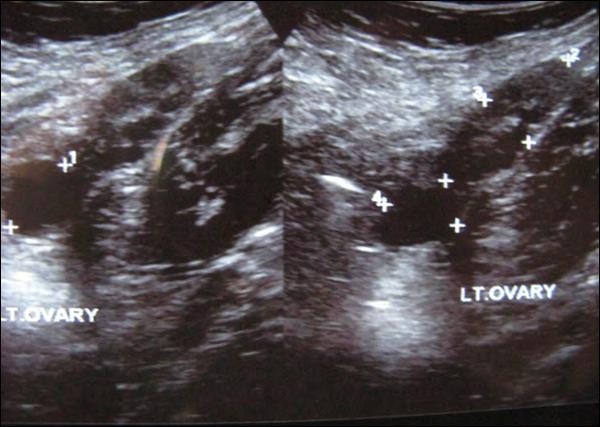
Case presentation: We report a case of PCOS in a fourteen years old obese adolescent girl with secondary amenorrhea whose lifestyle modification alone leading to weight loss was effective to treat PCOS, restore ovulation and regular menstrual cycles. This case is reported in order to emphasize the importance to implement a step wise approach for the management of PCOS in adolescence. Primarily a modification of dietary habits and increased physical activity is recommended and in case of no improvement, medical treatment follows.
Conclusions: The diagnosis of PCOS should be contemplated in adolescent girls with menstrual disturbances, hyperandrogenemia and obesity. Other causes should be excluded. Therapeutic approach as a first step aims to a decrease of body mass index following adherence to a healthy diet and increased physical activity.
Dept. of EndocrinologyGrowth and Development, Children’s Hospital “P&A Kyriakou”
Anatoli Fotiadou
2nd Department of Paediatrics, University of Athens, “P&A Kyriakou” Children's Hospital, Athens, Greece
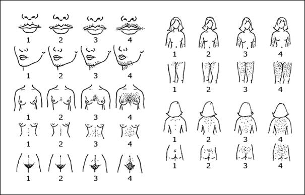
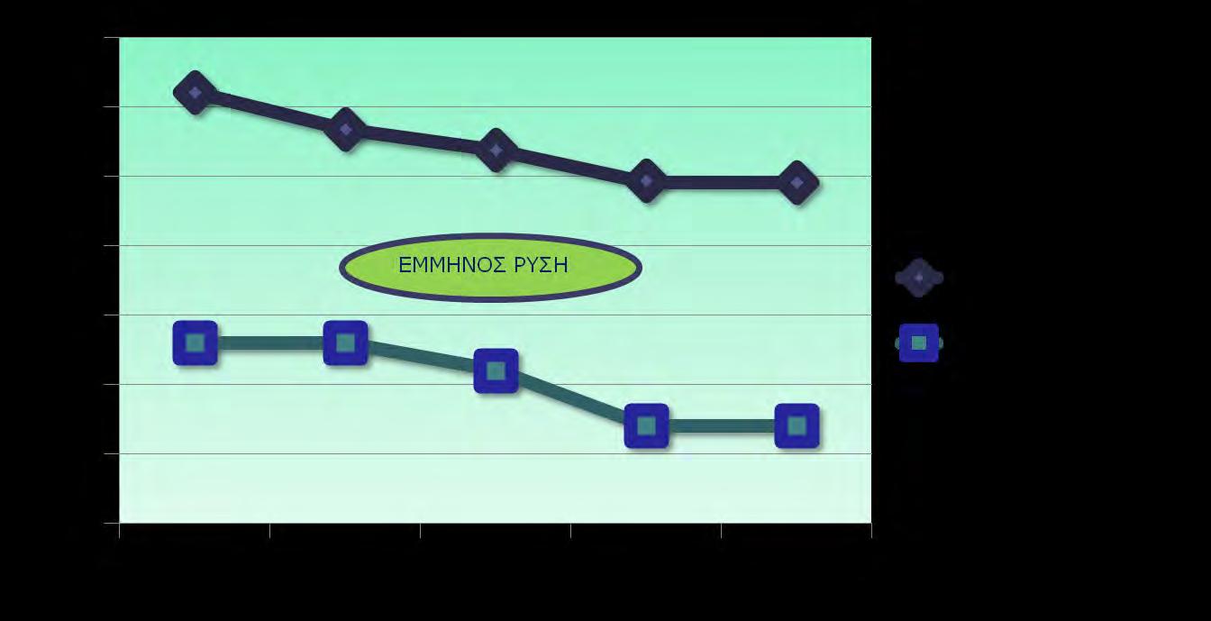
ΕΘ), ύψος σώματος: 161,5 εκατοστά (90η ΕΘ), ΔΜΣ: 31,1kg/m² (>95η ΕΘ), περίμετρος μέσης: 102cm (>90η ΕΘ), στάδιο ενήβωσης κατά Tanner V (μασχάλη, μαστοί, εφήβαιο). Η ασθενής παρουσίαζε δασυτριχισμό σε ανδρογονοεξαρτώμενες περιοχές (πρόσωπο, μαστοί, μέση γραμμή και κοιλιακή χώρα- Ferriman Gallwey score 12), μελανίζουσα
ΔΜΣ:
ΕΘ:
υδροξυπρογεστερόνη
ΣΠΩ:
DHEAS:
FG: Ferriman Gallwey
FSH:
Hb1Ac:
HOMA IR: Homeostatic Model Assessment for Insulin Resistance
LH:
ΡCOS: Polycystic Ovary Syndrome
SHBG:
TSH:
SHBG, DHEAS,
Βιβλιογραφία
1. Witchel SF, Oberfield SE, Peña AS. Polycystic Ovary Syndrome: Pathophysiology, Presentation, and Treatment with Emphasis on Adolescent Girls. J Endocr Soc. 2019;3(8):1545‐1573
Published 2019 Jun 14. doi:10.1210/js.2019-00078
2. Witchel SF, Oberfield S, Rosenfield RL, et al. The Diagnosis of Polycystic Ovary Syndrome during Adolescence [published online ahead of print, 2015 Apr 1]. Horm Res Paediatr. 2015;83:376–38910.1159/000375530
3. Ibáñez L, Oberfield SE, Witchel S, Auchus RJ, Chang RJ, Codner E et al. An International Consortium Update: Pathophysiology, Diagnosis, and Treatment of Polycystic Ovarian Syndrome in Adolescence. Horm Res Paediatr. 2017;88(6):371‐395. doi:10.1159/000479371
4. Rothenberg SS, Beverley R, Barnard E, Baradaran- Shoraka M, Sanfilippo JS. Polycystic ovary syndrome in adolescents. Best Pract Res Clin Obstet Gynaecol. 2018;48:103‐114. doi:10.1016/j. bpobgyn.2017.08.008
5. Anderson AD, Burt Solorazano CM, McCartney CR Childhood Obesity and its Impact on Adolescent PCOS Semin Reprod Med 2014 May;32(3):202-13 doi: 10.1055/s-0034-1371092
6. Moran LJ, Pasquali R, Teede HJ, Hoeger KM, Norman RJ. Treatment of obesity in polycystic ovary syndrome: a position statement of the Androgen Excess and Polycystic Ovary Syndrome Society. Fertil Steril. 2009;92(6):1966–1982.
7. Diamanti-Kandarakis E, Dunaif A. Insulin resistance and the polycystic ovary syndrome revisited: an update on mechanisms and implications. Endocr Rev. 2012;33(6):981–1030
8. Dunaif A. Insulin resistance and the polycystic ovary syndrome: mechanism and implications for pathogenesis. Endocr Rev. 1997;18(6):774–800.
9. Rosenfield RL. The Diagnosis of Polycystic Ovary Syndrome in Adolescents. Pediatrics.
2015;136(6):1154‐1165. doi:10.1542/peds.2015-1430
10. Al Khalifah RA, Florez ID, Dennis B, Thabane L, Bassilious E. Metformin or Oral Contraceptives for Adolescents With Polycystic Ovarian Syndrome: A Meta-analysis. Pediatrics. 2016;137(5):e20154089. doi:10.1542/peds.2015-4089
email:
Correspondence
Vasileios Giapros
Stavrou Niarchou Avenue, Ioannina, 45110, Greece
T. +30 2651099326
M. +30 6974754893
email: vgiapros@uoi.gr
A case report of an infantile hepatic haemangioma
D. Rallis, F. Balomenou, A. Drougia, V. Xydis, N. Giantzouli, I. Basoukas, A. Zellou4, V. Giapros
Abstract
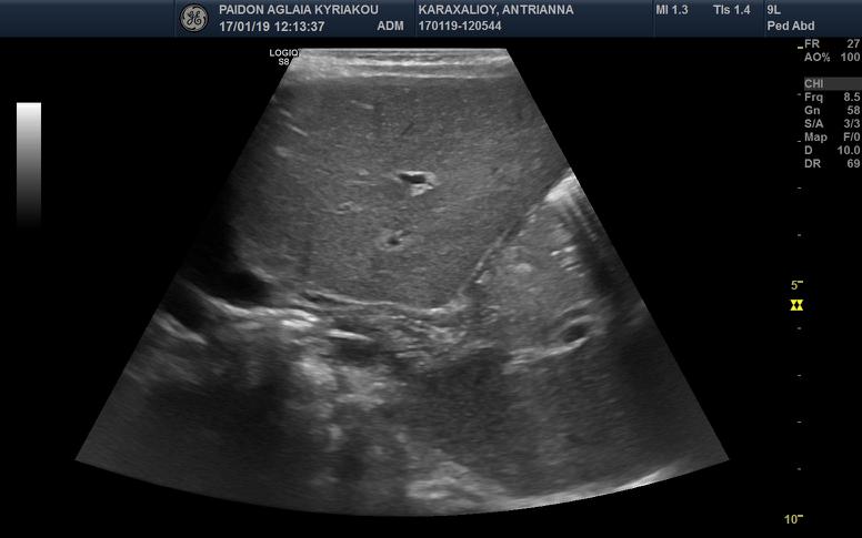
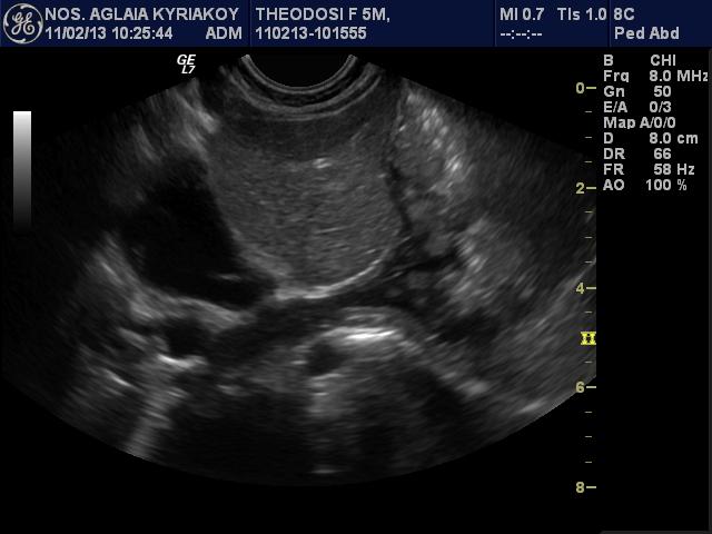
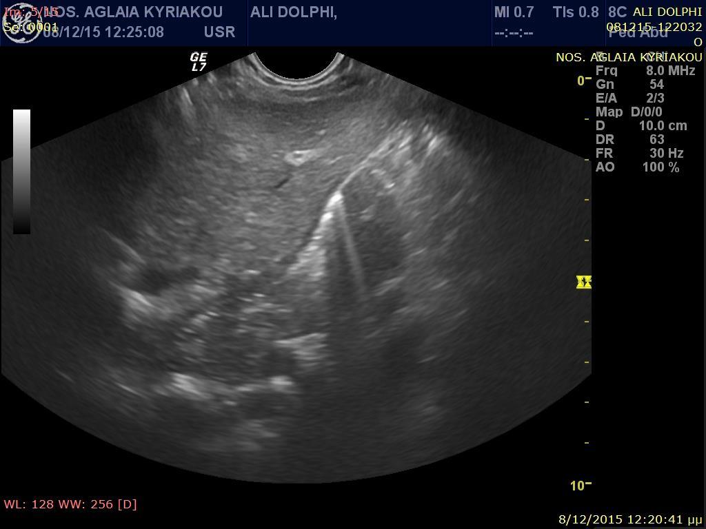
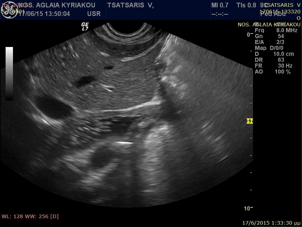
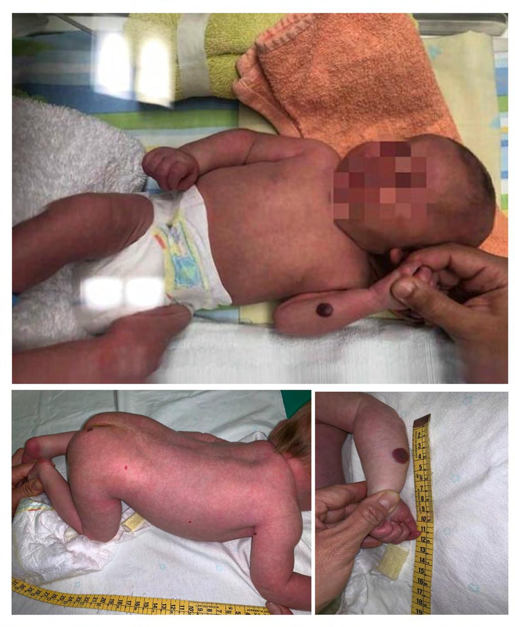
Introduction: Infantile hepatic haemangiomas are the most common neonatal benign vascular lesions, the prognosis of which is depended on the size and the location. We describe the case of an infant with multiple cutaneous haemangiomatosis and hepatic haemangiomas.
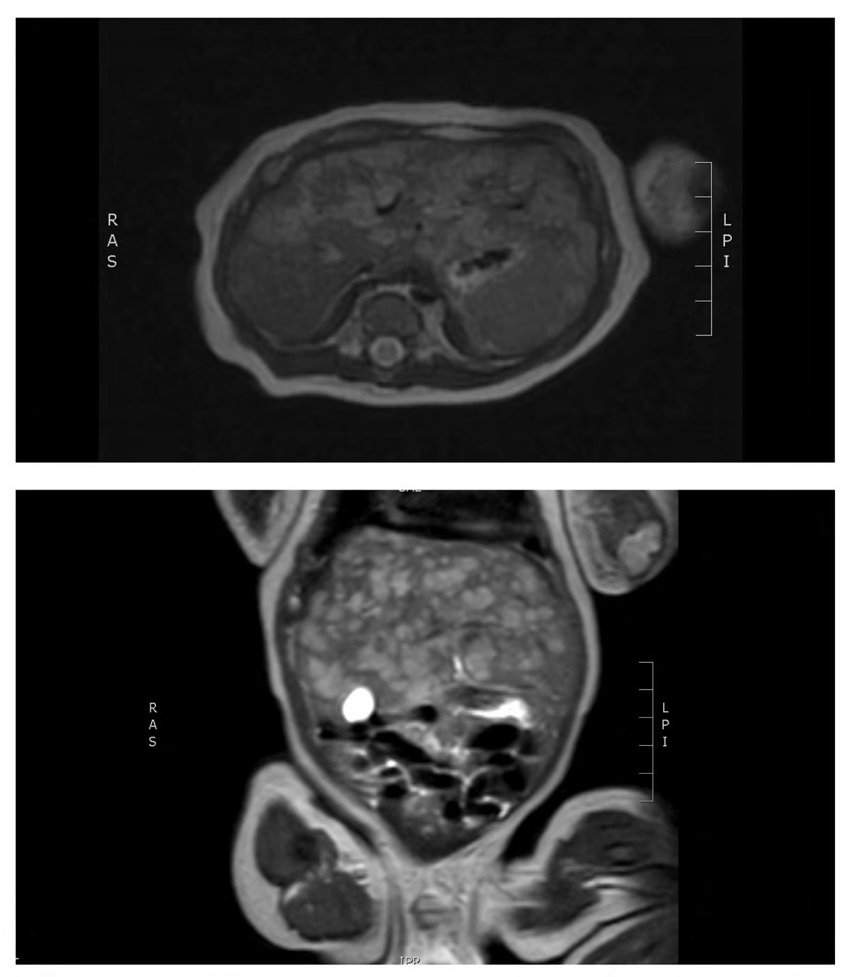
Case report: A male term infant was diagnosed since birth with multiple pigmented cutaneous lesions. A further evaluation detected multiple hepatic haemangiomas. The thyroid function was normal, while the a-fetoprotein values were gradually decreased. The infant received orally propranolol, without presenting any side effects.
Discussion: Infantile hepatic haemangiomas may present asymptomatic, or they may be related with significant systematic complication. The diagnosis requires further imaging in cases were a strong suspicion arises due to multiple cutaneous lesions. Currently propranolol is considered as the first line treatment, and has replaced the previous options of steroids or interferon-a. Finally, the invasive approach has been limited to only specific cases.
Radiology Department, University Hospital of Ioannina, Ioannina
I. Basoukas
Dermatology Department, University Hospital of Ioannina, Ioannina
A. Zellou
Division of Pediatric
Gastroenterology at Children’s Hospital
“Mitera”, Athens
1. Rawal N, Yazigi N. Pediatric Liver Transplantation. PediatrClin North Am. 2017;64(3):67784.
2. van der Meijs BB, Merks JH, de Haan TR, Tabbers MM, van Rijn RR. Neonatal hepatic haemangioendothelioma: treatment options and dilemmas. PediatrRadiol. 2009;39(3):277-81.
3. Cabrita SV, Goncalves S, Rodrigues H, Guerra N, Moura P. Antenatal diagnosis of congenital hepatic hemangioma: a case report. Cases J. 2009;2:6829.
4. Christison-Lagay ER, Burrows PE, Alomari A, Dubois J, Kozakewich HP, Lane TS, et al. Hepatic hemangiomas: subtype classification and development of a clinical practice algorithm and registry. J Pediatr Surg. 2007;42(1):62-7; discussion 7-8.
5. Itinteang T, Withers AH, Davis PF, Tan ST. Biology of infantilehemangioma. Front Surg. 2014;1:38.
6. Gallego C, Miralles M, Marin C, Muyor P, Gonzalez G, Garcia-Hidalgo E. Congenital hepatic shunts. Radiographics. 2004;24(3):755-72.
7. Huang SA, Tu HM, Harney JW, Venihaki M, Butte AJ, Kozakewich HP, et al. Severe hypothyroidism caused by type 3 iodothyronine deiodinase in infantile hemangiomas. N Engl J Med. 2000;343(3):185-9.
8. Gnarra M, Behr G, Kitajewski A, Wu JK, Anupindi SA, Shawber CJ, et al. History of the infantile hepatic hemangioma: From imaging to generating a differential diagnosis. World J ClinPediatr. 2016;5(3):273-80.
9. Zhu Z, Cai P, Zhu J, Chen J, Wu B, Gu Z, et al. Neonatal giant hepatic hemangioma: A case report. Medicine (Baltimore). 2018;97(42):e12863.
10. Krowchuk DP, Frieden IJ, Mancini AJ, Darrow DH, Blei F, Greene AK, et al. ClinicalPracticeGuideline for the Management of InfantileHemangiomas. Pediatrics. 2019;143(1).
11. Drolet BA, Frommelt PC, Chamlin SL, Haggstrom A, Bauman NM, Chiu YE, et al. Initiation and use of propranolol for infantile hemangioma: report of a consensus conference. Pediatrics. 2013;131(1):128-40.
12. Darrow DH, Greene AK, Mancini AJ, Nopper AJ, Section On Dermatology SOO-H, Neck S, et al. Diagnosis and Management of InfantileHemangioma. Pediatrics. 2015;136(4):e1060104.
13. Barlow CF, Priebe CJ, MullikenJB, Barnes PD, Mac Donald D, Folkman J, et al. Spastic diplegia as a complication of interferon Alfa-2a treatment of hemangiomas of infancy. J Pediatr. 1998;132(3 Pt 1):527-30.
14. Enjolras O, Riche MC, Merland JJ, Escande JP. Management of alarming hemangiomas in infancy: a review of 25 cases. Pediatrics. 1990;85(4):491-8.
15. Draper H, Diamond IR, Temple M, John P, Ng V, Fecteau A. Multimodal management of endangering hepatic hemangioma: impact on transplant avoidance: a descriptive case series. J PediatrSurg. 2008;43(1):120-5; discussion 6.
email: atosmedicals@yahoo.
Βιβλιογραφία
1. Dickison P, Christou E, Wargon O A prospective study of infantile hemangiomas with a focus on incidence and risk factors. Pediatr Dermatol 2011;28:663–669
2. Filippi L, Cavallaro G, Bagnoli P, DalMonteM, Fiorini P, Donzelli G, et al Oral propranolol
for retinopathy of prematurity: risks, safety concerns, and perspectives. J Pediatr 2014;163:1570–1577
3. Munden A, Butschek R, Tom WL, Marshall JS, Poeltler DM, Krohne SE et al Prospective study of infantile haemangiomas: incidence, clinical characteristics and association with placental anomalies. Brit J Dermatol 2014;170:907–913
4. Darrow DH, Greene AK, Mancini AJ, Nopper AJ. Diagnosis and Management of Infantile Hemangioma. Pediatrics. 2015 Oct;136(4):e1060-104.
5. Hermans DJ, Boezeman JB, Van de Kerkhof PC, Rieu PN, Van der Vleuten CJ (2009) Differences between ulcerated and nonulcerated hemangiomas, a retrospective study of 465 cases. Eur J Dermatol 2009;19:152–156
6. Wedgeworth E, Glover M, Irvine AD, Neri I, Baselga E, Clayton TH et al Propranolol in the treatment of infantile haemangiomas: lessons from the European Propranolol In the Treatment of Complicated Haemangiomas (PITCH) Taskforce survey. Br J Dermatol. 2016 Mar;174(3):594-601
7. Vergine G, Marsciani A, Pedini A, Brocchi S, Marsciani M, Desiderio E, et al Efficacy of propranolol treatment in thyroid dysfunction associated with severe Infantile hepatichaemangioma. Horm Res Paediatr 2014;78:256–60
8. Chen TS, Eichenfield LF, Friedlander SF Infantile hemangiomas: an update on pathogenesis and therapy. Pediatrics 2013;131:99–108
9. Marqueling AL, Oza V, Frieden IJ, Puttgen KB Propranolol and infantile hemangiomas four years later: a systematic review. Pediatr Dermatol 2013;30:182–191
10. McAinsh J, Cruickshank JM (1990) Beta-blockers and central nervous system side effects. Pharmacol Ther 1990;46:163–97
11. Parikh S, Darrow DH, Grimmer JF, Manning SC Propranolol use for infantile hemangiomas. American Society of Pediatric Otolaryngology Vascular Anomalies Task Force Practice Patterns. JAMA Otolaryngol Head Neck Surg 2013;139:153–156
Dixieland Band
Bernardo Nadal Ginard,
Roberta Williams,
ΝΕΟΕΛΛΗΝΕΣ
2.
3.
4.
5.
6.
7.
8.
9. Ενδιαφέρουσες
10. Σύντομα
11. Βραχείες
12.
13.
14.
(Uniform Requirements for Manuscripts Submitted to Biomedical Journals),
International Committee of Medical Journal Editors/Uniform Requirements for Manuscripts Submitted to Biomedical Journals, (www.icmje.org και www.icmje.org/icmje.
Cumulated Index Medicus [List of Journals Indexed in Index Medicus (www.nlm.nih.goν/bsd/uniform_requirements.html)].
1999;59:272-279.
Proesmans W. Bartter syndrome and its neonatal νariant. Eur J Pediatr 1997;156:669-679.
Flyvbjerg Α. Role of growth hormone, insulin-like growth factors (IGFs) and IGF-binding proteins in the renal complications of diabetes. Kidney Ιnt 1997;52 (60 Suppl):S12-S19.
Χωρίς συγγραφέα:
National Institutes of Health Consensus Deνelopment Conference. Neurofibromatosis conference statement. Arch Neurol1988;45:575-578.
Προσδιορισμός τύπου άρθρου: Schreiner GF, Lange L. Ethanol modulation of macrophage influx in glomerulonephritis [Abstract]. J Am Soc Nephrol 1991;2:562.
Should antileukotriene therapies be used instead of inhaled corticosteroids in asthma? [Editorial]. Am J Respir Crit Care Med 1998;158:1697-1701.
Clark AG, Barratt ΤΜ. Steroid-responsiνe nephrotic syndrome. Ιn: Barratt ΤΜ, Arner ED, Harmon WE, editors. Pediatric Nephrology. 4th ed. Baltimore: Lippincott William Wilkins; 1999. p. 742.
Σύγγραμμα
Gorlin RJ, Cohen ΜΜ, Leνin LS. Syndromes of the head and neck. 3rd ed. New York: Oxford Uniνersity Press; 1990.
Δημοσίευση
Bauer ΑW. The two definitions of bacterial resistance. In: Smith AJ, Rogers CA, eds. Proceedings of the Third International Congress of Chemotherapy; 1962 May 29-31; New York: International Society of Chemotherapy; 1963. p. 484-500.
Διδακτορική
Αθηνών; 1979.
Kaplan SJ. Post hospital home health care: the elderly’s access and utilization [dissertation]. St. Louis (Μο): Washington Univ.;1995.
III. CD-ROM
Andersoη SC, Poulsen ΚΒ. Anderson’s electronic atlas of hematology [CD-ROM]. Philadelphia: Lippincott Williams & Wilkins; 2002.
IV. ΣΤΟ ΔΙΑΔΙΚΤΥΟ
Abood S. Quality improνement initiatiνe in nursiηg homes: the ΑΝΑ acts in an adνisory role. Am J Nurs [Internet]. 2002 Jun: