Υποβολή Εργασιών e-mail: hps@ath.forthnet.gr
Οδηγίες προς τους συγγραφείς: http://www.e-child.gr/paediatriki/ iae.pdf
Φιλολογική
Eκδότης
K. Γριβέας
Communications In Practice Α.Ε.
Tηλ.: 210 87 78 884
Fax: 210 87 78 822
Tηλ.:
Fax:
e-mail: hps@ath.forthnet.gr
262 Neurophysiological studies during early life
D. Neubauer, D. Osredkar, D. Paro-Panjan, M. Derganc
An international annual conference presenting the latest, most insightful and authoritative overview of key developments in paediatrics.
Thematic streams cover all primary axes of paediatrics and are of interest to general and specialist paediatricians, general practitioners and family doctors
Τhe Scientific Committee of Excellence in Paediatrics is mainly comprised of the editors of the internationally acclaimed Paediatric Journals and Books.
Evidence Based Medicine and Child Nutrition
It is co-organised by the Cochrane Collaboration/Child Health Field and the European Paediatric Association (EPA/UNEPSA) on 3 December On the eve of the conference, special one-day event
British Journal of Haematology
Child: Care, Health and Development
Congenital Heart Disease
Developmental Medicine and Child Neurology
Evidence-Based Child Health: A Cochrane Review Journal
Evidence-Based Paediatric and Adolescent
Diabetes
JDDG Journal der Deutschen
Dermatologischen Gesellschaft
Journal of Paediatrics and Child Health
A scientific meeting focusing on children’s nutrition and particularly the ongoing challenge to help paediatricians acquire the necessary skills to integrate evidence based medicine into their day to day practice
Maternal & Child Nutrition
Pediatric Allergy & Immunology
Paediatric and Perinatal Epidemiology
Pediatric Pulmonology
Pediatric Transplantation
Pediatric Diabetes
Call for abstracts: Deadline for abstract submission: Deadline for early registration: June 2009 14 September 2009 05 October 2009
Περσεφόνη Αυγουστίδου-Σαββοπούλου, Mεταβολικά νοσήματα, Θεσσαλονίκη
Γεώργιος Βαρλάμης, Καρδιολογία, Θεσσαλονίκη
Εμμανουήλ Γαλανάκης, Ηθική και Δεοντολογία, Ηράκλειο
Λωρέττα Θωμαΐδου, Αναπτυξιακές διαταραχές, Αθήνα
Μαρία Κανάριου, Ανοσολογία, Αθήνα
Αντώνης Καττάμης, Αιματολογία - Oγκολογία, Αθήνα
Σοφία Κίτσιου-Τζέλη, Γενετική, Αθήνα
Αλεξάνδρα Παπαδοπούλου, Γαστρεντερολογία - Διατροφή, Αθήνα
Βασιλική Παπαευαγγέλου, Λοιμωξιολογία, Αθήνα
Αντιγόνη Σιαμοπούλου-Μαυρίδου, Ρευματολογία, Ιωάννινα
Αντιγόνη Συρίγου-Παπαβασιλείου, Νευρολογία, Αθήνα
Ευαγγελία Χαρμανδάρη, Ενδοκρινολογία, Αθήνα
Editor-in-Chief
Constantinos Stefanidis, Athens
Section Editors
Stella Andronikou, Neonatology, Ioannina
Michael Anthracopoulos, Pneumonology, Patras
Persefoni Avgoustides-Savvopoulou, Metabolic Disorders, Thessaloniki
George Varlamis, Cardiology, Thessaloniki
Emmanouil Galanakis, Ethics and Deontology, Heraklion
Loretta Thomaidou, Developmental Pediatrics, Athens
Maria Kanariou, Immunology, Athens
Antonis Kattamis, Haematology - Oncology, Athens
Sophia Kitsiou-Tzeli, Genetics, Athens
Alexandra Papadopoulou, Gastroenterology - Nutrition, Athens
Vassiliki Papaevagelou, Infectious Diseases, Athens
Antigoni Siamopoulou-Mavridou, Rheumatology, Ioannina
Antigone Syrigou-Papavasiliou, Neurology, Athens
Evangelia Charmandari, Endocrinology, Athens
Μέλη της Διέθνούς ςύντακτικης έπιτροπης • MeMbers of the InternatIonal edItorIal board
Alexis Arzimanoglou, Paris, France
Ellis D. Avner, Milwaukee, USA
Swati Bhave, New Delhi, India
Alberto Bissot, Panama, Panama
David Branski, Jerusalem, Israel
Francesco Chiarelli, Chieti, Italy
Chok-Wan Chan, Hong Kong, China
Denis Daneman, Toronto, Canada
Jochen Ehrich, Hannover, Germany
Demetrius Ellis, Pittsburgh, USA
Yoshikatsu Eto, Tokyo, Japan
Richard N. Fine, Stony Brook, USA
Margaret C. Fisher, Philadelphia, USA
Raif Geha, Boston, USA
Adenike Grange, Lagos, Nigeria
Judith G. Hall, Vancouver, Canada
Patricia Hamilton, London, UK
Enver Hasanoglu, Ankara, Turkey
Christer Holmberg, Helsinki, Finland
Lewis B. Holmes, Boston, USA
Peter Hoyer, Essen, Germany
Jan Janda, Prague, Czech Republic
Jan Kimpen, Ultrecht, Netherlands
Craig B. Langman, Chicago, USA
John Manis, Boston, USA
Manuel Moya, Alicante, Spain
Hugh O'Brodovich, Toronto, Canada
Ross Petty, Vancouver, Canada
Willem Proesmans, Leuven, Belgium
Jose Ramet, Antwerp, Belgium
Nikolai Shabalov, St. Petersburg, Russia
Alan Sinaiko, Minneapolis, USA
Nick J. Spencer, Coventry, UK
Alfred Tenore, Udine, Italy
Alkis Togias, Bethesda, USA
Eva Tsalikian, Iowa City, USA
Catherine Weil-Olivier, Paris, France
Max Zach, Graz, Austria
Zheng-Yan Zhao, Hangzhou, China
Johannes Zschocke, Heidelberg, Germany
Εvaluation of a child with nephrolithiasis
C. Kollios1, C. Stefanidis2
Abstract: Nephrolithiasis, which is recognized increasingly in children, occurs following a complex interaction of genetic, metabolic and environmental factors. Inherited metabolic diseases are identified more frequently in children than in adults. Chemical analysis of stones passed in the urine or removed surgically is very helpful, as the identification of stones consisting of uric acid, struvite and cystine may lead to specific diagnoses. The evaluation of a child with nephrolithiasis should be directed towards identifying physicochemical, anatomic and genetic factors predisposing to nephrolithiasis. Current concepts of metabolic disorders and environmental factors that predispose to the development of calcium, uric acid, struvite, cystine and oxalate stones in children are discussed in this review.
Key words: Kidney stone, calcium oxalate, calcium phosphate, cystine, uric acid, struvite, hypercalciuria.
1 3rd Paediatric Clinic, Aristotle University of Thessaloniki, Hippokration Hospital, Thessaloniki
2 Division of Nephrology, “P. & A. Kyriakou” Children’s Hospital, Athens
Correspondence: Constantinos Kollios kkollios@auth.gr
3rd Paediatric Clinic, “Hippokration” Hospital, 49, Constantinoupoleos St., 546 42, Thessaloniki, Greece
Κυστίνη
* κρ.=κρεατινίνης
3.
4. Ενδοκρινικές
5.
6.
(chloride/proton antiporter, CLC-5) (23).
1. Νόσος
Dent CLC5 300009
2. Νόσος Lowe OCRL 309000
3. Οικογενής υποπαραθυρεοειδισμός CaSR 601198
4. Γλυκογονίαση τύπου 1α G6PC 232200
5. Σύνδρομο Bartter (πέντε τύποι) NKCC2 601678 ROMK 241200 CLC-Ka-b 607364 Barttin 602522
6. Άπω σωληναριακή
1.
CaSR 601199
ATP6V1B1 267300
ATP6V0A4 602722
SLC4A1 179800
CAII 259730
COL1A1
166200 COL1A2
2. Σύνδρομο McGune Albright GNAS1 174800
1.
2.
τύπου I
3. Ανεπάρκεια αδενινο-φωσφοριβοσυλοτρανσφεράσης (ΑPRT)
Β. Υπερπαραγωγή
1. Μυελοϋπερπλαστικές νόσοι
2. Σύνδρομο λύσης όγκου
Ουρικοζουρία με φυσιολογικό ουρικό οξύ στο αίμα
1. Κετογόνος
2.
3.
4.
3.
ενζύμου GRHPR (αναγωγάση
γλυοξυλικού/ υδροξυπυρουβικού (τύπος IΙ) (34). Η ανεπάρκεια ενός άλλου ηπατικού ενζύμου, της οξειδάσης του γλυοξυλικού,
Βιβλιογραφία
1. Seftel A, Resnic MI. Metabolic evaluation of urolithiasis. Urol Clin North Am 1990;17:159-169.
2. Yoshida O, Terai A, Ohkawa T, Okada Y. National trend of the incidence of urolithiasis in Japan from 1965 to 1995. Kidney Int 1999;56:1899-1904.
3. Miyake O, Kakimoto K, Tsujihata M, Yoshimura K, Takahara S, Okuyama A. Strong inhibition of crystal-cell attachment by pediatric urinary macromolecules: a close relationship with high urinary citrate secretion. Urology 2001;58(3):493-497.
4. Milliner DS. Urolithiasis. In: Avner ED, Harmon WE, Niaudet P, editors. Pediatric Nephrology, 5th ed. Philadelphia: Lippincott Williams & Wilkins 2004; pp. 1091-1111.
5. Sasinka M. Urolithiasis. In: ESPN handbook Ed. P. Cochat on behalf of the European Society for Paediatric Nephrology, 2002; pp. 170-172.
6. Hulton SA. Evaluation of urinary tract calculi in children Arch Dis Child 2001;84:320-323.
7. Ryall RL. Glycosaminoglycans, proteins, and stone formation: adult themes and child's play. Pediatr Nephrol 1996; 10:656-666.
8. Pak CY. Citrate and renal calculi: an update Miner Electrolyte Metab 1994;20:371-377.
9. Bartosh SM. Medical management of pediatric stone disease. Urol Clin North Am 2004;31:575-587.
10. Milliner DS, Murphy ME. Urolithiasis in pediatric patients. Mayo Clin Proc 1993;68:241-248.
11. Stapleton FB. Clinical approach to children with urolithiasis. Semin Nephrol 1996;16:389-397.
12. Polinsky MS, Kaiser BA, Baluarte HJ. Urolithiasis in childhood. Pediatr Clin North Am 1987;34:683-709.
13. Noe HN, Stapleton FB, Jerkins GR, Roy S 3rd. Clinical experience with pediatric urolithiasis. J Urol 1983;129: 1166-1168.
14. Gillespie RS, Stapleton FB. Nephrolithiasis in children. Pediatr Rev 2004;25(4):131-138.
15. Nicoletta JA, Lande MB. Medical evaluation and treatment of urolithiasis. Pediatr Clin North Am 2006;53:479-491.
16. Cameron MA, Sakhaee K, Moe OW. Nephrolithiasis in children. Pediatr Nephrol 2005;20:1587-1592.
17. Jones C, Mughal Z. Disorders of mineral metabolism and nephrolithiasis. In: Webb N, Postlethwaite R, editorss. Clinical Pediatric Nephrology, 3rd ed. Oxford: University Studio Press, 2003; pp.73-101.
18. Moe OW, Bonny O. Genetic hypercalciuria. J Am Soc Nephrol 2005;16:729-745.
19. Pak CY, Kaplan R, Bone H, Townsend J, Waters O. A simple
test for the diagnosis of absorptive, resorptive and renal hypercalciurias. N Engl J Med 1975;292:497-500.
20. Bai S, Favus MJ. Vitamin D and calcium receptors: links to hypercalciuria. Curr Opin Nephrol Hypertens 2006;15: 381-385.
21. Vezzoli G, Soldati L, Gambaro G. Update on primary hypercalciuria from a genetic perspective. J Urol 2008; 179: 1676-1682.
22. Srivastava T, Schwaderer A. Diagnosis and management of hypercalciuria in children. Curr Opin Pediatr 2009;21: 214-219.
23. Stechman MJ, Loh NY, Thakker RV. Genetics of hypercalciuric nephrolithiasis: renal stone disease. Ann N Y Acad Sci 2007;1116:461-484.
24. Scheinman SJ. X-linked hypercalciuric nephrolithiasis: clinical syndromes and chloride channel mutations. Kidney Int 1998;53:3-17.
25. Ludwig M, Utsch B, Balluch B, Fründ S, Kuwertz-Bröking E, Bökenkamp A. Hypercalciuria in patients with CLCN5 mutations. Pediatr Nephrol 2006;21:1241-1250.
26. Wiebers DO, Wilson DM, McLeod RA, Goldstein NP. Renal stones in Wilson’s disease. Am J Med 1979;67:249-254.
27. Eriksson P, Denneberg T, Eneström S, Johansson B, Lindström F, Skogh T. Urolithiasis and distal renal tubular acidosis preceding primary Sjögren's syndrome: a retrospective study 5-53 years after the presentation of urolithiasis. J Intern Med 1996;239:483-488.
28. Chen YT. Type I glycogen storage disease: kidney involvement, pathogenesis and its treatment. Pediatr Nephrol 1991;5:71-76.
29. Frassetto L, Morris RC Jr, Sellmeyer DE, Todd K, Sebastian A. Diet, evolution and aging -- the pathophysiologic effects of the post-agricultural inversion of thepotassium-tosodium and base-to-chloride ratios in the human diet. Eur J Nutr 2001;40:200-213.
30. Kielb S, Koo HP, Bloom DA, Faerber GJ. Nephrolithiasis associated with the ketogenic diet. J Urol 2000;164:464-466.
31. Turner MA, Goldwater D, David TJ. Oxalate and calcium excretion in cystic fibrosis. Arch Dis Child 2000;83:244-447.
32. Cameron JS, Moro F, Simmonds HA. Gout, uric acid and purine metabolism in paediatric nephrology. Pediatr Nephrol 1993;7:105-118.
33. Holmes RP, Goodman HO, Assimos DG. Contribution of dietary oxalate to urinary oxalate excretion. Kidney Int 2001;59:270-276.
34. Rumsby G. An overview of the role of genotyping in the diagnosis of the primary hyperoxalurias. Urol Res 2005;33: 318-320.
35. Leumann E, Hoppe B. The primary hyperoxalurias. J Am Soc Nephrol 2001;12:1986-1993.
36. Dello Strologo L, Pras E, Pontesilli C, Beccia E, RicciBarbini V, de Sanctis L et al. Comparison between SLC3A1 and SLC7A9 cystinuria patients and carriers: a need for a new classification. J Am Soc Nephrol 2002;13: 2547-2553.
37. Knoll T, Zollner A, Wendt-Nordahl G, Michel MS, Alken P. Cystinuria in childhood and adolescence: recommendations for diagnosis, treatment, and follow-up. Pediatr Nephrol 2005;20:19-24.
38. Font-Llitjós M, Jiménez-Vidal M, Bisceglia L, Di Perna M, de Sanctis L, Rousaud F, et al. New insights into cystinuria: 40 new mutations, genotype-phenotype correlation, and digenic inheritance causing partial phenotype. J Med Genet 2005;42:58-68.
39. Feliubadaló L, Font M, Purroy J, Rousaud F, Estivill X, Nunes V, et al; International Cystinuria Consortium. Nontype I cystinuria caused by mutations in SLC7A9, encoding a subunit (bo,+AT) of rBAT. Nat Genet 1999;23:52-57.
40. Delvecchio FC, Preminger GM. Medical management of stone disease. Curr Opin Urol 2003;13: 229-233.
41. Pak CY. Citrate and renal calculi: new insights and future directions. Am J Kidney Dis 1991;17:420-425.
42. Tiselius HG, Berg C, Fornander AM, Nilsson MA. Effects of citrate on the different phases of calcium oxalate crystallization. Scanning Microsc 1993;7:381-389.
43. Osther PJ, Mathiasen H, Hansen AB, Nissen HM. Urinary acidification and urinary excretion of calcium and citrate in women with bilateral medullary sponge kidney. Urol Int 1994;52:126-130.
44. Torres VE, Erickson SB, Smith LH, Wilson DM, Hattery RR, Segura JW. The association of nephrolithiasis and autosomal dominant polycystic kidney disease. Am J Kidney Dis 1988;11:318-325.
45. Narendra A, White MP, Rolton HA, Alloub ZI, Wilkinson G, McColl JH, et al. Nephrocalcinosis in preterm babies. Arch Dis Child Fetal Neonatal Ed. 2001;85:207-213.
46. Monge M, García-Nieto VM, Domenech E, Barac-Nieto M, Muros M, Pérez-González E. Study of renal metabolic disturbances related to renal lithiasis at school age in verylow-birth-weight children. Nephron 1998;79:269-273.
47. Hufnagle KG, Khan SN, Penn D, Cacciarelli A, Williams P. Renal calcifications: a complication of long-term furosemide therapy in preterm infants. Pediatrics 1982;70: 360-263.
48. Cochat P, Cochat N, Jouvenet M, Floret D, Wright C, Martin X, et al. Ceftriaxone-associated nephrolithiasis. Nephrol Dial Transplant 1990;5:974-976.
49. Noble CB, Klein LT, Staiman VR, Neu N, Hensle TW, Berdon WE. Ureteral obstruction secondary to indinavir in the pediatric HIV population. Pediatr Radiol 1998;28: 627-629.
Quiz
1.
2.
3.
4.
5.
1
2
filiolga@hotmail.com
Β.
1, 166 73, Βούλα
1 Paediatric Department, “Asklipeion Voulas” General Hospital
2 Medical School, University of Athens
3 Paediatric Intensive Care Unit, University Hospital of Heraklion, Crete
Correspondence: Olga Filippou filiolga@hotmail.com
1, V. Pavlou St., 166 73, Voula, Greece
Drowning in children
O. Filippou1, B. Paouri1, K. Kapodistrias1, E. Briasouli2, G. Briasoulis3
Abstract: Recent data define drowning as “the process of experiencing respiratory impairment from submersion or immersion in a liquid”. It is the leading cause of accidental death among children and adolescents of 0-15 years of age of both genders worldwide and the second leading cause in Greece. The key pathophysiological feature in drowning is hypoxia-ischemia resulting in numerous systemic effects. The presence of fluid into the alveoli leads to the development of atelectasis, decreased lung compliance, ventilation/perfusion mismatch and finally to acute lung injury and ARDS. The duration and severity of the initial hypoxic-ischemic insult determines the brain damage and the subsequent quality of life. Therefore, the primary goal of treatment is to restore adequate oxygen delivery to tissues. Immediate resuscitation should be initiated while still in the water and should not be interrupted before the arrival at the hospital. Cardiopulmonary support, gradual rewarming and monitoring of cerebral function are crucial for survival. Neurological prognosis of a drowning victim is multifactorial and cannot be precisely determined from initial clinical presentation. Considering that drowning represents a serious cause of morbidity in otherwise healthy children, prevention strategy is a national priority. Preventive interventions are dependent on the age of the child, the site of submersion and the circumstances surrounding the event, and should involve parents, paediatricians and the authorities.
Key words: Drowning, immersion, submersion, ARDS, children.
(20).
2.
3.
4.
1.
2.
3. Καρδιοαναπνευστική
4.
Ringers Lactate.
(Ηigh Frequency Oscillatory Ventilation)
(ECMO), με
(32,33).
θετικού παράγοντα (surfactant) έχει
αποτελέσματα,
3.
4. Περιτοναϊκή
5. Πλευριτική
6. Ενδοφλέβια επαναθέρμανση
7. Εξωσωματική επαναθέρμανση
3.
3.
Βιβλιογραφία
1. Modell JH. Drown versus near-drown: discussion of definitions. Crit Care Med 1981;9:351-352.
2. Van Dorp JC, Knape JT, Bierens JJ. Recommendations: World Congress on Drowning, 2002, June 26-28; Amsterdam, The Netherlands. [Webpage]: http://www.drowning.nl/2003
3. Hwang V, Shofer F, Durbin D, Baren J. Prevalence of traumatic injuries in drowning and near-drowning in children and adolescents. Arch Ped Adolesc Med 2003;157:50-53.
4. Peden MM, McGee U. The epidemiology of drowning worldwide. Inj Control Saf Promot: 2003;10(4):195-199.
5. Safe kids worldwide – Preventing accidental injury [Website]: http://www.safekids.org
6. Centers for Disease Control and Prevention, National Center for Injury, Prevention and Control. Web-based injury statistics Query and reporting system. (WISQARS). [Webpage]: http://www.cdc.gov/ncipc/wisqars
7. Somers GR, Chiasson DA, Smith CR. Paediatric drowning: a 20-year review of autopsied cases: Bathtub drownings. Am J Forensic Med Pathol 2006;27(2):113-116.
8. Yuma P, Carroll J, Morgan M. A guide to personal flotation devices and basic open water safety for paediatric health care practitioners. J Ped Health Care 2006;20(3):214-218.
9. Rimza ME, Schackner RA, Bowen KA, Marshall W. Can child deaths be prevented? The Arizona Child Fatality Review Program experience. Pediatrics 2002;110:e11.
10. Yoshinaga M, Kamimura J, Fukushige T, Kusubae R, Shimago A, Nishi J, et al. Face immersion in cold water induces prolongation of the QT interval and T waves changes in children with nonfamilial long QT syndrome. Ann J Cardiol 1999;83:1494-1497.
11. Shavelle RM, Strauss DJ, Pickett J. Causes of death in autism. J Autism Dev Disord 2001;6:569-578.
12. Ryan CA, Dowling G. Drowning deaths in people with epilepsy. CMAJ 1993;148:781-784.
13. Besag FMC. Tonic seizures are a particular risk factor for drowning in people with epilepsy. BMJ 2001;322:975-976.
14. Crume TL, Diguiseppi C, Beyers T, et al. Underascertainment of child maltreatment fatalities by death certificates, 1990-1998. Paediatrics 2002;110:e18.
15. Quan L. Near-drowning. Paediatrics in review 1999;20(8): 255-259.
16. Orlowski JP, Abulleil MM, Phillips JM. The hemodynamic and cardiovascular effects of near-drowning in hypotonic, isotonic, or hypertonic solutions. Ann Emerg Med: 1989;18(10):1044-1049.
17. Hasibeder W. Drowning. Cur Opin in Anaesthesiology 2003;16:139-146.
18. Hildebrand CA, Hartmann AG, Arcinue EL, Gomez RJ, Bing RJ. Cardiac performance in pediatric near-drowning. Crit Care Med 1988;16:331-335.
19. Spicer ST, Quinn D, Nyi Nyi NN, Nankivell BJ, Hayes JM, Savdie E. Acute renal impairment after immersion and near-drowning. J Am Soc Nephrol 1999;10:382-386.
20. Bonnor R, Siddiqui M, Ahuja TS. Rhabdomyolysis associated with near- drowning. Ann J Med Sci 1999;318: 201-202.
21. Modell JH. Drowning. N Engl J Med 1993;328:253-256.
22. Stoelting RK, Miller RD. Basics of anaesthesia. New York, NY: Churchill Livingstone; 1984, p. 348.
23. Biggart MJ, Bohn D. Effect of hypothermia and cardiac arrest on outcome of near-drowning accidents in children. J Pediatr 1990;117:179-183.
24. Bohn DJ, Biggar WD, Smith CR, Conn AW, Barker GA. Influence of hypothermia, barbiturate therapy and intracranial pressure monitoring on morbidity and mortality after near-drowning. Crit Care Med 1986;14:529-534.
25. Biggar W, Bohn DJ, Kent F. Neutrophil circulation and release from bone marrow during hypothermia. Infect Immun 1983;40:708-712.
26. Salomez F, Vincent JL. Drowning: a review of epidemiology, pathophysiology, treatment and prevention. Resuscitation 2004;63:261-268.
27. Bolte RG, Black PG, Bowers RS, Thorne JK, Corneli HM. The use of extracorporeal rewarming in a child submerged for 66 minutes. JAMA 1988;260(3):377-379.
28. Αρμαγανίδης Απόστολος.
2007,
41-48.
29. Orlowski JP, Szpilman D. Drowning. Rescue, resuscitation and reanimation. Pediatr Clin North Am 2001;48:627-646.
30. Causey AL, Tilelli JA, Swanson ME. Predicting discharge in uncomplicated near-drowning. Am J Med 2000;18:9-11.
31. Burford AE, Ryan LM, Hirshon JM, Klein B. Drowning and near-drowning in children and adolescents. Ped Emerg Care 2005; 21(9):610-616.
32. Thalmann M, Trampitsch E, Haberfellner N, et al. Resuscitation in near-drowning with extracorpeal membrane oxygenation. Ann Thorac Surg 2001;72:607-608.
33.
34. Van Berkel M, Bierens JJ, Lie RL. Pulmonary oedema, pneumonia and mortality in submersion victims; a retrospective study in 125 patients. Int Care Med 1996;22:101-107.
35. \Lowson SM. Inhaled alternatives to nitric oxide. Anesthesiology 2002;96:1504-1513.
36. Ibsen LM, Koch T. Submersion and asphyxial injury. Crit Care Med 2002;30:5402-5408.
37. Conn AW, Edmonds JF, Barker GA. Cerebral resuscitation in near-drowning. Pediatr Clin North Am 1979;26:691-701.
38. Taylor MJ, Farell EJ. Comparison of the prognostic utility of VEPs and SEPs in comatose children. Pediatr Neurol 1989;5:145-150.
39. Bierens JLM, Knape JTA, Gelissen H. Drowing. Curr Opin Crit Care 2002;8:578-586.
40. Orlowski JP. It’s time for pediatricians to «rally around the pool fence». Pediatrics 1989;83:1065-1066.
41. Allman FD, Nelson WB, Pacentine GA, McComb G. Outcome following cardiopulmonary resuscitation in severe paediatric near-drowning. Am J Dis Child 1986;140:571-575.
42. Suominen P, Baillie C, Korpela R, Rautanen S, Ranta S, Olkkola KT. Impact of age, submersion time and water temperature on outcome in near drowning. Resuscitation 2002;52:247-254.
43. Pepe PE, Wigginton JG, Mann DM, et al. Prospective, decade-long, population-based study of paediatric drowning related incidents. Acad Emerg Med 2002;9:516-517.
44. Modell JH, Idris AH, Pineda JA, Silverstein JH. Survival after prolonged submersion in fresh water in Florida. Chest 2004;125:1948-1951.
45. Fisher DH. Near-drowning. Paediatrics in review 1993;14(4): 148-151.
46. Quan L, Cummings P. Characteristics of drowning by different age groups. Injury Prevention 2003;9:163-168.
47. American Academy of Pediatrics. Prevention of drowning in infants, children and adolescents. Pediatrics 2003;112(2):437-440.
48. Brenner RA and American Academy of Pediatrics, Committee on Injury, Violence and Poison Prevention. Technical Report: Drowning in infants, children and adolescents. Pediatrics 2003;112:440-445.
49. Orlowski JP. Prognostic factors in paediatric cases of drowning and near-drowning. JACEP 1979;8:176-179.
1 University Medical Centre
Ljubljana, Division of Paediatrics, Department of Child, Adolescent & Developmental Neurology, Ljubljana
2 University Medical Centre
Ljubljana, Division of Paediatrics, Department of Neonatology, Ljubljana
3 University Medical Centre
Ljubljana, Division of Surgery, Department of Paedaitric Surgery and Intensive Care, Ljubljana
Correspondence:
Prof. Dr. David Neubauer, david.neubauer@mf.uni-lj.si
University Medical Centre
Ljubljana, Division of Paediatrics, Department of Child, Adolescent and Developmental Neurology, Vrazov trg 1, 1525
Ljubljana, Slovenia
Neurophysiological studies during early life
D. Neubauer1, D. Osredkar1, D. Paro-Panjan2, M. Derganc3
Abstract: The main aims in managing sick newborn infants are to prevent or at least minimize brain injury and to establish optimal neuro-developmental outcome. Electroencephalography (EEG), which reflects brain electrical activity, is regarded as the most reliable tool for the recognition of possible central nervous system (CNS) dysfunction (e.g. seizures) and for predicting outcome. The best surveillance of the neonate and young infant can be achieved by continuous functional monitoring, but long term EEG is of limited availability in many centres and requires interpretation by a skilled paediatric electroencephalographer. Amplitude-integrated EEG (aEEG) is a technique for simplified EEG monitoring that has clinical potential in neonatal intensive care (IC), and which has recently been reintroduced in IC settings as a valuable and reliable tool for the continuous monitoring of brain activity. Cardiorespiratory function is usually continuously monitored in IC units (ICUs), neonatal, paediatric and adult. The technique of continuous monitoring of cardiorespiratory function (CMCRF) comprises 24-hour detection of various cardiorespiratory functions, including bi-level respiration (nasal and thoraco-abdominal), heart rate, electrocardiograph (ECG), oxygen saturation, actimetry and snoring. This technique is often called simplified polysomnography (PSG) and is easily applied in neonates and infants. This article is composed of two parts, the first of which discusses data from recent literature and presents the experience of the authors with the diagnostic possibilities of both standard (digitalized and video-assisted) EEG and aEEG, stressing the importance of using new generation machines that incorporate a display of the raw EEG, along with the aEEG signal, for better seizure detection. The second part deals with the problem of sleep disordered breathing (SDB) during early life. Pathophysiological mechanisms, aetiological factors, diagnostic procedures and management options are discussed briefly. Some early syndromes of cardiorespiratory dysfunction are presented, particularly idiopathic apnoea of prematurity (IAP), apparent life-threatening events (ALTE) and infantile and early childhood breathing disturbances, such as idiopathic apnoea of infancy, early obstructive sleep apnoea syndromes (OSAS) and congenital central hypoventilation syndrome (CCHS).
Key words: Neurophysiological studies, neonates and infants, EEG and aEEG, seizures, apnoea, sleep disordered breathing.
Abbreviations
EEG Electroencephalography
aEEG Amplitude-integrated EEG
CMCRF continuous monitoring of cardiorespiratory function
PSG Simplified polysomnography
SDB Sleep disordered breathing
IAP Idiopathic apnoea of prematurity
ALTE apparent life-threatening events
OSAS Obstructive sleep apnoea syndromes
CCHS congenital central hypoventilation syndrome
Part I
Electroencephalography (EEG) and amplitude-integrated electroencephalography (aEEG)
At the Department of Child, Adolescent and Developmental Neurology of the University Medical Centre Ljubljana we have been using electroencephalography (EEG) since 1954 when the first EEG machine (the first in the whole of former Yugoslavia) was introduced in the department by Professor Jože Jeras. The machine was
produced by Eden and donated by the American Women's Association (the practice of buying machines from donations is still employed) and was used for more than 20 years as a supplementary diagnostic tool for clinical follow-up of children and adolescents with epilepsy. The good clinical and EEG diagnostic techniques and their correlation enabled Professor Jeras and his coworker Dr Ivica Tivadar to write the first textbook on epilepsies in children in the English language (1). At the end of the 1990s a non-governmental organisation was founded, aiming to help the Foundation of Child Neurology and this enabled us to purchase the most sophisticated digital machines for registering EEG. One after another the following techniques were introduced for EEG recording in neonates, infants, children and adolescents: digital EEG, digital video EEG, ambulatory EEG, ambulatory polysomnography and video-telemetry. The machines used were first produced by Oxford-Medelec, thereafter by Nicolet and then both companies merged to form Viasys. Amplitude-integrated-electroencephalography (aEEG) is a technique for one- or two-
channel EEG monitoring that is increasingly used in neonatal intensive care units (NICU). In neonatology it has started to play an important role in the consideration of therapeutic possibilities in infants with perinatal hypoxia. The best practical guidelines on the use of aEEG, including the classification and interpretation for newborn and preterm infants have been compiled by al Naqeeb et al. (2), Rosen (3) and Hellström-Westas et al. (4). The application of these first steps resulted in the PhD thesis in 2006 of one of the authors (D.O.) (5). Our first machine (Lectromed), using paper, was introduced into everyday clinical practice in the departments of Neonatology, Child Neurology, Paediatric Surgery, ICU and NICU, followed about five years ago by new machines which are completely digital and also provide an integrated raw EEG signal (BrainZ monitor and NicoletOne monitor). These enable simultaneous registration of several channels of the standard EEG, and can even completely substitute the standard EEG recording in neonates and infants (NicoletOne). However, the aEEG monitor has no simultaneous video recording, and classical video EEG is still considered the gold standard for electrographic seizure detection and quantification (6). Many reports on the use of aEEG in cases of hypoxic-ischaemic encephalopathy confirm that it is an excellent tool for assessing the current status of a neonate, aiding decision-making about further diagnostic possibilities and predicting later neurodevelopmental outcome (7,8), especially the risk for development of postneonatal epilepsy (9). Monitoring of cerebral function also played an important role in multicentre neonatal neuroprotective trials of selective head cooling versus full-body cooling and predicting the outcome of neonates with neonatal encephalopathy (10). Its use is being evaluated as a diagnostic tool for other disease entities encountered in the IC setting (11,12).
Some of the most common uses of classical EEG (associated with video) and aEEG are presented below.
Classical, digital EEG with concomitant video recording (video EEG)
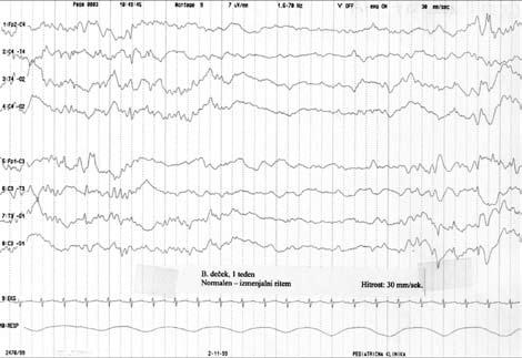
Classical EEG is traditionally based on visual interpretation of various criteria (gestalt) that usually include at least: continuity/discontinuity, amplitude –especially its symmetry and synchronicity, lability to behavioural states and background EEG composition (13). One of the most characteristic features of the neonatal EEG is its discontinuity, where during quiet sleep high-amplitude bursts are intermingled with low-amplitude interburst intervals, giving the typical pattern of tracé alternant. This pattern is physiological
Figure 1. Normal tracé alternant pattern in a one week-old neonate: A period of slow and sharp high-amplitude background activity (BA) followed by low-amplitude BA and again highamplitude BA.
and is observed in all neonates at around 35 weeks of postconceptional age and persists until 44 to 46 weeks of postconceptional age (Figure 1). However, this pattern (especially when higher amplitudes are intermingled) is frequently confused with one of the most pathological patterns, that of of burst-supression, which usually accompanies moderate to severe hypoxic brain insult (Figure 2). In such a situation when it is unclear whether we are dealing with a normal or a severely abnormal pattern, simultaneous video recording can be of great help; in the first instance the neonate (or preterm infant) will be sleeping quietly, while in the pathological state the video can demonstrate a comatose or even convulsing, severely ill newborn. While
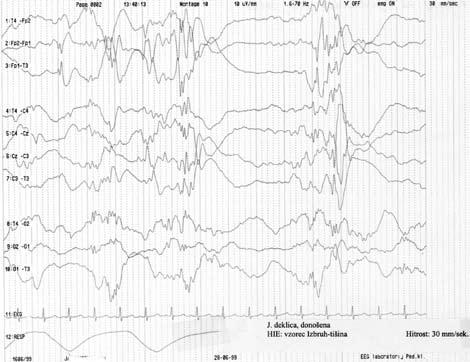
Figure 2. A characteristic pattern of hypoxic-ischaemic encephalopathy in a neonate (so called burst-suppression pattern. In contrast with the physiological pattern of tracé alternant this record presents clear-cut low amplitude background activity (BA) period (in the centre) with bilateral bursts of high-amplitude sharp waves and spike and wave complexes.
D. Neubauer et al.
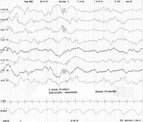
Figure 3. Asymmetrical delta brushes (superimposed fast and sharp rhythms on delta waves) which were considered as normal on this recording, despite the asymmetrical appearance. This neonate has since shown completely normal neurodevelopmental outcome at one year of age.
symmetry of background activity is a must in normal infants and older children, in neonates asymmetry of 20% to 30% is still acceptable, especially when asymmetry of otherwise physiological graphoelements is seen, e.g., asymmetry of delta brushes in preterm infants or asymmetry of frontal sharp transients in fullterm infants (Figure 3). Asynchrony means a timeframe shift of appearance of certain elements, e.g., when sleep spindles in infants appear first over one hemisphere only and then after few seconds also over the other. Some degree of asynchrony is quite acceptable up to the age of 6-12 months. Lability and composition of EEG background activity mean that certain graphoelements will prevail and be characteristic for certain postconceptional ages, e.g. delta brushes in preterm infants of 32-35 weeks postconceptional age. Such visual interpretation can be divided or subdivided into certain categories, such as: normal and abnormal; mildly, moderately and markedly (severely). When serial or multiple EEGs are performed within the first few weeks of life, persistence of such abnormalities not only can confirm the diagnosis of severe brain damage but also may be prognostic of unfavourable outcome. Some authors even distinguish between abnormal patterns that are consistent with cerebral palsy and those that are more often associated with future mental retardation (14). The most important diagnostic role of video EEG during early life is its ability to detect clinical, subtle and EEG-only seizures. It is well documented that the specificity and sensitivity of EEG for seizure detection are at the highest level
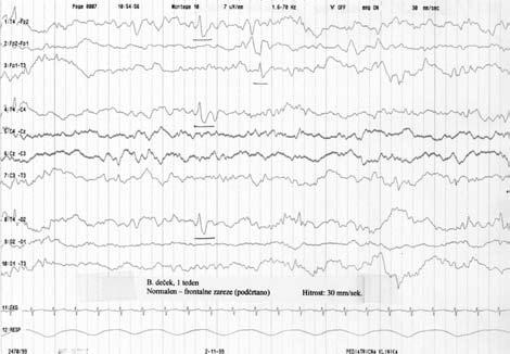
Figure 4. Physiological frontal sharp transients (biphasic sharp waves), which are the typical pattern of a neonate during so-called active sleep. This neonate has normal neurodevelopmental outcome at one year of age.
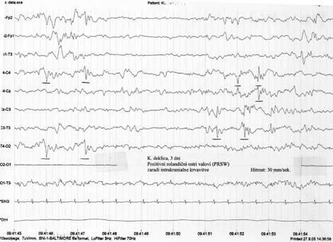
during the neonatal period, with one obligatory provision, i.e., that the EEG recording must be interpreted by an experienced reader of neonatal EEGs. Sometimes it is not difficult to distinguish between physiological graphoelements (e.g. sharp frontal transients) (Figure 4) and clear electrographic spikes (Figure 5). Sometimes graphoelements of certain abnormal conditions can be identified, such as repetitive spikes in the temporal regions in herpetic encephalitis. In neonates, positive rolandic sharp waves represent either intracranial haemorrhage or, even more frequently, periventricular leukomalacia, which may (especially when persisting) be prognostic of future cerebral palsy (Figure 6). Since video, and especially serial video, EEG was introduced into neonatal EEG evaluation, the positive predictive value for seizures
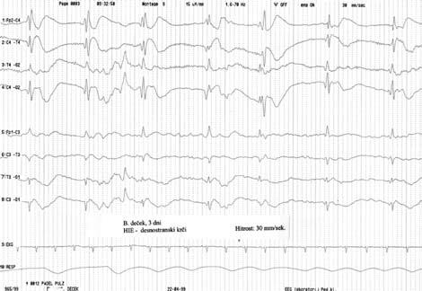
Figure 5. This EEG reveals high-amplitude sharp waves and spikes, localized in the right hemisphere (first four derivations). Clinically, ipsilateral clonic seizures were observed in a neonate with brain hypoxia. Signs of moderate cerebral palsy and left-sided hemiplegia were present in the second year of life.
Figure 6. Characteristic positive (downward) sharp waves over central (rolandic) areas in a female neonate with grade III intracranial haemorrhage (so-called positive rolandic sharp waves - PRSW). Mild cerebral palsy (level I) was observed at the age of two years.
has become even higher because a higher percentage of subtle seizures can be detected and treated accordingly (15). The limited availability of expensive technology and professional staff on the one hand and the technical challenges on the other are among the major reasons why, especially during the last decade, an alternative method has become so popular – that of cerebral function monitoring.
Cerebral function monitoring by amplitudeintegrated electroencephalography (aEEG)
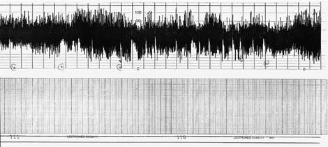
Some authors have found aEEG to be of excellent predictive value for neurodevelopmental outcome in newborn infants with hypoxic-ischaemic encephalopathy (7,10,16,17), especially in the case of moderate and severe hypoxic-ischaemic brain damage. One case series also showed that aEEG can be quite a reliable tool for assessment of the risk for brain damage in newborn infants without need for ventilatory support (18). In a recent report by Shany et al., which included data from, besides their own study, four other similar and comparable studies, it is confirmed that significantly abnormal patterns, such as flat tracings or very low voltage and the so-called burst-suppression pattern are highly predictive for later adverse neurodevelopmental outcome (10). The positive predictive value was 69-86%, while sensitivity was 75-100%. The interpretation of aEEG is simple and reliable and it has been shown to be well correlated between different profiles of readers (e.g., medical students, NICU nurses, child neurologists). It is of utmost importance, however, that cerebral function monitoring is performed during the early neonatal period, and is best within the first 3-6 hours of life (19). Monitoring as-
phyxiated infants with aEEG (and possibly also by standard EEG or a combination of both) is becoming an accepted practice in many NICUs and is recognized by some authors as probably the best, or at least a very important, tool for selecting patients for various neuroprotective studies. It is also proving to be an excellent tool for identifying those newborn infants with a potentially good outcome, which may help to reduce the anxiety of their parents at an early stage (10). The use of aEEG has also been found to be excellent for detecting seizures, especially those which are clinically silent. EEG seizures are common in severelyill newborn infants and aEEG may provide important information concerning the neurological status of these babies and may help to confirm or refute the presence of seizures in clinically suspected cases and detect infants with silent seizures (11,12). It has been shown that the early detection and treatment of clinical, subtle or EEG-recorded neonatal seizures by aEEG is of great importance, as in the series under study the incidence of postneonatal epilepsy after early institution of treatment was only 9.4%, which is lower than previously reported (9). We have also shown recently that aEEG can be applied successfully for other conditions in sick newborn infants in the NICU, e.g. for those neonates who are treated by extracorporeal membrane oxygenation, because aEEG in such cases will provide additional data for better treatment of
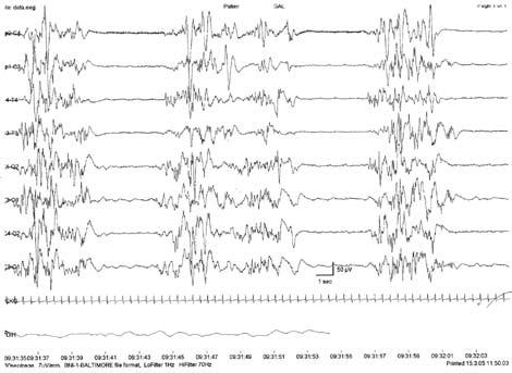
Figure 7. Burst-suppression pattern suspected by aEEG (a) and confirmed by standard EEG (b).
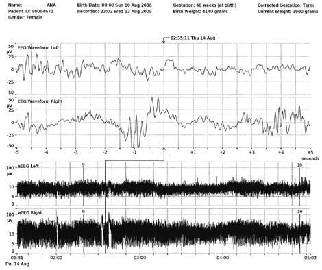
such infants (11). Finally, according to both our experience and literature reports (3,4,6,20), the best results can be achieved by a combination of aEEG and standard EEG. Some newer aEEG monitors have already incorporated both techniques in one machine (e.g. NicoletOne, BrainZ) and even allow the option of recording multiple EEG channels for better seizure detection. In those instances where from the aEEG it is not completely clear that a seizure has occurred, the concomitant EEG tracing can clearly confirm it (Figure 7a and b, Figure 8). This new generation of aEEG machines may achieve improved seizure detection in suspected cases and may increase the ability to detect seizure activity in high-risk neonates and in those infants where the differentiation between possible subtle seizures and other paroxysmal (non-convulsive) events was formerly difficult. In the near future it will be possible to confirm whether it is really important to treat as early as possible, not only clinically recognizable seizures, but also those subtle seizures with subclinical symptomatology. It is hoped that future guidelines on the management of neonatal seizures will also provide the impetus for improved pharmacotherapeutic intervention for sick newborn infants in this field.
Conclusion
The early and reliable determination of prognosis in neonates with acute encephalopathy is extremely important for all persons caring for these infants, and may in favourable cases help to reduce the anxiety of parents at an early stage. It is acknowledged that early recognition of clinical – and most probably also subtle
– seizures in neonates is imperative, because it will ensure early intervention which, as has been shown, may substantially decrease future epilepsy. Both standard EEG, digital and coupled with video, and aEEG have already demonstrated their capacity to predict neurological outcome and postneonatal epilepsy in infants with hypoxic-ischaemic encephalopathy. The recognition that both techniques have shortcomings has prompted the development of a new generation of recorders that display simultaneously aEEG and raw EEG and we believe that these will further increase our management options for severely ill newborn infants in the NICU.
Part II
Continuous monitoring of cardiorespiratory functions (CMCrf)
Many abnormal neurological and behavioural symptoms have been observed in infants and children with disordered breathing during sleep. Among the best studied of these are: declining school performance, morning headaches, mood changes, nocturnal enuresis, attention deficit – hyperactivity disorder, learning disabilities, aggressiveness and strange behaviour. Improvement has been reported following successful treatment of upper airway obstruction, suggesting a clear link between abnormal breathing during sleep and these neurobehavioural and cognitive problems (21-24). It is still not known if there is long-term impact on behaviour from such early respiratory dysfunction. Recently it has been shown that caffeine therapy for apnoea of prematurity improves the rate of survival without neurodevelopmental disability at 18 to 21 months in infants with very low birth weight (VLBW) (25). The fact that methylxanthines are the treatment of choice for reducing the frequency of apnoeic episodes in premature infants was recognized more than three decades ago, and the so called “caffeination of prematurity” has become popular because of the long half-life of caffeine in the neonate (26,27), although the mechanism is not fully understood. The most probable answers are primary stimulation of the respiratory centre, adenosine receptor blockade and improved function of the respiratory muscles (28). This article will deal with the definition, pathophysiology, aetiology, classification, diagnostic procedures and management of sleep disordered breathing (SDB), especially during early life. Some syndromes of early cardiorespiratory dysfunction are discussed briefly, and some of the authors’ experience with cardiorespiratory studies since the 1980s, when the use of cardiorespiratory diagnostic
D. Neubauer
Figure 8. Silent seizures are represented on CFM tracing as incisures (lower trace), while on EEG signal clear-cut spike and wave graphoelements are recorded (upper trace).
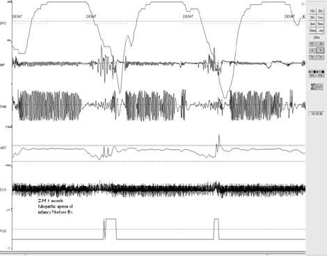
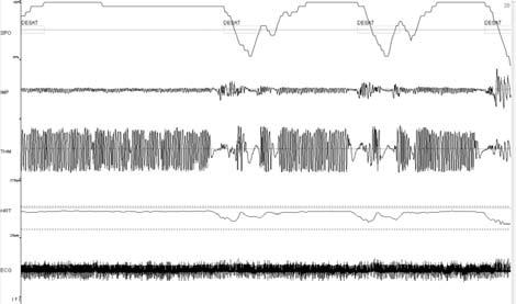
Figure 9. Typical recording of continuous monitoring of cardiorespiratory functions (CMCRF) in a one month-old infant with a typical pattern of idiopathic apnoea: the oxygen desaturation lasts as long as the apnoea (best represented at nasal level, some unsuccessful respirations are present in the thoracic breathing).
devices was introduced in the department, are presented.
The definition of SDB refers mainly to obstructive sleep apnoea syndrome (OSAS) in children, although studies in neonates and infants who experienced an apparent life-threatening event (ALTE) have shown that in many instances no clear-cut differentiation can be made between obstructive and central apnoea during sleep, especially regarding the long term consequences. There is also growing evidence that central and obstructive apnoea can have common aetiologies. It is well recognized that pharyngeal occlusion often occurs during central apnoea. If arousal does not occur, central apnoea may progress to a mixed apnoea. In addition, episodes of central apnoea with associated fluctuations in chemical drive to the brainstem structures can lead to central respiratory system oscillations, which may in turn predispose to obstructive apnoea. Cases in adults have been reported where upper airway narrowing led to central apnoea, and a similar situation has been described in infants and children with micro- and retrognathia.
OSAS in children is defined as a “disorder of breathing during sleep characterized by prolonged partial upper airway obstruction and/or intermittent complete obstruction (obstructive apnoea) that disrupts normal ventilation during sleep and normal sleep patterns” (29). Obstructive sleep apnoea is the most common category of SDB. Mild, occasional sleep apnoea, such as many children experience during an upper respiratory infection, may not be important, but chronic, severe obstructive sleep apnoea requires treatment in order to prevent low blood oxygen
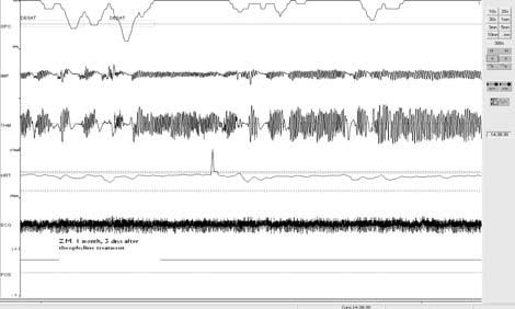
10. The infant in Figure 9, after theophylline treatment, when the pattern became completely normalized.
levels (hypoxaemia), sleep deprivation, and other complications, the most serious of which is a severe form of congestive heart failure or cor pulmonale
ALTE, an “apparent life-threatening event”, is a poorly defined term to describe an acute, unexpected change in an infant's breathing behaviour that is frightening to the infant's carer and includes some combination of the following features (30):
- Apnoea: usually no respiratory effort (central), or sometimes effort with difficulty (obstructive)
- Colour change: usually cyanotis or pallor, but occasionally erythema or plethora
- Marked change in muscle tone: usually limpness, more rarely rigidity
- Choking or gagging.
In some cases, the observer fears that the infant has died. Recovery occurs only after stimulation or resuscitation. However, an episode is often mislabelled as “ALTE” even when a parent reports that the child resumed normal breathing after simply being picked up and patted. Following early anecdotal reports of deaths from sudden infant death syndrome (SIDS) in infants with recurrent apnoea, enormous amounts of attention, research, and clinical resources were focused on the problem of ALTE in infants. Although various cardiorespiratory, autonomic, and neurophysiologic differences have been demonstrated in infants with ALTE as a group, these findings have not distinguished individual infants with ALTE from normal controls or provided premortem markers for the risk of SIDS (31). Infants with ALTE represent an extremely heterogeneous group of patients of varying ages with diverse pathophysiology.
The pathophysiology of SDB in infants and children remains poorly understood, but clinical studies have revealed distinctive differences between children and adults, the most striking of which are:
1. Infants are obligate nasal breathers, and only
Figure
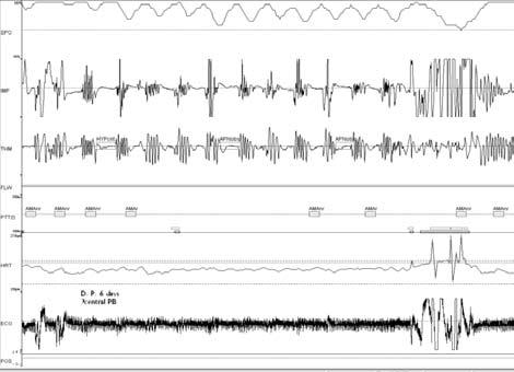
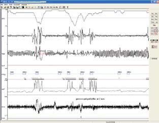
Figure 11. A 6 day-old neonate with extreme periodic breathing. It is clear that the central component (breathing on thoracic) is more obvious than breathing through the nose. It can also be seen that so called ventilation/apnoea ratio is below 1.0, in this case even below 0.5.
breathe through the mouth when crying or if the nose is obstructed.
2. Infants and children appear to have more severe clinical problems and consequences associated with milder forms of OSAS. This may be due to significant desaturation which occurs even with brief episodes of apnoea, because of the faster respiratory rate and smaller functional residual capacity in infants and children.
3. Infants and children have less cortical arousals in response to OSAS, because of their higher threshold, which decreases with increasing age.
4. Children may show a pattern of persistent partial upper airway obstruction causing hypercapnia and/or hypoxaemia, which has been termed obstructive hypoventilation
5. In infants and children OSAS occurs predominantly during the rapid-eye-movement (REM) stage of sleep, while it is quite normal that no obstruction is observed in the other stages of sleep. These REM-related obstructive apnoeas are usually longer and more numerous, and therefore also more troublesome, during the later REM periods (i.e., during the later phases of sleep, towards the early morning time).
The aetiology of OSAS in infants and children is multifactorial, and often it results from a combination of structural and neuromotor factors (22,23, 27,28). Among the structural factors are adenotonsillar hypertrophy, which is by far the commonest condition associated with childhood OSAS (radiological assessment, however, does not differentiate children with OSAS, who present major obstruction only during sleep, with no correlation between upper airway/ adenotonsillar size and OSAS), and craniofacial ab-
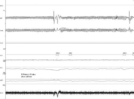
Figure 12. The neonate in Figure 11: Although the newborn infant exhibited no oxygen desaturation, caffeine was adminstered and immediately the pattern of periodic breathing disappeared.
normalities (some children with severe craniofacial dysmorphism require tracheostomy soon after birth because of fixed upper airway narrowing, but less affected neonates, infants and children may develop upper airway obstruction only during sleep). OSAS has been reported in a large number of dysmorphic syndromes, mainly in those with nasal obstruction and midfacial hypoplasia, micro- and retrognathia, macroglossia or in combination with obesity and/or hypotonia. The representatives of these abnormalities are genetic syndromes such as CHARGE, Goldenhar, Pierre-Robin, Pickwick and Prader-Willi. Obese children tend to have narrow upper airways due to both the deposition of adipose tissue within the muscles and soft tissue surrounding the airway and external compression from the neck and jaws. Many obese children also have restrictive lung disease which contributes to hypoxaemia, and they show decreased central ventilatory drive. Even in obese children, however, adenoidectomy and/or tonsillectomy will improve or even eliminate OSAS.
Among the neuromotor factors the most important is the so-called central ventilatory drive which, in a subtle abnormal form, is most probably present also in all children with OSAS (it is recognized that adult patients with OSAS have decreased ventilatory drive in response to hypercapnic challenge). Some authors speculate that the abnormality in hypercapnic ventilatory drive may be related to nocturnal hypoxia and/or hypercapnia or vice vers a. Other factors that influence neuromotor control can be predominantly “peripheral”, affecting mostly upper airway muscles (e.g., in children with neuromuscular disorders) or “central”, affecting mainly neuromotor coordination (e.g., in children with cerebral palsy and other encephalopathies) or a mixture of
D. Neubauer et al.
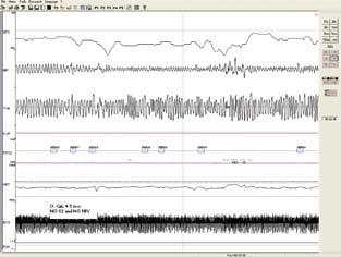
Figure 13. A 3 month-old infant with mixed apnoea and desaturation. The pattern resembles that in Figure 14, but the obstructive component is more obvious. The condition resolved completely after theophylline treatment for a few months.
both (e.g., in progressive neurodegenerative diseases). Several other factors can play an important role, including genetic predisposition, hormonal factors (the role of testosterone may be the reason for the increased incidence of OSAS in male children) and drugs (mainly those influencing or directly depressing upper airway muscle tone, such as sedatives, general anaesthetics and even some drugs used for sleep induction in infants and children for neurophysiological diagnostic studies, such as chloral hydrate).
The classification of SDB includes OSAS appearing in normal children and those problems of breathing during sleep that are commonly encountered in certain syndromes. Among the former are: primary snoring, which is a benign condition with no physiological abnormalities, and which requires no diagnostic and/or therapeutic management; upper airway resistance syndrome (UARS), which is a variant of classical OSAS characterized by a combination of increased upper airway resistance and breathing effort during sleep, snoring, frequent microarousals from sleep and daytime sleepiness and/or diminished neurocognitive functioning; obstructive hypoventilation (OH), which is partial upper airway obstruction characterized by increased breathing effort, snoring and high end-tidal CO2 without obvious lung disease; and classical OSAS, which is characterized by prolonged partial or total airway obstruction resulting in hypoxaemia, marked snoring, disturbed sleep and abnormal daytime symptoms and signs.
Among the syndromes (32-35) affecting respiratory control during sleep, that which has been by far the most intensively studied is congenital central hypoventilation syndrome (CCHS) or Ondine's curse, affecting infants (and children as well as some adults) who breathe completely normally during the waking
Figure 14. An infant of 2 months admitted because of an apparent life threatening event (ALTE) and proved to have obstructive hypopnoea with desaturation due to gastro-oesophageal reflux, which improved after preventive measures (semi-sitting position, special milk formula), and no more episodes of hypopnoea were observed.
state but severely hypoventilate or are even apnoeic during sleep (usually in phase 2 of non-REM sleep). The most probable reason is abnormal control of breathing due to a dysfunction in the central and/or autonomic nervous system. It is considered to be one of the human neurocristopathies (36). Genetic studies have confirmed a genetic marker in some children and families with CCHS (PHOX2B gene), although it is unlikely to be due to a simple inherited pattern involving only one gene, and the risk of recurrence in subsequent children in the same family is generally low (37), but in our department we have encountered a sister and brother, both with genetically confirmed CCHS. Another well documented syndrome with breathing difficulties, particullary during sleep, is the Arnold-Chiari malformation, where myelomeningocoele is combined with hydrocephalus and often causes herniation (displacement through the foramen magnum) of the medulla oblongata and cerebellum. This structural abnormality affects the brainstem respiratory centres and the neurological control of breathing. Abnormalities of the cardiorespiratory pattern during sleep are central sleep apnoea, hypoventilation and oxygen desaturation. Such abnormalities are easily recognized with simple two-channel pneumograms, which have proved to be a reliable diagnostic tool for detection of abnormal patterns and thus permit fast and easy recognition of those infants and children who are in need of further intervention (either surgical, i.e., shunting of hydrocephalus and/or posterior fossa decompression, pharmaceutical, with medications such as acetazolamide, theophylline, etc.). In some infants and
children long-term ventilation support will be needed also. All such children should have long-term home monitoring of cardiorespiratory function as sudden deaths, presumably due to apnoea, have been documented. Among other well documented (some genetically determined) syndromes causing problems of breathing during sleep of a greater or lesser severity are: Prader-Willi syndrome (deletion of the long arm of chromosome 15), achondroplasia and some of the mucopolysaccharidoses, Leigh's disease (mitochondrial ATP-ase deficiency), Joubert syndrome (agenesis of the cerebellar vermis), acquired central hypoventilation syndrome (e.g. from brain tumour, and after encephalitis or trauma, congenital vascular malformation) and other rare childhood syndromes where respiratory control can be disturbed, especially during sleep (Rett and Angelman syndrome).
The diagnostic approach usually depends on factors influencing upper airway resistance and/or compliance and factors influencing neuronal control (3640). In addition to a careful medical history (including extensive search for possible similarly affected family members), sleep history should be included. A special questionnaire completed by the parents can be very helpful for the recognition of possible subtle symptoms (this may include different clinical scoring systems which can also be useful for primary care providers – a child with clearly negative score is very unlikely to have significant breathing disturbances during sleep). In our department we use the adapted form of a questionnaire for OSAS children and the so-called taxonomy profile for ALTE cases. Physical examination should include careful evaluation of the upper airways (best performed by an experienced ear, nose and throat specialist), search for possible signs of
chronic hypoxia (digital clubbing is a rare consequence of OSAS) and craniofacial abnormalities and dysmorphic features. Among the neurophysiological techniques, the most important are polysomnography (PSG) and respiratory function monitoring (i.e., cardiorespirography, event-recording of oxycardiorespirographic functions, pulse oxymetry, end-tidal or transcutaneous CO2, pulse transit time analysis, capnography), diaphragmatic electromyography (EMG), oesophageal balloon, actimetry and chinEMG. The most frequently used techniques are those that use chest/abdominal impedance (strain gauge) and thermistors for nasal airway detection to differentiate obstructive from central apnoea. This can sometimes be problematic if the thermistor is not well applied, but with impedance obstruction is easy to distinguish since paradoxical chest and abdominal movements can be identified (so-called “out of phase” movements) and PSG should be used as the next diagnostic step (41).
PSG is considered to be the gold standard for diagnosis of SDB but it is expensive and very time and labour intensive (41-44). It is especially important either in cases of non-classical OSAS, such as upper airway resistance syndrome and obstructive hypoventilation (or obstructive hypopnoea) or where the sleep stage is important for establishing the diagnosis (e.g., in CCHS). In children, despite many unsolved questions and limitations, PSG is at present considered to be the diagnostic study of choice for suspected abnormal breathing during sleep. The recommendation of American Thoracic Society is that PSG is indicated in all children in whom diagnosis of OSAS cannot be made by direct observation of breathing during sleep (29). However some of the combined cardiorespiratory monitors can serve as supplementary devices in
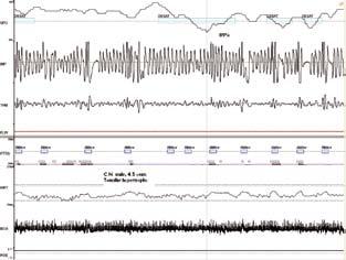
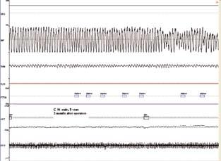
Figure 16. The boy in Figure 15, 6 months after tonsillectomy, when the breathing problems had disappeared completely.
Figure 15. A 4½ year-old boy with snoring and pallor during sleep, with quite frequent episodes of obstructive hypopnoea with desaturation. Tonsillar hypertrophy was present.
Figure 17. A boy of 4 months of age in whom central hypoventilation was suspected (breathing at the level of the chest was of lower amplitude than that at the nose) with low saturations during calm sleep and high values of pCO 2 measured by capnography.
those cases where PSG is not readily available and where continuous cardiorespiratory monitoring is needed (especially in the home or at the bedside in the children's department). These devices are also suitable in cases where frequent follow-up of the cardiorespiratory pattern is necessary, especially when different treatment regimes need to be evaluated (e.g., following adenotosillectomy or neurosurgical procedures, or after introducing treatment with various respiratory stimulants, e.g., theophylline) and when chronic ventilatory support is needed at home and breathing patterns have to be re-evaluated (42,43). Such devices usually offer continuous respiratory monitoring: abdominal and chest wall movements are detected by inductive plethysmography or strain gauges; airflow by either nasal/oral thermistors or nasal pressure sensors; ventilation by end-tidal CO 2, oxygenation by pulse oximetry and heart rate by ECG or beat-to-beat evaluation. Certain other functions may also be associated and are offered optionally, such as: non-invasive blood pressure measuring/monitoring, temperature monitoring, body movement detection by actimetric method, one or two channels of EEG (for approximation of sleep stage and/or detection of continuous function monitoring by amplitude change measurements), chin-EMG, capnography and others. With these techniques different abnormal patterns of respiration can be clearly recognized, classified and staged according to their severity. Usually differentiation can be made between: obstructive apnoea (absence of airflow in the presence of confirmed chest wall movements lasting longer than 2 respiratory cycle times, with or without significant hypoxaemia), central ap-
noea (absence of respiratory effort lasting more than 2 respiratory cycle times), hypopnoea (50% or more decrease in the amplitude of baseline nasal airflow signal, often accompanied by hypoxaemia and/or arousal. This term is frequently used in discussing paediatric sleep disorders and may be particularly useful when pCO2 is not being measured, since the reduction in effort or flow may be used as an indicator of hypoventilation and thus a possible marker of upper airway resistance syndrome, which can otherwise be recognized only by means of PSG); hypopnoea can be further classified into obstructive and non-obstructive types, hypoxaemia (baseline sleeping oxygen saturation <90% or one dip in the percentage of oxygen saturation of at least 10% below baseline per hour), and hypercapnia (end-tidal CO2 readings greater than 45 mm Hg have been associated with complications).
These techniques are particularly valuable in newborn infants in whom PSG is difficult to apply, and because newborns have a predominantly central type of SDB which can easily be detected by simple pneumographic techniques. However possible associated hypoxaemia should always be detected by pulse oxymetry. During infancy many patients experience ALTEs, which also are easily detected by cardiorespirographic techniques. In these cases usually an instant decision can be made about further investigation and/or management. It is very convenient that solutions can be devised soon after admission at the bedside on the basis of such cardiorespiratory functional evaluation. In older children these techniques can be very suitable for use either at home or in the general paediatric ward as a screening tool, before the use of more complicated techniques such as PSG. These techniques also present potential for development of a homebased video recording system as an effective way of screening for SDB and reducing the number of children who need full PSG evaluation.
The management of SDB problems in infants and children depends on a variety of factors involved in this condition and upon age. Not all infants and children with various cardiorespiratory abnormalities will need some sort of treatment. It is not known whether any episode of desaturation is harmful, nor what is the consequence of increased breathing effort and hypercarbia. Some experimental evidence even suggests that mild to moderate hypercapnia can improve tissue oxygenation and perfusion, which may ameliorate injury, especially to the immature lung and brain, but hypercapnia may also be associated with adverse outcome and the range of PaCO2 levels that are both safe and effective – especially in neonates – has yet to be determined (44).
At the present time, with the exception of those neonates and infants who have prolonged central apnoea with/without severe hypoxaemia and children with severe apnoea and nocturnal hypoxaemia and/or daytime abnormalities there are no clearcut guidelines for decision-making regarding the treatment. The decision to treat or to monitor clinically should be based on a combination of clinical findings and cardiorespiratory function tests and/or PSG results (45).
Recently, because of very promising outcomes in neonates from caffeine treatment (25), the use of xanthines during early life has again become quite popular. At the other end of the spectrum, severe cases have been quite “comfortably” managed by non-invasive ventilation techniques (46). We are currently managing, very successfully, three infants, all less than 10 months of age, with non-invasive ventilation, one with CCHS and two with spinal muscular atrophy.
A multiparametric study of data from the history, clinical signs and neurophysiological findings should permit delineation of the main features of importance for the differential diagnosis of SDB in infants and children. Meanwhile, for improving abnormal daytime symptoms and signs when they emerge in otherwise normal children, adenotonsillectomy has been shown to be the most effective therapeutic intervention (45,47).
Conclusion
Continuous monitoring of cardiorespiratory functions is one of the oldest forms of monitoring in the IC setting and in the units dealing with neonates, infants and small children with different cardiorespiratory problems. After the neonatal period, when central-drive respiratory problems are the most common (especially apnoea of prematurity), the obstructive causes of SDB become progressively more and more evident. During the infantile period many ALTEs may occur, the majority of which are quite benign in origin, often caused by simple gastro-oesophageal reflux, or are the consequence of some abnormality of the upper airways, usually due to a craniofacial syndrome. Later, during the preschool period, tonsillar hypertrophy can be the leading cause of SDB, as well as giving rise to daytime problems, and must be recognized and treated early. However it should be remembered that during this early period of development some rare causes of cardiorespiratory abnormalities may present, which must be identified and treated appropropriately to prevent the sudden development of severe life-threatening conditions.
references
1. Jeras J, Tivadar I. Epilepsies in children. New York: University Press of New England, 1973.
2. al Naqeeb N, Edwards AD, Cowan FM, Azzopardi D. Assessment of neonatal encephalopathy by amplitude-integrated electroencephalography. Pediatrics 1999;103:1263-1271.
3. Rosén I. The physiological basis for continuous electroencephalogram in the neonate. Clin Perinatol 2006;33: 593-611.
4. Hellström-Westas L, Rosén I, de Vries LS, Greisen G. Amplitude-integrated EEG classification and interpretation in preterm and term infants. Neoreviews 2006;7:e72-e87.
5. Osredkar D. EEG method of ampliude integration in neonatal neurology (in Slovenian language, English abstracts). PhD Thesis. Ljubljana: Medical Faculty, University of Ljubljana, 2006.
6. Shellhaas RA, Gallagher PR, Clancy R. Assessment of neonatal electroencephalography (EEG) background by conventional and two amplitude-integrated EEG classification systems. J Pediatr 2008;153:369-374.
7. Toet MC, Hellström-Westas L, Groenendaal F, Eken P, de Vries LS. Amplitude-integrated EEG 3 and 6 hours after birth in full-term neonates with hypoxic-ischaemic encephalopathy. Arch Dis Child Fetal Neonatal Ed 1999; 81:19-23.
8. van Rooij LGM, Toet MC, Osredkar D, van Huffelen AC, Groenendaal F, de Vries LS. Recovery of amplitude integrated electroencephalographic background patterns within 24 hours of perinatal asphyxia. Arch Dis Child Fetal Neonatal Ed 2005;90:F245-F251.
9. Toet MC, Groenendaal F, Osredkar D, van Huffelen AC, de Vries LS. Postneonatal epilepsy following amplitude-integrated EEG-detected neonatal seizures. Pediatr Neurol 2005;32:241-247.
10. Shany E, Goldtsein E, Khvatskin S, Friger MD, Heiman N, Goldstein M, Karplus M, Galil A. Predictive values of amplitude-integrated electroencephalography pattern and voltage in asphyxiated term infants. Pediatr Neurol 2006; 35:335-342.
11. Grosek Š, Osredkar D, Derganc M. Neubauer D, Primožič J. Detection of seizures with amplitude-integrated electroencephalography in a neonate treated with extracorporeal membrane oxygenation. Chin J Contem Pediatr 2008;10: 547-551.
12. Shany E, Khvastkin S, Golan A, Karplus M. Amplitude-integrated electroencephalography: A tool for monitoring silent seizures in neonates. Pediatr Neurol 2006;34:194-199.
13. Clancy RR, Bergvist AGC, Dlugos DJ. Neonatal electroencephalography. In: Ebersole J, Pedley T, editors. Current practice of clinical electroencepahlography. 3rd edition. Philadelphia: Lippincot Williams & Wilkins, 2003; pp. 160-234.
14. Hayakawa F, Okumura A, Kato T, Kuno K, Watanabe K. Determination of timing of brain injury in preterm infants with periventricular leukomalacia with serial neonatal electroencephalography. Pediatrics 1999;104:1077-1081.
15. Khan RL, Lahorgue Nunes M, Garcias da Silva LF, da Costa JC. Predictive value of sequential electroencephalogram (EEG) in neonates with seizures and its relation to neurological outcome. J Child Neurol 2008;23:144-150.
16. Shalak LF, Laptook AR, Velaphi SC, Perlman JM. Amplitude-integrated electroencephalography coupled with an early neurologic examination enhances predicition of term infants at risk for persistent encephalopathy. Pediatrics 2003; 111:351-357.
D. Neubauer
17. Hellström-Westas L, Rosen I, Svenningsen NW. Predicitve value of early contonuous amplitude integrated EEG recordings on outcome after severe brain asphyxia in fullterm infants. Arch Dis Child 1995;72:F34-38.
18. Osredkar D, Derganc M, Paro-Panjan D, Neubauer D. Amplitude-integrated electroencephalography in full-term newborns without severe hypoxic-ischemic encephalopathy: case series. Croat Med J 2006;47:285-291.
19. Osredkar D, Derganc M, Paro-Panjan D, Neubauer D. Continuous cerebral function monitoring in the newborn: comparison with standard EEG. Proceedings XXV Alpe Adria Meeting of Perinatal Medicine. Maribor: Splošna bolnišnica, 2003; pp. 119-224.
20. Shellhaas RA, Clancy RR. Characterization of neonatal seizures by conventional EEG and single-channel EEG. Clin Neurophysiol 2007;118:2156-2161.
21. Guilleminaut C, Pelayo R, Clerk A, Bocian RC. Recognition of sleep-disordered breathing in children. Pediatrics 1996;98:871-882.
22. Guilleminaut C, Winkle R, Korobkin R, Simmons B. Children and nocturnal snorring: evaluation of the effects of sleep related respiratory resistive load and daytime functioning. Eur J Pediatr 1982;139:165-171.
23. Guilleminaut C, Korobkin R, Winkle R. A review of 50 children with obstructive sleep apnea syndrome. Lung 1981; 159:275-287.
24. Neubauer D. Sleep disordered breathing in infants and children. Pediatrics Update 2000; (special issue):22-24.
25. Schmidt B, Roberts RS, Davis, P et al. Long-term effects of caffeine therapy for apnea of prematurity. N Engl J Med 2007;357:1893-1902.
26. Kuzemko JA, Paala J. Apnoeic attacks in newborn treated with aminophylline. Arch Dis Child 1973;48:404-406.
27. Stevenson DK. On the caffeination of prematurity. N Engl J Med 2007;357:1967-1968.
28. Lopes JM, Aranda JV. Pharmacologic treatment of neoanatl apnea. In: Yaffe SJ, Aranda JV, eds. Neonatal and pediatric pharmacology. 3rd ed. Philadelphia: Lippincott Williams & Wilkins, 2005; pp. 217-229.
29. American Thoracic Society. Standards and indications for cardiopulmonary sleep studies in children. Am J Respir Crit Care Med 1996;153:866-878.
30. National Institutes of Health Consensus Development Conference on Infantile Apnea and Home Monitoring. Pediatrics 1987;79:292.
31. Brooks JG. Apparent life-threatening events and apnea of infancy. Clin Perinatol 1992;19(4):809-838.
32. Gaultier C. Obstructive sleep apnea syndrome in infants
and children: established facts and settled issues. Thorax 1995;50:1204-1210.
33. Brouillette RT, Fernbach SK, Hunt CE. Obstructive sleep apnea in infants and children. J Pediatr 1982;100:31-40.
34. Marcus CL, Curtis S, Koerner CB, Joffe A, Serwint JR, Loughlin GM. Evaluation of pulmonary function and polysomnography in obese children and adolescents. Pediatr Pulmonol 1996;21:176-183.
35. Guilleminaut C, Partinen M, Praud JP, Quera-Salva MA, Powell N, Riley R. Morphometric facial changes and obstructive sleep apnea in adolescents. J Pediatr 1989;114: 997-999.
36. Etchevers HC, Amiel J, Lyonnet S. Molecular bases of human neurocristopathies. Adv Exp Med Biol 2006;589: 213-234.
37. Gaultier C, Amiel J, Dauger S, Trang H, Lyonnet S, Gallego J, Simonneau M. Genetics and early disturbances of breathing control. Pediatr Res 2004;55(5):729-733.
38. Gozal D, Arens R, Omlin KJ, Ben-Ari G, Aljadeff G, Harper RM, Keens TG. Ventilatory response to consecutive short hypercapnic challenges in children with obstructive sleep apnea. J Appl Physiol 1995;79:1608-1614.
39. Guilleminaut C, Partinen M, Hollman K, Powell N, Stoohs R. Familial aggregates in obstructive sleep apnea syndrome. Chest 1995;107:1545-1551.
40. Severinghaus J, Mitchell RA. Ondine's curse - failure of respiratory center automaticity while awake. Clin Res 1962; 10:122.
41. Elder DE, Russell L, Sheppard D, Purdie GL, Campbell AJ. Car seat test for preterm infants: comparison with polysomnography. Arch Dis Child Fetal Neonatal Ed 2007;92(6): F468-472.
42. Carroll JL, Loughlin GM. Diagnostic criteria for obstructive sleep apnea in children. Pediatr Pulmonol 1992;14:71-74.
43. Standards and indications for cardiopulmonary sleep studies in children. Am J Respir Crit Care Med 1996;153: 866-878.
44. Jankov RP, Tanswell AK. Hyperkapnia in a neonate. Acta Paediatrica 2008;97:1502-1509.
45. Brouillette RT, Jacob SV, Waters KA, Morielli A, Mograss M, Ducharme FM. Cardiorespiratory sleep studies for children can often be performed at home. Sleep 1996;19: S278-S280.
46. Wormald R, Naude A, Rowley H. Non-invasive ventilation in children with upper airway obstruction. Int J Pediatr Otorhinolaryngol 2009, Jan 12 [Epub ahead of print].
47. Fauroux B, Aubertin G, Clément A. What's new in paediatric sleep in 2007? Paediatr Respir Rev 2008;9(2):139-143.
1st Paediatric Clinic, Children’s General Hospital of Pendeli
Correspondence:
Eleana X. Stefanopoulou estefanopoulou@yahoo.gr
1st Paediatric Clinic, Children’s General Hospital of Pendeli 152 36, P. Pendeli, Greece
The metabolic syndrome in children
E.X.
Stefanopoulou,
N.E. Myriokefalitakis
Abstract: Metabolic Syndrome (MS) reflects a wide array of factors including adiposity, dyslipidaemia, hypertension, hyperinsulinaemia, impaired glugose metabolism, abnormalities in fibrinolysis and inflammation. Due to the increasing prevalence of childhood obesity worldwide, concern about the MS in children has skyrocketed. Clustering of these factors in children and adolescents identifies a patient population at high risk for future development of cardiovascular disease (CVD) and type 2 diabetes mellitus (type 2 DM). Components of the juvenile MS are often associated with their presence during adulthood. Prevalence of MS in obese pediatric patients in the USA ranges between 18% and 50% due to the lack of consensus regarding the definition of MS. Insulin resistance, which is strongly related to adiposity, plays a key role in pathogenesis of MS. Adipose tissue has a major endocrine function by expressing and secreting many hormones as well as other factors called adipokines. The release of adipokines is altered in obesity and these abnormalities are associated with the features of MS. Factors such as genetic influences and effects of foetal and infant environment predispose to the development of the MS. In conclusion, MS in children is a controversial and complex issue with many unanswered questions. Without doubt, prevention and treatment of childhood obesity should be the first line approach to this problem.
Key words: Metabolic syndrome, insulin resistance, obesity, adipokines, cardiovascular risk.
1. Fox R. Overweight children [Editorial]. Circulation 2003; 108:e9071.
2. Must A. Does overweight in childhood have an impact on adult health? Nutr Rev 2003;61:139-142.
3. St-Onge MP, Heymsfield SB. Overweight and obesity status are linked to lower life expectancy. Nutr Rev 2003;61: 313-316.
4. Reaven GM. Banting lecture 1988. Role of insulin resistance in human disease. Diabetes 1988; 37:1595-607.
5. World Health Organization: Definition, Diagnosis, and Classification of Diabetes Mellitus and its Complications: Report of a WHO Consultation. Geneva, World Health Organization, 1999.
6. Alberti KG, Zimmet P, Shaw J. The metabolic syndrome--a new worldwide definition. Lancet 2005;366:1059-1062.
7. Expert Panel on the Detection, Evaluation, and Treatment of High Blood Cholesterol in Adults: Executive summary of the Third Report of the National Cholesterol Education Program (NCEP) Expert Panel on Detection, Evaluation, and Treatment of High Blood Cholesterol in Adults (Adult Treatment Panel III). JAMA 2001;285:2486-2497.
8. Weiss R, Dziura J, Burgert TS, Tamborlane WV, Taksali SE, Yeckel CW, et al. Obesity and the Metabolic Syndrome in children and adolescents. N Engl J Med 2004;350:23622374.
9. Cook S, Weitzman M, Auinger P, Nguyen M, Dietz WH. Prevalence of metabolic syndrome phenotype in adolescents: findings from the third National Health and Nutrition Examination Survey, 1988-1994. Arch Pediatr Adolesc Med 2003;157:821.
10. Chi CH, Wang Y, Wilson DM, Robinson TN. Definition of metabolic syndrome in Preadolescent girls. J Pediatr 2006;148:788.
11. Zimmet P, Alberti G, Kaufman F, Tajima N, Silink M, Arslanian S, et al; International Diabetes Federation Task Force on Epidemiology and Prevention of Diabetes.The metabolic syndrome in children and adolescents. Lancet 2007; 369:2059-2061.
12. Caprio S. Insulin Resistance in childhood obesity. J Pediatr Endocrinol Metab 2002;15 (Suppl 1):487-492.
13. Miller J, Rosenbloom A, Silverstein J. Childhood Obesity. J Clin Endocrinol Metab 2004;89(9):4211-4218.
14. Hauner H. Secretory factors from human adipose tissue and their functional role. Proc Nutr Soc 2005;64:163-169.
15. Fried SK, Bunkin DA, Greenberg AS. Omental and subcutaneous adipose tissues of obese subjects release interleukin-6: depot difference and regulation by glycocorticoid. J Clin Endocrinol Metab 1998;83:847-850.
16. Koerner A, Kratzsch J, Kiess W. Adipocytokines: leptin-the classical, resistin-the controversial, adiponectin-the promising, and more to come. Best Pract Res Clin Endocrinol Metab 2005;19:525-546.
17. McTernan CL, McTernan PG, Harte AL, Barnett AH, Kumar S. Resistin central obesity and type 2 diabetes. Lancet 2002;35:46-47.
18. Chandran M, Phillips SA, Ciaraldi T, Henry RR. Adiponectin: more than just another fat cell hormone? Diabetes Care 2003;26:2442-2450.
19. Dietze–Schroeder D, Sell H, Uhlig M, Koenen M, Eckel J. Autocrine action of adiponectin on human fat cells prevents the release of insulin resistance –inducing factors. Diabetes 2005;54:2003-2011.
20. Gilardini L, McTernan PG, Girola A, da Silva NF, Alberti L, Kumar S, et al. Adiponectin is a candidate marker of metabolic syndrome in obese children and adolescents. Atherosclerosis 2006;189:401-440.
21. Böttner A, Kratzsch J, Müller G, Kapellen TM, Blüher S, Keller E, et al. Gender differences of adiponectin levels develop during the progression of puberty and are related to serum androgen levels. J Clin Endocrinol Metab 2004;89: 4053-4061.
22. Iwaki M, Matsuda M, Maeda N, Funahashi T, Matsuzawa Y, Makishima M, et al. Induction of adiponectin, a fatderived antidiabetic and antiatherogenic factor, by nuclear receptors. Diabetes 2003;52:1655-1663.
23. Degawa-Yamauchi M, Moss KA, Bovenkerk JE, Shankar SS, Morrison CL, LeIliott CJ, et al. Regulation of adiponectin expression in human adipocytes: effects of adiposity, glucocorticoids, and tumor necrosis factor {alpha}. Obes Res 2005;13:662-669.
24. Nilsson L, Binart N, Bohlooly Y, Brarnnert M, Egecioglu E, Kindblom J, et al. Prolactin and growth hormone regulate adiponectin secretion and receptor expression in adipose tissue. Biochem Biophys Res Commun 2005;331:1120-1126.
25. Xu A, Chan KW, Hoo RL, Wang Y, Tan KC, Zhang J, et al. Testosterone selectively reduces the high molecular weight form of adiponectin by inhibiting its secretion from adipocytes. J Biol Chem 2005;280:18073-18080.
26. Das UN. Is obesity an inflammatory condition? Nutrition 2001;17:953-966.
27. Sharma A, Staels B. Review: Peroxisome ProliferatorActivated Receptor γ and Adipose Tissue –Understanding Obesity –Related changes in regulation of lipid and glucose metabolism. J Clin Endocrinol Metab 2007;92(2):386-395.
28. Bruun JM, Lihn AS, Pedersen SB, Richelsen B. Monocyte chemoattractant protein -1 release is higher in visceral than subcutaneous human adipose tissue (AT): implication of macrophages resident in the AT. J Clin Endocrinol Metab 2005;90:2282-2289.
29. Gabay C, Kushner I. Acute-phase proteins and other systemic responses to inflammation. N Engl J Med 1999;340: 448-454.
30. Xu JW, Morita I, Ikeda K, Miki T, Yamori Y. C-reactive protein suppresses insulin signaling in endothelial cells.Role of Syk tyrosine kinase. Mol Endocrinol 2007;21:564-573.
31. Fernandez–Real JM, Ricart W. Insulin resistance and chronic cardiovascular inflammatory syndrome. Endocr Rev 2003;24:278-301.
32. Zouridakis E, Avanzas P, Arroyo-Espliguero R, Fredericks S, Kaski JC. Markers of inflammation and rapid coronary artery disease progression in patients with stable angina pectoris. Circulation 2004;110:1747-1753.
33. Berberoglu M, Evliyaoglu O, Adiyaman P, Ocal G, Ulukol B, Simsek F, et al. Plasminogen Activator Inhibitor -1(PAI-1) Gene Polymorphism (-675 4G/5G) associated with obesity and vascular risk in children. J Pediatr Endocrinol Metab 2006;19:741-748.
34. Torres JL, Ridker PM. Clinical use of high-sensitivity C-reactive protein for the prediction of adverse cardiovascular events. Curr Opin Cardiol 2003;18:471-478.
35. Pepys MB, Hirschfield GM. C-reactive protein:a critical update. J Clin Invest 2003;111:1805.
36. Chrousos GP. The hypothalamic-pitutary-adrenal axis and immune mediated inflammation. N Engl J Med 2005;332: 1351-1362.
37. Bjorntorp P, Rosmond R. The Metabolic Syndrome – a neuroendocrine disorder? Br J Nutr 2000;83 (Suppl 1): S49-57.
38. Sacks FM, Campos H. Cardiovascular endocrinology: Low-density lipoprotein size and cardiovascular disease: a reappraisal. J Clin Endocrinol Metab 2003;88:4525-4532.
39. Cnop M, Havel PJ, Utzschneider KM, Carr DB, Sinha MK, Boyko EJ, et al. Relationship of adiponectin to body fat distribution, insulin sensitivity and plasma lipoproteins: evidence for independent roles of age and sex. Diabetologia 2003;46:459-469.
40. Sarafidis P, Bakris G. Review: Insulin and Endothelin: An interplay contributing to hypertension development? J Clin Endocrinol Metab 2007;92(2):379-385.
41. Hanley AJ, Karter AJ, Festa A, D'Agostino R Jr, Wagenknecht LE, Savage P, et al. Factor analysis of metabolic syndrome using directly measured insulin sensitivity: The Insulin Resistance Atherosclerosis Study. Diabetes 2002;51:26422647.
42. Vanhala M, Vanhala P, Kumpusalo E, Halonen P, Takala J. Relation between obesity from childhood to adulthood and the metabolic syndrome: population based study. BMJ 1998;317:319.
43. Zou CC, Liang L, Hong F, Fu IF, Zhao ZY. Serum adiponectin, resistin levels and non-alcoholic fatty liver disease in obese children. Endocr J 2005;52:519-524.
44. Chan DF, Li AM, Chu WC, Chan MH, Wong EM, Liu EK, et al. Hepatic steatosis in obese Chinese children. Int J Obes Relat Metab Disord 2004;28:1257-1263
45. Lavine JE, Schwimmer JB. Nonalcoholic fatty liver disease in the pediatric population. Clin Liver Dis 2004;8(3):549558, viii-ix. [Review].
46. McCullough AJ. Pathophysiology of nonalcoholic steatohepatitis. J Clin GastroenteroI 2006;40:S17-529.
47. Coviello AD, Legro RS, Dunaif A. Adolescent girls with polycystic ovary syndrome have an increased risk of the metabolic syndrome associated with increasing androgen levels independent of obesity and insulin resistance. J Clin Endocrinol Metab 2006;91:492-497.
48. Lastra G, Manrique C, McFarlane SI, Sowers JR. Cardiometabolic syndrome and chronic kidney disease. Curr Diab Rep 2006;6(3):207-212.
49. Weitzman M, Cook S, Auinger P, Florin TA, Daniels S, Nguyen M, et al. Tobacco smoke exposure is associated with the metabolic syndrome in adolescents. Circulation 2005;112:862-869.
50. Cappola AR, Ladenson PW. Hypothyroidism and atherosclerosis. J Clin Endocrinol Metab 2003;88:2438-2444.
51. Raitakari OT, Juonala M, Kähönen M, Taittonen L, Laitinen T, Mäki-Torkko N, et al. Cardiovascular risk factors in childhood and carotid artery intima-media thickness in adulthood: the Cardiovascular Risk in Young Finns Study. JAMA 2003;290:2277-2283.
52. McGill HC Jr, McMahan CA, Herderick EE, Zieske AW, Malcom GT, Tracy RE, et al; Pathobiological Determinants of Atherosclerosis in Youth (PDAY) Research Group. Obesity accelerates the progression of coronary atherosclerosis in young men. Circulation 2002; 105:27122718.
53. Raitakari OT, Porkka KV, Rasanen L, Ronnemaa T, et al. Clustering and six-year cluster tracking or serum total cholesterol, HDL-cholesterol and diastolic blood pressure in children and young adults. Circulation 2002;106:143-160.
54. Kaufman F, Sharr J. Type 2 Diabetes in youth: rates, antecedents treatment, problems and prevention. Pediatrics Diabetes 2007;8 (Suppl 9):4-6.
55. Weiss R, Taksali SE, Tamborlane WV, Burgert TS, Savoye M, Caprio S. Predictors of changes in glucose tolerance status in obese youth. Diabetes Care 2005;28:902-909
56. Rich SS, Bowden DW, Haffner SM, Norris JM, Saad MF, Mitchell BD, et al. A genome scan for fasting insulin and fasting glucose identifies a quantitative trait locus on chromosome 17p: the insulin resistance atherosclerosis study (IRAS) family study. Diabetes 2005;54:290-295.
57. Pankow JS, Jacobs DR Jr, Steinberger J, Moran A, Sinaiko AR. Insulin resistance and cardiovascular disease risk factors in children of parents with the insulin resistance (metabolic) syndrome. Diabetes Care 2004; 27:775-780.
58. Sinaiko AR, Gomez-Marin 0, Prineas RJ. Relation of fasting insulin to blood pressure and lipids in adolescents and parents. Hypertension 1997;30:1554-1559
59. Saelens BE, Sallis JF, Nader PR, Broyles SL, Berry CC, Taras HL. Home environmental influences on children’s television watching from early to middle childhood. J Dev Behav Pediatr 2002;23:127-132.
60. Poulsen P, Vaag A. The impact of genes and pre- and postnatal environment on the metabolic syndrome. Evidence from twin studies. Panminerva Med 2003;45:109-115.
61. Maes HH, Neale MC, Eaves LJ. Genetic and environmental factors in relative body weight and human adiposity. Behav Genet 1997;27:325-351.
62. Boney CM, Verma A, Tucker R, Vohr BR. Metabolic syndrome in childhood: association with birth weight, maternal obesity, and gestational diabetes mellitus. Pediatrics 2005;115:e290-e296.
63. Finken MJ, Keijzer-Veen MG, Dekker FW, Frölich M, Hille ET, Romijn JA, et al; Dutch POPS-19 Collaborative Study Group. Preterm birth and later insulin resistance: effects of birth weight and postnatal growth in a population based longitudinal study from birth into adult life. Diabetologia 2006;49:478-485.
64. Leeson CP, Whincup PH, Cook DG, Donald AE, Papacosta O, Lucas A. Flow-Mediated Dilation in 9- to 11-Year-Old Children. The Influence of Intrauterine and Childhood Factors. Circulation 1997;96:2233-2238.
65. Clinical Guidelines on the Identification, Evaluation, and Treatment of Overweight and Obesity in Adults –The Evidence Report. National Institutes of Health. Obes Res 1998;6 (Suppl 2):51S-209S.
66. Sung RY, Yu CW, Chang SK, Mo SW, Woo KS, Lam CW. Effects of dietary intervention and strength training on blood lipid level in obese children. Arch Dis Child 2002; 86:407-410.
67. Kelly AS, Wetzsteon Rl, Kaiser DR, Steinberger 1, Bank AJ, Dengel DR. Inflammation, insulin, and endothelial function in overweight children and adolescents: the role of exercise. J Pediatr 2004;145:731-736.
68. Esposito K, Marfella R, Ciotola M, Di Palo C, Giugliano F, Giugliano G, et al. Effect of a mediterranean-style diet on endothelial dysfunction and markers of vascular Inflammation in the metabolic syndrome: a randomized trial. JAMA 2004;292:1440-1446.
69. Fu JF, Liang L, Zou CC, Hong F, Wang CL, Wang XM, et al. Prevalence of the metabolic syndrome in Zhejiang Chinese obese children and adolescents and the effect of metformin combined with lifestyle intervention. Int J Obes (Lond) 2007;31(1):15-22 [Epub 2006 Sep 5].
70. Styne D. Third Annual World Gongress on the Insulin Resistance Syndrome (IRS) –Meeting report. J Pediatr Endocrinol Metab 2006;19:863-867.
71. Farooki A, Schneider SH. Insulin resistance and cancerrelated mortality. J Clin Oncol 2007;25(12):1628-1629 [author reply 1629-30].
72. Kahn R, Buse J, Ferrannini E, Stern M. The metabolic syndrome: time for a critical appraisal: joint statement from the American Diabetes Association and the European Association for the Study of Diabetes. Diabetes Care 2005;28:2289-2304.
2.
3.
5.
6.
Metabolic syndrome in children and adolescents
E. Stefanaki, S. Stefanaki, A. Tsilimigaki - Christaki
Abstract: A world epidemic of obesity takes place the last 2 decades both in adults and in children. Ιncrease in the incidence of obesity in children is followed by increase in the incidence of the traditional cardiovascular risk factors that compose the metabolic syndrome (MS). In children and adolescents there is still no established definition for the MS and thus the adults definitions are adapted for children. Pathogenetic mechanisms of the components of MS in children (insulin resistance, obesity, dyslipidaemia, and hypertension) are reviewed. Emerging correlations of MS with proinflammatory cytokines (IL-6, TNF-a), cytokines of the adipose tissue (leptin, adiponectin) and other molecules that are correlated with the endothelium and the adipose tissue ((ICAM-1, VCAM-1 PAI-1, resistin, RBP4, etc.) are recorded. Disorders that coexist but are not components of the MS such as fatty liver disease, hyperuricaemia, microalbuminuria and risk groups for MS such as children born with low birth weight etc., are also reported. Risk factors associated with the development of atherosclerosis have been shown to begin in childhood making effective and early intervention absolutely necessary for the prevention and treatment of MS. The future epidemic of cardiovascular diseases in today’s overweight and obese children may be restrained only with coordinated efforts.
Key words: Metabolic syndrome, children, adolescents, pathogenesis.
2nd Department of Paediatrics, “Venizelio” General Hospital of Heraklion
Correspondence: Amalia Tsilimigaki - Christaki tsilimam@yahoo.gr 214, Herodotou St., 716 01, Heraklion, Greece
(Adult Treatment Panel III, ATP)
(National Cholesterol Educational Program, NCEP)
ΗΠΑ, το 2001 (5).
(2005)
Διαβήτη (International Diabetes Federation, IDF) (6)
Αίμα (American Heart Association / National Heart, Lung and Blood Institute, AHA/NHLPI) (7).
2.
ή ≤40 mg/dl σε εφήβους
4. Συστολική (ΣΠ) ή διαστολική πίεση (ΔΠ) αίματος >90ή ή 95η εκατοστιαία θέση, ανάλογα με την
5. Επίπεδα γλυκόζης
ΜΣ, σακχαρώδη διαβήτη
τύπου 2, δυσλιπιδαιμίας, καρδιαγγειακής νόσου, υπέρτασης ή παχυσαρκίας. Ηλικίες 10 ως <16 ετών: ΜΣ, αν έχουμε παχυσαρκία + ≥2 κριτήρια
1. Παχυσαρκία: >90ή εκατοστιαία θέση (ή πάνω
από το όριο των ενηλίκων αν είναι χαμηλότερο) εκτιμώμενη με την περίμετρο μέσης
2. Τριγλυκερίδια ≥1,7 mmol/L=140 mg/dl
3. HDL-C <1,03 mmol/L=40 mg/dl
4. ΣΠ ≥130 mmHg ή ΔΠ ≥85 mmHg
5. Γλυκόζη νηστείας ≥5,6 mmol/L=100 mg/dl (συνιστάται δοκιμασία ανοχής γλυκόζης) ή ήδη γνωστός σακχαρώδης διαβήτης τύπου 2. Ηλικίες >16 ετών Εφαρμογή των κριτηρίων IDF για ενήλικες
(Tumor Necrosis Factor-a,
Molecule-1 (ICAM-1),
κυττάρων, Vascular Cell Adhesion Molecule-1 (VCAM-1), E-σελεκτίνη),
(παράγοντας von Willebrand,
(Endothelial
Progenitor Cells, EPC) (52).
(56).
1. Kylin E. Studien. Hypertonie – Hyperglykamie - Hyperurikamiesyndrome. Zentralblatt fur innere Medizin (44), 1923.
2. Reaven GM. Banting lecture 1988. Role of insulin resistance in human disease. Diabetes 1988;37:1595-1607.
3. Alford FP. Syndrome X (insulin resistance metabolic syndrome): a deadly quartet or an awesome foursome? Med J Aust 1996 1;164 (1):4-5.
4. World Health Organization. Definition, diagnosis and classification of diabetes mellitus and its complications; Part 1: diagnosis and classification of diabetes mellitus. Geneva: Department of Non-communicable Disease Surveillance, 1999.
5. Expert panel on Detection, Evaluation and Treatment of High Blood Cholesterol in Adults 2001. Executive summary of the third report of The National Cholesterol Education Program (NCEP) Expert Panel on Detection, Evaluation, and Treatment of High Blood Cholesterol in Adults (Adult Treatment Panel III) JAMA 285:2486-2497.
6. International Diabetes Federation (IDF). The IDF consensus worldwide definition of the metabolic syndrome. [Webpage]: http://www.idf.org/webdata/docs/IDF_Meta syndrome_definition.pdf
7. Grundy SM, Cleeman JI, Daniels SR, Donato KA, Eckel RH, Franklin BA, et al. American Heart Association; National Heart, Lung, and Blood Institute. Diagnosis and management of the metabolic syndrome. An American Heart Association/National Heart, Lung, and Blood Institute Scientific Statement. Executive summary. Cardiol Rev 2005;13(6):322-327.
8. Λυμπερόπουλος
2005;46:311-316.
9. Eckel RH, Grundy SM, Zimmet PZ. The metabolic syndrome. Lancet 2005;365(9468):1415-1428.
10. Reaven GM. The metabolic syndrome: is this diagnosis necessary? Am J Clin Nutr 2006;83(6):1237-47. Erratum in: Am J Clin Nutr 2006;84(5):1253.
11.
2005;46:496-503.
12. International Obesity Task Force. EU childhood obesity “out of control” – IOTF childhood workshop 2004 [Webpage]: http://www.iotf.org/media/ IOTFmay28.pdf
13. Lobstein T, Frelut ML. Prevalence of overweight among children in Europe. Obes Rev 2003;4(4):195-200.
14. Βλαχοπαπαδοπούλου Ε, Καραχάλιου
15. Λιναρδάκης
16.
2007;70:97-106.
17. Papadopoulou-Alataki E, Papadopoulou-Legbelou K, Doukas L, Karatzidou K, Pavlitou-Tsiontsi A, Pagkalos E. Clinical and biochemical manifestations of syndrome X in obese children. Eur J Pediatr 2004;163:573-579.
18. Srinivasan SR, Myers L, Berenson GS. Changes in metabolic syndrome variables since childhood in prehypertensive and hypertensive subjects: the Bogalusa Heart Study. Hypertension 2006;48(1):33-39.
19. Morrison JA, Friedman LA, Gray-McGuire C. Metabolic syndrome in childhood predicts adult cardiovascular disease 25 years later: the Princeton Lipid Research Clinics Follow-up Study. Pediatrics 2007;120(2):340-345.
20. Cook S, Weitzman M, Auinger P, Nguyen M, Dietz WH. Prevalence of a metabolic syndrome phenotype in adolescents: findings from the third National Health and Nutrition Examination Survey, 1988-1994. Arch Pediatr Adolesc Med 2003;157(8):821-827.
21. Weiss R, Dziura J, Burgert TS, Tamborlane WV, Taksali SE, Yeckel CW, et al. Obesity and the metabolic syndrome in children and adolescents. N Engl J Med 2004;350(23):23622374.
22. López-Capapé M, Alonso M, Colino E, Mustieles C, Corbatón J, Barrio R. Frequency of the metabolic syndrome in obese Spanish pediatric population. Eur J Endocrinol 2006;155(2):313-319.
23. Jolliffe CJ, Janssen I. Development of age-specific adolescent metabolic syndrome criteria that are linked to the Adult Treatment Panel III and International Diabetes Federation criteria. J Am Coll Cardiol 2007;49 (8):891-898.
24. Zimmet P, Alberti G, Kaufman F, Tajima N, Silink M, Arslanian S, et al; International Diabetes Federation Task Force on Epidemiology and Prevention of Diabetes. The metabolic syndrome in children and adolescents. Lancet 2007;369 (9579):2059-2061.
25. Ford ES, Li C, Zhao G, Pearson WS, Mokdad AH. Prevalence of the metabolic syndrome among U.S. adolescents using the definition from the International Diabetes Federation. Diabetes Care 2008;31(3):587-589.
26. Ruderman N, Chisholm D, Pi-Sunyer X, Schneider S. The metabolically obese, normal weight individual revisited. Diabetes 1998;47:699-713.
27. Reaven GM, Brand RJ, Chen Y-DI, Mathur AK, Goldfine I.
Insulin resistance and insulin secretion are determinants of oral glucose tolerance in normal individuals. Diabetes 1993;42:1224-1232.
28. Lillioja S, Mott DM, Spraul M, Ferraro R, Foley JE, Ravussin E, et al. Insulin resistance and insulin secretory dysfunction as precursors of non-insulin-dependent diabetes mellitus. Prospective studies of Pima Indians. N Eng J Med 1993;329:1988-1992.
29. Invitti C, Guzzaloni G, Gilardini L, Morabito F, Viberti G. Prevalence and concomitants of glucose intolerance in European obese children and adolescents. Diabetes Care 2003;26(1):118-124.
30. Valle M, Gascón F, Martos R, Ruz FJ, Bermudo F, Morales R, Cañete R. Metabolic cardiovascular syndrome in obese prepubertal children: the role of high fasting insulin levels. Metabolism 2002;51(4):423-428.
31. Alberti KG, Zimmet PZ. Definition, diagnosis and classification of diabetes mellitus and its complications. Part 1: diagnosis and classification of diabetes mellitus provisional report of a WHO consultation. Diabet Med 1998;15(7):539-553.
32. Balkau B, Charles MA. Comment on the provisional report from the WHO consultation. European Group for the Study of Insulin Resistance (EGIR). Diabet Med 1999;16(5):442-443.
33. Matthews DR, Hosker JP, Rudenski AS, Naylor BA, Treacher DF, Turner RC. Homeostasis model assessment: insulin resistance and beta-cell function from fasting plasma glucose and insulin concentrations in man. Diabetologia 1985;28(7):412-419.
34. Cole TJ, Bellizi MC. Assessment of childhood and adolescent obesity; results from an International Obesity Task Force Workshop, Dublin. Am J Clin Nutr 1999;70:117-175.
35. Nicholson AJ, Del Torso S, Hadjipanyidis Α, Van Esso D. Childhood obesity - Α public health crisis across the European Uniοn. Παιδιατρική 2008;71:92-95.
36. Χιώτης Δ, Κρίκος Ξ, Τσίφτης Γ, Χατζησυμεών Μ, ΜανιάτηΧρηστίδη Μ, Δάκου-Βουτετάκη Α. Δείκτης
37. Maffeis C, Pietrobelli A, Grezzani A, Provera S, Tato L. Waist circumference and cardiovascular risk factors in prepubertal children. Obes Res 2001;9:179-187.
38. Moreno LA, Pineda I, Rodriguez G, Fleta J, Sarria A, Bueno M. Waist circumference for the screening of the metabolic syndrome in children. Acta Paediatr 2002;91:1307-1312.
39.
2007;70:300-307.
40. Maffeis C, Banzato C, Talamini G. Waist-to-height ratio, a useful index to identify high metabolic risk in overweight children. J Pediatr 2008;152:207-213.
41. Savva SC, Tornaritis M, Savva ME, Kourides Y, Panagi A, Silikiotou N, et al. Waist circumference and waist-to-height ratio are better predictors of cardiovascular disease risk factors in children than body mass index. Int J Obes Relat Metab Disord 2000;24:1453-1458.
42. Steinberger J. Diagnosis of the metabolic syndrome in children. Curr Opin Lipidol 2003;14:555-559.
43. Cruz ML, Goran MI. The metabolic syndrome in children and adolescents. Current Diabetes Reports 2004;4:53-62.
44. Rocchini AP. Obesity Hypertension. Am J Hypertens 2002;15:50S-52S.
45. Halle M, Korsten-Reck U, Wolfarth B, Berg A. Low-grade systemic inflammation in overweight children: impact on physical fitness. Exerc Immunol Rev 2004;10:66-74.
46. Tryhurn P, Wood IS. Adipokines: inflammation and the pleiotropic role of white adipose tissue. Br J Nutr 2004;92: 347-355.
47. Dovio A, Angeli A. Cytokines and type 2 diabetes mellitus. JAMA 2001;286(18):2233.
48. Cho L, Tsunoda M, Sharma RP. Effects of endotoxin and tumor necrosis factor alpha on regional brain neurotransmitters in mice. Nat Toxins 1999;7(5):187-195.
49. Yasmin, Mc Eniery CM, Wallace S, Mackenzie IS, Cockroft JR, Wilkinson IB. C-reactive protein is associated with arterial stiffness in apparently healthy individuals. Arterioscler Thromb Vasc Biol 2004;24:969-974.
50. Xu JW, Morita I, Ikeda K, Tamori Y. C-reactive protein suppresses insulin signaling in endothelial cells. Role of Syk tyrosine kinase. Mol Endocrinol 2007;21:564-573.
51. Tounian P, Aggoun Y, Dubern B, Varille V, Guy-Grand B, Sidi D, et al. Presence of increased stiffness of the common carotid artery and endothelial dysfunction in severely obese children: a prospective study. Lancet 2001;358(9291):1400-1404.
52. Aggoun Y. Obesity, metabolic syndrome, and cardiovascular disease. Pediatr Res 2007;61(6):653-659.
53. Tershakovec AM, Kuppler KM, Zemel BS, Katz L, Weinzimer S, Harty MP, et al. Body composition and metabolic factors in obese children and adolescents. Int J Obes Relat Metab Disord 2003;27(1):19-24.
54. Safar ME, Czernichow S, Blacher J. Obesity, arterial stiffness, and cardiovascular risk. J Am Soc Nephrol 2006;17: S109-111.
55. Loffreda S, Yang SQ, Lin HZ, Karp CL, Brengman ML, Wang DJ, et al. Leptin regulates proinflammatory immune responses. FASEB J 1998;12(1):57-65.
56. Retnakaran R, Zinman Β, Connelly PW, Harris SB, Hanley Α. Nontraditional cardiovascular risk factors in pediatric metabolic syndrome. J Pediatr 2006;148(2):176-182.
57. Valle M, Martos R, Gascón F, Cañete R, Zafra MA, Morales R. Low-grade systemic inflammation, hypoadiponectinemia and a high concentration of leptin are present in very young obese children, and correlate with metabolic syndrome. Diabetes Metab 2005;31(1):55-62.
58. Febbraio M, Hajjar DP, Silverstein RL. CD36: a class B scavenger receptor involved in angiogenesis, atherosclerosis, inflammation, and lipid metabolism. J Clin Invest 2001; 108(6):785-791.
59. Graham ΤΕ, Yang Q, Bliher Μ, Hammarstedt Α, Ciaraldi ΤΡ, Henry RR, et al. Retinol-binding protein 4 and insulin resistance in lean, obese, and diabetic subjects. Ν Engl J Med 2006 15;354 (24):2552-2263.
60. Hillege HL, Janssen WM, Bak ΑΑ, Diercks GF, Grobbee DE, Crijns Η, et al. Microalbuminuria is common, also in a nondiabetic, nonhypertensive population, and an independent indicator of cardiovascular risk factors and cardiovascular morbidity. J Intern Med 2001;249(6):519-526.
61. Jossa F, Farinaro Ε, Panico S, Κrogh Υ, Celentano Ε, Galasso R, et al. Serum uric acid and hypertension: the Olivetti heart study. J Hum Hypertens 1994;8(9):677-681.
62. Περβανίδου Π, Κανακά-Gantenbein X, Παπασωτηρίου
2007;54(1):41-52.
63. Corbould A. Effects of androgens on insulin action in women: is androgen excess a component of female metabolic syndrome? Diabetes Metab Res Rev 2008;24(7):520-532.
64. Arends NJ, Boonstra ΥΗ, Duivenvoorden HJ, Hofman PL, Cutfield WS, Hokken-Koelega AC. Reduced insulin sensitivity and the presence of cardiovascular risk factors in short prepubertal children born small for gestational age (SGA). Clin Εndοcrinοl (Oxf) 2005;62(1):44-50.
65. Singhal Α, Kattenhom Μ, Cole TJ, Deanfield J, Lucas A. Preterm birth, vascular function, and risk factors for atherosclerosis. Lancet 2001;358 (9288):1159-1160.
66. Leeson CP, Whincup ΡΗ, Cook DG, Donald ΑΕ, Papacosta Ο, Lucas Α, et al. Flow-mediated dilation in 9- to 11-yearold children: the influence of intrauterine and childhood factors. Circulation 1997;96(7):2233-2238.
67. Βitsοri Μ, Kafatos Α. Dysmetabolic syndrome in childhood and adolescence. Acta Paediatr 2005;94(8):995-1005.
68. Flynn ΜΑ, McNeil DA, Maloff Β, Mutasingwa D, Wu Μ, Ford C, et al. Reducing obesity and related chronic disease risk ίn children and youth: a synthesis of evidence with 'best practice' recommendations Obes Rev 2006;7 (Suppl 11):1-5.
69. Kavey RE, Daniels SR, Lauer RM, Atkins DL, Hayman LL, Taubert Κ. American Heart Association. American Heart Association guidelines for primary prevention of atherosclerotic cardiovascular disease beginning ίn childhood. Circulation 2003 25;107 (11):1562-1566.
70. Williams CL, Hayman LL, Daniels SR, Robinson TN, Steinberger J, Paridon S, et al. Cardiovascular health in childhood: A statement for health professionals from the Committee on Atherosclerosis, Hypertension, and Obesity in the Young (AHOY) of the Council on Cardiovascular Disease in the Young, American Heart Association. Circulation. 2002;106(1):143-160.
71. Daniels SR, Green FR; Committee on Nutrition. Lipid screening and cardiovascular health in childhood. Pediatrics 2008;122:198-208.
Αre
parents aware of HPV infection and vaccination?
Α. Sakellaropoulou1, Μ. Karantza2, D. Agoritsa3, S. Agoritsa3, A. Sarra2, Κ. Theodoridou1, C. Chatzichristodoulou4, M. Theodoridou1
Abstract
Introduction: The vaccination of children and young adolescents against sexually transmitted diseases (STD) targets their protection. This study records the parental knowledge about and attitudes towards vaccination of their children against human papillomavirus (HPV).
Patients and methods: Questionnaires concerning HPV infection and vaccination were completed by parents visiting the Centre for Health and Prevention in Adolescence of the Agia Sophia Children’s Hospital in Athens (group 1) and the General Hospital of Trikala (group 2). Social-demographic variables, knowledge and experience of HPV infection, and health beliefs about STD and vaccination were recorded.
results: Data were collected from 232 parents in group 1 and 245 parents in group 2. Parental knowledge on STDs and particularly HPV infection characteristics appears to be extremely poor in both groups. The primary sources of knowledge were, in group 1, books and newspapers, and in group 2, television and radio. Οnly 20.7% and 17.6% respectively of parents of the two groups had received information from a paediatrician. Almost half of the parents considered the HPV infection of their child unlikely or rare, but most of them (93.5% and 91.4%) believe that vaccination will reduce the risk of transmission of HPV, but also that vaccination will increase STD-related high-risk behaviour.
Conclusions: The majority of parents in both groups accept vaccination against ΗΡV, but they express excessive fears about possible changes in the sexual behaviour of their children if they are vaccinated. Improved information by doctors appears to be necessary.
Key words: Parental knowledge, attitudes, HPV vaccine, STD.
1 First Department of Paediatrics, “Aghia Sophia” Children’s Hospital, Athens
2 Centre for Health and Prevention in Adolescence, “Aghia Sophia” Children’s Hospital, Athens
3 Paediatric Department, General Hospital of Trikala, Trikala
4 Department of Hygiene and Epidemiology, Medical School, University of Thessaly, Larissa
Correspondence:
Afroditi Sakellaropoulou wx@otenet.gr
First Department of Pediatrics, “Aghia Sophia” Children’s Hospital, Thivon & Papadiamandopoulou St., Goudi 115 27, Athens, Greece
HPV: το Gardasil® (Sanofi Pasteur),
L1 (VLPs, Virus Like Proteins) των
οροτύπων 6,11,16,18, και το Cervarix® (GlaxoSmith Kline) με VLPs των οροτύπων
18 (11-14).
Όχι
1p=0,309, 2p=0,001, 3p=0,021, 4p=0,029
Πίνακας 2.
Φύλο των παιδιών
Άρρεν 81 (35,2%) 86 (35,1%)
Θήλυ 149 (64,8%) 156 (63,7%)
2 (0,9%) 3 (1,2%)
(έτη) 7,5-19 5-24
Ηλικιακό εύρος (Μέσος όρος ± Τυπική
14,53 ± 2,16 14,42 ± 3,017
193 (83,1%)1 230 (93,87%)1 Ιδιωτικό 39 (16,8%)1 7 (2,85%)1
καταγράφηκε 1 (0,43%)1 8 (3,26%)1
1p=0,000
των παιδιών. Αναλυτικότερα, στην ομάδα 2, το μορφωτικό επίπεδο του πατέρα ήταν χαμηλότερο σε σχέση με την ομάδα 1 (p=0,001). Αντιθέτως, στην ομάδα 1 (Αθήνα) σε σύγκριση με την ομάδα 2 (επαρχία), τα περισσότερα παιδιά είχαν δημόσια περίθαλψη-ασφάλιση (p=0,021), μεγαλύτερο ποσοστό παρακολουθούσε ιδιωτικά σχολεία (p=0,000),
Ραδιόφωνο 55 (23,9%)2 27 (11%)2
Ιατροί 127 (44,8%) 134 (54,7%)
Φίλοι 49 (21,3%) 45 (18,4%)
Γνώσεις
Έρπης 177 (76,3%) 177 (72,2%)
Μεγαλοκυτταροϊός 35 (15,1%) 42 (17,1%)
Χλαμύδια 130 (56%) 115 (46,9%)
Σύφιλη 196 (84,5%) 179 (73,1%)
Μύκητες 199 (85,8%) 206 (84,1%) HIV 204 (87,9%) 220 (89,8%)
ο HPV προκαλεί 106 (45,7%) 118 (48,2%)
122
109
156 (67,2%) 134 (54,7%)
102
1p=0,000, 2p=0,000
100
(37,1% και 45,7%, p=0,1, 95% CI=0,682-1,033), β)
σωστή
(34,1% και 41,6%, p=0,159, 95% CI=0,681-1,055), γ)
(13,4% και 20,4%, p=0,082, 95% CI=0,464-1,04). Τέλος, μεγαλύτερο ποσοστό
(17,2%
Σύνολο
1p=0,162, 2p=0,1, 3p=0,159, 4p=0,04, 5p=0,082
*Odds Ratio (OR) = 0,839, 95% Confidence Interval (CI) = 0,682-1,033, **OR = 0,848, 95% CI = 0,681-1,055, #OR = 0,697, 95% CI = 0,494-0,983, §OR = 0,695, 95% CI = 0,464-1,04
1p=0,019, 2p=0,001
*Odds Ratio (OR) = 0,518, 95% Confidence Interval (CI) = 0,302-0,89, **OR = 1,079, 95% CI = 1,033-1,127
1. Mays RM, Sturm LA, Zimet GD. Parental perspectives on vaccinating children against sexually transmitted infections. Soc Sci Med 2004;58:1405-1413.
2. Zimet GD, Mays RM, Sturm LA, Ravert AA, Perkins SM, Juliar BE. Parental attitudes about sexually transmitted infection vaccination for their adolescent children. Arch Pediatr Adolesc Med 2005;159:132-137.
3. Davis K, Dickman ED, Ferris D, Dias JK. Human papillomavirus vaccine acceptability among parents of 10 to 15-yearsold adolescents. J Low Genit Tract Dis 2004;8:188-194.
4. Lazcano-Ponce E, Rivera L, Arillo-Santillán E, Salmerón J,
Hernández-Avila M, Muñoz N. Acceptability of a human papillomavirus (HPV) trial vaccine among mothers of adolescents in Cuernavaca, Mexico. Arch Med Res 2001;32: 243-247.
5. Zimet GD. Improving adolescent health: focus on HPV vaccine acceptance. J Adolesc Health. 2005;37 (6 Suppl): S17-S23.
6. Palefsky JM. Cutaneous and genital HPV-associated lesions in HIV-infected patients. Clin Dermatol 1997;15:439-447.
7. Centers for Disease Control and Prevention, Workowski KA, Berman SM. Sexually transmitted diseases treatment guidelines, 2006. MMWR Recomm Rep 2006;55:1-94.
8. Mammas IN, Vageli D, Spandidos DA. Geographic variations of human papilloma virus infection and their possible impact on the effectiveness of the vaccination programme. Oncol Rep 2008;20:141-145.
9. Yapijakis C, Adamopoulou M, Antonopoulos G, Koufaliotis N, Vairaktaris E. Prevalence of HPV types in a cohort of Greeks with clinical indication of infection. Anticancer Res 2008;28(4B):2233-2237.
10. Kroupis C, Thomopoulou G, Papathomas TG, Vourlidis N, Lazaris AC. Population-based study of human papillomavirus infection and cervical neoplasia in Athens, Greece. Epidemiol Infect 2007;135:943-950.
11. Gall SA, Teixeira J, Naud P, et al. Substantial impact on precancerous lesions and HPV infections through 5.5 years in women vaccinated with the HPV-16/18 L1 VLP AS04 candidate vaccine. Annual American Association for Cancer Research Meeting, Los Angeles, CA. April 14-18, 2007. Abstract 4900.
12. Garland SM, Hernandez-Avila M, Wheeler CM, Perez G, Harper DM, Leodolter S, et al. Quadrivalent vaccine against human papillomavirus to prevent anogenital diseases. N Engl J Med 2007;356:1928-1943.
13. Villa LL, Costa RL, Petta CA, Andrade RP, Paavonen J, Iversen OE, et al. High sustained efficacy of a prophylactic quadrivalent human papillomavirus types 6/11/16/18 L1 virus-like particle vaccine through 5 years of follow-up. Br J Cancer 2006;95:1459-1466.
14. Paavonen J, Jenkins D, Bosch FX, Naud P, Salmerón J, Wheeler CM, et al. Efficacy of a prophylactic adjuvanted bivalent L1 virus-like-particle vaccine against infection with human papillomavirus types 16 and 18 in young women: an interim analysis of a phase III double-blind, randomised controlled trial. Lancet 2007;369:2161-2170.
15. Karlsson R, Jonsson M, Edlund K, Evander M, Gustavsson A, Bodén E, et al. Lifetime number of partners as the only independent risk factor for human papillomavirus infection: a population-based study. Sex Transm Dis 1995; 22:119-127.
16. Sedlacek TV. Advances in the diagnosis and treatment of human papillomavirus infections. Clin Obstet Gynecol 1999;42:206-220.
17. Waller J, McCaffery K, Forrest S, Szarewski A, Cadman L, Wardle J. Awareness of human papillomavirus among women attending a well woman clinic. Sex Transm Infect 2003;79:320-322.
18. Allan Guttmacher Institute: Rachel Benson Gold. Challenges and opportunities for U.S. Family Planning Clinics in Providing the HPV Vaccine. Guttmacher Policy Review 2007;10 (3):8-14. [Internet] http://www.guttmacher. org/pubs/gpr/10/3/gpr100308.pdf
19. Waller J, McCaffery K, Wardle J. Beliefs about the risk factors for cervical cancer in a British population sample. Prev Med 2004;38:745-753.
20. Pitts M, Clarke T. Human papillomavirus infections and risks of cervical cancer: what do women know? Health Educ Res 2002;17:706-714.
21. Paraskevopoulou E, Economou A, Panagopoulos P, Zikopoulos M, Petrakos G, Koutras I. Cervix cancer screening among Greek and immigrant women: the experience of a Greek District Hospital. Clin Exp Obstet Gynecol 2005;32:52-54.
22. Iatrakis G, Zervoudis S, Peitsidis P, Nikolaki MM, Biba D, Sotiropoulou E. Κnowledge and general consideration about Pap test screening among women from Finland and Greece. Clin Exp Obstet Gynecol 2008;35:211-214.
23. Zimet GD, Mays RM, Fortenberry JD. Vaccines against sexually transmitted infections: promise and problems of the magic bullets for prevention and control. Sex Transm Dis 2000;27:49-52.
24. Hak E, Schönbeck Y, De Melker H, Van Essen GA, Sanders EA. Negative attitude of highly educated parents and health care workers towards future vaccinations in the Dutch childhood vaccination program. Vaccine 2005;23:3103–3107.
25. Padilla Paz LA. Human Papillomavirus vaccine: History, Immunology, Current Status, and Future Prospects. Clin Obstet Gynecol 2005;48:226-240.
26. American Academy of Pediatrics, Committee on Adolescence. The adolescent’s right to confidential care when considering abortion. Pediatrics 1996;97:746-751.
27. Abma JC, Martinez GM, Mosher WD, Dawson BS. Teenagers in the United States: sexual activity, contraceptive use, and childbearing, 2002. Vital Health Stat 23. 2004;24:1-48.
28. Constantine NA, Jerman P. Acceptance of Human Papillomavirus vaccination among Californian parents of daughters: a representative state-wide analysis. J Adolesc Health 2007;40: 108-115.
29. Mahdavi A, Monk BJ. Vaccines against Human Papillomavirus and cervical cancer: promises and challenges. Ongologist 2005;10:528-538.
1
1 Third Department of Paediatrics, Aristotle University of Thessaloniki
2 First Department of Paediatrics, Aristotle University of Thessaloniki
3 Microbiology Department, Hospital for Infectious Diseases, Thessaloniki
Correspondence:
Konstantinos D. Kollios
kkollios@auth.gr
3rd Department of Paediatrics, Aristotle University of Thessaloniki
49, Constantinoupoleos St., 146 44, Thessaloniki, Greece
E. coli O157:H7 haemolytic uraemic syndrome: report of three cases
K. Kefala - Agoropoulou1, S. Sarafidou1, A. Platnaris1, A. Kansouzidou - Kanakoudi3, N. Printza2, E. Roilides1, K.D. Kollios1
Abstract: Three children with Haemolytic Uraemic Syndrome caused by Escherichia coli O157:H7, who were diagnosed during the course of one year are presented. Diagnosis was confirmed by stool cultures and serotyping or by verotoxin detection in the stools and PCR. Peritoneal dialysis was performed in 2 out of 3 patients. Laboratory methods for detection of E. coli Ο157:H7 infections are presented and the presentation of signs mimicking intussusception in one patient as well as the favourable outcome in all patients are emphasized.
Key words: Haemolytic uraemic syndrome, E. coli O157:H7, shiga-toxins, stx1 and stx2 genes.
2).
Δεύτερη περίπτωση Νήπιο άρρεν, ηλικίας 2,5 ετών,
(
(STX1, STX2) (ΕLISA)
υλικό σορβιτόλη-MacConkey agar (OXOID). Μετά
Vitek (Vitek GNI, BioMerieux).
Welcolex E.
Remel Europe.
(STX1, STX2)
ELISA-Premier EHEC (Meridian Diagnostics). Για
αναζήτηση των γονιδίων stx1
PCR (4).
STEC (Shiga toxin-producing E.
δειγμάτων κοπράνων. Βέβαια, καλύτερα αποτελέσματα
(19).
1. Gasser C, Gautier E, Steck A, Siebenmann RE, Oechslin R. Hemolytic-uremic syndrome: bilateral necrosis of the renal cortex in acute acquired hemolytic anemia. Schewiz Med Wochenschr 1955;85:905-909.
2. Andreoli. The pathophysiology of the hemolytic uremic syndrome. Curr Opin Nephrol Hypertension 1999;8:459-464.
3. Andreoli SP, Trachtman H, Acheson DW, Siegler RL, Obrig TG. Hemolytic uremic syndrome: epidemiology, pathophysiology, and therapy. Pediatr Nephrol 2002;17:293-298.
4. Schmidt H, Geitz C, Tarr PI, Frosch M, Karch H. NonO157:H7 pathogenic Shiga toxin-producing Escherichia coli: phenotypic and genetic profiling of virulence traits and evidence for clonality. J Infect Dis 1999;179:115-123.
5. Paton JC, Paton AW. Pathogenesis and diagnosis of Shiga toxin-producing Escherichia coli infections. Clin Microbiol Rev 1998;11:450-479.
6. Mahon BE, Griffin PM, Mead PS, Tauxe RV. Hemolytic uremic syndrome surveillance to monitor trends in infection with Escherichia coli O157:H7 and other shiga toxin-producing E. coli. Emerg Infect Dis 1997;3:409-412.
7. Lansbury LE, Ludlam H. Escherichia coli O157: lessons from the past 15 years. J Infect 1997;34:189-193.
8. Elliott EJ, Robins-Browne RM, O'Loughlin EV, et al. Nationwide study of haemolytic uraemic syndrome: clinical, microbiological, and epidemiological features. Arch Dis Child 2001;85:125-131.
9. Gerber A, Karch H, Allerberger F, Verweyen HM, Zimmerhackl LB. Clinical course and the role of shiga toxin-producing Escherichia coli infection in the hemolytic-uremic syndrome in pediatric patients, 19972000, in Germany and Austria: a prospective study. J Infect Dis 2002;186:493-500.
10. Kανσουζίδου Α, Μήτκα Σ, Υφαντίδου Α, Χαιδούλη Ε, Ψυλλάκη
11. Γεωργάκη Ε, Τσουμάκας Κ, Νίτσα Α, et al. Το ΑιμολυτικόΟυραιμικό Σύνδρομο στην παιδική ηλικία.
μιας 20ετίας. 38ο Πανελλήνιο Παιδιατρικό Συνέδριο. 2000; Βιβλίο περιλήψεων, σ. 274.
12. Παπαχρήστου Φ, Τριανταφύλλου Π, Πρίντζα Ν, Βαλερή
Παιδιατρική 2004;424-429.
13. Klein EJ, Stapp JR, Clausen CR, Boster DR, Wells JG, Qin X, et al. Shiga toxin-producing Escherichia coli in children with diarrhea: a prospective point-of-care study. J Pediatr 2002;141:172-177.
14. Nataro JP, Kaper JB. Diarrheagenic Escherichia coli. Clin Microbiol Rev 1998;11:142-201.
15. Smith CD, Schuster SR, Gruppe WE, Vawter GF. Hemolyticuremic syndrome: a diagnostic and therapeutic dilemma for the surgeon. J Pediatr Surg 1978;13:597-604.
16. Kendall PA, Reid I, Wright JE, Nanra RS. Gangrenous colitis in the haemolytic-uraemic syndrome. Med J Aust 1984;140:543-544.
17. Cohen MB. Shiga toxin-producing E coli: two tests are better than one. J Pediatr 2002;141:155-156.
18. Wong CS, Jelacic S, Habeeb RL, Watkins SL, Tarr PI. The risk of the hemolytic-uremic syndrome after antibiotic treatment of Escherichia coli O157:H7 infections. N Engl J Med 2000;342:1930-1936.
19. Ake JA, Jelacic S, Ciol MA, Watkins SL, Murray KF, Christie DL, et al. Relative nephroprotection during Escherichia coli O157:H7 infections: association with intravenous volume expansion. Pediatrics 2005;115:e673-680.
20. Bhimma R, Coovadia HM, Adhikari M, Connolly CA. Reevaluating criteria for peritoneal dialysis in “classical” (D+) hemolytic uremic syndrome. Clin Nephrol 2001;55:133-142.
21. Niaudet P. Hemolytic and uremic syndrome in the child. Nephrol Ther 2008;4:34-40.
22. Michael M, Elliott EJ, Craig JC, Ridley G, Hodson EM. Interventions for hemolytic uremic syndrome and thrombotic thrombocytopenic purpura: a systematic review of randomized controlled trials. Am J Kidney Dis 2009;53:259-272.
23. Chang HG, Tserenpuntsag B, Kacica M, Smith PF, Morse DL. Hemolytic uremic syndrome incidence in New York. Emerg Infect Dis 2004;10:928-931.
24. Garg AX, Suri RS, Barrowman N, Rehman F, Matsell D, Rosas-Arellano MP, et al. Long-term renal prognosis of diarrhea-associated hemolytic uremic syndrome: a systematic review, meta-analysis, and meta-regression. JAMA 2003;290:1360-1370.
25. Oakes RS, Siegler RL, McReynolds MA, Pysher T, Pavia AT. Predictors of fatality in postdiarrheal hemolytic uremic syndrome. Pediatrics 2006;117:1656-1662.
26. Fitzpatrick MM, Shah V, Trompeter RS, Dillon MJ, Barratt TM. Long term renal outcome of childhood haemolytic uraemic syndrome. BMJ 1991;303:489-492.
Πίνακας
Ht: 17%, Hb: 5,9 g/dl, Αιμοπετάλια: 42.900/mm3, ΔΕΚ: 0,4%,
άμεση Coombs (+),
Ουρία: 103 mg/dl, κρεατινίνη: 1 mg/dl, ουρικό οξύ: 10,5 mg/ dl, Νa: 127mEq/L,
LDH: 3587 UI/l, AST: 168 mg/dl, λευκωματίνη ορού: 2,6 g/l
PT: 13,7΄΄, PTTK: 76΄΄, ινωδογόνο: 619 mg/L, FDP: 684 μg/L,
Κλάσματα συμπληρώματος: κ.φ., ψυχροσυγκολλητίνες (-)
Π-ΑΟΣ,
κλινδαμυκίνη (40 mg/kg/24h).
Ο εργαστηριακός έλεγχος
(100 mg/kg/24h) και
CRP:
231 mg/L, Λευκά: 15.000/mm3 (Π: 70%), Hb: 10 gr/ dl, Ht: 30%, Αιμοπετάλια: 292.000/mm 3, ουρία: 33 mg/dl, κρεατινίνη: 0,5 mg/dl. Την 3η ημέρα νοσηλείας παρουσίασε επιδείνωση της κλινικής εικόνας με
ταχύπνοια, αναπνευστική δυσχέρεια, ωχρότητα, ολιγουρία, καθώς και επιδείνωση των εργαστηρια-
κών ευρημάτων με αναιμία (Hb: 5,9 gr/dl και Ht: 17% ΔΕΚ: 0,4%), παρουσία
ερυθρών (burr cells) στο περιφερικό αίμα, θρομβοπενία
58 mg/dl και κρεατινίνης 0,6 mg/dl, αλλά με σταθερή αιματουρία
οι
(6B, 14, 23F)
(4, 6B, 9V, 14, 18C, 19F, 23F).
(8).
Nathanson
Deschenes
(positive minor cross-match).
1. Νoris M, Remuzzi G. Hemolytic uremic syndrome. J Am Nephrol 2005;16 (4):1035-1050.
2. Copelovitch L, Kaplan B. Streptococcus pneumoniae-
associated hemolytic uremic syndrome. Pediatr Nephrol 2008;23(11):1951-1956.
3. Geary D. Hemolytic uremic syndrome and Streptococcus pneumoniae: Improving our understanding. J Pediatrics 2007;151(2):113-114.
4. Huang DT. T-antigen for prediction of pneumococcusinduced hemolytic uremic syndrome and hemolytic anemia. Pediatric Infec Dis J 2006;25(7):608-610.
5. Gray B. Is pneumococcal hemolytic – uremic syndrome a new disease? Infect Med 2001;18(5):251-258.
6. Cabrera G, Butler J, Cooperstone B. Hemolytic uremic syndrome associated with invasive Streptococcus pneumoniae infection. Pediatrics 1998;101(4):699-704.
7. Brandt J, Wong C, Mihm S, Roberts J, Smith J, Brewer E et al. Invasive pneumococcal disease and hemolytic uremic syndrome. Pediatrics 2002;101:371-376.
8. Waters A, Kerecuk L, Luk D, Haq MR, Fitzpatrick M,
Gilbert R et al. Hemolytic uremic syndrome assosciated with invasive pneumococcal disease: The United Kingdom Experience. J Pediatrics 2007;151(2):140-144.
9. Vigier R, Seibel K, Bianchetti M. Positive Coombs test in pneumococcus-associated hemolytic uremic syndrome. Nephron 1999;82:183-184.
10. Huang YH, Lin TY, Wong KS, Huang YC, Chiu CH, Lai SH et al. Ηemolytic uremic syndrome associated with pneumococcal pneumonia in Taiwan. Eur J Pediatric 2006; 165(5):332-335.
11. Nathanson S, Deschenes G. Prognosis of Streptococcus pneumoniae-induced hemolytic uremic syndrome. Pediatr Nephrol 2001;16:362-365.
12. Vanderkooi O, Kellner J, Wade A, Jadavji T, Midgley J, Louie T, et al. Invasive Streptococcus pneumoniae infection causing haemolytic uremic syndrome in children: Two recent cases. Can J Infect Dis 2003;14(6):339-343.
Clinical findings and molecular genetic analysis in a family with ectodermal dysplasia
A. Katana1, I. Giannatou1, E. Leze1, F. Sarifi2, P. Makrythanasis1, T. Tsivitanidou - Kakourou3, S. Kitsiou - Tzeli1
Abstract: Hypohidrotic Ectodermal dysplasia is characterized by a triad of signs comprising hypotrichosis, oligo/anodontia and hypo/anhidrosis. The case is reported of a 23 month-old male infant who was referred to the Genetics Department for evaluation because of peculiar facial characteristics. The infant had atopic dermatitis, hypotrichosis, anodontia and anhidrosis, with normal psychomotor development. The mother had a similar but milder phenotype. The clinical presentation of the infant and mother led to the diagnosis of X-linked ectodermal dysplasia (XLHED: OMIM 305100). DNA analysis identified a hemizygous c.463C>T mutation in exon 2 of the ED1 gene in the Xq12q13.1 locus, compatible with the diagnosis of X-linked hypohidrotic ectodermal dysplasia. The same mutation was present in the heterozygous state in the mother. The characteristic clinical presentation of the disease, and its rarity, raise a strong clinical suspicion of XLHED. Because the associated heat intolerance and hyperthermia due to hypo/anhidrosis in male patients may lead to a life-threatening situation, confirmation of the diagnosis with molecular analysis is essential for the family to be provided with appropriate clinical advice and genetic counselling.
Key words: Ectodermal dysplasia, anhidrosis, anodontia, hypotrichosis, hyperthermia.
1 Medical Genetics Laboratory, Kapodistian University of Athens, “Aghia Sophia” Children’s Hospital
2 Second Pediatric Department, Athens Pediatric Centre
3 First Pediatric Department,, Kapodistrian University of Athens, “Aghia Sophia” Children’s Hospital
Correspondence: Sophia Kitsiou - Tzeli skitsiou@med.uoa.gr
Medical Genetics Laboratory, Kapodistrian University of Athens, “Aghia Sophia” Children’s Hospital, Thivon and Levadias, 115 27, Athens, Greece
XLHED X-Linked Hypohidrotic Ectodermal Dysplasia
ED1 Ectodysplasin 1
OMIM Online Mendelian Inheritance in Man
EEC Ectrodactyly-Ectodermal dysplasiaClefting syndrome
(1,4).
(HED) ή Christ-SiemensTouraine syndrome (OMIM:305100) (4,5). Η HED
με συχνότερη μορφή την Χ-φυλοσύνδετη (XLHED), η οποία
δυσπλασία (1,3,4). Η XLHED οφείλεται σε
μετάλλαξη του γονιδίου ED1 που εντοπίζεται στο
σκέλος του Χ χρωμοσώματος (Xq12q13.1) (5).
Στους προσβεβλημένους άρρενες μπορεί να
σήμερα έχουν περιγραφεί περισσότερες από 100 μεταλλάξεις στο γονίδιο ED1 (HGMD database) (6,8). Στην XLHED οι περισσότερες μεταλλάξεις είναι παρερμηνεύσιμες (missence), ενώ το 1/5 από αυτές είναι ενθέσεις (insertions) ή εξαλείψεις (deletions) (5,6,8-10). Στον ασθενή
c.463C>T
(p.Arg155Cys) (1).
Witkop
1. Lamartine J. Towards a new classification of ectodermal dysplasias. Clin Exp Dermatol 2003;28(4):351-355.
2. Mehta U, Brunworth J, Fete TJ, Sindwani R. Head and neck manifestations and quality of life of patients with ectodermal dysplasia. Otolaryngol Head Neck Surg 2007;136(5):843-847.
3. Jones.MD. Smith’s Recognizable Patterns of Human Malformation. 6th ed. Philadelphia, Pensylvania: Elsevier Saunders 2006.
4. Itin PM, Fistarol SK. Ectodermal dysplasias. Am J Med Genet C Semin Med Genet 2004;131C(1):45-51.
5. Online Mendelian Inheritance In Man, OMIM (ΤΜ). McKusick-Nathans, John Hopkins University (Baltimore, MD) and National Center for Biotechnology Information, National Library of Medicine (Bethesda, MD), 2000. World Wide Web URL: http://www.ncbi.nlm.nih.gov/omim
6. Zhao J, Hua R, Zhao X, Meng Y, Ao Y, Liu Q et al. Three novel mutations of the EDA gene in Chinese patients with X-linked hypohidrotic ectodermal dysplasia. Br J Dermatol 2008;158(3):614-617.
7. Ørstavik KH, Knudsen GP, Nordgarden H, Ormerod E, Strømme P, Lazarou LP et al. Severe hypohidrotic ectodermal dysplasia in a girl caused by a de novo 9;X insertion that includes XIST and disrupts the EDA gene. Am J Med Genet A 2007;143(13):1510-1513.
8. Huang C, Yang Q, Ke T, Wang H, Wang X, Shen J et al. A novel frame-shift mutation of the EDA1 gene in a Chinese Han family with X-linked hypohidrotic ectodermal dysplasia. J Hum Genet 2006;51(12):1133-1137.
9. Fan H, Ye X, Shi L, Yin W, Hua B, Song G et al. Mutations in the EDA gene are responsible for X-linked hypohidrotic ectodermal dysplasia and hypodontia in Chinese kindreds. Eu J Oral Sci 2008;116(5):412-417.
10. Vincent MC, Biancalana V, Ginisty D, Mandel JL, Calvas P. Mutational spectrum of the ED1 gene in X-linked hypohidrotic ectodermal dysplasia. Eur J Hum Genet 2001; 9(5):355-363.
11. Van der Hout AH, Oudesluiijs GG, Venema A, Verheij JB, Mol BG, Rump P et al. Mutation screening of the Ectodysplasin-A receptor gene EDAR in hypohidrotic ectodermal dysplasia. Eur J Hum Genet 2008;16(6): 673-679.
12. Firth HV, Hurst JA, Hall GJ. Unusual hair, teeth, nails and skin In: Oxford Desk Reference, Clinical Genetics, 1st ed. New York: Oxford University Press; 2005, pp. 256-258.
Episodes of recurrent syncope in a young child
D. Georgakopoulos
Department of Cardiology, “P. & A. Kyriakou” Children’s Hospital
Correspondence: D. Georgakopoulos geodim4@hotmail.com
Department of Cardiology, “P. & A. Kyriakou” Children’s Hospital, 1, Thivon St., 115 27, Athens, Greece
Department of Paediatrics, University Hospital of Heraklion
Correspondence: Maria Bitsori bitmar@hol.gr
Department of Paediatrics, University Hospital of Heraklion, P.O. Box 1352, 715 00 Heraklion, Greece
Νεφρολογία
Nephrology
M. Bitsori
National Institute for Health and Clinical Excellence (NICE) (www.nice.org.uk/CG54)
Bruce Edmonson
Ellen Wald ( Pediatrics 2009;123: e544),
( Pediatrics 2008;122: S233).
(“We do not simply need more studies. We need the right studies done right.”).
ουλή. Σύμφωνα με τα ευρήματα της μελέτης που συμπεριέλαβε 121 παιδιά με παλινδρόμηση και 169 υγιείς μάρτυρες, οι ομοζυγώτες ως προς τον
Oncology Department, Children's Hospital
“P. & A. Kyriakou”
Correspondence: Marina Servitzoglou marinaser@live.com
Oncology Department, Children's Hospital
“P. & A. Kyriakou” Thivon and Levadias St., 115 27 Athens, Greece
Paediatric Oncology Sites
M. Servitzoglou
torsades des pointes) (1).
1. Moric-Janiszewska E, Markiewicz-Loskot G, Loskot M, Weglarz L, Hollek A, Szydlowski L. Challenges of diagnosis of long-QT syndrome in children. Pacing Clin Electrophysiol 2007;30:1168-1170.
2. Roden DM. Clinical practice. Long-QT Syndrome. N Engl J Med 2008;358:169-176.
3. Schwartz PJ, Priori SG, Spazzolini C, Moss AJ, Vincent GM, Napolitano C, et al. Genotype-phenotype correlation in the long-QT syndrome: gene-specific
triggers for life-threatening arrhythmias. Circulation 2001;103:89-95.
4. Quaglini S, Rognoni C, Spazzolini C, Priori SG, Mannarino S, Schwartz PJ. Cost-effectiveness of neonatal ECG screening for the long QT syndrome. Eur Heart J 2006;27:1824-1832.
5. Schwartz PJ, Moss AJ, Vincent GM, Crampton RS. Diagnostic criteria for the long QT syndrome. An update. Circulation 1993;88:782-784.
6. Collins KK, Van Hare GF. Advances in congenital long QT syndrome. Curr Opin Pediatr 2006;18:497-502.
7. Zipes DP, Camm AJ, Borggrefe M, Buxton AE, Chaitman B, Fromer M, et al. ACC/AHA/ESC 2006 Guidelines for management of patients with ventricular arrhythmias and the prevention of sudden cardiac death. Circulation 2006;114: e385-484.
8. Sauer AJ, Moss AJ, McNitt S, Peterson DR, Zareba W, Robinson JL, et al. Long QT syndrome in adults. J Am Coll Cardiol 2007;49:329-337.
9. Daubert JP, Zareba W, Rosero SZ, Budzikowski A, Robinson JL, Moss AJ. Role of implantable cardioverter defibrillator therapy in patients with long QT syndrome. Am Heart J 2007;153 (4 Suppl): 53-58.
10. Tan HL, Bardai A, Shimizu W, Moss AJ, Schulze-Bahr E, Noda T, et al. Genotype-specific onset of arrhythmias in congenital long-QT syndrome: possible therapy implications. Circulation 2006;114:2096-2103.
28 Αυγούστου - International Meeting of the World florence, Italy
1 Σεπτεμβρίου 2009 federation of Pediatric Intensive and Critical Care Societies
Πληροφορίες: Key Congressi srl
Tel.: +39 040 660352
Fax: +39 040 660353
E-mail: florence2009@keycongressi.it
Website: www.wfsiccm-florence2009.it
2-5 Σεπτεμβρίου 2009 35th Annual Meeting of the International ljubljana, Slovenia
Society of Pediatric and Adolescent Diabetes
Tel.: +49 30 246 03-0
Fax: +49 30 246 03-200
Website: www.ispad2009.com
2-5 Σεπτεμβρίου 2009 42nd Annual Meeting of the European Society Birmingham, UK for Pediatric Nephrology
University of Birmingham
Tel: +44 (0) 870 458 4138
Fax: +44 (0) 870 442 9940
E-mail: ESPN2009@mci-group.com
Website: www.espn2009.co.uk
3-6
Τηλ.: 210 7499305
Fax: 210 7705752
Ε-mail: congress@triaenatours.gr
Website: www.child-egypt2009.com
Mdcongress
Τηλ.: 210 6074200
Ε-mail: md@mdcongress.gr
Website: www.15perinatal.mdcongress.gr
Aldemar Olympian Village 5*
Τηλ.: 210 8228950
Fax: 210 8228901
E-mail: info@firstevent.gr
Website: www.firstevent.gr
igermanakis@pagni.gr
Tηλ.: 2810 229712, εσωτ. 54
E-mail: book@conference.gr
25-29 Σεπτεμβρίου 2009 1st South-Eastern European Pediatric ljubljana, Slovenia
Gastroenterology Meeting
Tel.: +386 1 561 13 41
Fax: +386 1 561 13 42
E-mail: info@seepeg.org
Website: www.seepeg.org/
26
Τηλ.: 210 7210052, 7210001
Fax: 210 7210051
E-mail: info@congressworld.gr
Website: www.congressworld.gr
30 Σεπτεμβρίου - 32nd UMEMPS Congress Dubrovnik, Croatia
3 Οκτωβρίου
3-4 Οκτωβρίου 2009
(Union of Middle Eastern and Mediterranean Pediatric Societies)
Tel.: +385 21 231 977, +385 21 231 655
Fax: +385 21 231 397
E-mail: eridan@eridan.hr
Website: www.umemps.com
igermanakis@pagni.gr
15
Tηλ.: 2810 229712, εσωτ. 54
E-mail: book@conference.gr
2009
9-12 Οκτωβρίου 2009 The 50th Annual Meeting of the European Hamburg, Germany Society for Pediatric research - ESPr 2009 Πληροφορίες: Liraz Bregman
Τηλ.: +41-229-080-488
Fax: +41-227-322-850
E-mail: espr09@kenes.com
17-20
2009
American Academy of Pediatrics (AAP) Washington, DC, USA
National Conference Website: www.aapexperience.org
24-25
Τηλ.: 210 6722354, 462334-5
Fax: 210 6722345, 430098
E-mail: info@1medical.gr
Website: www.1medical.gr