Mycoplasma bovis pneumonia in marsh deer (Blastocerus dichotomus) from a natural reserve of Argentina
D. Balbuena1, C. Margineda2,3, E. Sticotti4, S. Fanti5, P. Tamiozzo4 & JA García1
Mycoplasma bovis is a well-known respiratory pathogen in cattle and has also been reported in bison, white-tailed deer and free-ranging pronghorn. Here, we describe an outbreak of pulmonary mycoplasmosis in marsh deer within a wildlife reserve in Buenos Aires Province, Argentina. The affected marsh deer were part of an extensive grazing system, cohabiting with Pampas deer and separated by a fence from cattle and red deer. Over a five-month period, a cumulative incidence and mortality rate of 21.15% (11/52) resulted, with all affected animals found dead. Overall, six animals presented caseonecrotic and occasionally fibrinous bronchopneumonia. Histopathological examination of lung sections from two analyzed animals revealed caseonecrotic bronchopneumonia closely resembling pulmonary mycoplasmosis in cattle, and Mycoplasma was isolated through bacteriological culture. M. bovis was confirmed via immunohistochemistry and PCR. In one of the lungs, Trueperella pyogenes was also isolated, suggesting synergistic mechanism with M. bovis, impairing respiratory defense, facilitating infection and exacerbating lung lesions. This study represents the first report of M. bovis-associated pneumonia in marsh deer, demonstrating that these deer can develop pulmonary disease and lung lesions like those seen in bovine mycoplasmosis. Given the widespread presence of M. bovis in cattle in Argentina, we hypothesize that fence-line contact with cattle may have been the source of transmission.
Keywords Marsh deer, Respiratory disease, Pneumonia, Mycoplasma bovis
Mycoplasma bovis is a common inhabitant of the respiratory tract in both, healthy and diseased cattle1,2 However, infections with M. bovis are often associated to chronic respiratory disease and polyarthritis in calves, as well as mastitis in dairy cattle1,3,4. Other clinical manifestations include otitis, keratoconjunctivitis, meningitis and endocarditis5. Pneumonia associated with M. bovis is characterized by subacute or chronic caseonecrotic or suppurative bronchopneumonia3. This pneumonia has been reported in various species, including beef and dairy cattle, bison (Bison bison), white-tailed deer (Odocoileus virginianus) and free-ranging pronghorn (Antilocapra americana)6–9. Additionally, in North American bison, M. bovis infections were associated to polyarthritis, necrotic pharyngitis and abortion7. Notably, outbreaks of M. bovis-induced mastitis in cattle have demonstrated the bacterium’s ability to cross the species barrier when coexist with pigs, posing a potential challenge to eradication programs targeting this pathogen10. Epidemiological studies indicate a rising incidence of M. bovis pneumonia in cattle across several countries, including the United States of America11,12, Canada13, Mexico14, France15, Italy16, Australia17, Pakistan18, Argentina4,19 and Brazil20. The increasing prevalence of M. bovis highlights the importance of pathogen surveillance, particularly in wild species, where the identification of infectious agents contributes valuable insights into the ecoepidemiology of infectious diseases21. In deer species, pneumonia has been associated with Trueperella pyogenes, Escherichia coli, Fusobacterium necrophorum, Klebsiella pneumoniae, Mannheimia haemolytica, Mycobacterium spp., Streptococcus bovis, as well as parasitic infections caused by Dictyocaulus spp. and Protostrongylus spp22. Furthermore, viral pathogens such as epizootic
1Instituto Nacional de Tecnología Agropecuaria (INTA) EEA Balcarce, Balcarce, Buenos Aires, Argentina. 2Laboratorio de Patología, Estación Experimental Agropecuaria INTA Marcos Juárez, Córdoba, Argentina. 3Cátedra de Enfermedades Infecciosas, Facultad de Ciencias Veterinarias, Universidad Nacional de Rosario, Casilda, Santa Fe, Argentina. 4Departamento Patología Animal, Facultad de Agronomía y Veterinaria, Universidad Nacional de Río Cuarto., Río Cuarto, Córdoba, Argentina. 5Laboratorio Azul, Azul, Buenos Aires, Argentina. email: garcia.juanagustin@inta.gob.ar
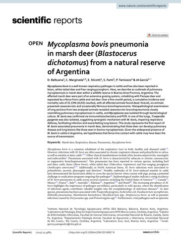
Fig. 1 Pulmonary mycoplasmosis pathological findings in marsh deer (Blastocerus dichotomus). A. Gross lung findings. Cranioventral caseonecrotic bronchopneumonia characterized by multiple coalescing protruding white nodules (black arrows). B and C. Microscopic lung findings. Multifocal caseonecrotic bronchopneumonia with foci in bronchioles and alveoli characterized by a hypereosinophilic core of necrotic debris (asterisk) surrounded by neutrophils, a rim of macrophages, occasional lymphocytes, and fibroblast proliferation. Hematoxylin and eosin. 10x (B) and 40x (C). D. Immunohistochemistry. Positive immunolabeling for M. bovis in lungs staining the margin of the caseonecrotic foci and, to a lesser extent, at the center of the necrotic foci. 10x.
hemorrhagic disease viruses, blue tongue viruses and herpesviruses remain epidemiologically significant23 However, in Argentina, no surveillance has been conducted to detect these viruses in deer populations.
While much is known about M. bovis infections in domestic species, little research has explored its presence in wildlife, particularly in cervids such as marsh deer. This species, the largest native cervid in South America, primarily inhabits the Esteros del Iberá (Corrientes) and the Paraná Delta (Santa Fe, El Chaco, Formosa, Buenos Aires, and Entre Ríos) in Argentina24. Marsh deer populations face several threats, including habitat destruction, disease, and hunting25. As a result, the species is classified as vulnerable on the International Union for Conservation of Nature’s Red list26, while in Argentina, it is hold an endangered status24. Diseases has played a significant role in recent mortality events. A study investigating marsh deer die-offs in Argentina from 2014 to 2017 identified multiple infectious agents, including Fasciola hepatica, gastrointestinal parasitosis and hemoparasites (Trypanosoma spp., Anaplasma spp.), as potential causes25. Given the ongoing conservation concerns for marsh deer, understanding the role of infectious diseases, including M. bovis, is critical for management and protection efforts.
To the best of our knowledge, there are no reports of respiratory disease associated with M. bovis in wildlife animals in South America affecting deer. This study represents the first report of M. bovis-associated pneumonia in marsh deer in a nature reserve in Argentina, highlighting that these deer can develop pulmonary disease and lung lesions similar to those observed in bovine mycoplasmosis.
Results
In a 5-month period, between June and October 2024, 11 of 52 (21.15%) marsh deer were found dead in a wildlife reserve located in north-central Buenos Aires Province, Argentina. The deer, along with pampas deer (Ozotoceros bezoarticus), were bred extensively on natural land for later wildlife reintroduction. Red deer (Cervus elaphus) and cattle also live in the reserve, but they are kept in separate paddocks fenced off from the marsh deer. Grossly, in all 6 autopsies, the lungs presented bilateral, cranioventral red, clear to dark consolidation (approximately 30 to 60%), and multiple coalescing protruding white nodules (Fig. 1A). At the cut surface, the lung parenchyma presented multiple caseous nodules characterized by white exudate surrounded by fibrosis. In 2 cases, fibrinous diffuse pleuritis was also observed. Microscopically, in the two lung samples analyzed,
multifocal caseonecrotic bronchopneumonia was observed (Fig. 1B). These foci were centered in bronchioles and alveoli characterized by eosinophilic material with mild admixed cellular debris delimited by a variable number of degenerated leukocytes that maintain cellular figures (ghost-like remnants) and a small number of active macrophages with scattered lymphocytes and some fibroblasts (caseonecrotic centers), surrounded by variable amounts of neutrophils, a rim of macrophages with scattered lymphocytes, and, in some areas, mild fibroblast proliferation (Fig. 1C). Some bronchioles and bronchi presented multifocal segmental epithelial necrosis. In 1 case, multiple foci of necrosis with abundant neutrophils, intralesional bacteria and a thin rim of macrophages (abscesses) as well as interlobular enlargement by fibrinous exudate and fewer mixed inflammatory cells (neutrophils, macrophages and lymphocytes) were observed. Additionally, the latter had fibrinous diffuse pleuritis. Gram staining of the lungs revealed short, round-tipped, gram-positive bacilli within abscesses and intravascularly. Ziehl–Neelsen staining of both lungs was negative. Immunohistochemistry for M. bovis in both lungs revealed positive immunolabeling, characterized by intense, dark brown, granular, stained structures in the margin of the caseonecrotic foci and, to a lesser extent, at the center of the necrotic foci (Fig. 1D). Positive staining was also observed within bronchioles and bronchiolar epithelial cells and the cytoplasm in some host cells. Few macrophages with positive staining were present within the airway lumina, peribronchiolar septa, or alveoli. Through routine bacteriological culture, Trueperella pyogenes was identified from the lungs of case 1. In selective culture for Mycoplasma, typical fried egg-shaped colonies were observed in both lung samples. Colonies of Mycoplasma from lung culture were detected as Mycoplasma spp. and M. bovis by both PCRs.
Discussion and conclusion
The diagnosis of caseonecrotic bronchopneumonia by M. bovis was based on pathological features and confirmed by immunohistochemistry (IHC), bacteriological culture and PCR in the two lungs analyzed. To our knowledge, this is the first report of pulmonary mycoplasmosis in marsh deer. Although we cannot confirm that mycoplasmosis was the primary cause of death in the 11marsh deer, we strongly suggest its crucial role as both a primary and/or secondary pathogen, facilitating opportunistic infections by other bacteria like T. pyogenes27isolated from one lung. This interaction between M. bovis and T. pyogenes could be synergistic, with M. bovis impairing the respiratory defense system and facilitating T. pyogenes infections, as seen in other ruminants8,21.
The pathological findings in the lungs, caseonecrotic bronchopneumonia, are characteristic of mycoplasmosis, as thoroughly reported in cattle and bison3,6,7,19. Caseonecrotic nodules are distinctive lesions with caseous necrosis that fill small bronchioles, alveoli or interlobular septa, where leukocytes undergo a distinctive form of necrosis maintaining their ghost-like cellular outlines3,19. Also, the abundant M. bovis detected in necrotic foci of both affected lungs via IHC and distribution, evidences strong influence of the bacteria in the pneumonic process, associated to fatal mycoplasmosis in cattle3,11. Although, M. bovis pneumonia is well documented in cattle3,11, cases in wildlife are rare6–8, often leading to an underestimation of its potential impact. In deer, pneumonia is a significant disease associated to a range of bacterial, parasitic, and viral pathogens22,23. While M. bovis has been rarely reported in these animals, its role in pneumonia outbreaks may be overlooked. Nevertheless, it has been detected from both captive white-tailed deer, free-ranging pronghorn and free-ranging mule deer (Odocoileus hemionus)6,7,9,23,28,29. Alongside our findings, this evidence highlights the importance of considering M. bovis in the differential diagnosis of respiratory diseases in deer.
T. pyogenes is likely a secondary opportunistic pathogen30 commonly associated with subacute to chronic pneumonia in ruminants31however, its presence correlates positively with the isolation of Mycoplasma spp. in bovine respiratory diseases. This interaction suggests a possible synergistic effect, exacerbating lung disease3,12 Particularly, in wildlife, T. pyogenes is an important emergent pathogen associated with pneumonia and extrapulmonary abscesses29,30. However, no such findings have been reported in marsh deer30. Dyer et al.6 described an outbreak of respiratory disease in white-tailed deer coinfected with M. bovis and T. pyogenes as herein. Additionally, in cattle, most cases of M. bovis pneumonia involve coinfections with other pathogens, including T. pyogenes, such as P. multocida, E. coli, M. haemolityca and H. somni27. These coinfections intensify the severity of respiratory disease in cattle, where M. bovis causes degeneration and impairment of ciliated respiratory epithelial cells, predisposing the lungs to secondary infection32,33, in which the typical caseonecrotic foci of M. bovis infection develop into abscesses with a fluid purulent center in coinfection with T. pyogenes3. The latter could be the case herein, exacerbating lung lesions with subsequent high mortality.
Other pathogens such as viral and parasitic agents are commonly involved in respiratory diseases in coinfection with M. bovis3,27. Unfortunately, these tests were not carried out as samples were not conserved after bacteriological analysis.
The host species of origin in this outbreak is unknown. Given, that in areas where marsh deer are distributed, such as national or provincial parks of Argentina, cattle and marsh deer often cohabit. In this outbreak, although cattle and marsh deer did not share the same paddocks, they were separated only by a fence. Given the high prevalence of asymptomatic infections in cattle and the rare detection of M. bovis in wildlife, transmission from a livestock reservoir to marsh deer seems likely. Cattle is known to act as carrier in clinically healthy animals, with variable disease expression, and intermittent shedding, maintaining M. bovis in populations3,34. This situation generates a possible scenario where cattle could transmit the disease to deer, implicating potential risk of transmission to wildlife with subsequent outbreaks as described. Mycoplasmosis, in other species, such as bison, has been shown to cause outbreaks with high mortality35, and for this reason, monitoring marsh deer in Argentina is important. Serological studies monitoring M. bovis prevalence in marsh deer and nearby cattle could provide valuable insights into the presence of subclinically infected deer and help identify potential risk factors associated with elevated antibody levels and subsequent outbreaks. Specifically, determining the prevalence of M. bovis in coexisting cattle and deer would enable the implementation of preventive measures
based on the findings. Whenever possible, these efforts should be complemented by postmortem examinations and laboratory testing to enhance diagnostic accuracy.
This report highlights the circulation of M. bovis in animal species other than cattle. A high incidence of M. bovis has been recently reported in cattle in Argentina4,19,36, reinforcing the high circulation of the bacteria, which could easily reach other species, such as the marsh deer described herein. Further studies are necessary to gain knowledge of M. bovis behaviour as primary or secondary agent in wildlife species, together with detection of other agents, such as viruses and parasites, potentially involved in respiratory diseases as co-infections.
The occurrence of M. bovis-associated pneumonia in wildlife highlights the importance of pathogen surveillance, as identifying infectious agents provides valuable insights into the ecoepidemiology of infectious diseases. This is especially critical herein for marsh deer, an endangered species, where eradication programs are essential for its conservation.
Methods
Epidemiological data, including location, affected animal species, rearing system, incidence and mortality and coexistence with other species, were collected. Autopsy was carried out in 6 animals, but only samples of 2 were collected for bacteriological and pathological studies. Sections of the lung, heart, kidney and small and large intestine were fixed by immersion in 10% neutral-buffered formalin (pH 7.2) for 48 h and embedded in paraffin. Four-micrometer sections were prepared routinely and stained with H&E. Sections of the lungs were selected for Gram staining, Ziehl–Neelsen staining and immunohistochemistry for the detection of M. bovis in both cases33. Briefly, blocks of selected paraffin-embedded tissues were subsequently subjected to M. bovis IHC. Epitope retrieval was performed by autoclaving the samples in citrate buffer at pH 6.0 for 10 min at 105 °C. IHC staining of M. bovis tissues was performed with a rabbit anti-M. bovis polyclonal antibody applied for 18 h. at a dilution of 1:5000. Negative controls were prepared by replacing the primary antibody with nonimmune rabbit serum. In addition, lung samples from both animals were collected for bacteriological culture and specialized Mycoplasma culture4. First, the samples were cultured on Columbia agar plates supplemented with 15% sterile equine blood (ASC) (Laboratorios Britania SA, Los Patos, CABA, Argentina). These plates were incubated for 24–72 h at 37 °C in an atmosphere with 10% CO2. For selective Mycoplasma culture, lung samples were inoculated onto Mycoplasma Base Medium with Selective Mycoplasma supplement -MM-(Oxoid Ltd., Wad Road, Basingstoke, UK) and Columbia Blood Agar -CBA- (Oxoid Ltd., Wad Road, Basingstoke, UK) with 7% bovine blood and MacConkey agar -MC- (Oxoid Ltd., Wad Road, Basingstoke, UK). All the plates were incubated at 37 °C, with MM under 5% CO2, with CBA under 10% CO2 and with MC under aerobiosis and examined at 96, 48 and 24 h, respectively. The genera were classified in accordance with Bergey’s Manual of Systematic Bacteriology. Plates containing colonies compatible with Mycoplasma were stained with Dienes´ stain (Dienes, 1967) and observed via binocular stereomicroscopy. For molecular analysis, DNA was extracted with a commercial kit according to the manufacturer’s instructions (PuriPrep-S, Inbiohighway, Argentina) and processed first by no-species specific nested PCR targeting the 16–23 S rRNA intergenic spacer region37 and then by M. bovis-specific PCR38.
Data availability
The data will be provided upon request to the corresponding author, Juan Agustin Garcia (garcia.juanagustin@ inta.gob.ar).
Received: 6 March 2025; Accepted: 11 June 2025
References
1. Maunsell, F. P. et al. Mycoplasma bovis infections in cattle. J. Vet. Intern. Med. 25, 772–783 (2011).
2. Perez-Casal, J. Pathogenesis and virulence of Mycoplasma bovis Vet. Clin. N Am. Food Anim. Pract. 36 (2), 269–278 (2020).
3. Caswell, J. L., Bateman, K. G., Cai, H. Y. & Castillo-Alcala, F. Mycoplasma bovis in respiratory disease of feedlot cattle. Vet. Clin. North. Am. Food Anim. Pract. 26 (2), 365–379 (2010).
4. Cantón, G. et al. Mycoplasma bovis-pneumonia and polyarthritis in feedlot calves in argentina: first local isolation. Rev. Argent. Microbiol. 54 (4), 299–304 (2022).
5. Dudek, K., Nicholas, R. A. J., Szacawa, E. & Bednarek, D. Mycoplasma bovis infections-occurrence, diagnosis and control. Pathogens 9 (8), 640 (2020).
6. Krogh, N. W., Schaan, L. P. & D.F. & Pulmonary mycoplasmosis in farmed white-tailed deer (Odocoileus virginianus). J. Wildl. Dis. 40 (2), 366–370 (2004).
7. Register, K. B. et al. Comparison of multilocus sequence types found among North American isolates of Mycoplasma bovis from cattle, bison, and deer, 2007–2017. J. Vet. Diagn. Invest. 31 (6), 899–904 (2019).
8. Deeney, A. S., Collins, R. & Ridley, A. M. Identification of Mycoplasma species and related organisms from ruminants in England and Wales during 2005–2019. BMC Vet. Res. 17, 325 (2021).
9. Malmberg, J. L. et al. Mycoplasma bovis infections in free-ranging pronghorn, wyoming, USA. Emerg. Infect. Dis. 26 (12), 2807–2814 (2020).
10. Spergser, J. et al. re-emergence, spread and host species crossing of Mycoplasma bovis in the Austrian alps caused by a single endemic strain. Vet. Microbiol. 64 (3–4), 299–306 (2013).
11. Gagea, M. I. et al. Naturally occurring mycoplasma bovis—associated pneumonia and polyarthritis in feedlot beef calves. J. Vet. Diagn. Invest. 18 (1), 29–40 (2006).
12. Fulton, R. W. et al. Lung pathology and infectious agents in fatal feedlot pneumonias and relationship with mortality, disease onset, and treatments. J. Vet. Diagn. Invest. 21 (4), 464–477 (2009).
13. Booker, C. W. et al. Microbiological and histopathological findings in cases of fatal bovine respiratory disease of feedlot cattle in Western Canada. Can. Vet. J. 49, 473–481 (2008).
14. Ramírez-Romero, R. et al. Demostración histoquímica de Mycoplasma bovis En lesiones neumónicas crónicas En Ganado de corral de Engorda. Vet. Mex 41, 289–296 (2010).
15. Arcangioli, M. A. et al. The role of Mycoplasma bovis in bovine respiratory disease outbreaks in veal calf feedlots. Vet. J. 177, 89–93 (2008).
16. Radaelli, E., Luini, M., Loria, G. R., Nicholas, R. A. J. & Scanziani, E. Bacteriological, serological, pathological and immunohistochemical studies of Mycoplasma bovis respiratory infection in veal calves and adult cattle at slaughter. Res. Vet. Sci. 85, 282–290 (2008).
17. Horwood, P. F. et al. Is Mycoplasma bovis a missing component of the bovine respiratory disease complex in australia?? Aust Vet. J. 92, 185–191 (2014).
18. Yilmaz, R. et al. Histopathological, immunohistochemical and bacteriological characterization of Mycoplasma bovis pneumonia in cattle. Pakistan Vet. J. 36, 316–321 (2016).
19. Margineda, C. A. et al. Mycoplasma bovis pneumonia in feedlot cattle and dairy calves in Argentina. Brazilan J. Vet. Pathol. 10 (2), 79–86 (2017).
20. Oliveira, T. E. S. et al. Mycoplasma bovis and viral agents associated with the development of bovine respiratory disease in adult dairy cows. Transbound. Emerg. Dis. 67 (2), 82–93 (2020).
21. Tompkins, D. M., Carver, S., Jones, M. E., Krkošek, M. & Skerratt L.F. Emerging infectious diseases of wildlife: a critical perspective. Trends Parasitol. 31, 149–159 (2015).
22. Navas-Suárez, P. E. et al. A retrospective pathology study of two Neotropical deer species (1995–2015), brazil: marsh deer (Blastocerus dichotomus) and brown brocket deer (Mazama gouazoubira). Plos One 13(6), 1–26 (2018).
23. Smith, A. C. et al. Causes of mortality in farmed white-tailed deer in the Midwestern united states, 2004–2023. J. Vet. Diagn. Invest. 36 (6), 809–815 (2024).
24. Pereira, J. et al. Blastocerus dichotomus. Categorización 2019 de los mamíferos de Argentina según su riesgo de extinción. Lista Roja de los mamíferos de Argentina. Secretaría de Ambiente y Desarrollo Sustentable de la Nación y Sociedad Argentina para el Estudio de los Mamíferos. (2019). https://cma sarem org ar/index php/es/especie-nativa/blastocerus-dichotomus Accessed 21 Jan 2025.
25. Orozco, M. M. et al. A participatory surveillance of marsh deer (Blastocerus dichotomus) morbidity and mortality in argentina: first results. BMC Vet. Res. 16, 321 (2020).
26. Duarte, J. M. B., Varela, D., Piovezan, U., Beccaceci, M. D. & Garcia, J. E. Blastocerus dichotomus. IUCN Red List. Threatened Species https://doi org/10 2305/IUCN UK 2016-1 RLTS T2828A22160916 (2025).
27. Carella, E. et al. Identification of Mycoplasma species in cattle associated with bovine respiratory disease mortality. Microorganisms 12 (11), 2340 (2024).
28. Hattel, A. L. et al. A retrospective study of mortality in Pennsylvania captive white-tailed deer (Odocoileus virginianus): 2000–2003. J. Vet. Diagn. Invest. 16 (6), 515–521 (2004).
29. Clarke, L. L. Postmortem diagnoses and factors influencing diagnoses in captive white-tailed deer in wisconsin, 2009–2021. J. Vet. Diagn. Invest. 35 (6), 782–788 (2023).
30. Rzewuska, M. et al. Pathogenicity and virulence of Trueperella pyogenes: a review. Int J. Mol. Sci 20(11), 1–33 (2019).
31. Ribeiro, M. G. et al. Trueperella pyogenes multispecies infections in domestic animals: a retrospective study of 144 cases (2002 to 2012). Vet. Q. 35 (2), 82–87 (2015).
32. Gourlay, R. N. & Houghton, S. B. Experimental pneumonia in conventionally reared and gnotobiotic calves by dual infection with Mycoplasma bovis and Pasteurella haemolytica Res. Vet. Sci. 38, 377–382 (1985).
33. Rodriguez, F., Bryson, D. G., Ball, H. J. & Forster, F. Pathological and immunohistochemical studies of natural and experimental Mycoplasma bovis pneumonia in calves. J. Comp. Pathol. 115, 151–162 (1996).
34. Caswell, J. L. & Archambault, M. Mycoplasma bovis pneumonia in cattle. Anim. Health Res. Rev. 8, 161–186 (2008).
35. Martin, K., Jones, L., Browne, A. & Buttke, D. An epidemiological investigation into Mycoplasma bovis infection in U.S. bison (Bison bison): A case–control survey. American Association of Bovine Practitioners Conference Proceedings. 56,230 (2023). Available: https://doi.org/10.21423/aabppro20238942
36. Margineda, C. A., Diab, S., Quiroga, M. A. & López, A. Facial paralysis in feedlot cattle associated with otitis caused by Mycoplasma bovis: first report in Argentina. Braz J. Vet. Pathol. 16 (3), 213–218 (2023).
37. Tang, J., Hu, M., Lee, S. & Roblin, R. A polymerase chain reaction based method for detecting Mycoplasma/Acholeplasma contaminants in cell culture. J. Microbiol. Methods 39 (2), 121–126 (2000).
38. Thomas, A. et al. Conservation of the UvrC gene sequence in Mycoplasma bovis and its use in routine PCR diagnosis. Vet. J. 168, 100–102 (2004).
Acknowledgements
We thank Valeria Scioli for histopathological processing.
Author contributions
Delfina Balbuena wrote the main manuscript and performed the formal analysis. Carlos Margineda: writing, conceptualization and methodology. Erika Stokati, Sofia Fanti and Pablo Tamiozzo: methodology. Juan Agustín García: writing, editing, methodology and conceptualization. All the authors reviewed the manuscript.
Funding
The authors received no financial support for the research, authorship, and/or publication of this article.
Declarations
Competing interests
The authors declare no competing interests.
Additional information
Correspondence and requests for materials should be addressed to J.G.
Reprints and permissions information is available at www.nature.com/reprints
Publisher’s note Springer Nature remains neutral with regard to jurisdictional claims in published maps and institutional affiliations.
Open Access This article is licensed under a Creative Commons Attribution-NonCommercial-NoDerivatives 4.0 International License, which permits any non-commercial use, sharing, distribution and reproduction in any medium or format, as long as you give appropriate credit to the original author(s) and the source, provide a link to the Creative Commons licence, and indicate if you modified the licensed material. You do not have permission under this licence to share adapted material derived from this article or parts of it. The images or other third party material in this article are included in the article’s Creative Commons licence, unless indicated otherwise in a credit line to the material. If material is not included in the article’s Creative Commons licence and your intended use is not permitted by statutory regulation or exceeds the permitted use, you will need to obtain permission directly from the copyright holder. To view a copy of this licence, visit http://creativecommo ns org/licenses/by-nc-nd/4 0/
© The Author(s) 2025