1 Physiolo G y of n E r V ous syst EM
The nervous system contains nerve cells (neurons) and their supporting cells (glial cells or glia).
1.1 N E u R o N
Neuron is the fundamental structural and functional unit of the nervous system. There are between 300 and 500 billion neurons in the nervous system. The principal role is to conduct information to and from the rest of the body. Neurons are capable to accept, integrate and conduct information in the form of electrical impulses. There are three principal functions of neurons: specific – neurons as conductors, ability to produce and conduct discharges, to generate a propagate nerve impulses, troffic effects (proteins synthesized in the neuronal cell body are moved to axon terminals by axonal transport) and secretory – release of substances.
Some neurons have a sensory function; they conduct information to the CNS from the environment or from the inside of the organism. Other neurons are motor, they conduct information from the CNS to muscle cells and are responsible for contraction of striated muscles. Some neurons belong to the autonomic nervous system; they conduct information to inner organs and glands and are responsible for example for contraction of smooth muscles and secretion by glands.
The neuron is organized into 1) a receptive segment (input from other neurons – dendrites and cell body) 2) a conductive segment (single axon) and 3) an effector segment (synapse e.g. to the muscle). This neuron is called multipolar.
The cell body (soma) of the neuron contains cell nucleus, mitochondria, endoplasmic reticulum, neurotubules, and neurofilaments.
Dendrites are short branched cellular processes. They receive input (afferent impulses) from other neurons.
An axon (or nerve fiber) is a single long cell process which starts from a cell body at an axon hillock and sends output (efferent impulses) from a cell body to other neurons or effector cells (e.g. a skeletal muscle fiber). Axons can be extraordinarily long. Those from motor neurons may be more than 1m long.
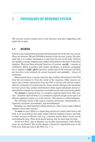
Axons less than 1 µm in diameter are usually unmyelinated, while thicker axons are myelinated. The myelin sheath is formed by oligodendrocytes in the CNS or by Schwann cells in the PNS (fig. 1-1 ).
Nucleus
Axon hillock
Axon
Myelinated internode
Initial segment (unmyelinated) Nodes
Schwann cell nucleus Node
Schwann cell
Schwann cell nucleus
Axon
Neurilemma
Axons
Myelin covering internode
Axolemma
Schwann cell nucleus
Neurilemma
Myelinated axon
Unmyelinated axon
Fig. 1-1 Myelinated and unmyelinated axon
Fig. 1-1 Myelinated and unmyelinated axon
The myelin sheath consists of multiple layers of cellular membrane containing the lipid substance sphingomyelin, which is an excellent insulator. However many Schwann cells are required to myelinate the entire length of an individual axon. At the junction between two adjacent Schwann cells, a small unmyelinated segment remains (only 1–3 µm in length) which is called the node of Ranvier. Thus individual segments of myelin, which can be up to 1 mm long, are separated by nodes of Ranvier.
1.2 Axo NAL t RANSP o R t
rates of approximately 1 and 3 mm/day respectively. Retrograde transport (backwards) moves molecules destined for degradation from periphery back to cell body, where they are broken down by lysosomes. It moves only materials packaged in vesicles at rate up to 200 mm/day (fig. 1-2). Since the axon depends on axoplasmic transport for vital proteins and materials, axonal injury that interrupts the transport will cause the distal axon to degenerate in a process called Wallerian degeneration.
GLIAL CELLS
Nerve impulses resent an electrical which is called The concentration either side of inside of the
Axonal transport (also called axoplasmic transport) is critical for the survival of neurons. All synthesis of protein takes place in the cell body. Functions such as growth, development, repair, that depend on movement of molecules or organelles to another part of neuron requires transport. Receptors, signaling proteins, enzymes for synthesis of neurotransmitter must be moved to sites of use in distant axon terminals or dendrites. There are two principal transport sys-
Neurons are surrounded by supportive cells, which are collectively called glial cells (neuroglial cells, neuroglia).
Astrocytes are the most frequent glial cells in the CNS. Their predominant role is to help form the
Rough endoplasmic reticulum Cell body Nucleus DendriteLysosome
Microtubule network
Axon terminal
Golgi apparatus
Synaptic or secretory vesicle or mitochondrion
Unmyelinated axon
unmyelinated axon
Fig. 1-2 Axonal transport respectively. moves molecules periphery back to cell lysosomes. It vesicles at rate up axon depends on and materials, transport will cause process called Walcells, which (neuroglial cells, glial cells in the help form the capillaries and
Fig. 1-2 Axonal transport
tems. Anterograde transport is movement of molecules/organelles from cell body toward the nerve terminal. Fast anterograde transport is specific for membrane bound organelles or materials and can transport a variety of molecules, including complex proteins, but must be packaged in vesicle and carries at maximal rates 300–400 mm/day. Slow anterograde transport moves mostly soluble proteins not bound in vesicle at rates of approximately 1 and 3 mm/day respectively. Retrograde transport (backwards) moves molecules destined for degradation from periphery back to cell body, where they are broken down by lysosomes. It moves only materials packaged in vesicles at rate up to 200 mm/day (fig. 1-2 ). Since the axon depends on axoplasmic transport for vital proteins and materials, axonal injury that interrupts the transport will cause the distal axon to degenerate in a process called Wallerian degeneration.
ACTION POTENTIAL
Nerve impulses that are conducted along axons represent an electrical change on an axonal membrane, which is called an action potential.
The concentration of various ions is different on either side of the cell membrane. Thus at rest the inside of the cell is negatively charged compared
1.3 G LIAL CELLS
Neurons are surrounded by supportive cells, which are collectively called glial cells (neuroglial cells, neuroglia).
Astrocytes are the most frequent glial cells in the CNS. Their predominant role is to help form the blood-brain barrier between brain capillaries and brain tissue. Thus many molecular substances do not pass from the blood into the interstitial
fluid of the brain. Exchange of materials between the CNS and the rest of the body is tightly regulated.
Oligodendocytes (in the CNS) and Schwann cells (in the PNS) wrap their specialized cell membranes around axons, which is called myelination. The myelination increases the conduction velocity of nerve impulses along the axon.
Microglia are extremely small glial cells. Microglia are immune cells in the brain, similar to macrophages in peripheral tissues. They are resident phagocytes in the brain.
Ependymal cells form a lining of cerebral ventricles and the central canal of the spinal cord.
1.4 AC t I o N P ot EN t IAL
Nerve impulses that are conducted along axons represent an electrical change on an axonal membrane, which is called an action potential.
The concentration of various ions is different on either side of the cell membrane. Thus at rest the inside of the cell is negatively charged compared to the outside of the cell. This potential difference is known as the resting membrane potential. As the membrane potential is increased, sodium ion channels open, allowing the entry of Na+ ions into the cell. This is followed by the opening of potassium ion channels that permit the exit of K+ ions from the cell. The inward flow of Na+ increases the concentration of positively-charged cations in the cell and causes depolarization, where the potential of the cell is higher than the resting potential. The Na+ channels close at the peak of the action potential, while K+ continues to leave the cell (fig. 1-3 ). The efflux of K+ decreases the membrane potential or hyperpolarizes the cell. The transmembrane potential difference falls again into the negative value, which is called repolarization.
The action potential is initiated at the axon hillock and is then conducted along the axonal membrane by successive opening of voltage-gated sodium channels. In myelinated axons, the neuronal cell membrane is very well insulated between the nodes of Ranvier so that action potentials “jump” from one node of Ranvier to the next. This process is known as saltatory conduction. Saltatory conduction occurs, because the action potential is generated only at the nodes. The cell membrane below the myelin sheaths has hardly any Na+ channels and is therefore inexcitable.
1.5 Sy NAPSES
Neurons transmit nerve impulses (action potentials) from one neuron to another or to an effector organ (muscle fiber) at specialized formation of contact called
vesicles at rate up axon depends on and materials, transport will cause process called Walsupportive cells, which (neuroglial cells, glial cells in the help form the capillaries and substances do not interstitial
uid of the the CNS and and Schwann specialized cell memmyelination. conduction velocity of cells. Microgsimilar to macroresident phagocerebral ventricord.
is called an action potential. The concentration of various ions is different on either side of the cell membrane. Thus at rest the inside of the cell is negatively charged compared
Fig. 1-3 Propagation of action potential – inward flow of Na+ and efflux of K+ ions
synapses. Synapses are either electrical or chemical. The most common synapses are chemical.
Each synapse is consisted of three main components. The end of an axon forms a small dilated bulblike region called synaptic terminal or synaptic button. The synaptic button contains a variety of organelles, including mitochondria, and synaptic vesicles with molecules of a chemical neurotransmitter. The synaptic button is separated from an adjacent dendrite or soma of the next neuron by a narrow space called the synaptic cleft. The adjacent membrane of the next neuron is called the postsynaptic membrane. When an action potential reaches the synaptic button, voltage-gated calcium channels (P/Q type) in the surface membrane of the synaptic button are opened,
Fig. 1-3 Propagation of action potential – inward flow of Na+ and efflux of K+ ions 11
13.10.2010 13:22:33
an effector organ formation of contact either electrical or synapses are chemical. three main comforms a small dilated terminal or synapcontains a variety mitochondria, and synaptic chemical neurotransseparated from an the next neuron by cleft. The adjacent called the postsynapreaches the synaptic channels in the surbutton are opened, The influx of Ca++ neurotransmitter is released across the synapthe postsynaptic postsynaptic neuron is then neurotransmitters
muscle fi ber. The nerve ending contains secretory vesicles with acetylcholine. When an action potential reaches the nerve terminal, acetylcholine is released into the synaptic cleft and then binds to specific recep-
Somatic motor neuron
Axon terminal
Axon potential
Fig. 1-4 Chemical synapse. ACh – acetylcholine, AChE – acetylcholinesterase
Fig. 1-4 Chemical synapse. ACh – acetylcholine, AChE –acetylcholinesterase
and Ca++ moves into the terminal. The influx of Ca++ enables that the chemical neurotransmitter is released from the synaptic button, diffuses across the synaptic cleft and binds to receptors on the postsynaptic membrane (fig. 1-4 ). The postsynaptic neuron is then excited or inhibited. The action of neurotransmitters is terminated by reuptake, enzymatic degradation or diffusion away from the synapse.
Neurotransmitters include acetylcholine, various amines, such as dopamine, norepinephrine, epinephrine and serotonin (5-hydroxytryptamine), aminoacids such as glutamate, aspartate, gamma-aminobutyric acid (GABA) and glycine. Other neurotransmitters are various peptides, such as enkephalins, and also nitric oxide.
Postsynaptic receptors may be ionotropic or metabotropic. Ionotropic receptors mediate neurotransmission over milliseconds. These receptors consist of various subunits with a binding site localized around a central ion channel. When the binding site is activated by a neurotransmitter, opens the gate to permit ion flow via the channel into the cell.
Metabotropic receptors are coupled via G-proteins to a transducer – an ion channel or a second messenger system. When the receptor is activated by a neurotransmitter, G-protein either opens a membrane channel specific for a certain ion or activates cyclic adenosine monophosphate (cAMP) or cyclic guanosine
13:22:34
monophosphate (cGMP), which stimulates then a specific metabolic process in the postsynaptic neuron. This results in changes in the excitability or functional activity of the postsynaptic neuron.
The typical example of a simple synapse is the neuromuscular junction (or motor end plate). The neuromuscular junction is a specialized synapse (relay station) between the motor neuron and the skeletal muscle fiber. The nerve ending contains secretory vesicles with acetylcholine. When an action potential reaches the nerve terminal, acetylcholine is released into the synaptic cleft and then binds to specific receptors on the postsynaptic membrane. This leads to excitation (depolarization) of the sarcolemma with the subsequent muscle contraction. Acetylcholine is inactivated by breakdown into its two components, choline and acetate. This step is catalyzed by the enzyme acetylcholinesterase.
In simple synapses, such as the neuromuscular junction of skeletal muscle, each action potential that reaches the presynaptic terminal evokes an action potential in the postsynaptic cell. However, in the CNS, the situation is more complex and such a direct information transmission is rare.
A typical neuron in the CNS receives thousands of synaptic contacts from other neurons on its dendrites and cell body (soma). At each of these synapses, the neurotransmitter activates postsynaptic receptors and causes a local change in membrane potential. These miniature potentials are called excitatory or inhibitory postsynaptic potentials (EPSPs and IPSPs).
The EPSP is the result of the opening of Na+ channels in the postsynaptic membrane. Na+ enters the postsynaptic neuron and the membrane potential becomes more positive (depolarized).
The IPSP is the result of the opening of chloride channels, Cl- ions move into the cell and membrane potential becomes more negative (hyperpolarized).
The character of the postsynaptic potential is determined by the neurotransmitter and receptor. Certain neurotransmitters such as glutamate are always excitatory; they evoke EPSPs by facilitating Na+ entry. Other neurotransmitters such as GABA open Cl- channels and induce only IPSPs. Many neurotransmitters such as acetylcholine or dopamine can evoke both types of postsynaptic potential, depending on the receptor upon which they act.
EPSPs and IPSPs are summated in the neuron over time and space.
Temporal summation is a process by which individual postsynaptic potentials (excitatory or inhibitory) may add together (summate) when they arrive within a short interval one after another. Temporal summation occurs when a second postsynaptic potential arrives at the same synapse before the postsynaptic membrane has returned to its resting level.
Spatial summation occurs when more postsynaptic potentials (excitatory or inhibitory) are present simultaneously at different synapses of the neuron arriving from different sources (neurons).
medial system
lateral system
anterior cingulate
thalamus
receptors
Fig. 4-2 Central pain pathways: lateral and medial systems – green, proprioception, vibration sense and touch – red. Lateral and medial spinothalamic tract run together to the thalamus (green), here they form synapses with third sensory neurons in different nuclei. The lateral spinothalamic tract (lateral sys‑ tem) forms synapses in the ventral posterolateral nucleus. From here the third sensory neurons lead to the primary (and secondary) somatosensory cortex. This pathway is used to perceive the location and intensi‑ ty of pain. The neurons of the medial spinothalamic tract (medial system) terminate in the ventral medial nucleus of the thalamus and from there project to the insular cortex (not shown in the figure). This path‑ way is responsible for the cognitive component of pain. Another part of the medial spinothalamic tract projects into the mediodorsal nucleus of the thalamus. A third sensory neuron projects to the cingulate cortex. This pathway is responsible for the affective ‑ emotional aspect of pain (the pain is unpleasant).
Fibers of the lateral spinothalamic tract system form synapses in the ventral posterolateral nucleus of the thalamus. From there, third order neurons lead to the primary and secondary somatosensory cortex. This pathway is responsible for the discriminative aspect of pain (the precise location and intensity of pain).
Neurons of the medial spinothalamic tract terminate in the ventral medial nucleus of the thalamus and then project to the insular cortex. This pathway is responsible for the cognitive-evaluative component of the pain. Other neurons of the medial spinothalamic tract project to the mediodorsal nucleus of the thalamus. Third-order neurons from the mediodorsal thalamus project to the cingulate cortex. This component is responsible for the affective-emotional aspect of pain. This means that we perceive pain unpleasantly (suffering).
Finally, there are also spinobulbar or spinoreticular neurons that terminate in various subcortical structures such as the reticular formation of the brainstem, the periaqueductal gray, the hypothalamus and the amygdala. These nerve fibres are responsible for arousal, endocrine and autonomic responses to pain and help modulate pain.
4.6 S ENSI t I z At I o N o F t HE NER vou S S y S t EM
Intense nociceptive stimuli at the site of the damaged tissue with the formation of tissue inflammation lead to various changes at the level of the peripheral and central nervous system, which amplify the pain signal and can also lead to persistent pain. These changes are referred to as sensitization of the nervous system. Depending on the level at which sensitisation manifests itself, it is distinguished between peripheral and central.
4.6.1 Peripheral sensitization
In peripheral sensitization, there is an increased sensitivity of peripheral nociceptors (free nerve endings) at the site of the noxious stimulus. In addition to the sensitization of free nerve endings by chemical mediators at the site of tissue inflammation due to the release of serotonin, histamine, prostaglandins, and K+ ions, the number of certain ion channels on free nerve endings at the site of damage is increased, which lowers the threshold for nociceptor activation and leads to enhanced peripheral nociceptive stimulation into the dorsal horn via primary nociceptive afferent neurons.
Peripheral sensitization also occurs when primary afferent nociceptive neurons are damaged or injured. These neurons then produce spontaneous activity that is generated outside the sites of normal impulse generation. This activity is referred to as ectopic discharge. Increased expression of sodium channels in the course of damaged nerve fibres plays an important role in the generation of spontaneous ectopic activity. This results in a lowered threshold for action potential generation and increased sensitivity of the damaged peripheral nerves to various subthreshold and non-painful stimuli.
4.6.2 Central sensitization
Central sensitization predominantly affects the dorsal horns of the spinal cord, and secondarily also nociceptive structures in the higher levels of the brain (e.g. thalamus), where this process is less well studied. Sensitization of central pain pathways is triggered by continuous peripheral nociceptive afferent impulses that arrive in the dorsal horn of the spinal cord during peripheral sensitization. These continuous nociceptive impulses from the periphery result in processes that lead to increased excitability of posterior horn neurons. Repeated nociceptive stimuli
of the same intensity and duration induce an increasing increase in electrical response, i.e. a progressive rise in action potential firing of dorsal horn neurons. This progressive increase in the electrical response of dorsal horn neurons is called the wind-up phenomenon.
This cumulative effect is caused by two processes. Increased peripheral nociceptor firing results in increased glutamate release in the dorsal horn of the spinal cord and activation and proliferation of dorsal horn glial cells – microglia and astrocytes.
Increased glutamate release from the central endings of primary nociceptive afferent neurons in the dorsal spinal cord horn causes, in addition to activation of AMPA glutamate receptors, a secondary marked activation of NMDA glutamate receptors on dorsal spinal cord horn neurons, leading to increased activity on central pain pathways.
Activated glial cells in the dorsal horn of the spinal cord (microglia and astrocytes) release pronociceptive neuropeptides, including SP, CGRP, tumor necrosis factor alpha and others. All of these substances lead to increased sensitivity of the dorsal horn neurons of the spinal cord and thus to their increased excitability.
Continuous nociceptive impulses from the periphery also activate cellular processes within WDR neurons, which cause translocation of AMPA and NMDA receptors from intracellular stores to the synaptic membrane of WDR neurons. The consequence is increased sensitivity of WDR neurons to glutamate. The increased sensitivity of WDR neurons results in the ability of normally non-painful stimuli to activate central pain pathways such as the spinothalamic tract via Abeta fibers. The non-painful stimulus thus causes pain, which is called allodynia.The increased sensitivity of WDR neurons can similarly lead to hyperalgesia, which means that a normal painful stimulus is perceived with excessive intensity. The presence of allodynia and hyperalgesia in a patient is indicative of central sensitization.
The transmission of pain signals in the spinal dorsal horns further facilitates (promotes) sprouting of damaged fibres from the touch receptors. As a result of this sprouting, tactile fibers form synapses with spinal dorsal horn neurons that normally receive only nociceptive stimuli. This explains why innocuous stimuli can cause pain when peripheral nerve fibres are damaged.
4.7 Mo D u LAt I o N o F PAIN t RANSMISSI o N
Pain impulses that run along the nociceptive pathway to the cerebral cortex are modulated at both the spinal and supraspinal levels.
Transmission of pain impulses at synapses between primary nociceptive afferent neurons and dorsal horn neurons (second order neurons) in the dorsal horns of the spinal cord may be inhibited or facilitated. The exact mechanism of modulation of pain transmission in the dorsal spinal cord horn is not yet fully understood. The gatekeeper theory of pain (Melzack and Wall) has attempted to explain it, but it is no longer completely valid in its original form. Local
inhibitory and excitatory interneurons play an important role in modulating the transmission of pain impulses in the dorsal spinal cord horn, as they perform integration of incoming nociceptive and non-nociceptive impulses in the posterior spinal cord horn and may suppress (inhibit the transmission of) some painful stimuli and facilitate others.
In addition to spinal interneurons, the transmission of nociceptive impulses in the dorsal horn of the spinal cord is also influenced by the descending modulation of nociception at the supraspinal level, which may also have both inhibitory and facilitative effects on dorsal horn neurons (second order neurons). This descending modulation is carried out by neurons in the brainstem, which mainly comprise the periaqueductal gray (PAG) and rostral ventromedial medulla (RVM). The PAG and RVM receive descending projections from cortical and limbic regions. The PAG projects to the RVM containing catecholaminergic neurons. The latter sends either inhibitory or excitatory impulses to dorsal horn neurons, modulating the transmission of nociceptive impulses at synapses in the dorsal horn of the spinal cord. PAG neurons also project to the raphe nuclei, whose serotoninergic neurons in turn project to the dorsal horn, where they release serotonin. By acting on spinal serotonin (5-HT7 ) receptors, they inhibit pain transmission, whereas by acting on other serotonin (5-HT2 a 3 ) receptors, they facilitate nociception. In addition, the noradrenergic neurons of the locus coeruleus are also located in the pons, which in the spinal cord carry out descending inhibition of nociception.
4.8 D EEP AND v ISCERAL PAIN
In deep tissues, Adelta fibers are relatively less represented, therefore the rapid and sharp first pain is less expressed. Deep and visceral pain is less easily localized, and is often accompanied by nausea, sweating, and changes in blood pressure.
Nociceptors in visceral organs are less numerous compared to somatic structures. Afferent fibres from visceral organs run to the brain via sympathetic and parasympathetic nerves, including cranial nerves such as the vagus nerve. The cell bodies of these nerve fibers are located in the dorsal root ganglia and in the cranial nerve ganglia.
Referred pain
When a visceral organ is irritated, the pain caused is often not felt in that organ, but is projected to some distant region from the original source. This pain is called referred pain. It is important for the physician to know these common sites where pain from visceral organs is transmitted. A typical example is pain originating in the heart, which is most often referred to the inside of the left arm. The most common sites of visceral pain are shown in the figure 4-3


Fig. 4-3 Referral patterns of visceral pain
Referred pain is explained by the convergence of somatic and visceral nociceptive fibers on the same second order neurons in the spinal dorsal horn.These second order neurons then go to the thalamus and then to the somatosensory cortex. This concept is called convergence-projection theory (fig. 4-4 ).
4.9 tyPES oF PAIN
Pain can be divided according to its duration into acute and chronic, according to its pathophysiology into several main types – nociceptive, neuropathic, dysautonomic, nociplastic and psychogenic.
4.9.1 Division of pain according to duration
ACutE PAIN
Acute pain is a symptom that, with the exception of childbirth, is part of many pathologies and diseases (for example, trauma, surgery, inflammation, some internal diseases). It has primarily a protective function, it has a warning meaning for the organism. It alerts the organism to actual or potential (impending) tissue damage. It lasts for the duration of the underlying cause. It disappears after the
ventrolateral pain pathway
midbrain medulla
periaqueductal gray matter
spinal cord
nucleus raphe magnus


afferent pain fibers
visceral structures
somatic structures


Fig. 4-4 Schematic illustration of the convergence‑projection theory


removal of the cause during the healing process. Pain may be considered acute for a maximum of 3-6 months.
C HR o NIC PAIN
Chronic pain lasts longer than 3-6 months. It is often characterized as pain lasting longer than the expected healing time. It is also referred to as ‚pathological‘ or ‚dysfunctional‘ because it loses its original meaning, no longer fulfils a defensive function and is a source of suffering for the patient.
Changes in metabolism, function and structure of the nervous system are called neuroplastic changes. These changes affect axons, synapses, receptors and the function of neurotransmitters. As a result of these changes, noxious stimuli are capable of causing more intense pain of longer duration (hyperalgesia) and normal stimuli (non-noxoius stimuli) can cause or contribute to pain.
In addition to the nociception itself, other components come to the fore, such as the psychological (anxiety, depression, loss of appetite, loss of libido, etc.), emotional (anger, fear, anxiety), cognitive (assessment of treatment, feelings of possible guilt – self or others), and behavioural (painful behaviour – avoiding certain physical activity, lying down, taking protective and relieving positions, overuse of medication, repeatedly seeking medical care or hospitalisation).
Chronic pain is considered a separate disease often with complicated aetiology, pathophysiology and symptomatology.
4.9.2 Division of pain according to its pathophysiology
No CICEP t I v E PAIN
Nociceptive pain is caused by damage to various tissues, such as a cut, burn, contusion, pressure from inside the body (e.g. a tumour), etc. The reason why we feel pain in these situations is due to the activation of receptors (free nerve endings) in these tissues. Pain impulses are then transmitted to the brain via nerves. The transmission of pain impulses from the site of injury or tissue damage is an adequate physiological response, an important biological defense system that warns and protects the body from further damage.
Traditional analgesics such as paracetamol (acetaminophen), nonsteroidal anti-inflammatory drugs (NSAIDs) and opioids are usually effective. Thus, we have non-opioid and opioid analgesics to treat nociceptive pain. Among the non-opioid analgesics are paracetamol (acetaminophen), which probably exerts inhibition at the spinal dorsal horns or supraspinal level, acetylsalicylic acid and other nonsteroidal anti-inflammatory drugs (NSAIDs). The essence of the analgesic effect of these substances is the inhibition of cyclooxygenase (COX). COX is present in the human body in two isoforms – COX-1 and COX-2. COX-1 is essential for normal functions and is involved in, for example, gastric protection. Inhibition of the COX-1 isoform is responsible for the side
effects of nonselective NSAIDs. The COX-2 isoform is an enzyme whose production increases sharply when the body is damaged by inflammatory mediators. It causes inflammation, pain and temperature. Inhibition of COX-2 therefore has anti-inflammatory effects, suppressing pain and increased temperature. Based on their selectivity for individual COX enzymes (isoforms), we classify nonsteroidal anti-inflammatory drugs into 3 categories. Non-selective COX inhibitors inhibit both COX-1 and COX-2. These include all the classical NSAIDs: acetylsalicylic acid, ibuprofen, diclofenac, indomethacin, naproxen and ketoprofen. They are burdened by gastrointestinal side effects: dyspepsia, epigastric pain, heartburn and gastrointestinal bleeding due to COX-1 inhibition. The second group consists of preferential COX-2 inhibitors, which are characterised by relative COX-2 selectivity. These include nimesulid and meloxicam. The third group consists of selective COX-2 inhibitors, including celecoxib.
Opioid analgesics can be used to control severe pain. There are weak opioids – tramadol, codeine, hydrocodeine and strong opioids – morphine, fentanyl, hydromorphone, oxycodone and buprenorphine. Opioids are associated with risks of addiction, physical dependence and tolerance.
N E u R o PAt HIC PAIN
Neuropathic pain is caused by a lesion or dysfunction of the nervous (somatosensory) system. The cause of neuropathic pain can be a disorder of both the peripheral and central nervous system Therefore, neuropathic pain is divided into peripheral and central neuropathic pain. Neuropathic pain is characterised as burning, stabbing, shooting or lancinating. Fatigue and emotions usually increase the intensity of the perception of this pain.
Examples of peripheral neuropathic pain are trigeminal neuralgia and postherpetic neuralgia, nerve root compression by herniated intervertebral disc, neuralgic amyotrophy (Parsonage-Turner syndrome), symmetrical diabetic polyneuropathy, Guillain-Barré syndrome. Central neuropathic pain occurs in spinal cord injury, multiple sclerosis and as post-stroke pain. Neuropathic pain often does not respond to common analgesics including paracetamol, NSAIDs and opioids.
The drugs of choice for neuropathic pain are tricyclic antidepressants (TCAs), especially amitriptyline. Other TCAs such as nortriptyline, imipramine, desipramine and maprotiline can also be used. Among other antidepressants, serotonin-norepinephrine reuptake inhibitors (SNRIs) such as duloxetine and venlafaxine are also used. An alternative to antidepressant medication is the antiseizure medications gabapentin or pregabalin.
For trigeminal neuralgia, carbamazepine or oxcarbazepine are the first-line treatment. Other options are gabapentin or pregabalin, TCAs or baclofen.
Dy SA uto N o MIC PAIN
The autonomic nervous system, especially the sympathetic nervous system, contributes significantly to the intensity and chronification of dysautonomic pain.
A typical example is complex regional pain syndrome (CRPS) types 1 and 2 (formerly known as reflex sympathetic dystrophy and causalgia).
CRPS type 1 develops after a minor or even significant injury to the affected limb, but without obvious nerve damage; type 2 develops after peripheral nerve injury. Both CRPS are characterised by continuous spontaneous and/or provoked regional pain that is seemingly disproportionate to the degree of injury or other lesion.
Typical is the regional occurrence of pain, which, however, does not follow a specific territory of a peripheral nerve or dermatome. It usually has a distal predominance and progresses over time. In addition to regional pain, there are other sensory symptoms such as hyperesthesia and/or allodynia, vasomotor changes (discolouration of the skin of the affected area and asymmetry in the temperature of the extremities), sudomotor changes (such as hyperhidrosis or hypohidrosis), motor symptoms (weakness, tremor or dystonia) and trophic changes (hair, nail and skin disorders). Patients often have radiographic changes such as patchy osteoporosis of the small bones of the hands or feet and distal metaphysis of the forearm or tibia.
A multidisciplinary approach is applied in the treatment. It includes pharmacological treatment with antidepressants (amitriptyline, nortriptyline, desipramine, venlafaxine), antiseizure therapy (gabapentin, pregabalin, carbamazepine) and possibly opioids. Interventional therapy is also used, such as sympathetic ganglion blockades, e.g. stellate ganglion, intrathecal baclofen in dystonia patients and neuromodulation therapy (spinal cord stimulation).
o t HER t y PES o F PAIN
Nociplastic pain is caused by alteration of the nociceptors, although there is no clear evidence of tissue damage or impending tissue damage causing activation of peripheral nociceptors or disease or lesions of the somatosensory system causing pain. Examples include fibromyalgia and irritable bowel syndrome. In psychogenic pain, the psychological component predominates; for example, in depressive disorders and some neuroses, pain is a somatic projection of primary psychological difficulties. Selective serotonin reuptake inhibitors (SSRIs), SNRIs and TCAs are used in treatment.
