International Research Journal of Engineering and Technology (IRJET)
e-ISSN: 2395-0056
Volume: 05 Issue: 06 | June-2018
p-ISSN: 2395-0072
www.irjet.net
MULTIMODAL IMAGE FUSION BY USING NON-SUBSAMPLED CONTOURLET TRANSFORM CH.CHANDRIKA1, Dr.CH. RAMESH2 1 M.Tech
2nd year, Department of CSE, AITAM, Tekkali, Srikakulam, AP, India. Professor, Department of CSE , AITAM, Tekkali, Srikakulam, AP, India. ---------------------------------------------------------------------***---------------------------------------------------------------------2
Abstract - Medical image fusion is the process of combining two different modality images into single image. This resultant image is helpful in medical field for efficient disease diagnoses, retrieval of images, undergo surgery treatment, tumor identification etc. This single image features cannot be obtained from the single modality medical images and it can be resolved by image fusion.
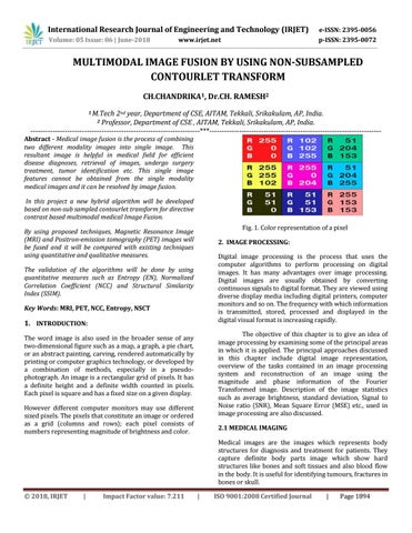
In this project a new hybrid algorithm will be developed based on non-sub sampled contourlet transform for directive contrast based multimodal medical Image Fusion. Fig. 1. Color representation of a pixel
By using proposed techniques, Magnetic Resonance Image (MRI) and Positron-emission tomography (PET) images will be fused and it will be compared with existing techniques using quantitative and qualitative measures.
2. IMAGE PROCESSING: Digital image processing is the process that uses the computer algorithms to perform processing on digital images. It has many advantages over image processing. Digital images are usually obtained by converting continuous signals to digital format. They are viewed using diverse display media including digital printers, computer monitors and so on. The frequency with which information is transmitted, stored, processed and displayed in the digital visual format is increasing rapidly.
The validation of the algorithms will be done by using quantitative measures such as Entropy (EN), Normalized Correlation Coefficient (NCC) and Structural Similarity Index (SSIM). Key Words: MRI, PET, NCC, Entropy, NSCT
1. INTRODUCTION:
The objective of this chapter is to give an idea of image processing by examining some of the principal areas in which it is applied. The principal approaches discussed in this chapter include digital image representation, overview of the tasks contained in an image processing system and reconstruction of an image using the magnitude and phase information of the Fourier Transformed image. Description of the image statistics such as average brightness, standard deviation, Signal to Noise ratio (SNR), Mean Square Error (MSE) etc., used in image processing are also discussed.
The word image is also used in the broader sense of any two-dimensional figure such as a map, a graph, a pie chart, or an abstract painting, carving, rendered automatically by printing or computer graphics technology, or developed by a combination of methods, especially in a pseudophotograph. An image is a rectangular grid of pixels. It has a definite height and a definite width counted in pixels. Each pixel is square and has a fixed size on a given display. However different computer monitors may use different sized pixels. The pixels that constitute an image or ordered as a grid (columns and rows); each pixel consists of numbers representing magnitude of brightness and color.
2.1 MEDICAL IMAGING Medical images are the images which represents body structures for diagnosis and treatment for patients. They capture definite body parts image which show hard structures like bones and soft tissues and also blood flow in the body. It is useful for identifying tumours, fractures in bones or skull.
Š 2018, IRJET
|
Impact Factor value: 7.211
|
ISO 9001:2008 Certified Journal
|
Page 1894