Infectious disease diagnosis often requires expensive and complex lab equipment, skilled personnel, and significant time, all of which can be barriers to effective outbreak response in low-resource settings. To address these challenges, scientists have
Cont’d on page 9
First-of-its-Kind Device Profiles Newborn Immune Function from Single Drop of Blood
Premature infants are highly susceptible to severe and life-threatening conditions, such as sepsis and necrotizing enterocolitis (NEC). Newborn sepsis, which is a bloodstream infection occurring in the first weeks of life, is a significant global health
issue, contributing to approximately one million infant deaths annually. NEC, a severe intestinal disease causing intense inflammation, is one of the top causes of death among premature babies, with up to 50% of low birth weight neonates diagnosed with
Cont’d on page 13
SPredicting Sepsis At 90% Accuracy
AI Detects Gene Combinations That Cause Complex Illnesses
AI Detects Gene Combinations That Cause Complex Illnesses
In an analytical breakthrough that links genotype to phenotype, a new computational tool called Transcriptome-Wide Conditional Variational Auto-Encoder (TWAVE) uses generative artificial intelligence (AI) to uncover how combinations of genes contribute to disease development.
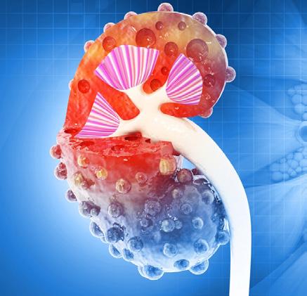
Low-Cost Portable Screening Test to Transform Kidney Disease Detection
Millions of individuals suffer from kidney disease, which often remains undiagnosed until it has reached a critical stage. This silent epidemic not only diminishes the quality of life for those affected but also imposes significant financial strain on healthcare systems due to the costs associated with
Mass Spectrometry Detects Bacteria Without Time-Consuming Isolation and Multiplication
Speed and accuracy are essential when diagnosing diseases. Traditionally, diagnosing bacterial infections involves the labor-intensive process of isolating pathogens and cultivating bacterial cultures, often resulting in several days of waiting before targeted treat-
ment can begin. Researchers have now developed a groundbreaking method that can identify bacteria with unprecedented speed, reducing the waiting time from several days to just a few minutes.
The innovative approach, developed by researchers at Technical
epsis is a severe response to an infection that causes the immune system to attack the body’s own organs and tissues, potentially leading to organ failure and death if not addressed promptly. It is responsible for approximately 20% of all global deaths. Predicting sepsis remains challenging, as its early symptoms are vague and current diagnostic tests can take up to
Cont’d on page 10
Smartphone Detects Colorectal Cancer
Despite the availability of colorectal cancer screening programs, participation remains low, especially for fecal immunochemical tests (FIT), a non-invasive method to detect hidden blood in the stool. With only about 20% of the eligible population regularly using FIT, researchers are exploring digital alternatives to improve uptake. Now, a new smartphone-based stool test
Cont’d on page 2
Smartphone Detects Colorectal Cancer
could serve as a more convenient and patient-friendly option, potentially increasing participation in early detection efforts.
Scientists at the German Cancer Research Center (DKFZ, Heidelberg, Germany; www.dkfz.de) sought to evaluate whether smartphone-based testing could meaningfully supplement traditional laboratory-based FIT screening. Their goal was to leverage everyday technology to reduce barriers and enhance accessibility for individuals hesitant to undergo standard screening procedures. The new method uses a commercially available rapid hemoglobin test paired with a smartphone app.
The process is simple: users collect a stool sample at home using a test stick, mix it in a solution, and place three drops on a test cassette. After a 15-minute wait, the test cassette is photographed using a smartphone. The app then analyzes the color intensity on the cassette to determine the presence of blood in the stool, displaying results instantly. Unlike traditional FIT, this approach eliminates the need for doctor visits, laboratory processing, and long wait times, offering a seamless, private, and immediate at-home experience.
To evaluate the effectiveness and acceptance of the smartphone test, researchers offered it to participants in the BLITZ study, who were already scheduled for colonoscopy appointments across gastroenterology practices in southern Germany between 2021 and 2023. Out of 654 participants, 361 (55%) chose to take the smartphone-based test in addition to the standard FIT. The results were promising: 89% of those who used the smartphone method rated it as a useful alternative to the conventional test, according to a standardized questionnaire.
A comparison with the colonoscopy findings showed that advanced, potentially cancerous mucosal changes were detected by the app in 28% of cases. A comparable sensitivity of 34% was determined for laboratory testing. The specificity—the second important criterion for the informative value of a diagnostic test—was 92% for both methods, suggesting a low rate of false-positive test results. This study suggests that digital tools like smartphone-based stool testing could help overcome longstanding participation barriers in colorectal cancer screening efforts. By making FIT more accessible and user-friendly, such innovations may improve early detection rates and reduce mortality associated with the disease.
“Based on our results, smartphone-based FIT testing could be a meaningful alternative or supplement to traditional laboratory tests,” said co-author Herrmann Brenner. “I see a realistic chance that this additional option will enable us to get more people on board to participate in early colon cancer detection and take advantage of the associated opportunities for colon cancer prevention.”
Image: The new technology combines a rapid hemoglobin test with a smartphone app (Photo courtesy of 123RF)
27 th - 31 st July 2025
INTRODUCING
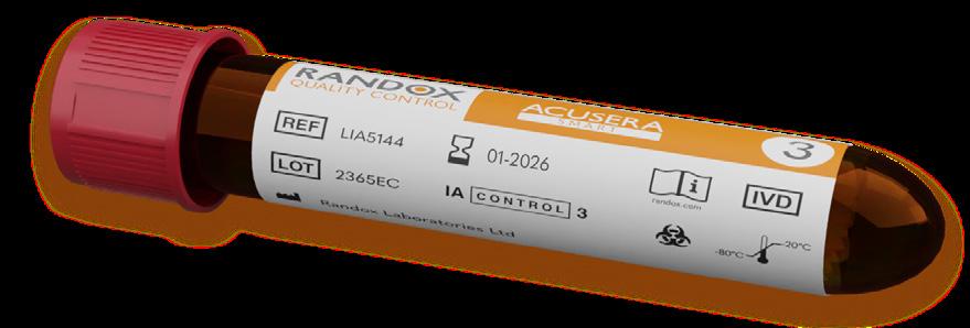
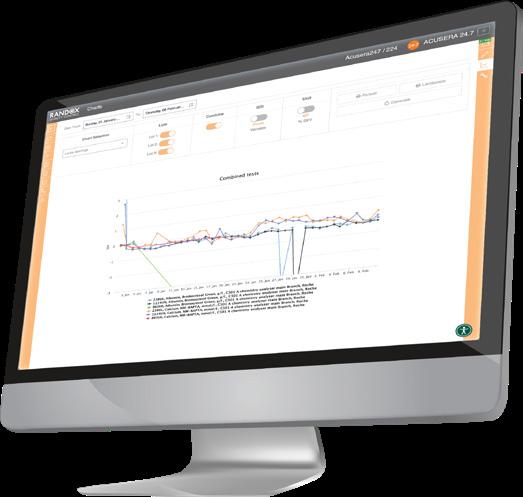
ACUSERA SMART
STREAMLINE YOUR QC, THE SMART WAY
The Acusera Smart controls range is designed to fit directly onto a wide range of test systems without the need to aliquot material, streamlining the QC process by minimizing human error and optimizing workflow.
STREAMLINE WORKFLOW
With the ability to load the Acusera Smart QC and walk away, it optimizes workflow by removing manual steps.
AUTOMATE PROCESSES
Automating the quality control process reduces turnaround time and increases efficiency.
SIMPLIFY PROCESSES
Our SmartScan controls are linked to an XML file from Randox.com, where QC target values can be uploaded directly to the analyzer, simplifying the onboarding process.
MINIMISE HUMAN ERROR
The Smart QC workflow minimizes the potential for human error by reducing the operator handling time.
REDUCE STORAGE
Can be conveniently stored onboard analyzers with refrigerated compartments, for added convenience.
CONVENIENT DESIGN
Our Smartload controls can be conveniently loaded directly into the analyzer, eliminating the need to aliquot QC material.
Stem Cell Test Predicts Treatment Outcome for Patients
with Platinum-Resistant Ovarian Cancer
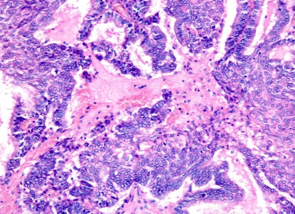
Image: The cancer stem cell test can accurately choose more effective treatments (Photo courtesy of Shutterstock)
Epithelial ovarian cancer frequently responds to chemotherapy initially, but eventually, the tumor develops resistance to the therapy, leading to regrowth. This resistance is partially due to the activation and selection of cancer stem cells (CSCs) that repair and rebuild the tumor after chemotherapy damage. New findings from a Phase 3 trial, published in the journal npj Precision Oncology, reveal that a test for cancer stem cells can help identify more effective treatments and improve outcomes for patients with platinum-resistant ovarian cancer (those who experience recurrence within six months after receiving platinumbased chemotherapy).
The ChemoID platform, developed by researchers at the University of Cincinnati (Cincinnati, OH, USA; med.uc.edu), works by testing cancer stem cells found in individual patients' tumors against specific anticancer drugs to determine the most effective treatment options. The platform operates under the premise that by evaluating both tumor cells and cancer stem cells for chemosensitivity, one can target and eliminate CSCs that could otherwise repopulate the tumor. In the trial, 81 patients with platinum-resistant ovarian cancer were randomly assigned to have their chemotherapy treatment chosen using the ChemoID platform or based on the physician's standard methods. Physicians typically select treatments based on approved therapies, their previous experiences, past treatments the patient has undergone, and the side effects or toxicity that the patient has previously experienced.
The primary outcome measured was the patients’ objective response rate (ORR),
which refers to the percentage of patients in each group who achieved either a partial or complete response to the treatment within a designated period. In addition, progressionfree survival (PFS)—the duration of time a patient lives without the cancer worsening during and after treatment—and the duration of the response were measured. Results showed that patients who were treated using the ChemoID assay had significantly better clinical and statistical outcomes compared to those who received the physician’s choice of therapy. The ORR for patients in the ChemoID group was 50%, while only 5% of patients in the physician-choice group responded positively.
Median PFS for patients in the ChemoID group was 11 months, compared to only three months in the physician-choice group. Furthermore, patients in the ChemoID group had a median duration of response of eight months, whereas those in the physicianchoice group experienced just five-and-a-half months. These improved response rates could potentially reduce healthcare costs associated with ineffective therapies and unnecessary toxicity, according to researchers.
The findings from this study suggest that using assay-guided treatment approaches could reduce financial toxicity for ovarian cancer patients, particularly those with platinumresistant disease who often struggle with treatment decisions. Future studies are needed to further validate the ChemoID platform and explore how it works with different molecular subgroups, such as patients with BRCA mutations. Additionally, the testing of emerging biologic therapies will help refine the best uses for this test.
Graham
Hernán Fares
Argentina
Bernard Gouget France
Maurizio Ferrari Italy
Tahir S. Pillay South Africa
Andreas Rothstein Colombia
Praveen Sharma India
Rosa I. Sierra-Amor Mexico
Peter Wilding United States
Andrew Wootton United Kingdom
LabMedica lnternational is published eight times a year and is circuIated worldwide (outside the USA and Canada) without charge and by written request, to clinical laboratory specialists and administrators, and other qualified professionals allied to the field.
To all others: Paid Subscription is available for a two-year subscription charge of US$120. Single copy price is US$20. Mail your paid subscription order accompanied with payment to Globetech Media, P.O.B. 800222, Miami, FL 33280-0222.
For change of address or questions on your subscription, write to: LabMedica lnternational, Circulation Services at above address; or visit: www.LinkXpress.com
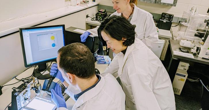
New POC Biosensing Technology Improves Detection of Molecular Biomarkers
Traditional diagnostic procedures in medicine typically involve sending a patient’s blood or tissue samples to clinical laboratories, where trained scientists perform testing and data interpretation. In contrast, point-of-care diagnostics are conducted and produce results directly at the site of patient care, be it at home, in a clinic, or elsewhere. This approach enables rapid, cost-effective, and user-friendly tests that guide immediate medical decisions. Common examples already in everyday use include urinebased pregnancy tests, COVID-19 antigen kits, and glucose meters that help diabetes patients manage fluctuations in blood sugar throughout the day. Researchers are exploring innovative ways to implement such technologies within clinical environments, such as during visits to oncologists or oral surgeons. Doing so could reduce the logistical and financial strain on patients while enhancing physicians’ ability to make timely, informed decisions. Researchers are now a step closer to realizing this goal through the integration of machine learning-based analysis into point-ofcare biosensing platforms.
Researchers at the Carl R. Woese Institute for Genomic Biology (Urbana, IL, USA; igb.illinois.edu) have developed a new approach called LOCAPRAM that simplifies biomarker detection by removing the need for skilled professionals to analyze the resulting images. This development builds upon the team’s earlier work introducing Photonic Resonator Absorption Microscopy, or PRAM—a biosensing technique capable of identifying individual biomarker molecules, such as nucleic acids, antigens, and antibodies. In contrast to conventional methods, which measure signals from large groups of molecules, PRAM offers the sensitivity to detect and analyze single molecules.
PRAM images appear as a red field dotted with black specks. Though visually straightforward, determining which specks represent AuNP-tagged biomarker molecules requires expert analysis. To make this method suitable for point-of-care settings, the researchers proposed incorporating machine learning into the image analysis. Unlike other biosensing methods that primarily detect optical signals, PRAM produces microscope images, making it wellsuited for deep learning applications. To ensure high-quality data for algorithm training, the team imaged identical samples using both PRAM and scanning electron microscopy.
The AuNPs—nanoparticles roughly 1,000 times thinner than a strand of hair—appear as tiny black spots in PRAM images but are more distinctly
visible under electron microscopy. Through a labor-intensive process, the researchers meticulously cross-referenced every black spot seen in the PRAM images with the corresponding features in the electron microscope images to create an accurate training dataset for the machine learning model. This resulted in a deep learning-enhanced method known as Localization with Context Awareness, integrated with PRAM. This combined platform delivers precise, real-time molecular biomarker detection without requiring the intervention of a technical specialist. When evaluated, the findings published in Biosensors and Bioelectronics showed that LOCA-PRAM outperformed standard techniques by identifying lower concentrations of biomarkers and reducing the likelihood of false positives and negatives.
“Current technologies require patients to visit hospitals to get diagnostics which takes time. A
lot of people also have barriers where more appointments may not be financially or spatially feasible,” said Han Lee, first author of the study and a graduate student in the Nanosensors research group. “I think that we can make a difference by developing more point-of-care technologies that are available for people.”
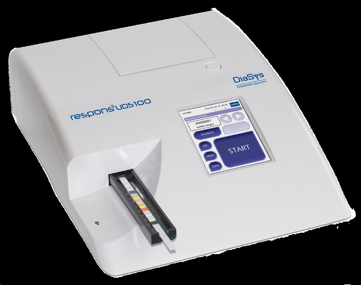
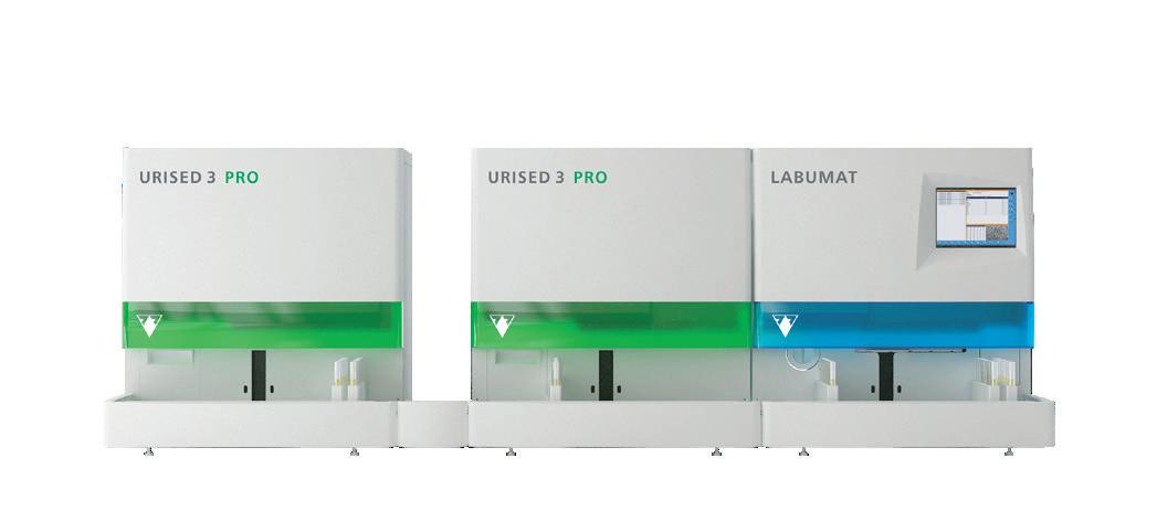
High performance solution for advanced urine determination
•
• Accurate and consistent results by automated reading, interpretation and documentation
• Reliable and cost-effective alternative through multiple system advantages
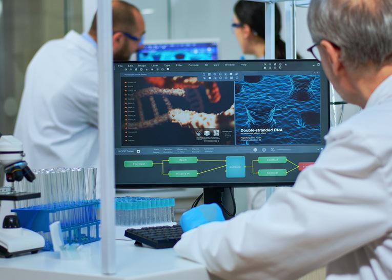
Image: A machine learning method is helping to bring diagnostic testing out of the lab (Photo courtesy of 123RF)
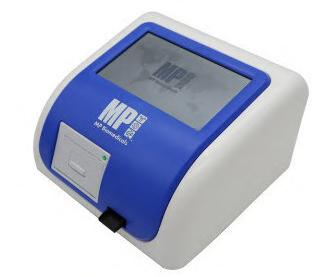
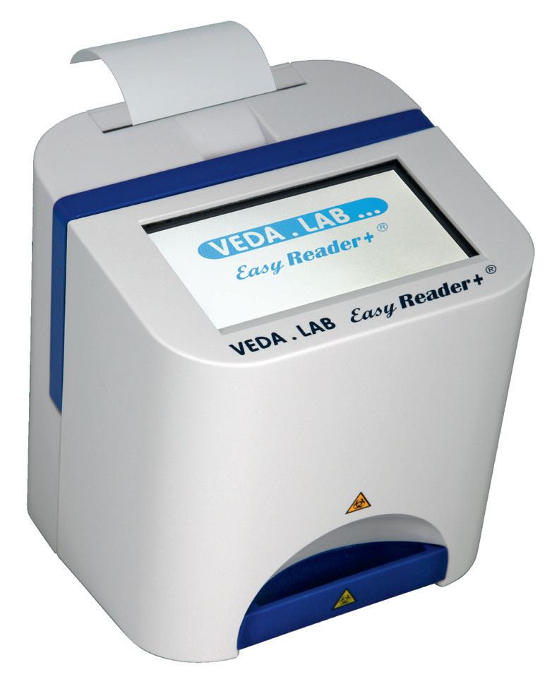
IMMUNOFLUORESCENCE ANALYZER MP BIOMEDICALS
The MPQuanti Immunofluorescence Analyzer is a fast, compact device delivering quantitative and qualitative results in under 20 seconds.
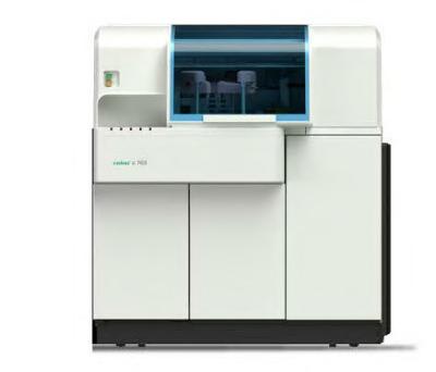
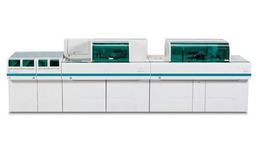
The extensive test menu includes cardiac markers, infections, diabetes, and tumor screening. AUTO CLINICAL CHEMISTRY ANALYZER ROCHE DIAGNOSTICS
AI Detects Gene Combinations that Cause Complex Illnesses
For years, scientists have faced significant challenges in decoding the genetic basis of complex diseases like diabetes, asthma and cancer. Traditional methods such as genome-wide association studies often fall short in identifying how multiple genes interact to influence these traits, primarily due to limited statistical power and the vast number of possible gene combinations. Now, researchers have developed a new computational tool that uses generative artificial intelligence (AI) to uncover the genetic networks driving complex human conditions.
The tool, known as Transcriptome-Wide Conditional Variational Auto-Encoder (TWAVE), was created by a team of biophysicists at Northwestern University (Evanston, IL, USA; northwestern.edu) to improve understanding of how combinations of genes contribute to disease development. This model represents a breakthrough in linking genotype to phenotype by shifting the analytical focus from individual genes to coordinated gene groups.
TWAVE uses a generative AI model that amplifies limited gene expression data to detect patterns of gene activity associated with disease
states. It works by simulating both healthy and diseased cellular environments and identifying how specific changes in gene expression can alter those states. Through a combined machine learning and optimization framework, TWAVE pinpoints the gene changes most likely to transition a cell from healthy to diseased or vice versa. By analyzing gene expression rather than DNA sequences, TWAVE circumvents patient privacy concerns and captures the influence of environmental factors that can modulate gene activity. This focus allows the model to provide a dynamic view of cellular function and better reflect the complexity of real-world disease mechanisms. In the study published in PNAS on June 12, TWAVE was tested across a variety of complex diseases. It successfully identified relevant gene combinations, including some that had been missed by existing techniques.
Notably, the model revealed that the same disease could arise from different gene sets in different individuals, highlighting the potential for more personalized therapeutic strategies. By revealing gene networks instead of isolated markers, TWAVE offers a powerful tool for de-
The cobas c 703 analytical unit enhances efficiency with a throughput of up to 2000 tests/hour and 70 onboard reagent positions. Ideal for high-volume labs, it reduces manual intervention while maintaining operational precision.
veloping targeted treatments that consider the specific genetic drivers in each patient. It paves the way for new interventions designed to modify the collective gene expression patterns that underlie chronic and multifactorial diseases.
“Many diseases are determined by a combination of genes — not just one,” said Northwestern’s Adilson Motter, the study’s senior author. “You can compare a disease like cancer to an airplane crash. In most cases, multiple failures need to occur for a plane to crash, and different combinations of failures can lead to similar outcomes. This complicates the task of pinpointing the causes. Our model helps simplify things by identifying the key players and their collective influence.”
Advanced Predictive Algorithms Identify Risk of Undiagnosed Cancer
Two newly developed advanced predictive algorithms leverage a person’s health conditions and basic blood test results to accurately predict the likelihood of having an undiagnosed cancer, including challenging-to-diagnose liver and oral cancers. These innovative models have the potential to transform cancer detection in primary care, making it easier for patients to receive treatment at much earlier stages.
Currently, the UK’s NHS uses prediction
tools like the QCancer scores, which integrate various patient data to identify individuals at high risk for undiagnosed cancer, allowing general practitioners and specialists to refer them for further testing. Researchers from Queen Mary University of London (London, UK) and the University of Oxford (Oxford, UK) utilized anonymized electronic health records from over 7.4 million adults in England to develop two new algorithms. These models are more sensitive than existing tools and could
lead to improved clinical decision-making and earlier cancer detection. Significantly, the new algorithms incorporate not only patient details like age, family history, medical diagnoses, symptoms, and general health, but also include the results of seven routine blood tests. These blood tests, which measure full blood count and liver function, serve as biomarkers to enhance early cancer diagnosis.
When compared with the current QCancer
Cont’d on page 8
Image: The new method uses a generative AI model to amplify limited gene expression data (Photo courtesy of Camila Felix/Northwestern University)
Low-Cost Portable Screening Test to Transform Kidney Disease Detection
dialysis and organ transplants. However, early diagnosis can significantly alter a patient’s course of treatment, offering better options and potentially halting or slowing the progression of the disease. The challenge lies in the fact that current kidney function tests typically require visits to a laboratory, expensive equipment, skilled personnel, and several days to obtain results. These requirements make routine screenings difficult, particularly for those in rural and Indigenous communities. Now, researchers have created an innovative tool that could revolutionize how kidney disease is diagnosed, especially in underserved areas.
A team of researchers from the University of Manitoba (Winnipeg, Canada; www.umanitoba. ca) has developed a device known as the uCR-Chip, a low-cost, portable diagnostic tool designed to make kidney function testing faster, more convenient, and widely accessible. The results are available in less than seven minutes, and the test does not require specialized laboratory equipment. Their research, published in Microsystems & Nanoengineering, showcases how this device could play a crucial role in enhancing early detection and improving outcomes for individuals living with chronic kidney disease (CKD). The uCR-Chip addresses the challenges of current testing methods by utilizing a color-based chemical reaction to measure creatinine, a key biomarker of kidney health, from a small urine sample. Since it does not require advanced lab equipment, it can be used directly in health clinics or mobile healthcare settings.
The uCR-Chip has the potential to relieve pressure on healthcare systems by identifying kidney issues at earlier stages, thereby reducing the number of patients who progress to more advanced stages of disease. This could lead to fewer patients requiring costly treatments such as dialysis or transplants, allowing for more effective management with early interventions. In many rural, remote, and Indigenous communities, where access to advanced diagnostic equipment is limited, the uCR-Chip offers a portable, cost-effective alternative that could greatly improve access to vital testing. As healthcare systems worldwide seek affordable solutions to manage chronic diseases, innovations like the uCRChip offer not just hope, but tangible solutions that bring life-saving diagnostics to those who need them most.
“Traditiona lab test can take days and may delay diagnosis,” said Dr. Francis Lin who led the research team. “Our new test method will lead to faster, more accessible and reliable diagnostic results to prevent irreversible kidney damage.”
Image: The research team has developed the uCR-Chip device to enhance kidney function testing (Photo courtesy of University of Manitoba)
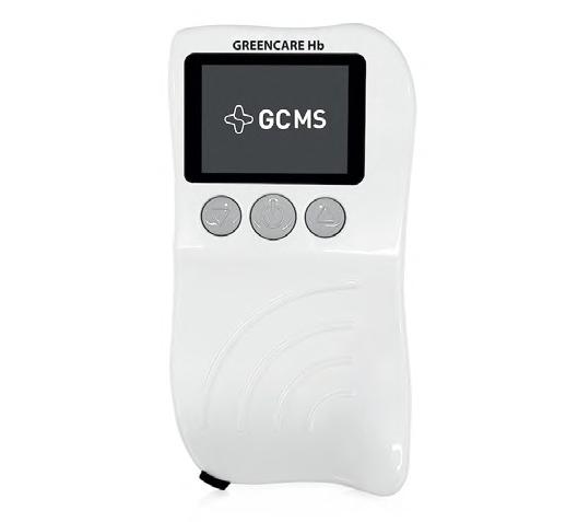
BGREENCARE Hb is an IVD medical analyzer designed for the quantitative measurement of total hemoglobin concentration in blood, helping to prevent anemia, iron deficiency, and other blood disorders through regular
Autolas Bi1000 is a modular clinical chemistry system integrating biochemistry and immunoassay modules. Features include 150–300 sample inlet capacity, an overtaking track for urgent tests, and SDM sample management.
New Test Diagnoses Bacterial Meningitis Quickly and Accurately
acterial meningitis is a potentially fatal condition, with one in six patients dying and half of the survivors experiencing lasting symptoms. Therefore, rapid diagnosis and treatment are critical. However, distinguishing bacterial meningitis from other similar conditions is often challenging for healthcare providers. Currently, it can take a considerable amount of time to diagnose meningitis, which delays the initiation of appropriate treatment. Until now, there have been no reliable diagnostic tests available that could quickly differentiate bacterial meningitis from other conditions. Researchers have now developed a new diagnostic test capable of quickly and accurately diagnosing bacterial meningitis by measuring the CRP protein in cerebrospinal fluid, a protein commonly tested in blood to detect bacterial infections.
Researchers at Amsterdam UMC (Amsterdam, Netherlands; www.amsterdamumc.org) found that the CRP protein in cerebrospinal fluid is a highly reliable indicator of bacterial meningitis. While CRP is often tested in blood to detect bacterial infections, its value in cerebrospinal fluid had not been extensively
studied until now. After successful laboratory tests, the researchers demonstrated that the device used to measure CRP in blood is also sensitive enough to measure CRP in cerebrospinal fluid. Since June 2024, this new test has been incorporated into daily practice at Amsterdam UMC. In their latest, published in The Lancet Regional Health Europe, the researchers described how the test was introduced and assessed its effectiveness in a clinical setting.
The results showed that all patients diagnosed with bacterial meningitis had elevated CRP levels in their cerebrospinal fluid, whereas only a few patients without bacterial meningitis showed similar elevations. The test was also proven to be reliable in additional studies conducted with children and patients in Denmark. It is expected that more hospitals will begin to adopt this test, as it can be performed in laboratories with existing equipment. The cost of the test ranges from three to five euros, offering an affordable and accessible solution
Image: CRP protein in cerebrospinal fluid was found to be a very reliable indicator of bacterial meningitis (Photo courtesy of 123RF)
for diagnosing and treating bacterial meningitis more quickly.
“Any laboratory in which CRP is measured in blood can introduce this test for cerebrospinal fluid tomorrow. We could not have predicted in advance that a new diagnostic test would be used in patients within a year of its discovery,” said last author and Amsterdam UMC neurologist, Matthijs Brouwer.
Advanced Predictive Algorithms Identify Risk of Undiagnosed Cancer
Cont’d from page 6
models, the new algorithms identified four additional medical conditions associated with an elevated risk of 15 different types of cancer, including those affecting the liver, kidneys, and pancreas. The new models also discovered two additional links between family history and lung or blood cancer, along with seven new symptoms—such as itching, bruising, back pain, hoarseness, flatulence, abdominal mass, and dark urine—that were associated
with various types of cancer. The findings, published in Nature Communications, show that these new algorithms significantly improve diagnostic capabilities and are currently the only models applicable in primary care settings to assess the likelihood of undiagnosed liver cancer.
“These algorithms are designed to be embedded into clinical systems and used during routine GP consultations,” said Professor Julia Hippisley-Cox, Professor of Clinical
Epidemiology and Predictive Medicine at Queen Mary University of London, and lead author of the study. “They offer a substantial improvement over current models, with higher accuracy in identifying cancers — especially at early, more treatable stages. They use existing blood test results which are already in the patients’ records making this an affordable and efficient approach to help the NHS meet its targets to improve its record on diagnosing cancer early by 2028.”
Diagnosing Infectious Diseases in 10 Minutes
Cont’d from cover
developed a breakthrough paper-based diagnostic device that can detect COVID-19 and other infectious diseases in under 10 minutes, without the need for sophisticated lab equipment or trained personnel.
The Radially Compartmentalized Paper Chip (RCP-Chip), developed by a team of scientists at NYU Abu Dhabi (Abu Dhabi, UAE; nyuad.nyu.edu), offers a fast, affordable, and portable solution for on-site screening. The RCP-Chip was conceived during the early COVID-19 lockdowns and was developed to detect even minute traces of viral genetic material. It operates without the need for electricity or specialized equipment, requiring only a source of mild heat (around 65°C, similar to warm water).
The chip is made from a single sheet of paper, incorporating miniature components such as sample ports, vents, fluidic resistors, and reaction chambers pre-loaded with primers, enzymes, and gold nanoparticles to facilitate the detection process. In their research published in Advanced Sensor Research, the team has detailed the development and validation of the RCP-Chip. The device functions as a rapid, multiplexed diagnostic platform that allows for the detection of multiple infectious diseases, making it particularly useful for low-resource settings.
The RCP-Chip’s ability to detect SARS-CoV-2 genetic targets in under 10 minutes has been validated in tests, and the device’s effectiveness was demonstrated through its ability to perform multiplexed testing, detecting multiple gene targets in a single run, which increases efficiency while reducing sample volume and cost. The RCP-Chip can be reconfigured to detect a variety of pathogens, including bacteria, viruses, and oth ers, across various sample types such as saliva, blood, and environmental sources.
The device’s compact, user-friendly design makes it a powerful tool for field deployment, especially in regions lacking access to traditional diagnos tic laboratories. Looking forward, the research team aims to enhance the chip’s plasmonic detection capabilities and expand its use for point-of-care applications. They also plan to explore smartphone connectivity for real-time data sharing and outbreak tracking, further increasing the device’s utility in public health surveillance.
“This is a fast, affordable, lab-free test that detects multiple gene targets in under 10 minutes,” said Pavithra Sukumar, NYUAD Research Assistant and co-first author of the study. “What makes it truly impactful is its real-world potential. This portable test could significantly improve outbreak response by enabling faster isolation, treatment, and control.”
Image: The paper-based diagnostic tool is designed for rapid, affordable infectious disease detection (Photo courtesy of Advanced Microfluidics and Microdevices Laboratory, NYU Abu Dhabi)
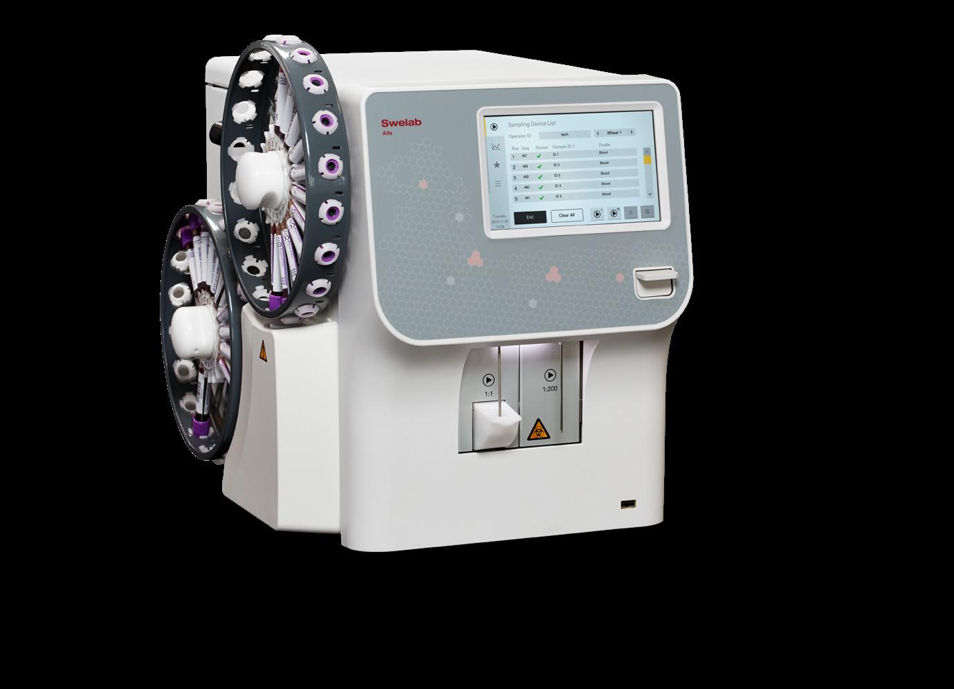
Swelab Alfa Plus Sampler
3-part hematology system, with autosampler design that provides constant mixing of queued samples.
Autolader
Pre-load up to 2x 20 samples and let it do the work.
Cap-piercing device
For analysis of samples with decreased risk of blood contact.
Pre-dilution mode
Specialized channel for analyzing
pre-diluted whole blood, particularly when the available sample volume is limited.
Shear valve sample aspiration
Maintenance-free shear valve to deliver
Micro-pipet adapter (MPA)
MPA method allows analysis of capillary blood from a finger-stick sample taken directly from the patient.
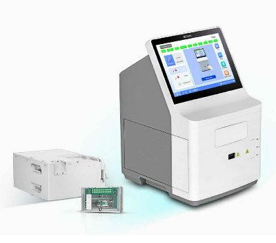
BLOOD GAS AND CHEMISTRY ANALYSIS
EDAN INSTRUMENTS
The Edan i500 delivers blood gas and chemistry results in 45 seconds using microfluidic and multi-use microchip technology. It features auto-sampling, a 10.1” color touchscreen, barcode scanner, and up to 600 tests per cartridge.
RESPIRATORY SYNCYTIAL VIRUS TEST SEKISUI DIAGNOSTICS
The OSOM® RSV Test is a CLIA-waived, point-ofcare test designed to detect RSV using a painless anterior nasal swab, in just 15 minutes. It is suitable for both children aged 6 months to 6 years and adults aged 60 and above.
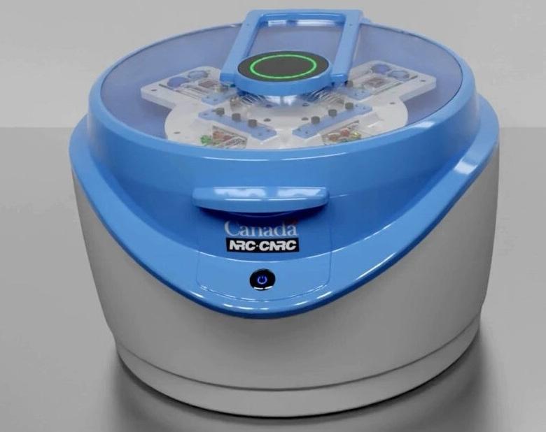
Predicting Sepsis At 90% Accuracy
Cont’d from cover
18 hours, often requiring specialized labs. This delay in treatment increases the risk of death by nearly 8% per hour. Now, researchers have developed a new blood test and portable device that can detect the onset of sepsis more quickly and accurately than existing methods.
The test, developed by scientists at the University of British Columbia (UBC, Vancouver, BC, Canada; www.ubc.ca) and Sepset Biosciences (Victoria, BC, Canada; www.sepset.ca), has demonstrated over 90% accuracy in identifying individuals at high risk of developing sepsis, marking a significant advancement in how doctors will diagnose and treat this condition. The study, published in Nature Communications, involved analyzing blood samples from more than 3,000 hospital patients with suspected sepsis. Using machine learning, the researchers identified a six-gene expression signature, dubbed “Sepset,” which successfully predicted sepsis nine times out of ten, even before a formal diagnosis was made. When tested with an additional 248 blood samples using RTPCR (Reverse Transcription Polymerase Chain Reaction), a common hospital laboratory technique, the test showed 94% accuracy in detecting early-stage sepsis in patients whose condition was likely to worsen.
rithm for assessing sepsis risk. To make this test more accessible in clinical settings, the National Research Council of Canada (NRC) has developed a portable device, PowerBlade, which uses a drop of blood and an automated sequence of steps to detect sepsis efficiently. In trials with 30 patients, PowerBlade demonstrated 92% accuracy in identifying patients at high risk of sepsis and 89% accuracy in ruling out those not at risk. The device provided results in under three hours, making it suitable for use in various environments, including emergency rooms and remote healthcare settings.
This success highlights the powerful role of AI in handling complex data, pinpointing the critical genes necessary for predicting sepsis, and developing a highly accurate algo-
“Sepsis accounts for roughly 20% of all global deaths,” said lead author Dr. Claudia dos Santos, a critical care physician and scientist at St. Michael’s Hospital. “Our test could be a powerful game changer, allowing physicians to quickly identify and treat patients before they begin to rapidly deteriorate.”
Image: PowerBlade is a portable device that uses a drop of blood and an automated sequence of steps to efficiently detect sepsis (Photo courtesy of NRC)
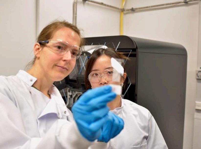
Mass Spectrometry Detects Bacteria Without Time-Consuming Isolation
and Multiplication
University of Munich (TUM, Munich, Germany; www.tum.de) and Imperial College London (London, UK; www.imperial.ac.uk) uses mass spectrometry to detect specific metabolic products of bacteria directly from tissue and stool samples. Central to this process is a database that currently includes 232 medically relevant bacterial species and their associated metabolic products. From this database, biomarkers are extracted to allow for the direct identification of specific bacteria.
The new method is capable of identifying bacteria responsible for a variety of serious conditions, including stomach cancer, pneumonia, meningitis, preterm birth, gonorrhea, and sepsis. To ensure that this method becomes a regular tool in clinical settings, the biomarker database needs to be expanded. The researchers note that over 1,400 bacterial pathogens are known, and their specific metabolic products need to be identified and added to the database. The team also sees significant potential for this method in personalized medicine, where treatments
Machine Learning-Enabled Blood Test Predicts Immunotherapy Response in Lymphoma Patients
Chimeric antigen receptor (CAR) T-cell therapy has emerged as one of the most promising recent developments in the treatment of blood cancers. However, over half of non-Hodgkin lymphoma (NHL) patients who fail to respond to conventional treatments also experience relapse or disease progression within six months after undergoing CAR T therapy. In response, a new tool using machine learning has been developed to predict how well an NHL patient might respond to CAR T-cell therapy prior to starting the treatment.
Called InflaMix (Inflammation Mixture Model), this innovative tool was developed by researchers at City of Hope (Duarte, CA, USA; www.cityofhope.org) to evaluate inflammation, which is considered a potential cause of CAR T failure, by testing for various blood biomarkers in 149 patients with NHL. Using machine learning, a form of artificial intelligence that analyzes data through algorithms to identify patterns and draw conclusions, the model was able to identify an inflammatory biomarker through a set of blood tests that are not typically used in standard clinical practice. By examining the inflammatory signature identified by InflaMix, the researchers found a significant association with an increased risk of CAR T treatment failure, including a higher risk of death or relapse. Notably, InflaMix is an unsupervised model, meaning it was trained without prior knowledge of clinical outcomes.
can be tailored precisely to individual patients based on the specific bacteria detected.
“Our innovative approach is not to look directly for the pathogenic bacteria, but only for their metabolic products. This allows us to detect them indirectly, but much more quickly,” said first author Wei Chen.
“This is one of the most important future topics in biotechnology and medicine. Targeted interventions can dramatically improve the chances of successful treatment. As analysts, we develop modern tools and methods for doctors to do this,” added Prof. Nicole Strittmatter.
Designed + manufactured with P E O P L E in mind.
The research team noted that the machine learning model is highly adaptable, showing good performance even when using just six commonly available blood tests—tests that are typically evaluated for lymphoma patients—to assess InflaMix’s functionality with less data. This is an important feature because it suggests that the test could be accessible to a broad range of lymphoma patients. To validate their initial findings, the researchers studied three independent cohorts, comprising 688 NHL patients with diverse clinical characteristics and disease subtypes who had received different CAR T products. Moving forward, the team plans to explore whether the inflammation identified by InflaMix directly impacts CAR T-cell function and to investigate the underlying sources of this inflammation.
“These studies demonstrate that by using machine learning and blood tests, we could develop a highly reliable tool that can help predict who will respond well to CAR T cell therapy,” said Marcel van den Brink, M.D., Ph.D., president of City of Hope Los Angeles and City of Hope National Medical Center, and a senior author of the paper published in Nature Medicine. “InflaMix could be used to reliably identify patients who are about to be treated with CAR T and are at high risk for the treatment not working. By identifying these patients, doctors may be able to design new clinical trials that can boost the effectiveness of CAR T with additional treatment strategies.”
Puritan® specimen collection and transport devices. Our extensive selection of swabs and transport systems — including foam and patented flock swabs, as well as liquid Amies and universal transport media — are crafted for both convenience and patient comfort.
LEARN MORE ONLINE info.puritanmedproducts.com/specimen-2025
VISIT US IN PERSON ADLM 2025, Booth #422
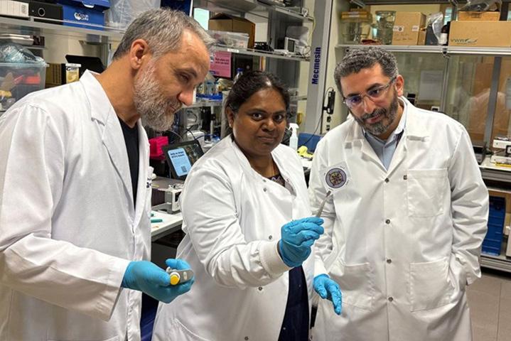
Image: Prof. Nicole Strittmatter (left) and first author Wei Chen stand in front of the mass spectrometer with a tissue sample (Photo courtesy of Robert Reich/TUM)
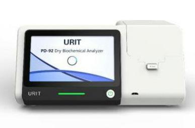
The PD–92 ALT/AST is a compact, high-throughput dry chemistry analyzer designed for precise liver function testing. It delivers accurate measurements of ALT and AST enzymes, essential for assessing
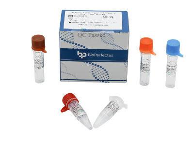
VIRUS TEST BIOPERFECTUS TECHNOLOGIES
The Dengue Virus Type 3 & Type 4 Real Time PCR Kit is designed for the qualitative detection of Dengue virus types 3 and 4 in clinical specimens such as whole blood, serum, plasma, and urine.
Gut Microbes Could Enable Early Detection and Treatment of Pancreatic Cancer
Pancreatic cancer remains one of the most serious and challenging diseases in oncology due to its difficulty in detection and limited treatment options. Now, a new international collaborative study suggests that in the future, early-stage pancreatic cancer could potentially be identified through the analysis of gut microbes. These microbes may also present new opportunities for developing therapies. In this study, researchers from the University of Jyväskylä (Jyväskylä, Finland; www.jyu.fi/en) and their collaborators analyzed the gut microbiota of over 180 pancreatic cancer patients from Finland and Iran, along with healthy individuals from the same regions. They discovered consistent microbial patterns associated with pancreatic cancer regardless of the patients’ geographic or ethnic backgrounds. Compared to the healthy group, the intestinal microbiota of pancreatic cancer patients contained significantly more facultative pathogens and notably fewer beneficial bacteria. Specifically, a decline was observed in helpful bacteria from the Clostridia class, such as the butyric acid–producing Lachnospiraceae, Butyricicoccaceae, and Ruminococcaceae. In contrast, higher levels of potential pathogens like Enterobacteriaceae, Enterococcaceae, and
Fusobacteriaceae were found in the gut flora of those with pancreatic cancer.
Based on these findings, the researchers propose that future investigations should explore whether these beneficial microbes could be developed into a new class of probiotics—live microbial products—that could be used alongside standard chemotherapy. This combination could potentially offer a more precise and effective method for treating pancreatic cancer. At the same time, the researchers stress the importance of conducting additional studies in varied populations to validate these results. Furthermore, they created a statistical model using microbiome data, which may eventually assist in predicting pancreatic cancer. This marks a promising initial step toward achieving earlier diagnosis of the disease.
“The findings on beneficial Clostridia are interesting because it has been shown earlier that ordinary Clostridiales populations effectively mediate anti-canceric immune reactions against solid tumors,” said Satu Pekkala, Senior Lecturer from the Faculty of Sport and Health Sciences, University of Jyväskylä.
Adrenal Function & Hypertension
Alzheimer’s Diseases
Bone Metabolism
Cancer
Cardiovascular Diseases
Diabetes & Metabolism
Gastrointestinal Metabolism
Growth factors
Inflammation
Kidney Function
Thyroid Function
First-of-its-Kind Device Profiles Newborn Immune Function from Single Drop of Blood
NEC not surviving. The symptoms in infants are often vague, making it difficult to diagnose these conditions. However, both conditions can deteriorate rapidly and require immediate medical treatment to improve recovery chances.
Current diagnostic methods to identify and prevent these critical conditions in newborns rely on large blood samples—up to 1 ml, a substantial amount for a newborn—and lengthy laboratory processes. This is particularly problematic for premature infants, whose total blood volume may be as low as 50 mL, restricting the ability to perform repeated or high-volume sampling and potentially leading to complications like anemia. Additionally, traditional tests, such as blood cultures or inflammatory panels, may take hours or even days to produce actionable results, delaying necessary clinical interventions. Researchers have now created an innovative device to profile the immune function of newborns.
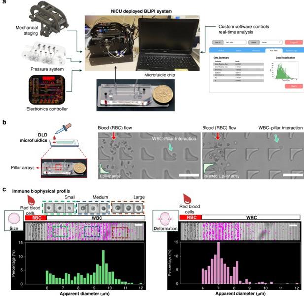
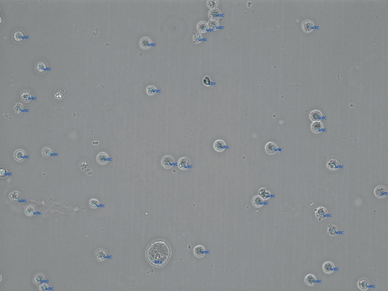
The BiophysicaL Immune Profiling for Infants (BLIPI) system developed by researchers from Singapore-MIT Alliance for Research and Technology (SMART, Singapore; smart.mit.edu) and KK Women’s and Children’s Hospital (KKH, Singapore; kkh.com.sg) uses just a single drop of blood to provide real-time insights into the immune responses of newborns, allowing for the early detection of severe inflammatory conditions and facilitating timely intervention. This groundbreaking device addresses the critical need for rapid and minimally invasive diagnostic tools to safeguard vulnerable newborns, especially those born prematurely. The BLIPI device requires only 0.05 ml of blood and delivers results within 15 minutes. In their study published in Pediatric Research, the researchers demonstrated how BLIPI employs microfluidic technology to assess how immune cells change in response to infection by evaluating their size and flexibility.
Unlike traditional tests that merely detect the presence of pathogens, BLIPI directly shows how a newborn’s immune system is responding. The cellular changes detected by BLIPI align with standard medical tests, such as C-reactive protein (CRP) levels, white blood cell counts, and immature-to-total neutrophil ratios. This rapid testing format can immediately indicate whether a baby’s immune system is combating an infection. In the study, BLIPI was used to screen 19 infants at various time points—8 full-term and 11 preterm—and revealed clear differences in the immune cell characteristics between the babies. Importantly, when one premature infant developed a severe blood infection, the device detected significant changes in immune cells, demonstrating its potential for early infection detection.
BLIPI is a portable device capable of providing results directly at the ward or in neonatal intensive care units (NICUs), eliminating the need to transport blood samples to a laboratory. This makes the device par ticularly useful in resource-limited or rural healthcare settings. Notably, BLIPI requires just one drop of blood, which is 20 times less than the amount needed by existing methods. The rapid results offered by BLIPI can help clinicians make life-saving decisions in critical situations, such as sepsis or NEC, where early intervention is essential.
Future research will focus on larger clinical trials to further validate BLIPI’s diagnostic accuracy across diverse neonatal populations, including various age groups and medical conditions. Additionally, the researchers plan to refine the device’s design to ensure its global adoption in hospitals, providing a much-needed diagnostic solution for vulnerable infants at their bedsides. Beyond hospitals, pharmaceutical companies and researchers may also use BLIPI in clinical trials to assess immune responses to neonatal therapies in real-time, potentially revolutionizing research and develop ment in pediatric medicine.
“Our goal was to create a diagnostic tool that works within the unique constraints of neonatal care — minimal blood volume, rapid turnaround, and high sensitivity,” said Dr Kerwin Kwek, Research Scientist at SMART
Cont’d on page 14 Image: Custom hardware and software for the real-time detection of immune cell biophysical signatures in NICU (Photo courtesy of Pediatric Research, DOI:10.1038/s41390-025-03952-y)
Cont’d
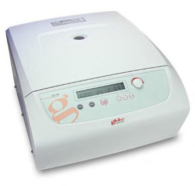
MULTI-PURPOSE CENTRIFUGE
GLOBE SCIENTIFIC
The GCC-MP multi-purpose centrifuge is a reliable, high-performance laboratory device designed for various applications. It features adjustable speed and time settings, allowing for efficient separation of
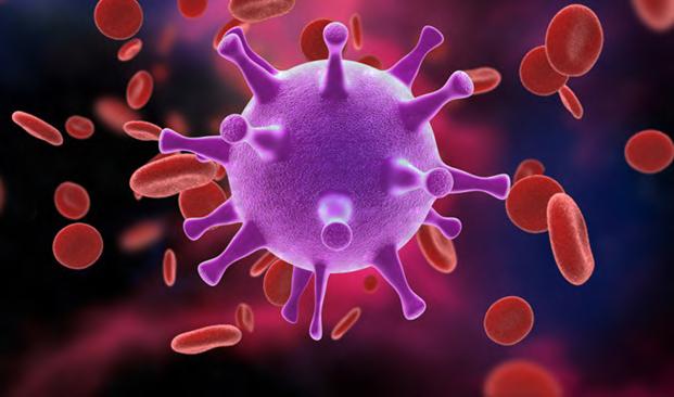
Disposable Microchip Technology Could Selectively Detect HIV in Whole Blood Samples
As of the end of 2023, approximately 40 million people globally were living with HIV, and around 630,000 individuals died from AIDS-related illnesses that same year. Despite a substantial decline in deaths from previous years, AIDS-related deaths continue to represent a critical global health concern. While antiretroviral therapy (ART) has significantly increased life expectancy for people living with HIV, the lack of effective diagnostic tools and disease management strategies has hindered widespread implementation. Only 59% of those in need have access to ART, and approximately one in four individuals with HIV remain unaware of their status. Moreover, people on ART who maintain uninterrupted treatment might experience viral rebound between viral load tests, staying unaware of treatment failure and remaining potentially infectious for weeks or even months.
One of the key challenges in managing HIV is the absence of self-testing technologies capable of detecting new infections during the early acute phase (the first two weeks after infection) or identifying viral rebound in patients on ART. To date, no self-test has been developed to detect HIV during the acute phase or viral rebound in patients with suppressed viral loads on ART. In response to the pressing need for a rapid, affordable, and reliable self-test for early HIV detection, researchers at Florida Atlantic University (Boca Raton, FL, USA; www.fau.edu), in collaboration with other experts, are developing an innovative disposable microchip technology for HIV-1 self-testing. This technology would detect HIV within the first two weeks post-infection and monitor viral rebound in ART-suppressed patients. Additionally, the technology could be adapted for use in detecting other infectious diseases
such as flaviviruses, hepatitis, tuberculosis, malaria, and SARS-CoV-2.
Unlike current HIV tests, which can be expensive, the new technology is expected to cost less than USD 5 per test. Present HIV testing methods, such as PCR-based assays, are costly, requiring expensive lab equipment, skilled technicians, and reagents, making them unsuitable for self-testing. Although miniaturized versions exist, they tend to be labor-intensive, and other tests are costly, require refrigeration, and have long turnaround times. Moreover, rapid tests that detect HIV antibodies are ineffective for early detection since antibodies typically appear three to four weeks after infection. Drawing on expertise in microchip fabrication, microfluidics, isothermal amplification, imaging, and microelectronics, the research team is working on an HIV-1 self-testing chip that can selectively detect HIV in whole blood samples.
The assay being developed will enable early detection of HIV during the acute infection phase or viral rebound, provide rapid results, and remain stable without the need for refrigeration. This handheld device will be battery-powered and fully automated, providing true “sample-in-answer-out” functionality with minimal user involvement. After loading the blood sample into the magnetic actuation platform, results will be available within 40 minutes, a substantial improvement over conventional assays that take several hours and require clinical lab infrastructure.
“Development of this novel self-testing technology is a game changer because it addresses a fundamental gap in HIV detection and management, particularly in impoverished areas where access to health care is limited,” said Stella Batalama, Ph.D., dean of the College of Engineering and Computer Science. “By
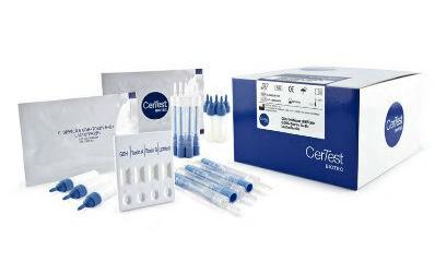
GDH+TOXIN A+B+LACTOFERRIN TEST
CERTEST BIOTEC
The CerTest Clostridium Difficile GDH+Toxin A+B+Lactoferrin One Step Combo Card Test is a chromatographic immunoassay designed to detect Clostridium difficile GDH, Toxin A, Toxin B, and human lactoferrin (hLf) in stool samples.
developing an affordable, easy-to-use self-testing platform, we can empower individuals to detect HIV early, reduce transmission and improve treatment outcomes. This innovation has the potential to save countless lives, offering hope to those who otherwise might not have access to timely care, ultimately helping to curb the global HIV epidemic.”
First-of-its-Kind Device Profiles Newborn Immune Function from Single Drop of Blood
Cont’d from page 13
CAMP and SMART AMR, and co-lead author of the study. “BLIPI represents a major step forward by providing clinicians with fast, actionable immune health data using a non-invasive method, where it can make a real difference for newborns in critical care.”
“BLIPI exemplifies our vision to bridge the gap between scientific innovation and clinical need,” added Prof Jongyoon Han, colead Principal Investigator at SMART CAMP, Principal Investigator at SMART AMR. “By leveraging microfluidic technologies to extract real-time immune insights from whole blood, we are not only accelerating diagnostics but also redefining how we monitor immune health in fragile populations. Our work reflects a new paradigm in point-of-care diagnostics: rapid, precise, and patient-centric.”
Image: The HIV-1 self-testing chip will be capable of selectively detecting HIV in whole blood samples (Photo courtesy of Shutterstock)
Treatment Switching Guided by Liquid Biopsy Blood Tests Improves Outcomes for Breast Cancer Patients
Standard treatment for patients with advanced estrogen receptor (ER)-positive, HER2-negative breast cancer, a subtype driven by estrogen receptors that fuel tumor growth, often involves aromatase inhibitors, which suppress estrogen production. However, resistance frequently develops when tumors acquire ESR1 gene mutations, keeping estrogen receptors active even without hormones. A major clinical challenge in treating advanced ER-positive breast cancer is detecting resistance mutations early enough to adjust treatment before tumor progression occurs. A new international trial has now shown that using liquid biopsy blood tests to identify these mutations, specifically in the ESR1 gene, followed by a timely switch to a novel drug, can significantly extend the period of tumor control compared to standard treatment.
The findings come from the SERENA-6 study, a large-scale, randomized clinical trial conducted across 264 clinical sites in 23 countries, including major contributions from Weill Cornell Medicine (New York, NY, USA; weill.cornell.edu). The study, overseen by a team of international researchers and clinicians, focused on patients with advanced ER-positive, HER2-negative breast cancer. The researchers explored whether switching patients to an experimental drug— camizestrant—at the first detection of ESR1 mutations via liquid biopsy, rather than waiting for imaging or symptoms, would improve outcomes. Camizestrant works by directly reducing the number of estrogen receptors on cancer cells, targeting the root of the resistance. Of more than 3,300 pa-
of 23.0 months in the camizestrant group, versus just 6.4 months in the control group. These results were considered both statistically and clinically significant. Camizestrant was also well tolerated, with a few patients discontinuing it due to side effects. The findings mark one of the first successful demonstrations that treatment adaptation based on liquid biopsy results can improve patient outcomes in real-world clinical settings. This trial not only validates liquid biopsy as a viable early monitoring tool in breast cancer but also paves the way for similar approaches in other cancers where resistance mutations can be detected in blood. For ER-positive breast cancer, in particular, the strategy could
Image: Switching to an experimental drug after liquid biopsy detection of breast cancer recurrence can improve outcomes (Photo courtesy of Shutterstock)
redefine how clinicians monitor recurrence and make proactive treatment decisions.
“The main message here is that liquid biopsy technology allows us to intervene sooner when the tumor burden is lower and the chance of a good outcome is higher,” said study co-author Dr. Massimo Cristofanilli, who helped design and oversee the SERENA-6 study.
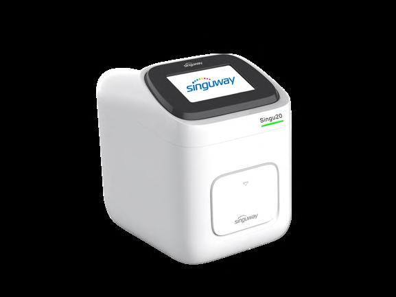
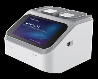
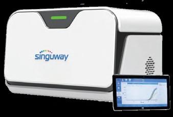
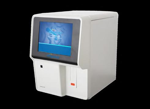
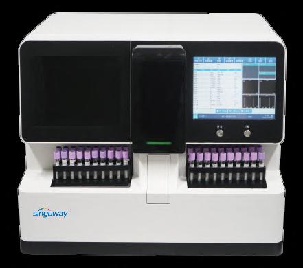
For small and medium labs, @SINGUWAY is your answer.
≤30 minutes
≤35 minutes
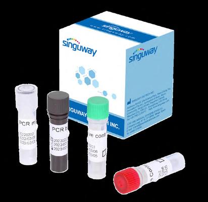
qPCR Detection Kits (PCR fluorescence method):
Respiratory Diseases: COVID-19, fluA, fluB, AdV, TB and multiplex test
Blood Diseases: HBV, HCV, HIV and multiplex test
Sexually Transmitted Diseases: HPV, CT, NG, UU and multiplex test
Viral Zoonotic Diseases: MPV
Vector-borne Diseases: PF, ZIKV
Genetic Diseases: MTHFR
Animal Diseases: Swine/Avian/Aquatic
Animal/Ruminant/Companion Animal Diseases
Singu20 Nucleic Acid Extractor AccuRa-32 Real-Time PCR System (32 samples)
Singu20 Nucleic Acid Extractor AccuRa mini Real-Time PCR System (8-16 samples)
The StripSpin 12D is a digital mini centrifuge designed specifically for PCR strips. It features a rotor that holds up to four 12-position PCR strips or 48 x 0.2 ml tubes and offers digital speed control up to
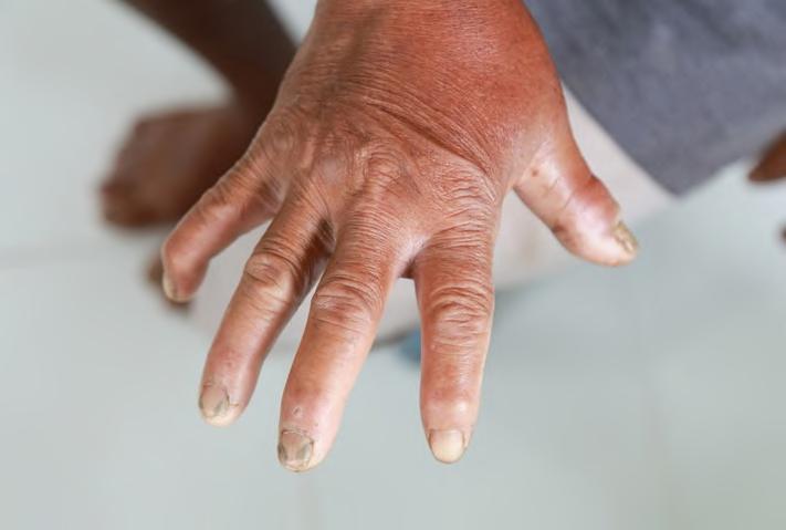
Identify Patients at Risk for Severe Scleroderma
Systemic sclerosis, also known as scleroderma, causes the hardening of the skin and connective tissues. In many cases, the disease can also damage vital organs, including the heart, kidneys, lungs, and gastrointestinal system, potentially leading to life-threatening complications. Among individuals diagnosed with systemic sclerosis, those with diffuse cutaneous systemic sclerosis generally experience a worse prognosis and higher mortality rate compared to those with limited cutaneous systemic sclerosis. Early diagnosis and intervention are crucial to slowing the progression of the disease, but currently, there are no clinical biomarkers to identify patients at higher risk for severe outcomes. Typically, doctors classify patients with suspected systemic sclerosis based on their symptoms: those with skin fibrosis limited to below the elbows and knees are diagnosed with limited cutaneous scleroderma, often experiencing less severe outcomes. On the other hand, patients with diffuse cutaneous systemic sclerosis, where skin fibrosis extends above the knees and elbows and affects other areas of the body, face a more aggressive disease course. Some of these patients may become disabled or develop progressive, debilitating conditions. A new test could now assist in identifying patients at risk for severe scleroderma, as highlighted in a study published in The Lancet Rheumatology.
In this groundbreaking study, researchers led by Yale School of Medicine (YSM, New Haven, CT, USA; medicine.yale.edu) demonstrated that type 1 interferons (IFNs)—a group of proteins that play a role in cell signaling—can serve as a blood biomarker for individuals with diffuse cutaneous systemic sclerosis. This discovery marks a significant advancement in early detection of high-risk patients. To find a reliable marker that could help clinicians predict poor outcomes in patients with diffuse cutaneous scleroderma, a team of clinician scientists from 11 academic centers across the United States collaborated to recruit patients with early-stage diffuse cutaneous systemic sclerosis. In 2012, they established the U.S. Prospective Registry of Early Systemic Sclerosis (PRESS), which includes patients who meet specific criteria for early diffuse cutaneous systemic sclerosis. The study involved 110 patients from the PRESS cohort. Simultaneously, researchers in the U.K. recruited a separate cohort of 32 healthy individuals and 72 patients with diffuse cutaneous systemic sclerosis as part of the Stratification for Risk of Progression in Scleroderma (STRIKE) study.
High levels of type 1 IFNs have been associated with worse outcomes in autoimmune diseases such as rheumatoid arthritis and lupus, but measuring blood serum levels of these IFNs is challenging. To address
The OnSite Malaria Pf/Pv Ab Combo Rapid Test is a lateral flow chromatographic immunoassay for simultaneous detection and differentiation of antibodies (IgG, IgM, IgA) to p.falciparum and p.vivax in serum, plasma or whole blood.
Image: A group of proteins involved in cell signaling can be used as a blood biomarker for patients with diffuse cutaneous systemic sclerosis (Photo courtesy of Shutterstock)
this, the researchers analyzed the concentrations of various molecules that respond to type 1 IFNs and are present in sufficient quantities for measurement, using them as indirect indicators of IFN activity. They discovered that participants in the PRESS cohort with high serum IFN scores tended to experience worse lung function and increased disability, including chronic joint pain, both at the start of the study and during follow-up. In the STRIKE cohort, patients with high IFN serum scores also exhibited poorer lung function, and these differences persisted throughout the study. Across both cohorts, individuals with elevated IFN scores had higher mortality rates than those with lower scores. As scleroderma-related lung disease is the leading cause of death in this patient population, identifying a blood biomarker that could predict patients at greater risk for lung disease is an important breakthrough. The researchers believe that the high serum IFN score could eventually be used to predict which patients, especially in the early stages of their disease, are at risk for developing severe conditions, facilitating more personalized and effective treatment strategies.
“Our results suggest that measuring type I IFN activity is akin to assessing the fuel driving autoimmune processes in systemic sclerosis patients,” said Monique Hinchcliff, MD, MS, at YSM. While additional validation and testing are necessary, “the ability to possibly discriminate between high-risk and low-risk patients with diffuse cutaneous systemic sclerosis using a blood test represents a large step forward for the community.”
Edited by Marilena Stamouli, BSc, MS
MESSAGE FROM THE PRESIDENT
By Tomris Ozben • President, IFCC
Dear
Colleagues, Dear Friends, It was truly a pleasure to see so many of you at the XXVI EuroMedLab 2025 this May in the beautiful city of Brussels. Following the great success of the congress, I’m also delighted to share that we achieved a record-breaking 9,004 participants and welcomed 115 exhibiting companies. A total of 114 countries were represented, with the highest number of registrations coming from Spain.
These outstanding results reflect the growing global interest in IFCC activities and the EuroMedLab Congress itself. During the congress in Brussels, a rich and diverse scientific programme brought together leading experts from around the world to explore the future of laboratory medicine. Through high-level plenary lectures, symposia, educational workshops, and round table sessions, participants engaged in in-depth discussions on the most pressing topics in the field, including innovations in clinical chemistry, advances in molecular diagnostics, digital pathology, the integration of artificial intelligence in laboratory workflows, and the role of laboratory medicine in personalized healthcare.
These sessions not only highlighted cutting-edge scientific developments and emerging trends but also provided valuable networking opportunities and a platform for open dialogue between participants, speakers, chairs, clinicians, researchers, and industry leaders. The success of the scientific programme highlights the strength of the collaboration between IFCC and its industry partners, a partnership that continues to play a crucial role in advancing innovation and progress in laboratory medicine.
This year’s congress also introduced several new features. For the first time, simultaneous translation from English to Spanish was provided, enhancing accessibility
Via Carlo Farini 81, 20159 Milan, ITALY Tel: (39) 02-6680-9912
World Environment Day, marked on 5 June, is a call to protect our environment and take urgent actions for a sustainable future. In this issue, you can read about One Health perspective, a collaborative, multidisciplinary approach that integrates human, animal, and environmental health. One Health aims to sustainably balance and optimize the health of people, animals and ecosystems, all of which are closely linked and interdependent.
In her message published in the present issue, our President, Prof. Tomris Ozben, expresses her sincere appreciation to the speakers, session chairs, IVD industry representatives, and all participants from around the world, that contributed to the success of IFCC General Conference in Bruges and XXVI IFCC-EFLM EuroMedLab Congress in Brussels.
We invite you to read about the Global MedLab Week 2025, coordinated by the IFCC and its six federations, which set a new global benchmark for public recognition and professional celebration of clinical laboratory workers.
News from member societies from Nepal, Ethiopia, Pakistan, Japan, and Jordan are also included in this issue. They share with us exciting information about their congresses, confer-
ences and other activities, which promote knowledge exchange and professional networking. The high quality of this scientific content shows once again the commitment of Medical Laboratory professionals to innovation, advanced patient care and continuous progress. Moreover, colleagues from Peru share with us the impact of applying a screening strategy that has allowed the timely diagnosis of patients with chronic kidney disease.
Interesting news from the IFCC Professional Exchange Program and from Task Force on Outcome Studies in Laboratory Medicine (TF-OSLM), are also included in this issue.
On August 1st, 2025, the UNIVANTS of Healthcare Excellence award program opens the 2026 application process, allowing to online submissions. In this issue you can find useful tips for when applying to the UNIVANTS of Healthcare Excellence award program.
Finally, I take the opportunity to remind you that IFCC Executive Board call for nominations for Executive Board Members will be open until the end of June, and to encourage appropriate candidates to apply for the various positions of the EB.
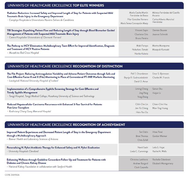
Nine New Integrated Clinical Care Teams Receive Recognition Through the UNIVANTS of Healthcare Excellence Awards
In June 2025, the IFCC, in partnership with Abbott and 6 other prestigious partner organizations, proudly announced the nine clinical care teams who have received recognition through the UNIVANTS of
Healthcare Excellence program for their measurable impact for patients, clinicians, health systems/ administrations and payors.
These 2025 winning teams are recognized for “UNIFYING” across disciplines to enable the development and implementation of “AVANT-GARDE” processes to enable measurably better health outcomes.
These impressive teams have made measurably improvements to care, with impact made across disease states and focuses. With 3 top global winners, 3 teams of distinction, and 3 teams of achievement (see table below), these diverse and innovative initiatives span geographies, health systems, patients and disease states. To learn more about these best practices, please visit www.UnivantsHCE.com
Are you interested in recognition through UNIVANTS? Start preparing your application now as applications for the 2025 UNIVANTS of Healthcare Excellence award program open Aug 1, 2025, through until Nov 15, 2025. To learn more about UNIVANTS, to gain insights on tips and tricks and/or apply, please visit www.UnivantsHCE.com.
The UNIVANTS of Healthcare Excellence award program is proudly comprised of the following program partners: International Federation of Clinical Chemistry (IFCC), Association for Diagnostics and Laboratory Medicine (ADLM, formerly AACC), Modern Healthcare, National Association for Healthcare Quality (NAHQ), European Health Management Association (EHMA), Institute of Health Economics (IHE), Healthcare Information and Management Systems Society (HIMSS); each in partnership with Abbott.
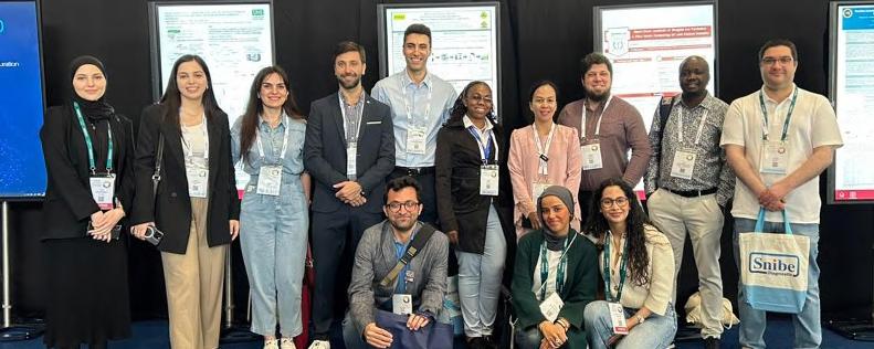
Young Scientists Poster Tour at EuroMedLab 2025: A Resounding Success in Brussels!
Marie Lenski, France (Société Française de Biologie Clinique, SFBC), IFCC TF-YS member
Aleksei Tikhonov, France (Société Française de Biologie Clinique, SFBC), EFLM C-YS member
The IFCC Task Force for Young Scientists (TF-YS), in collaboration with the EFLM Committee for Young Scientists (C-YS), proudly hosted the « Young Scientists Poster Tour » during the EuroMedLab Congress in Brussels, Belgium. This vibrant three-day initiative was designed to provide young researchers with a welcoming platform to present their scientific work, hone their presentation skills, and build lasting international connections.
Over the course of Monday to Wednesday, 40 young scientists representing 26 countries participated in daily onehour sessions. Each session was structured around small group interactions, guided by coordinators from IFCC TF-YS and EFLM C-YS. Presenters were given 5 minutes to introduce their research, followed by 5 minutes of lively and supportive peer discussion. This informal format fostered focused scientific exchange and encouraged open dialogue in a collegial atmosphere.
For many participants, the Poster Tour marked their first opportunity to present at an international congress—a milestone that made the experience especially meaningful. The event not only amplified the voices of emerging scientists but also attracted attention from the broader congress audience, further highlighting the innovative work of young professionals in laboratory medicine. Feedback from attendees was overwhelmingly positive. Participants valued the relaxed environment, the chance to connect with peers from around the world, and the opportunity to receive constructive feedback. Many noted that the experience boosted their confidence and inspired them to pursue future publication opportunities, particularly in the eJIFCC. Several participants have since subscribed to ongoing IFCC and EFLM activities, eager to remain engaged with this supportive community.
The Young Scientists Poster Tour truly exemplifies the mission of IFCC and EFLM: empowering and connecting the next generation of leaders in laboratory medicine. We are excited to continue this tradition at future congresses and to further expand opportunities for young professionals in our field.
ADLM 2025 BRING THE WONDER
JULY 29–31 • CHICAGO, IL • USA
PREPARE TO BE AMAZED
There’s only one Expo that brings the wonder of clinical laboratory medicine to life. Join lab medicine leaders from various specialties to explore innovative solutions, cutting-edge technologies, and essential products. Don’t miss this opportunity to discover what’s practical—and what’s possible—in the lab and beyond!
Join us at the ADLM 2025 Expo in Chicago for:
• 850+ exhibitors, 200+ product categories and live demos
• Lecture Series Presentations and Industry Workshops
• Networking opportunities to connect with industry peers and leaders
• New technologies competing for this year’s Disruptive Tech Award
• Exclusive member benefits at the ADLM booth + member lounge
Cont’d from page 17
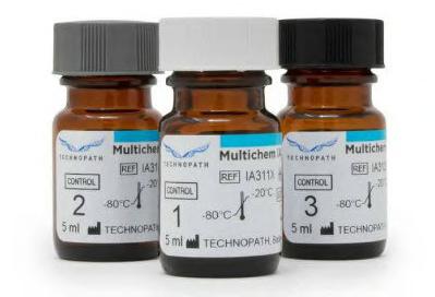
QUALITY CONTROL MATERIAL LGC CLINICAL DIAGNOSTICS
Multichem IA is a tri-level, liquid-stable control material for immunoassays with 83 analytes, including fertility, thyroid, cardiac, and metabolic markers, ensuring stable, reliable performance across multiple platforms. INTEGRATED
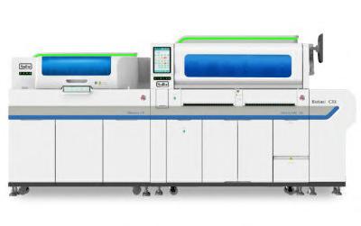
The Biolumi CX8 is an integrated chemistry and chemiluminescence system designed for high throughput with a minimal footprint. It combines the Biossays C8 and MAGLUMI X8 for simultaneous biochemical and immunological testing.
Message from the President
for a broader audience. Additionally, all sessions were recorded, a step forward in making the congress more accessible, inclusive, and impactful than ever before.
This year also marked the 4th edition of the IFCC Young Scientists Forum, which welcomed early-career professionals from across Europe and around the world. IFCC provided 52 scholarship grants, enabling young scientists from diverse regions to attend and actively participate in the FORUM and EuroMedLab congress. This initiative demonstrates IFCC’s strong commitment to supporting and nurturing the next generation of leaders in laboratory medicine.
The following satellite meetings were held before and after the EuroMedLab Congress.
1. 17th International Congress in Pediatric Laboratory Medicine (ICPLM) Emerging Technologies and Innovations in Pediatric Laboratory Medicine. 18th May 2025.
2. Joint Symposium between the RBLSM (Royal Belgian Society of Laboratory Medicine) and the SFBC (French Society of Clinical Chemistry). Preventive Diagnostics: the Power of Laboratory Medicine. 18th May 2025.
4. IFCC Satellite Symposium on Mass Spectrometry. 22nd May 2025, following the closure ceremony. It brought together seven
Sinternationally renowned speakers to explore the role, methodologies, and applications of mass spectrometry in laboratory medicine
As we move into the summer months, I encourage all our members, partners, and colleagues to stay actively engaged and continue participating in the many exciting initiatives and opportunities that lie ahead.
I would like to remind you about the WorldLab 2026: the 27th International Congress of Clinical Chemistry and Laboratory Medicine. This prestigious event — a joint initiative of the IFCC and the Asia and Pacific Federation of Clinical Chemistry (APFCB) — will be hosted by the Association of Clinical Biochemists of India (ACBI) in New Delhi, in conjunction with the 52nd Congress of the ACBI. We warmly encourage you to attend and participate in this global celebration of laboratory medicine.
It is my pleasure to announce and warmly invite you to the upcoming EuroMedLab Congress, which will be held in London in 2027. We look forward to welcoming you to this major scientific event, which will build on the success and momentum of previous congresses.
Thank you once again for your dedication and valuable contributions to the IFCC community.
Wishing you all a restful and enjoyable summer holiday.
With my best regards, Prof. Dr Tomris Ozben IFCC President
Saliva-Based Testing Enables Early Detection of Common Diseases
aliva is one of the most accessible biological fluids, yet it remains underutilized in clinical practice. While saliva samples are used to perform genetic tests to determine, for example, paternity, the potential of saliva-based clinical analysis goes far beyond this, as shown in new research published in npj Genomic Medicine.
A research team at the University of the Basque Country (UPV/EHU, Biscay, Spain; www.ehu.eus) analyzed saliva samples from more than 350 people and catalogued the common variations in DNA, known as genetic polymorphisms, or SNPs (single-nucleotide polymorphisms), that could influence genome function in saliva. Specifically, they found that these polymorphisms act as a switch that activates or deactivates the function of the genes they affect. Comparing the data from their study with the results of previous large international genetic studies on the risk of developing chronic diseases, they observed that many of the same polymorphisms that were detected in the saliva are associated with a higher risk of developing common diseases such as prostate cancer, coronary disease, Parkinson’s and Type 2 diabetes.
Furthermore, using advanced statistical tools, the research group demonstrated that these markers can explain a significant proportion of the genetic heritability of various diseases, in some cases with greater precision than traditional blood indicators. Although these findings need to be validated in larger cohorts for each of the diseases, they represent a significant advance in identifying non-invasive disease biomarkers. This work opens the door to developing saliva-based testing that could, in the future, be used for the early detection of diseases or for monitoring treatments, without the need to extract blood or perform other invasive procedures. Another contribution from this study is the creation of the largest public database of genetic data derived from saliva samples, which can be consulted via an open-access platform. The open-access nature of the database is expected to encourage new research studies and innovations in different biomedical disciplines.
“Our results show that molecular markers present in saliva can reflect systemic pathological processes beyond the oral cavity,” said José Ramón Bilbao, Professor of Medical Genetics at the EHU and one of the lead authors of the study.
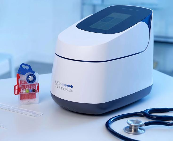
uidelOrtho Corporation (San Diego, CA, USA; quidelortho. com) has announced plans to acquire full ownership of LEX Diagnostics (Cambridgeshire, UK; lexdiagnostics.com) after 510(k) clearance by the U.S. Food and Drug Administration (FDA) for consideration at closing of approximately USD 100 million. LEX
Illumina Acquires SomaLogic to Accelerate Proteomics Business
Illumina, Inc. (San Diego, CA, USA; illumina.com) has entered into a definitive agreement to acquire SomaLogic (Boulder, CO, USA; somalogic.com), a leader in data-driven proteomics technology, and other specified assets for USD 350 million in cash payable at closing, subject to customary adjustments, plus up to USD 75 million in near-term performance-based milestones and performance-based royalties.
This transaction builds on a co-development agreement established in December 2021 to bring the SomaScan Proteomics Assay onto Illumina’s high-throughput next-generation sequencing (NGS) platforms. Illumina Protein Prep is currently in use with nearly 40 early-access customers globally and will become available to all customers starting in the third quarter of 2025. Combining SomaLogic’s proteomics technology with Illumina’s scalable NGS ecosystem, DRAGEN software, and Illumina Connected Multiomics will accelerate the technology development roadmap for proteomics and reduce the time and cost of proteomic research.
BioMérieux Expands Infectious Disease Diagnostics Range
ioMérieux (Marcy l’Étoile, France; biomerieux.com) has agreed to acquire the assets of Day Zero Diagnostics (Watertown, MA, USA; dayzerodiagnostics.com), an infectious disease diagnostics company using genome sequencing and machine learning to combat the rise of antibiotic-resistant infections. This strategic acquisition aims to enhance bioMérieux’s capabilities in next-generation sequencing (NGS) and rapid diagnostics, further solidifying its commitment to advancing healthcare and Antimicrobial Stewardship through innovative solutions. Day Zero Diagnostics has developed groundbreaking technologies that integrate direct-from-whole-blood sample preparation, sequencing, and advanced ID/AST analytics.
Qiagen in Korean Partnership to Develop Oncology PCR Platform
IAGEN (Venlo, Netherlands; www.qiagen.com) and GENCURIX (Seoul, South Korea; www.gencurix.com) have entered into a new partnership to develop oncology assays for use on the QIAcuityDx platform, a high-performance digital PCR system designed for clinical diagnostics.
GENCURIX is the first development partner under QIAGEN’s QIAcuityDx Partnering Program. This important advancement marks a significant step towards establishing a broad menu of in vitro diagnostic (IVD) assays on the QIAcuityDx Four platform, increasing access to digital PCR diagnostics. The new partnership combines QIAGEN’s QIAcuityDx digital PCR platform to advance sensitive, cost-effective oncology diagnostics with GENCURIX’s expertise in multiplex assay development. The aim is to enable the creation of oncology IVD assays for both tissue and liquid biopsy applications, with flexible commercialization options and global reach through QIAGEN’s Partnering Program.
The QIAcuityDx Four platform delivers a scalable and high-performance digital PCR solution for clinical laboratories. As global adoption of digital PCR grows, it is increasingly recognized as a complementary method to qPCR and NGS—particularly in oncology, infectious diseases, and rare genetic disorders. Through the QIAcuityDx Partnering Program, QIAGEN is opening its platform to third-party developers, encouraging innovation in order to offer laboratories a continuously expanding range of validated diagnostic tests.
Diagnostics’ ultra-fast molecular platform provides positive results in about six minutes and negative results in approximately 10 minutes using a single multiplex test for the detection and differentiation of Flu A, Flu B and COVID19. The system integrates into point-of-care workflows, bringing PCR-level sensitivity to urgent care centers, physician labs, hospitals, and other decentralized settings at a competitive cost.
QuidelOrtho’s initial investment in LEX Diagnostics on December 2023 included an option to acquire the whole company up to or shortly after 510(k) clearance by the FDA.
Image: The LEX system brings together high speed and high sensitivity in a PCR molecular system (Photo courtesy of LEX Diagnostics)
Thermo-Resistant (- 60 °C to 300 °C) Fully Washable & Flexible Suitable for central sterilization services Sterilizable STERILIZABLE INSTRUMENT & WORK-SURFACE MATS
2025
AUGUST
32nd Congress of the Latin American Society of Cytopathology (SLAC). Aug 5-8; Buenos Aires, Argentina; citologiala.org
23rd CNB International Congress 2025 – Colombian National College of Bacteriology. Aug 15-18; Bogota, Colombia; congresocnb.com
IUIS 2025 – International Union of Immunological Societies. Aug 17-22; Vienna, Austria; iuis2025.org
ICBMBLM 2025 – Joint Conference of the Malaysian Association of Clinical Biochemists (MACB) & Malaysian Society for Biochemistry and Molecular Biology (MSBMB). Aug 25-27; Petaling Jaya, Malaysia; conference.macb.org.my
SEPTEMBER
WHX Labs Cape Town 2025 (formerly Medlab Africa). Sep 2-4; Cape Town, South Africa; worldhealthexpo.com
Thailand Lab International 2025. Sep 3-5; Bangkok, Thailand; thailandlab.com
CUBRA 2025 – National Biochemical Congress of Argentina. Sep 4-6; Santiago del Estero, Argentina; cubra.org.ar
ECP 2025 – 37h Congress of the European Society of Pathology. Sep 6-10; Vienna, Austria; esp-pathology.org
47th Mexican National Congress of Clinical Chemists and Expoquím 2025. Sep 8-13; Oaxaca, Mexico; conaquic.com
EUROTOX 2025 – 59th Congress of the European Societies of Toxicology. Sep 14-17; Athens, Greece; eurotox2025.com
ESCV 2025 – 27th Annual Meeting of the European Society of Clinical Virology. Sep 17-20; Thessaloniki, Greece; escv2025.org
17th National Conference of the Bulgarian Society of Clinical Laboratory. Sep 18-20; Golden Sands, Bulgaria; bsclconference.com
23rd International Congress of Therapeutic Drug Monitoring & Clinical Toxicology. Sep 21-24; Singapore; iatdmct2025.org
MASCL 2025 – Congress of the Association for Mass Spectrometry & Advances in Clinical Lab. Sep 21-26; Montreal, Canada; msacl.org OCTOBER
54th Mexican Congress of Clinical Pathology – Mexican Federation of Clinical Pathology. Oct 1-4l Guadalajara, Mexico; fempac.org.mx
WHX Labs Nairobi 2025. Oct 6-8; Nairobi, Kenya; worldhealthexpo.com
ASHI 2025– 51st Annual Meeting of the American Society for Histocompatibility and Immunogenetics. Oct 6-10; Orlando, FL, USA; ashi-hla.org
JFBM 2025 – Journées Francophones de Biologie Médicale. Oct 8-10; Cannes, France; jfbm.fr
32nd Meeting of the Federation of Balkan Clinical Laboratories (BCLF) & 16th National Conference of the Romanian Association of Laboratory Medicine (AMLR). Oct 8-11; Sinaia, Romania; amlr.medical-congresses.ro
47th ISOBM Conference - International Society of Oncology and Biomarkers. Oct 13-15, Murnau am Staffelsee, Germany; isobm.org
62nd Annual Scientific Conference of the Australasian
Association of Clinical Biochemistry and Laboratory Medicine (AACB). Oct 13-16; Auckland, New Zealand; aacb.asn.au
34th WASPALM & 51st ACBICON 2025 – World Congress of Association of Societies of Pathology and Association of Clinical Biochemists of India. Oct 13-16; Pune, India; waspalm2025.org
ASHG 2025 – Annual Meeting of the American Society of Human Genetics. Oct 14-18; Boston, MA, USA; ashg.org
First Annual Meeting of the Emirates Clinical Chemistry Society (ECCS) and the Digital Health & Laboratory Medicine Forum. Oct 17-18; Dubai, UAE; eccsforum.com
DKLM 2025 – Annual Congress of the German Society for Clinical Medicine and Laboratory Medicine (DGKL). Oct 23-24; Leipzig, Germany; laboratoriumsmedizin-kongress.de
27th National Congress of the Society of Medical Laboratory Technology of South Africa (SMLTSA). Oct 24-26; Stellenbosch, South Africa; smltsa.org.za
ISBT Perth 2025 – Regional Congress of the International Society of Blood Transfusion. Oct 26-29; Perth, Australia; isbtweb.org
WSPID 2025 – 14th World Congress of the World Society for Pediatric Infectious Disease. Oct 28-31; Bangkok, Thailand; wspid2025.com
LMCE-KSLM 2025 – Laboratory Medicine Congress & Exhibition and 66th Annual Meeting of the Korean Society of Laboratory Medicine. Oct 29-31; Incheon, Korea; lmcekslm.org
23rd Greek National Congress of Clinical Chemistry. Oct 30 - Nov 2; Thessaloniki, Greece; eekx-kb.gr NOVEMBER
APCCMI 2025 – 20th Asia Pacific Congress of Clinical Microbiology and Infection. Nov 2-4; Bangkok, Thai-
land; apccmi2025.com
6th SMLC Congress – Chilean Medical Society of Clinical Laboratory (SMLC). Nov 5-6; Santiago, Chile; smlc.cl 57th SIBioC National Congress – Italian Society of Clinical Biochemistry and Molecular Biology. Nov 5-7; Bologna, Italy; sibioc.it
73rd Annual Scientific Meeting of the American Society of Cytopathology (ASC). Nov 5-9; St. Louis, MO, USA; cytopathology.org
65th Annual Academic Assembly of the Japan Society of Clinical Chemistry (JSCC). Nov 7-9; Nagoya, Japan; jscc-jp.gr.jp
AMP 2025 – Association for Molecular Pathology Annual Meeting & Expo. Nov 11-15; Boston, MA, USA; amp25.amp.org
LABCLIN 2025 – 19th Spanish National Congress of Laboratory Medicine. Nov 12-14; Valencia, Spain; labclin.org
Annual Meeting of the RBSLM – Royal Belgian Society of Laboratory Medicine. Nov 13-14; Brussels, Belgium; rbslm.be
47th Annual Conference of the Association of Clinical Biochemists in Ireland (ACBI). Nov 14-15; Athlone, Ireland; acbi.ie
46th Annual Meeting of the American College of Toxicology (ACT). Nov 16-19; Phoenix, AZ, USA; actox.org
MEDICA 2025. Nov 17-20; Dusseldorf, Germany; medica-tradefair.com
ASCP 2025 – Annual Meeting of the American Society for Clinical Pathology. Nov 17-20; Atlanta, GA, USA; ascp.org
DECEMBER
ASI 2025 – Annual Scientific Meeting of the Australian and New Zealand Society for Immunology. Dec 1-5; Perth, Australia; immunology.org.au
67th Annual Meeting & Exposition of the American Society of Hematology (ASH). Dec 6-9; Orlando, FL, USA; hematology.org
7th EFLM Conference on Preanalytical Phase. Dec 1213; Padua, Italy; eflmpreanalytical.org
2026
FEBRUARY
Labquality Days 2025 – International Congress on Quality in Laboratory Medicine. Feb 5-6; Helsinki, Finland; labqualitydays.fi
SLAS2026 International Congress & Exhibition. Feb 711; Boston, MA, USA; slas.org WHX Labs Dubai 2025. Feb 10-13; Dubai, UAE; worldhealthexpo.com
5th International Laboratory Diagnostics Congress. Feb 15-18; Virtual; ldcongress.com
MARCH
USCAP 115th Annual Meeting – United States and Canadian Academy of Pathology. Mar 21-26; San Antonio, TX, USA; uscap.org
APRIL
ESCMID Global 2026. Apr 17-21; Munich, Germany; escmid.org
MARCH
USCAP 115th Annual Meeting – United States and
Canadian Academy of Pathology. Mar 21-26; San Antonio, TX, USA; uscap.org
APRIL
Immunology 2026 – Annual Meeting of the American Association of Immunologists (AAI). Apr 15-19; Boston, MA, USA; aai.org
ESCMID Global 2026. Apr 17-21; Munich, Germany; escmid.org
ISLH 2026 Congress – International Society for Laboratory Hematology. Apr 17-19; Edinburgh, Scotland, UK; islh.org
MAY
SLAS Europe 2026 Conference and Exhibition - Society of Laboratory Automation and Screening. May 19-21; Vienna, Austria; slas.org
JUNE
WHX Labs Lagos 2025. Jun 2-4; Lagos, Nigeria;
worldhealthexpo.com
ICE 2026 – 22nd International Congress of Endocrinology. Jun 2-5; Kyoto, Japan; isendo.org
ASM Microbe 2026 – American Society for Microbiology. Jun 4-8; Washington, DC, USA; asm.org
FOCIS 2026 – Annual Meeting of the Federation of Clinical Immunology Societies. Jun 9-12; San Francisco, CA, USA; focisnet.org
ESHG 2026 Hybrid Conference – European Society of Human Genetics. Jun 13-16; Gothenburg, Sweden; 2026.eshg.org
11th FIDSSA Congress – Federation of Infectious Diseases Societies of Southern Africa. Jun 18-20. Cape Town, South Africa; fidssa.co.za
PREMIER MULTIMEDIA PLATFORM SERVING THE WORLD’S CLINICAL LABORATORY COMMUNITY
Anytime, Anywhere, On the Go...
LabMedica International LabMedica en Espanol PRINT & DIGITAL EDITIONS
ENGLISH
HemoCell delivers workcell efficiency, quality and standardization for Hemostasis labs. Featuring ACL TOP® 750 LAS and HemoHub™ Intelligent Data Manager, it’s the only workcell to combine the leading Hemostasis testing system with specialized lab automation. And, because it can be customized to your lab’s specific needs, HemoCell lets you design the automation layout that’s right for you.
Make your lab a HemoCell lab.
For more information, contact your local Werfen representative.