Liesbeth Harkema1#, Naomi de Bruijn1, Judith van Andel2, Eveline Dijkstra1 1Royal GD, Arnsbergstraat 7, 7418 EZ Deventer, The Netherlands 2Provinos sheep advisegroup, Venlo, The Netherlands #Corresponding author, e-mail: l.harkema@gddiergezondheid.nl
Introduction
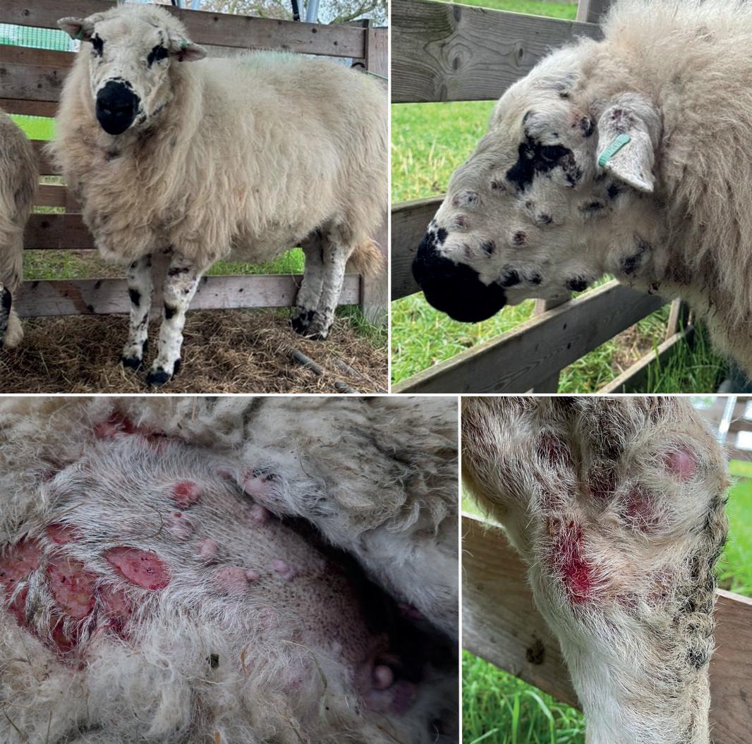
Skin diseases in sheep are common, but determining their cause based on clinical appearance can be challenging. This case report describes a rare instance of cutaneous lymphoma in a sheep, clinically indistinguishable from sheep and goat pox (SGP). It highlights the importance of recognizing atypical disease manifestations, disease surveillance, and the role of histopathological examination in determining the cause of skin diseases.
Materials and Methods
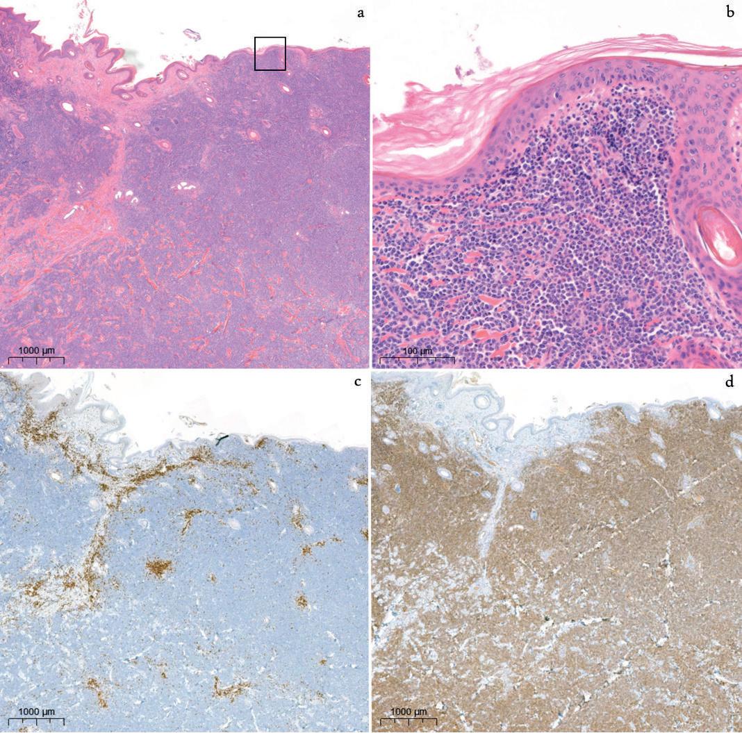
A six-year-old Welsh Hill Speckled Face ewe from a flock of 600 sheep presented with multiple cutaneous nodules. The practitioner consulted the GD Veekijker, a passive surveillance consultancy. Due to the similarity of the lesions to SGP and recent SGP outbreaks in Europe, the case was referred to official authorities under the suspicion of an OIE-listed disease. A capripoxvirus PCR on EDTA blood and an ELISA on a serum sample were performed. The ewe was later euthanized for post-mortem examination. Samples from the skin nodules were collected for bacteriological culture and DNA/RNA nanopore sequencing. Samples from the skin, caecum, liver, kidney, and mammary lymph node were collected for histopathology, fixed in 10% neutral-buffered formalin, processed, paraffin-embedded, and stained with haematoxylin and eosin. Immunohistochemical staining with CD3 and CD79a antibodies was performed on the skin lesions.
Results and Discussion
PCR and ELISA tests for Capripoxvirus were negative, ruling out SGP. Clinical observations demonstrated rapid progression of the lesions in size and number over the next month, and lack of response to treatment with meloxicam and oxytetracyclin. Macroscopic pathologic examination revealed nodular and ulcerated cutaneous lesions over the entire body, pale nodules in the kidneys, and prominent Peyer’s patches in the caecum.
Histopathological examination and immunohistochemical phenotyping with CD3 and CD79a antibodies confirmed a multicentric non-epitheliotropic cutaneous B-cell lymphoma. Bacterial culture identified Staphylococcus chromogenes as a secondary skin infection. Nanopore sequencing of the lesional tissue for viral and bacterial complexes was negative.
Conclusion
This case report documents the first case of cutaneous non-epitheliotropic B-cell lymphoma in sheep in the Netherlands, initially suspected to be SGP. It underscores the critical role of veterinary practitioners and pathological examination in disease identification and surveillance. Although the ewe in our case did not exhibit the full clinical spectrum of SGP, only a single animal in the flock was affected, and there was no history of recent introduction of animals or animal products, it is commendable that the veterinary practitioner promptly reported her findings to the official authorities. Many animal diseases can deviate from the expected clinical pattern due to factors such as host or vector conditions, strain variations, and local climate and these deviations make early identification more challenging. However, early and accurate diagnosis is essential for managing atypical disease presentations and ensuring effective disease control and prevention.
Lymphomas are sporadically diagnosed in sheep. Retrospective analysis of pathology data of sheep examined at GD between 2013 and 2024, revealed only two positive cases of lymphoma (unpublished data). Most of the reported cases describe the multicentric form involving multiple lymph nodes. Ovine cases of extranodal lymphomas are rarely described in literature, and cutaneous lymphoma is considered very rare in sheep.