CATARACT & REFRACTIVE
SMILE COMPLICATIONS Most SMILE complications are mild and diminish in frequency with experience. Roibeard Ó hÉineacháin reports
S
MILE has a rate of complications movement by the patient immediately similar to that of LASIK, and after docking the laser interface. The when complications occur they complication has been reported in 0.8-toare generally resolved with a good 4.4% of SMILE-treated eyes. visual outcome for the patient, “The incidence of suction loss decreases Catherine Albou-Ganem MD, with experience but it cannot be eliminated Paris, France, told the XXXV Congress of completely,” Dr Albou-Ganem stressed. the ESCRS in Lisbon, Portugal. Depending on when during the procedure “Most of SMILE’s complications are suction loss occurs, the surgeon can either related to experience and are included in abort the procedure or re-dock the eye the technique’s learning curve,” Dr Albouand continue. If suction loss occurs during Ganem reported. the creation of the lenticules, the possible She noted that an American Academy strategies include aborting the procedure, of Ophthalmology report in 2013 showed re-scheduling the procedure after the that the overall frequency of hazards reabsorption of the cavitation bubbles, and complications for SMILE was conversion to into thin-flap LASIK, 8.6%, which is comparable to or immediate re-docking and what is expected after LASIK. resumption of the procedure. Some complications that If suction loss occurs after occur in SMILE procedures the sculpting of the lenticules are the same as those that and during the creation of occur in LASIK, such as the incision, the best option decentration and epithelial is to restart the incision with a defects. She noted that epithelial decreased distance from the centre defects occur in around 40% of Catherine Albou-Ganem and an increased side-cut depth. patients. However, it is generally Black spots are reported in a mild problem that resolves in one or two 11-to-14% of cases. They result from the days with the use of artificial tears. It has no presence of water droplets, debris or air effect on visual acuity and is present only in between the laser and the coupling device the area around the incision. and the ocular surface. They leave an area Dr Albou-Ganem pointed out that dryness of untreated lenticule interface, making of the corneal surface is less frequent, less dissection more difficult. intense and it does not last as long after Opaque bubbles lead to the same SMILE as it does after LASIK. Diffuse consequence. They are more common in lamellar keratitis is also less frequent and eyes with a thicker cornea and a thinner there can be a hazy interface. There can also lenticule, but they do not affect the overall be epithelial in-growth, but more generally clinical outcome, she said. it is a case of epithelial “seeds” that are easily Lenticule ruptures often occur during rinsed away. Infection can also occur. the learning curve after difficult lenticule dissection. They can lead to remnant lenticule fragments between the stromal interfaces. SUCTION LOSS Incision ruptures during the enlargement An intraoperative complication that is of the small incision also occur more very specific to SMILE is suction loss. It frequently during the learning curve. generally results from excessive ocular
CALL FOR ENTRIES Entries to be sent to: henprize@eurotimes.org For further information visit: www.escrs.org EUROTIMES | MARCH 2018
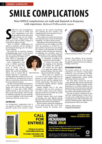
Courtesy of Catherine Albou-Ganem MD
18
Intraoperative black spots are reported in 11-to-14% of cases
However, the problem can be overcome by very careful removal of the lenticule through the ruptured incision and adding a bandage contact lens.
RETREATMENT OPTIONS The primary options for eyes that need retreatment for refractive enhancement include the circle, the side-cut and PRK, as recommended by Zeiss. Other options, suggested independently, include LASIK and subcap lenticule extraction. The circle option converts the cap into a large diameter flap with the femtosecond laser, followed by lifting of the flaps and the performance of a refractive ablation on the exposed stroma. The circle option is indicated for corneas where lenticule creation leaves a thin anterior wall and thick posterior walls. The side-cut option is indicated for eyes with thin residual posterior stroma, but it can only correct small refractive errors with a small optical zone. PRK is indicated in eyes with thin residual posterior walls where the final pachymetry will be over 350µm. Catherine Albou-Ganem: cati.ganem@wanadoo.fr
JOHN HENAHAN
CLOSING DATE FRIDAY 30 MARCH 2018
PRIZE 2018 Young ophthalmologists are invited to write an essay on
“Do We Need a Randomised Controlled Clinical Trial in Cataract Surgery?” First prize is a €1,000 travel bursary to the 36th Congress of the ESCRS in Vienna, Austria.