Dare to think differently Team
2017-2024

2017-2024

Cancer Grand Challenges is a global initiative that is building an elite, interdisciplinary community to take on and solve cancer’s most complex problems.
Co-founded by the two largest funders of cancer research in the world, Cancer Research UK and the National Cancer Institute in the US, Cancer Grand Challenges aims to accelerate high-impact research and translate discoveries for public and patient benefit by transforming how team science is conducted.

“By solving the 3D tumour mapping challenge, IMAXT captured the true complexity of intratumour architecture and provided an entirely new way to study cancer.”
Gemma Balmer-Kemp Head of Research, Cancer Grand Challenges
In 2015, Cancer Grand Challenges set the 3D tumour mapping challenge. In 2017, funded by Cancer Research UK, team IMAXT alongside team Rosetta, set out to tackle the challenge and find a way of mapping tumours at the molecular and cellular level.
It’s hard to imagine now but at the time, 2D technologies were still embryonic, while 3D spatial-multi-omics was mere science fiction. The limited singlecell technologies that were available remained difficult to interpret and often untested in more challenging tissue-types, let alone intact tissues, such as primary tumours and metastases.
IMAXT’s plan was ambitious, the team proposed to bring together advanced imaging approaches with emerging technologies for measuring DNA, RNA and protein on a single-cell level. The team would first have to optimise, develop and even invent these technologies, before going on to enable their implementation in a robust manner and then integrating them on an unprecedented scale. IMAXT also planned to take a novel approach to visualise these vast, complex data in an interactive 3D map.
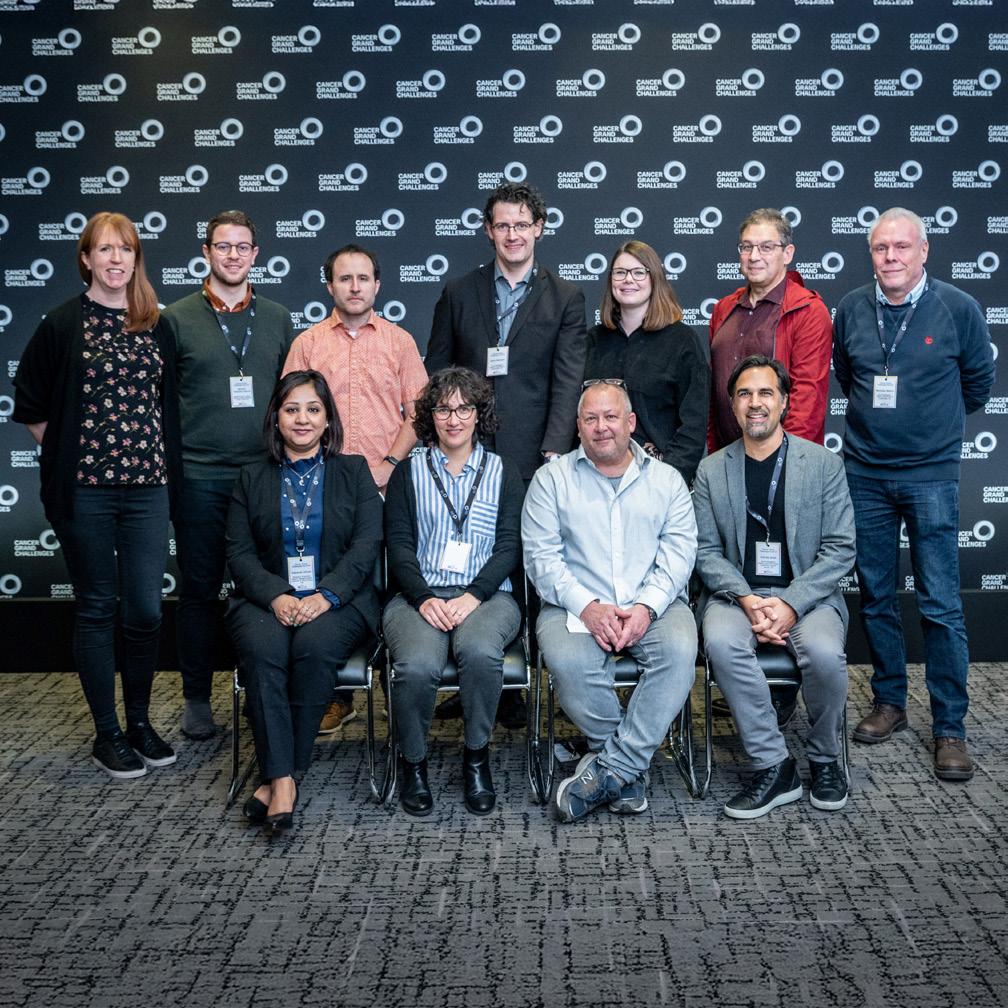
IMAXT built a global, multidisciplinary team, with experts from fields rarely brought together - from medicine and astronomy to VR and statistics, programming and molecular biology. Critically the team included those who had pioneered the techniques that would be further developed as part of IMAXT.
IMAXT was led by Greg Hannon with a hub of scientists at the Cancer Research UK Cambridge Institute, including Shankar Balasubramanian, Raza Ali, Dario Bressan and Nicholas Walton (Institute of Astronomy, University of Cambridge). The research team also comprised Sam Aparicio (University of British Columbia), Bernd Bodenmiller (University of Zürich), Edward Boyden (Massachusetts Institute of Technology), Owen Harris (Suil Interactive), Johanna Joyce (University of Lausanne), Sohrab Shah (University of British Columbia), Simon Tavaré (Columbia University), and Xiaowei Zhuang (Harvard University).
Cancer Grand Challenges


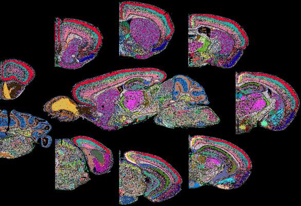
Since the challenge was set, the field of spatial biology has exploded, with the team at the forefront of this revolution, pushing the boundaries of what is possible. IMAXT invented, optimised and developed cutting-edge approaches, going on to create a platform which integrates a variety of technologies, from bulk-profiling and 3D whole organ imaging, to spatial proteomics and spatial transcriptomics, along with advanced data infrastructure and visualisation approaches.
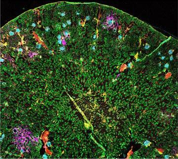
By using the IMAXT platform to produce comprehensive 3D maps of tumours, the team can identify rare but extremely important features, which would not be possible in 2D, such as micro-invasions which may lead to metastases. Upon identifying these rare events or cell types, visualising their neighbourhood and interactions in 3D can already point towards why these cells are present, how they got there and what they are doing.
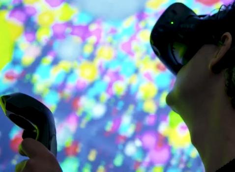
The team has pioneered a novel approach to visualise spatial data using virtual reality (VR), enabling researchers to interact with detailed 3D maps of the tumour microenvironment. Scientists and clinicians can immerse themselves inside a tumour, visualising the relationships between cells and conducting realtime analyses not feasible in 2D. Multiple researchers can examine a tumour simultaneously, enabling experts from different locations and specialties to work together to find ways to improve patient diagnosis and treatment. The VR viewer, Project Theia, is capable of real-time processing of single-cell spatial datasets containing millions of cells, enabling end-to-end analysis directly within the VR environment. These efforts have empowered the team to address fundamental questions in cancer biology that otherwise would not have been possible. Members of the team and Cancer Research Horizons have formed a company, Suil Vision, the first start-up from Cancer Grand Challenges. Suil Vision will develop VR software that enables immersive, multidimensional data visualisation and analysis for almost any type of biological data.
IMAXT has already used its novel technologies to dissect the clonal architecture of breast cancer and understand how it evolves over time and under treatment. Critically the team has gone on to demonstrate that tumour mapping can yield valuable prognostic and diagnostic insights.

Connecting the dots
Improving MERFISH
Creating and utilising DLP+
IMAXT awarded $25 million in funding Timeline
Spatial transcriptome profiling by MERFISH reveals subcellular RNA compartmentalization and cell cycle-dependent gene expression.
PNAS
Clonal decomposition and dna replication states defined by scaled single-cell genome sequencing.
JANUARY
The single-cell pathology landscape of breast cancer.
NATURE
Imaging mass cytometry and multiplatform genomics define the phenogenomic landscape of breast cancer.
NATURE CANCER
Genome-scale imaging of the 3D organization and transcriptional activity of chromatin. CELL
JANUARY
Expansion sequencing:
Spatially precise in situ transcriptomics in intact biological systems.
SCIENCE
Leveraging IMC
The clinical relevance of tumour mapping
Mechanisms of immune evasion and tumour evolution
Multiplexed error-robust fluorescence in situ hybridisation (MERFISH), was originally invented by the lab of Xiaowei Zhuang, building on single molecule FISH to allow copy number and spatial localisation of RNA molecules to be determined. The team has further developed this technology to allow whole-transcriptome analysis and to ensure compatibility with tumour tissues. These developments aided the commercial development of MERFISH, which is now available for purchase. The Zhuang lab have also adapted MERFISH to detect epigenetic information as well as genomic architecture.


Co-first author and IMAXT future leader Jean Fan is now an Assistant Professor of Biomedical Engineering at Johns Hopkins University, US.
The team improved upon DNA transposition single-cell library preparation (DLP) to create DLP+. IMAXT then leveraged this to dissect the clonal architecture of breast cancer and understand how it evolves over time and under treatment. Using DLP+ to track clonal dynamics and fitness in patient-derived xenografts of breast epithelium and triple-negative breast cancer, the team revealed a drug-driven inversion of the fitness landscape.


The team adapted expansion microscopy to develop expansion sequencing (ExSeq), which allows multiplexed mapping of RNA, and demonstrated its utility in the mouse brain to generate nanoscale-resolution maps.
Clonal fitness inferred from time-series modelling of single-cell cancer genomes.
NATURE
Exploration and analysis of molecularly annotated, 3D models of breast cancer at single-cell resolution using virtual reality.
BIORXIV
Three-dimensional imaging mass cytometry for highly multiplexed molecular and cellular mapping of tissues and the tumor microenvironment.
NATURE CANCER
Breast tumor microenvironment structures are associated with genomic features and clinical outcome.
NATURE GENETICS
Single-cell genomic variation induced by mutational processes in cancer.
NATURE
Spatially resolved epigenomic profiling of single cells in complex tissues.
CELL
Ovarian cancer mutational processes drive site-specific immune evasion.
NATURE
Clonal transcriptomics identifies mechanisms of chemoresistance and empowers rational design of combination therapies.

Named after the Titaness of sight in Greek mythology, the team describe Theia, the VR visualisation and analysis platform which provides a multi-user, 3D environment for the exploration of spatial datasets. The team published multimodal datasets to allow other academics to explore the utility of Theia, as well as detailing the IMAXT pipeline to generate such spatial 3D maps.

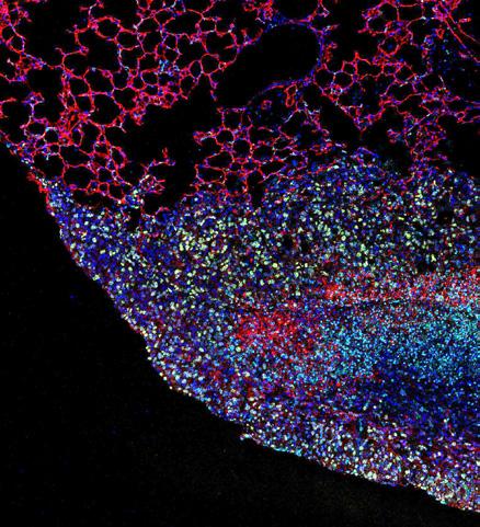
By leveraging imaging mass cytometry (IMC) in breast cancer patient samples to create detailed maps of the tumour microenvironment, the team identified new breast cancer subgroups that are associated with differential patient outcomes. The team went on to develop 3D-IMC which uncovered heterogeneity at both the cellular and microenvironmental levels that were not visible in 2D.

By leveraging IMC alongside multiplatform bulk genomics to profile breast tumours from hundreds of patients, the team demonstrated that the structure of the tumour neighbourhood correlated strongly with patient outcomes. The team extended this work from primary tumours through to lymphatic metastases, identifying a specific tumour microenvironment motif with significant prognostic potential.

By carrying out whole-genome sequencing plus single single-cell RNA sequencing, digital histopathology and multiplexed immunofluorescence the team created a multi-modal resource of the tumour microenvironment in high-grade serous ovarian cancer (HGSOC). The team’s approach allowed the identification of immune resistance mechanisms, by mapping cellular constituents and linking them to mutational processes in space. In a further Nature paper, by using single-cell whole genome sequencing the team uncovered whole-genome doubling (WGD) is an ongoing mutational process in HGSOC. The team went on to develop a computational approach to time WGD, DoubleTime and used it to reveal how WGD drives tumour evolution.
Multiplex imaging of breast cancer lymph node metastases identifies prognostic single-cell populations independent of clinical classifiers.

The team developed WILD-seq (Wholistic Interrogation of Lineage Dynamics by sequencing) a clonal transcriptomics approach which utilises genetic barcoding. By using WILD-seq in mouse models of triple-negative breast cancer the team revealed mechanisms of taxane chemotherapy resistance and go on to identify asparagine bioavailability as a targetable vulnerability of resistant cells.

Co-first author of both papers and IMAXT future leader Ignacio Vázquez-García is now an Assistant Professor at Harvard Medical School and Broad Institute, US.
APRIL 2023
Imaging and Molecular Annotation of Xenographs and Tumours (IMAXT): High throughput data and analysis infrastructure.
BIOLOGICAL IMAGING
Demonstrating the power of interdisciplinary collaboration, the team developed a method of co-embedding samples with fluorescent agarose beads and using the signal from these as reference landmarks. This approach was adapted from techniques originally built to map the cosmos and employed in the astronomy domain.
Review - The dawn of spatial omics.
SCIENCE
AUGUST 2023 MAY 2024
Spatially tuneable multi-omics sequencing using light-driven combinatorial barcoding of molecules in tissues.
BIORXIV
APRIL 2024
Microenvironmental reorganization in brain tumors following radiotherapy and recurrence revealed by hyperplexed immunofluorescence imaging.
NATURE COMMUNICATIONS
SEPTEMBER 2024
First Cancer Grand Challenges start-up company launched: SUIL VISION

The team present BALI (Barcoding by Activated Linkage of Indexes), which uses light to generate combinatorial spatial molecular barcodes directly onto target molecules in situ. This approach allows multi-omic profiling by next generation sequencing.


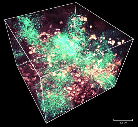
IMAXT published its Hyperplexed Immunofluorescence Imaging (HiFi) approach, which allows imaging of over 45 markers. The team set out to make HiFi accessible and economical to allow the approach to be widely adopted by anyone interested in spatial analysis of complex and fragile tissues. IMAXT demonstrated the utility of HiFi by showing that primary glioblastomas extensively reorganise their microenvironment in response to radiation, whereas metastatic glioblastomas do not.

Senior author and future leader Jean Hausser is now an Assistant Professor and SciLifeLab Fellow at the Karolinska Institute, Sweden.

In a first for the initiative, £5.25m in phase-two funding from Cancer Research UK through Cancer Grand Challenges was awarded to members of IMAXT to establish SPACE at the University of Cambridge. SPACE will bring the cuttingedge technologies of the IMAXT multi-omics profiling platform together with almost every existing technology, including most commercial platforms, under one roof.
This unique technological infrastructure and unified approach offers unparalleled advantages, with the ability to select the best method for each research question, compare technologies in an unbiased way and integrate them to generate even more exciting data and insights about cancer biology.
As part of Cancer Grand Challenges’ commitment to democratise these technologies, SPACE is designed to open up this platform to the wider research community.
“Together, IMAXT and Rosetta have given us ‘Google Earth’-like capabilities, allowing us to study the anatomy of cancers at an extraordinary level of detail. These technologies hold great promise to better understand tumours as a whole, and the interactions between tumour cells and their microenvironments.”
René Bernards
Cancer Grand Challenges Scientific Committee Member
The technologies developed and optimised by the team include Serial twophoton tomography (STPT), Ex-Seq, MERFISH, 3D IMC, DLP+, WILD-Seq, HiFi and BALI to name but a few. For STPT, this imaging technique had previously never been applied to tumours. The team has developed this approach to allow its high-throughput use, enabling high quality imaging of entire tumours and metastasis, from almost any tissue, while at the same time preserving biomolecules for analysis by further approaches. Together with the advanced data infrastructure and creative visualisation approaches, these fundamental tools and techniques add to the team’s scientific legacy and will continue to drive further discoveries in the cancer field and beyond.
Almost 100 people were a part of IMAXT in different capacities and the junior scientists involved were a major driving force in the team’s achievements. IMAXT established ‘tiger teams’ focused on key aspects of the challenge. These were run by post-doctoral scientists who were tasked with pushing the agenda, developing collaborative experimental plans across laboratories and coordinating data acquisition. The team also comprised ‘IMAXT associates’, junior group leaders who were assigned resources under the sponsorship of more established investigators. They were able to leverage the access to data, resources and contacts to push their careers and obtain independent grants. The team also encouraged intra-lab mobility, with trainees spending time in different labs to learn specific techniques.
Patient advocates are embedded into Cancer Grand Challenges teams and are important members of the teams we fund to address cancer’s toughest challenges.
The IMAXT team comprised four patient advocates over the course of the award, including Elaine Chapman, Lynn Dundas and Ellen Landsberger. A particular highlight was Elaine Chapman leading an interactive event aimed at the Cancer Patient Partnership Group in Cambridge. This event collected anonymous feedback from cancer patients on their perception of the VR developed by the team, and how it could be helpful for them to better understand their disease or if it could be detrimental in any way. The valuable insight from this event continues to guide the further development of the VR.
The IMAXT platform, alongside the expertise developed, represents cutting-edge technology, allowing unprecedented exploration of the complexity of the tumour microenvironment. The field is now at an inflection point, with IMAXT and SPACE poised to push the boundaries of spatial biology even further, far beyond descriptive studies. Application of the team’s innovative tools has already yielded novel insights into cancer biology – and can now be used to address important clinical questions. Through SPACE the transformative technologies of the IMAXT platform will be opened up, allowing the acceleration of groundbreaking findings for more researchers and further questions in cancer biology, and the team’s impact will only grow.