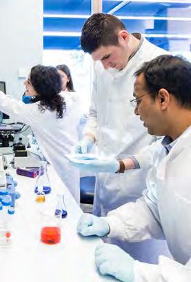
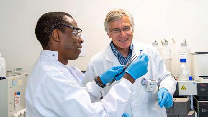
Uniting the world’s brightest minds against cancer’s toughest challenges
A growing global community of 700+ investigators $100m to take on four new challenges
Dr David Scott Director, Cancer Grand Challenges, Cancer Research UK
Dr Dinah Singer Deputy Director for Scientific Strategy and Development, National Cancer Institute, US
Cancer Grand
The best research is done through empowered global collaboration. Cancer Grand Challenges provides diverse, global teams with the time, space and funding to foster innovation and transcend traditional boundaries of geography and discipline – crucial to effectively tackle the toughest challenges in cancer research. Over the past year, our community has continued to apply new thinking to problems that have long hindered progress, making important discoveries across a broad pipeline of research.
This is showcased by the stories we’ve picked for this edition of Discover. For example, the team taking on our Unusual Mutation Patterns challenge (page 8) – a global consortium collecting samples from across five continents – has revealed the wealth of information on cancer that can be learned by studying normal, healthy tissue. These insights are now challenging the fundamental understanding of cancer biology. Research by other teams has now progressed to a point with clear paths to clinically benefitting patients. For example, surprising findings from the Lethal vs Non-lethal challenge team (page 6) on the progression of breast carcinomas may save many people from the burden of invasive overtreatment. Excitingly, novel emerging technologies from some of our teams are now available for the wider research community to use to probe cancer’s inner workings – read more about the global network of early-career researchers helping to develop a novel 3D tumour-mapping pipeline on page 20.
We hope that you enjoy these stories, which provide a striking demonstration of the power of global team science to drive high-impact research. We’re proud of our community for advancing these discoveries – an impressive feat against the backdrop of another challenging year of pandemic lockdowns and restrictions.
We’re also excited to be entering a new chapter for Cancer Grand Challenges by announcing $100M of funding to four new world-class teams seeking to address four of the most pressing challenges in cancer research. Two of the challenges seek to tackle major clinical problems: cachexia, the debilitating wasting syndrome that dramatically affects quality of life and survival for many people with advanced cancer, and extrachromosomal DNA, a major driver of tumour evolution and treatment resistance. The Normal Phenotypes challenge seeks to learn more about what triggers a cell to progress along the pathway to malignancy. The Solid Tumours in Children challenge aims to unlock new information about the fundamentally different biology of paediatric cancers, and to develop muchneeded novel therapies for these patients. Read more on page 26.
The four teams taking on these challenges are the first to be supported through our partnership between Cancer Research UK and the National Cancer Institute. Both organisations have a long history of establishing research networks in areas of emerging scientific opportunity. We’re delighted that new and existing partners have joined us for this new round of teams and, through Cancer Grand Challenges, we’re excited to further grow a global community of scientists working together to address some of cancer’s toughest challenges.
Cancer Grand Challenges: driving progress through global collaboration
Cancer Grand Challenges supports a global community of diverse world-class research teams coming together and thinking differently to take on some of cancer’s toughest challenges.
These challenges continue to impede research progress, and no one scientist, institution or country will be able to solve them alone. Cancer Grand Challenges teams are empowered to transcend the traditional boundaries of geography and discipline, and ultimately change outcomes for people with cancer.
Founded by the two largest funders of cancer research in the world – Cancer Research UK and the National Cancer Institute in the US – and uniting an international community of partners, Cancer Grand Challenges aims to make urgently needed progress against cancer.
The toughest challenges in cancer research
Cancer Grand Challenges works with the global research community and people affected by cancer to identify the toughest challenges in cancer research, then dares diverse world-class teams to take them on. Our research community is currently pursuing the 10 challenges outlined aside, following our announcement of four new challenges in June 2022; read more on page 26.
Cancer Causes
Understand how lifestyle factors, such as obesity, cause cancer
Lethal vs Non-lethal Cancers
Distinguish between lethal cancers that need treating and non-lethal cancers that don’t
Extrachromosomal DNA
Understand the biology of ecDNA generation and action, and develop approaches to target these mechanisms in cancer
Solid Tumours in Children
Develop novel therapies to target unique features in solid tumours in children
3D Tumour Mapping
Map the molecular and cellular tumour microenvironment to define new targets for therapy and prognosis
Tissue Specificity
Understand why mistakes in certain genes cause cancer in only specific parts of the body
Normal Phenotypes
Understand how cells and tissues maintain ‘normal’ phenotypes while harbouring oncogenic mutations and how they transition to become a tumour
Unusual Mutation Patterns
Discover how unusual patterns of mutation are induced by different cancer-causing events
Understand how microbes inside our bodies affect cancer treatment
Cachexia
Understand and reverse cachexia and declining performance status in cancer patients
Microbiota
Credit: NCI
Credit: Chris Tape
Credit Spencer Watson
Credit: NCI
Credit S Schuller
Credit: NCI
Credit: NCI
Credit: Tetiana Lazunova
Credit: NCI
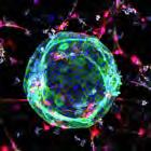
Credit Valeria Molinari, Louise Howell, Maria Vinci, Katy Taylor and Chris Jones, Institute of Cancer Research
Refuting the dogma surrounding breast cancer risk
Non-invasive breast carcinomas have long been assumed to be the precursors of any invasive cancers that follow. But new findings indicate that around 1 in 5 of these subsequent cancers are new primary tumours unrelated to the initial lesion. This understanding could cause a paradigm shift in how ductal carcinoma is managed in the clinic, helping to reduce the burden of overtreatment.
Challenge: Lethal vs Non-lethal Cancers: Distinguish between lethal cancers that need treating and non-lethal cancers that don’t
Team:
PRECISION (PREvent ductal Carcinoma
In Situ Invasive Overtreatment Now)
In ductal carcinoma in situ (DCIS), abnormal but non-invasive cells are present in the breast milk ducts. Although DCIS remains harmless in most people, some people will develop ipsilateral (same-breast) invasive breast cancer (IBC). Because predicting who will or will not develop IBC is impossible, treatment is generally recommended for all cases.
Each year, thousands of people with DCIS undergo surgery, radiation and hormone therapy, which may not be necessary and may be accompanied by unnecessary stress, adverse effects and anxiety. To avoid unnecessary treatment, a deeper understanding of DCIS biology is urgently needed, and predictive markers of progression must be identified. The Cancer Grand Challenges PRECISION team is taking on this pressing challenge.
The findings from the study may change clinical paradigms. “Our study indicates we can no longer consider DCIS solely as a precursor but rather also a risk factor for the development of a second invasive breast cancer later on in life,” says Elinor Sawyer, joint senior author of the study. “This important new information about DCIS biology and behaviour could change the way we manage and treat the condition in the clinic.”
A tour de force in integrating diverse data
Historically, research in the progression of DCIS to IBC has been logistically challenging because the proportion of people with DCIS who later develop IBC is relatively small, and recurrent IBC often occurs long after the initial DCIS diagnosis. PRECISION has addressed this challenge by analysing data and samples from well-annotated cohorts of people with DCIS throughout the UK, US and Netherlands, which are the largest of their kind in the world.
“Through Cancer Grand Challenges, we have access to three huge cohorts of people who were diagnosed with DCIS and later developed IBC,” describes Esther Lips, joint first author of the study. “We can match patient samples to follow up clinical information long after their DCIS diagnosis.”
Avoiding the burden of unnecessary treatment
“As doctors, we all train to do ‘something’ to help our patients. Taking a step safely back and choosing not to treat someone is really hard, so DCIS is almost always treated,” says Jelle. “Our findings published to date, and all the data we’ve collected through Cancer Grand Challenges so far, are all leading us closer to answering the question we posed at the start of the challenge: when is cancer not really cancer? Collectively, our work could ultimately save thousands of women the burden of invasive over-treatment.”
to consider a watch-and-wait approach”. He continues, “If we could understand the risk of a particular DCIS lesion in a particular individual, we could learn when it’s safe to undergo a moratorium on treatment and opt instead for active surveillance.”
The role of the microenvironment in individual risk
more prone to breast tissue tumorigenesis than the general population.
Answering this question by developing novel models and optimising existing models of DCIS is a major component of the team’s ambitious programme, which is being spearheaded by co-investigators Jos Jonkers, Jacco van Rheenen and Fariba Behbod. Many of these models have now been shown to be reliable, realistic tools for studying the progression of human DCIS. For example, the Mouse INtraDuctal (MIND) model – in which patients’ DCIS epithelial cells are injected into mouse mammary glands and observed as they progress naturally – has recently been reported to accurately mimic all histological subtypes of human DCIS.
The team is now able to visualise the entire mammary gland through in vivo models such as MIND, as enabled by the development of a workflow incorporating 3D imaging and intravital microscopy –a powerful tool for imaging biological processes in live animals. This technology is already revealing new information about the growth patterns and ductal architecture in indolent and invasive disease.
Professor Jelle Wesseling PRECISION team lead, Netherlands Cancer Institute, Netherlands
Elinor
PRECISION co-investigator, King’s College London, UK
A remaining question is why 1 in 5 people with IBC after a DCIS diagnosis can develop two clonally unrelated cancers in the same breast. Further research is ongoing. “But we can speculate,” says Elinor.
Jelle Wesseling PRECISION team lead “
Collectively, our work could ultimately save thousands of women the burden of invasive over-treatment.
Major findings from the team challenge the current dogma in which almost all cases of IBC after DCIS are believed to be related to the initial lesion and due to lesion progression. The study, published in Nature Genetics, shows that as many as 1 in 5 subsequent IBCs are unrelated to the initial DCIS and instead develop as second primary tumours.
“Our results are surprising, and I think some people might find it hard to believe that such a large proportion of subsequent IBCs are completely unrelated to the original DCIS,” says team lead Jelle Wesseling. “If we had only used bulk sequencing on a few samples, I think that would be a valid point. But our findings are backed up by such a breadth and depth of quality information.”
This rich source of data has been central to the team’s paradigmshifting insights, by allowing them to integrate clinical, pathological and epidemiological data, in addition to multiple layers of genomic, exomic and mutational analyses, even at the singlecell level. “This study is the culmination of a real tour de force,” Jelle says. “It’s wonderful that this full team effort could have real-world implications for the way we manage DCIS in the clinic.”
For oncologist Elinor, the clinical goal is decreasing the use of radiotherapy, which is often used to reduce the risk of local recurrence and progression. “If the invasive cancer is unrelated to the DCIS, radiation won’t help to prevent it, and the patient will receive no benefit,” she says. “Perhaps we should explore whether endocrine therapy could provide a more effective preventative tool in these people – to treat their DCIS and reduce the risk of both local recurrence and a new primary cancer.”
Meanwhile, for pathologist Jelle, the goal is “understanding when it’s safe
One possibility might be that inherited genetic changes may influence whether people are more likely to develop a recurrence or a new cancer after DCIS. To probe this possibility, her team is analysing a vast data bank of germline genetic information from people with DCIS, collected through ICICLE, a cohort study funded by Cancer Research UK.
Alternatively, the breast microenvironment may permit the emergence of a second, unrelated primary tumour, in a phenomenon termed the field cancerisation effect. According to this hypothesis, certain factors in the surrounding tissue – perhaps immune cells, stromal cells or the tissue architecture – provide an environment where clones of cells with pro-tumorigenic mutations thrive, resulting in large fields of cells that are more likely to develop into a tumour. This would make some people
Dr Esther Lips PRECISION co-investigator, Netherlands Cancer Institute, the Netherlands
Dr Jacco van Rheenen PRECISION co-investigator, Netherlands Cancer Institute, the Netherlands
Original article
Jos
PRECISION co-investigator, Netherlands Cancer Institute, the Netherlands
Professor Fariba Behbod PRECISION co-investigator, University of Kansas Medical Center, US
Lips E et al. Nat Gen 2022; doi: 10.1038/ s41588-022-01082-3
Hong Y et al. J Pathol 2021; doi: 10.1002/ path.5820
al. npj Breast Cancer 2021; doi: 10.1038/s41523-021-00232-w
Featured team members
Professor
Sawyer
Professor
Jonkers
The PRECISION team is funded by Cancer Research UK and the Dutch Cancer Society.
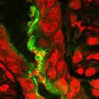
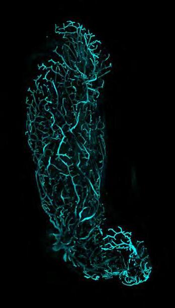
Image: Microscopic image of the ductal tree of the mouse mammary gland
Credit: Hendrik Messal and Jacco van Rheenen
Cancer’s secrets uncovered from non-cancerous tissue
What can studying non-cancerous tissue from people with cancer teach us about a tumour?
Peter Campbell reflects on the wealth of information that can be uncovered.
Challenge: Unusual Mutation
Patterns: Discover how unusual patterns of mutation are induced by different cancer-causing events
Team: Mutographs
What causes cancer incidence to vary in different parts of the world? When taking on the Unusual Mutation Patterns challenge, this is the question we set out to answer – and it fascinates me. Differences in regional cancer incidence have been systematically documented for upwards of 50 years. Occasionally, we’ve found the basis for that difference, but for the vast majority of cases – such as pancreatic cancer, which is common in Central Europe and Japan, but much rarer in East African countries and South America – we still have very little insight.
There must be other lifestyle, environmental or maybe inherited factors that we’ve yet to identify, which could be the key to preventing these cancers. By uniting experts from the worlds of epidemiology, cancer and genomics, we’ve developed a new way to study mutational signatures – characteristic patterns of damage left on DNA by
exposure to mutagens and genetic disorders. This incredibly powerful metaphorical microscope lets us build a high-resolution picture of a tumour’s molecular characteristics, allowing us to understand the roles of these exposures in the journey to malignancy.
While most of the Mutographs team has focused on the larger puzzle of cancer tissue, in my sub-team’s work, we’ve flipped it around to focus on normal tissue samples donated by people with breast, blood, liver and lung cancers. These cells have developed in the same context as the tumour, experienced the same exposures and have the same potential to succumb to inherited abnormalities, yet they haven’t gone through the same evolution as their cancerous neighbours.
So, as we begin to wrap up my package of work, what have we learned by analysing normal tissues? And how much can this information tell us about the early stages of tumour development?
An unexpected degree of complexity
We’ve certainly seen a degree of complexity in normal tissues that we didn’t anticipate. We know there are a bunch of mutagens that leave mutational signatures and are associated with an increased risk of cancer, for example, tobacco in lung cancer or aristolochic acid – a compound in some herbal products –in liver and urinary tract cancer. The really striking thing we’ve found is that we can see the same signatures of damage in normal tissues as in the tumour.
More surprising, however, is the extent to which these mutational signatures vary across the tissue. Neighbouring sections of liver less than a millimetre apart can possess the signature for aristolochic acid exposure but have 10-fold differences in its signal. Similarly, while nearly all cells in the lungs of heavy smokers carry the signature of tobacco exposure, there seems to be a small protected population of cells with a mutational landscape comparable that of to a non-smoker’s lung. It’s these uninjured cells that replace their damaged neighbours and repopulate the lung after a person quits smoking.
This finding was totally unexpected. I’d have thought that all cells exposed to these chemicals would possess the same catalogue of mutations and the same chance of becoming cancerous. What drives this difference in vulnerability
to these exposures? Are some cells protected from damage or more effective at neutralising carcinogens than their neighbours? Our research has certainly made us ask all sorts of interesting followup questions, which we’re just starting to explore.
Lessons from normal tissue
Ultimately, these unexpected findings mean that we can gather information about biology from normal tissue similarly to cancerous tissue.
First, looking for recurrent mutations in multiple cancer genomes can identify genes responsible for changing biology – and we can learn the same from non-cancerous tissue. In liver tissue, we identified three genes that affect how cells metabolise fat and respond to insulin, and consequently underpin the biology of obesity and type 2 diabetes. In some people, mutations in these genes lead to organ-wide changes in liver function and can result in chronic liver disease, which affects roughly 1.5 billion people worldwide. Although neither gene is linked to liver cancer, our findings provide important clues about diseased-liver biology and may prove useful in understanding the changes that a liver undergoes at it evolves to become cancerous.
Second, searching for mutational signatures in both cancerous and normal tissues can tell us which factors have influenced the environment in which the cancer evolved. To become cancerous, a cell must develop mechanisms to escape the normal constraints on its growth, as well as to invade tissue barriers and evade the immune system. These mechanisms are very important to study, but the genomes of these cells are dynamic. Our findings suggest that analysing normal tissue from people with cancer could provide a faithful representation of past exposures that might have been important early in the tumour’s development and therefore might identify new opportunities for intervention.
We’re currently exploring this possibility in breast cancer, which is epidemiologically a very interesting disease whose risk is influenced by a combination of many factors, including inherited mutations, age and reproductive history. By studying normal breast tissue from people with breast cancer, we’re starting to unpick the molecular
basis for some of these epidemiological observations – and the data we’re getting are amazing. We’re able to see some of that DNA damage in normal tissue and track it back to an exposure sometimes occurring decades before the initial diagnosis.
Finally, in both cancerous and normal tissues, we can pool this information to make relatively profound statements about how the tissue has evolved. With the liver, for example, we can map the early biological changes with which normal cells adjust to sustained insulin exposure and how this leads to a chronically diseased liver; we can also track how this process of damage and regeneration drives the tissue towards malignancy. Most exposed cells will never take another step down the road to malignancy, but in some people they will. Mapping out the roles that these changes play early in a tumour’s development is important for understanding cancer risk.
Tipping the balance to favour healthy cells
The Unusual Mutation Patterns challenge called for us to look beyond our own disciplines – to adopt a new philosophy in a holistic approach to answering a question that none of us could address individually. It’s been very rewarding to work in this way. As we begin to wrap up this package of our work, we are reflecting on how we’ve advanced our understanding of the question at hand.
It’s exciting to think about what we could do with this information. The idea that intrigues me the most is the possibility that we could tip the balance so that the growth of healthy cells without or with a limited mutational signature is favoured over that of their cancerous neighbours. This seems to be what happens when a person quits smoking: a niche of protected cells rises up to replace many of the damaged cells and is why people’s risk of lung cancer shrinks when they stop smoking.
Harnessing this biological phenomenon sounds like science fiction, but I can imagine a world where we work out how to tip the balance so that normal cells outcompete their neighbours as a novel strategy for cancer prevention and treatment. We’re a long way from reaching that stage. But we certainly can’t do it without understanding the underpinning biology – this is the stage we’re in now.
The
“
It sounds like science fiction, but I can imagine a world where we can tip the balance so that normal cells can out-compete their neighbours as a route to cancer prevention and treatment.
Peter Campbell Mutographs co-investigator
Yoshida K et al. Nature 2020; doi: 10.1038/s41586-020-1961-1
Ng SWK et al. Nature 2021; doi: 10.1038/s41586-021-03974-6
Featured team members
Dr Peter Campbell Mutographs co-investigator, Wellcome Sanger Institute, UK
“We can all be agents of change” – learnings from our first Future Leaders Conference
In October 2021, we held our first Future Leaders Conference, a virtual event bringing together the early-career members of our community. Here, Elee Shimshoni, postdoctoral fellow, shares her thoughts on the event.
An entirely new way to visualise tumours
Meet the global network of early-career researchers driving the development of cancer’s most intricate maps.
The Cancer Grand Challenges community brings together numerous types of expertise and techniques, all aimed at advancing cancer research. For those with their ‘boots on the ground’, communication between one another has the highest potential to lead to fruitful collaborations. This is why I joined the steering committee to help influence the content for the Cancer Grand Challenges Future Leaders Conference.
In addition to lightning talks and an insightful panel focused on career development, we used breakout rooms to build networking and discussion into both days. It was great to witness people taking a lot of interest in each other’s research. We were also excited that Uri Alon delivered our keynote address, accompanied by his guitar. A pioneer in the world of systems biology, Uri writes about, advocates for and even sings about the importance of human relationships in science.
A theme I felt emerging throughout the event was scientific environment and culture. Among us future leaders, there is significant thirst for a culture that is more geared toward fostering and sustaining both a healthy work-life balance and a collaborative, collegial research community in which researchers enable each other’s growth rather than impede it.
Elee’s top 5 takeaways from Uri Alon’s key note:
01
We can all be agents of change in our own realm: we can be better reviewers, grant committee members, mentors and peers.
03
Find what drives you. Let it guide you to the research questions that you would like to pursue.
05
It’s important to have conversations to describe the not-so-glamourous challenges of science to bring change to scientific culture.
02
Real-life research is never straightforward: you usually step into a ‘cloud’ of uncertainty in between your initial hypothesis and your final discovery or conclusion. This cloud is where true innovation lies.
04
Seek support from the people around you. Find and be grateful for that colleague who is a good listener, who can ‘hold your hand’ and help you get through the cloud.
Challenge: 3D Tumour
Mapping: Map the molecular and cellular tumour microenvironment to define new targets for therapy and prognosis
Team: IMAXT
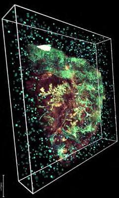
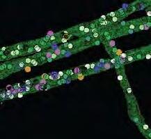
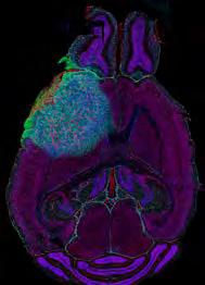
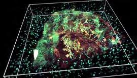
The tumour ecosystem, a web of interactions between the tumour’s microenvironment and the rest of the body, evolves over time, producing a highly interconnected structure of cancer cells, normal tissues, immune cells and structural support. A detailed 3D map of this complex ecosystem – including each cell’s type, location and genetic makeup, and, importantly, how it interacts with and influences its neighbours – would reveal hidden information about tumour biology. This map could guide researchers in search of novel ways to diagnose and treat cancer, and prevent metastasis or recurrence.
The Cancer Grand Challenges IMAXT team, in taking on the 3D Tumour Mapping challenge, set out to develop a new way to create a map of the molecular and cellular tumour environment, through an integrated pipeline of techniques, which can be explored in virtual reality. The power of their approach lies in the integration of these techniques – each powerful in its own right – to build a map that simultaneously reveals the complexity of tumour architecture and multi-omic information about each and every cell.
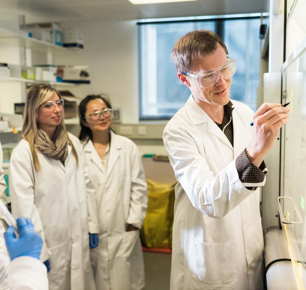
Five years into their programme, with the pipeline now mature and consistently producing high-quality data, the team is beginning to delve into biological questions that their pipeline is uniquely positioned to answer. The pipeline’s development has relied on a global network of earlycareer researchers who, together with their multidisciplinary team leads, have cleared technological hurdles and advanced the technology to its current working state. We met with some of them to learn more.
Resolving tumour architecture
The team’s mapping pipeline is illustrated on page 14. After progressing through core analysis technologies, data flows and highpowered processing infrastructure, a sample is presented as a computerised 3D model, which is annotated with detailed, interactive
information about each cell’s molecular characteristics.
All analysis begins with serial two-photon tomography (STPT) with a microscope that serially sections the sample into 15 µM slices, imaging four fluorescence channels simultaneously. During this step, physical sections are collected onto slides for further analyses throughout the rest of the pipeline. At the beginning of the team’s programme, this collection phase was a labour-intensive, manual process. “A major advancement of our workflow has been automating the collection of these sections,” explains Claire Mulvey, a principal scientific associate based in Cambridge, UK, who has been heavily involved in the STPT optimisation. “This automated section capture can now collect hundreds of slices from a single tissue sample and arrange them in the correct order, and is just one of the many ways we’ve upgraded the STPT instrument.”
STPT is an important step that produces a faithful 3D scaffold of the sample, upon which the results of all further analyses are annotated. This method has also allowed the team to explore many aspects of tumour architecture, including characterising the spatial relationships of cells within their environment, and it can be used to pinpoint physical regions of interest, such as areas showing vascular mimicry or metastasis, which can be further explored with higherresolution imaging techniques.
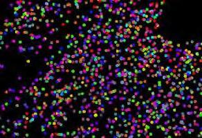
To produce ultra-high-resolution proteomic maps of specific regions of interest, samples can be cut into even thinner 2 µM slices for imaging mass cytometry (IMC) – a combination of mass cytometry and immunohistochemistry with high-frequency laser ablation, which can simultaneously image as many as 40 proteins and their modifications. IMC was originally pioneered for 2D imaging, but in late 2021, the team published a landmark dataset showcasing how the team optimised the technique for 3D image analysis. “The spatial arrangement
The Future Leaders Conference is supported by Bjorn Saven.
Elee Shimshoni
3D tumour sample.
Credit: Eduardo Gonzales Solares
of cells in a tumour is immediately clear when you map them in 3D,” says Laura Kütt, PhD student in Zurich, Switzerland, and lead author of the study. “With our 3D model, we could detect a cluster of epithelial cells that was potentially an early metastatic protrusion, which wouldn’t have been possible in 2D.”
IMC is an incredibly powerful technique, but challenges such as processing speed and throughput can limit its suitability for answering questions requiring large sample sizes and high throughput. To expand the pipeline’s applicability, the team has developed a new high-dimensional imaging approach: hyperplexed immunofluorescence imaging (HIFI). “HIFI is extremely gentle on tissue – meaning even very fragile samples can be investigated – and can be used to identify all major cell types in a tumour substructure, including their phenotype and localisation with respect to the wider tumour architecture,” says Spencer Watson, postdoctoral researcher in Lausanne, Switzerland.
HIFI, still a relatively novel technique, has yet to be fully integrated into the IMAXT pipeline. But in the future, Spencer hopes to perform HIFI on the full collection of slides from STPT, to first answer important questions about how cell biology changes across the entire tumour architecture, then zoom in on areas of interest by using IMC.
Aligning single-cell and spatial data
A novel aspect of this pipeline is the ability to link data from separately performed single-cell analyses and survey sequencing to build comprehensive expression profiles for each cell. Since the start of their programme, the team has optimised their pipeline of single-cell techniques, including optimising protocols, developing models to link RNA- and DNA-sequencing data enhancing direct library preparation for single-cell whole-genome sequencing and applying single-cell profiling to clinical samples. “We’ve used these methods to examine tumour evolution and chromosomal structural aberrations at the single-cell level, and learn more about the underlying biology,” says Ciara O’Flanagan, a research associate based in Vancouver, Canada. For example, matching cells’ single-cell DNA- and RNA-sequencing data has identified clonal populations with differential gene expression in triplenegative breast cancer, pinpointing new avenues of exploration to better understand the disease. “Integrating single-cell sequencing data with spatial transcriptomic profiling technologies reveals novel cell-cell interactions,” Ciara notes.
To map gene expression in space, two core technologies are used: expansion sequencing (ExSeq, a technique covered in last year’s report), which physically expands a sample to detect the subcellular localisation of specific RNA transcripts, and MERFISH (multiplexed error-robust fluorescence in situ hybridisation), which can achieve near-genome-wide, spatially resolved RNA profiling of individual cells with high accuracy and detection efficiency. “Profiling the transcriptomes of individual cells in their native environment allows one to uncover spatial gradients in gene expression across whole tissues, the spatial distribution of cell types, and even the subcellular localisation of DNA and transcripts,” says Leonardo Sepulveda Duran, a postdoctoral fellow based in Boston, US.
Learnings from astronomy
Although each of the techniques in the pipeline is valuable in itself, “their integration gives an unprecedented view of the spatial dissemination of cancer cells in their surrounding environment,” according to New York-based research fellow Ignacio Vázquez-García. Ignacio’s role is to integrate disaggregated single-cell sequencing data with in situ measurements and images. “3D molecular mapping poses a number of challenges that we’ve managed to overcome so far. A computational challenge we’re currently addressing is how to bridge between modalities so that we can resolve spatial patterns across the tumour at the single-cell level.”
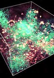
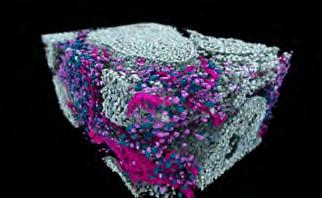
To build the final map, all spatial and single-cell data must be annotated on the 3D scaffold of the sample produced via STPT, but this process is challenging because each technique captures data with different characteristics and formats. Drawing inspiration from astronomical surveys, in which multi-wavelength images and data are stitched together to produce accurate maps of the sky, Eduardo Gonzales Solares, a senior research associate with a background in physics and astronomy, has helped overcome this challenge. “On several occasions, we’ve asked ourselves: how did we solve this problem in astronomy?” says Eduardo. The team has adopted a technique that develops a ‘star field’ for each tissue sample, using agarose-bead reference points in the STPT analysis. “We wouldn’t be able to achieve such accurate image registration without this crucial step,” Eduardo elaborates.
Because a single sample analysed on the pipeline produces multiple terabytes of data, another major hurdle faced by
the team is the phenomenal volume of data produced. “It’s not plausible to move such large quantities of data around the world,” Eduardo says. “Not everyone has access to high-end workstations, expensive software or local storage capacity.” Therefore, instead of bringing data to local computers with limited power, the team has developed automated workflows and analysis pipelines, as well as a new cloudbased compute lab allowing investigators to access data, run software and interpret results on demand. “We call it the IMAXT cloud,” Eduardo says. “It’s provided a new environment for scientific research and collaboration across the team.”
Answering important questions
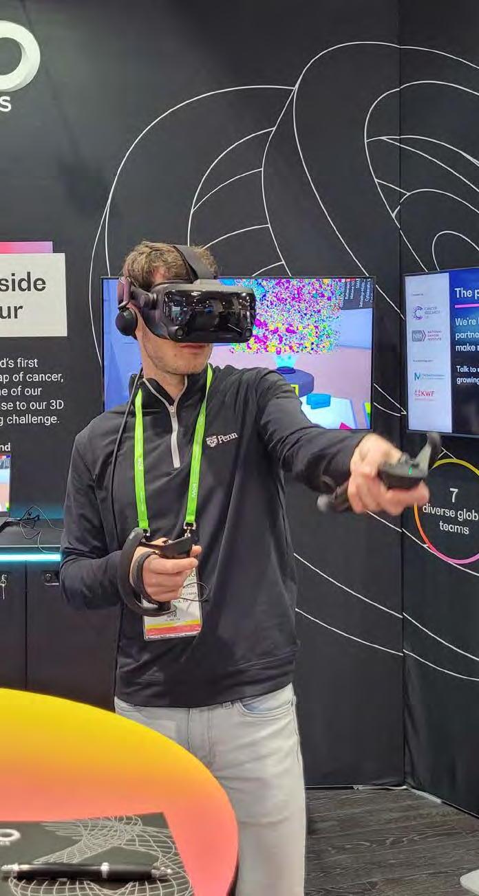
A final piece of the team’s puzzle is how to interact with the rich data produced by the pipeline. Although computers can handle such large amounts of information, initial exploration and interpretation of raw results by a human user is an indispensable practice for good science. To facilitate this process, the team has developed Project Theia, the world’s first virtual-reality cancer laboratory, in which users can ‘fly’ through and interact with their data in 3D, launch complex analyses and even extend analysis to 4D timeseries. Theia is planned to be released later this year and will equip researchers with an entirely new tool to investigate cancer biology.
“Now that our workflows are fully established, we can concentrate on using this pipeline of advanced techniques to answer key questions in tumour biology,” says Claire. In particular, the team is starting to use their pipeline to provide new ways to explore the poorly understood process of metastasis. Researchers have faced difficulties in studying the spread of cancer cells and in measuring metastasis-determining factors in situ long before the metastasis is detectable. The team’s integrated pipeline, with its ability to detect and investigate single cells within entire organs, should enable exploration of metastasis in unprecedented detail.
The team is also looking to understand the role of the immune system during tumour development, leveraging
the pipeline’s ability to detect tumour populations and local microenvironments in 3D to study the dynamic spatial selection and pruning of cancer cells under the selective pressure of the immune system. “Our aim is to identify stereotypical models of spatial cancer growth in 3D and how they influence clone competition and immune containment of the tumour,” says Ignacio.
Looking ahead, the team continues to optimise what they can achieve with their pipeline. ‘Tiger teams’ – specialist multidisciplinary groups originally named by NASA – have been formed across the consortium to address exceptionally difficult technical challenges. Spencer leads a tiger team focused on spatial proteomics and how to derive biologically relevant findings from detailed information on the 3D spatial organisation of a tumour. Conversations within this group ultimately helped achieve the development of HIFI.
“A great deal of our studies have been enabled by the highly collaborative environment across our consortium,” Spencer says. “I don’t think anything we do at this level of research would be possible without team science like this. Being part of this Cancer Grand Challenge has improved my work and the scope of my goals, just by being around the ambitious nature of the whole project. This kind of ambition is infectious and inspires us all to push harder.”
A new integrated pipeline to probe cancer’s inner workings
The sample is prepared for analysis as intact tissue or is disaggregated for single-cell analysis. The team has made major advances enabling a wide variety of samples to be analysed through the pipeline, from cultured cells to frozen biopsies or formalin-fixed paraffinembedded tissues, which have historically been difficult to image.
This map represents the journey of a sample through IMAXT’s 3D tumourmapping pipeline, progressing through multiple levels of analysis on intact tissue, as well as single-cell sequencing on disaggregated cells. Eventually, all data are integrated and mapped onto the same 3D scaffold, providing a 3D tumour map that can be explored in virtual reality.
Expansion sequencing: spatial transcriptomics
Physical expansion of a
Intact tissue
Serial two-photon tomography: spatial proteomics
By serially sectioning the sample as 15µM slices and imaging four fluorescence channels simultaneously, STPT provides the scaffold upon which all further annotations are projected.
Disaggregated cells
Survey sequencing and single-cell genomics and transcriptomics
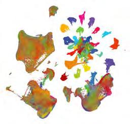
Image: Chromosomal copy number profile from scDNAseq and UMAP from scRNAseq data
A non-destructive, high-dimensional variant of cyclic immunofluorescence that can image 40 or more markers across whole-slide tissue sections.
“Thanks to discussion with our wider team, we’ve overcome a number of technical challenges with HIFI, including setting up a new analytical pipeline and solving how to align and combine all images from each round of labelling.”
– Spencer Watson, postdoctoral fellow
3D imaging mass cytometry: ultra-high-resolution spatial proteomics
Simultaneous detection of as many as 40 antigens and nucleic acid sequences. “We’ve overcome a number of technical difficulties in translating IMC from 2D to 3D, including using a diamond knife to cut 2 µM slices without distorting the tissue.”
– Laura Kütt, PhD student
Human
MERFISH:
spatial transcriptomics
Near-genome-wide, spatially-resolved RNA profiling of individual cells, with high accuracy and efficiency.
“We’ve significantly optimised our sample procurement and analysis protocols to obtain high-quality data and are continuing to optimise the technique to expand the repertoire of samples we can target with MERFISH.”
– Leonardo Sepulveda Duran, postdoctoral fellow
“These methods give us detailed information about the expression profiles of individual cells and populations, letting us examine tumour evolution and chromosome structural aberrations at the single-cell level. Combining this with spatial data lets us uncover novel cell-cell interactions and underlying biology.
Ciara O’Flanagan research associate
Image stitching, segmentation and re-alignment
Data gathered throughout the pipeline are projected onto the anatomical scaffold provided by STPT. “One of the technical challenges of our pipeline is the large volumes of imaging data and the variety of instrument measurement modalities. To achieve this integration, we’ve taken learnings from astronomy, including adopting approaches used for data handling and analysis of astronomical surveys.”
Featured Articles
Alon S et al. Science 2021; doi: 10.1126/ science.aax2656
Bressan D et al. bioRxiv 2021; doi: 10.1101/2021.06.28.448342
– Eduardo Gonzales Solares, senior research associate
Image: Beads around the 3D model of a tumour, which are used for image registration of sample slices as well as across different modalities
Credit: Eduardo Gonzales Solares
Output: a detailed, computerised, 3D map of the original sample that can be explored in virtual reality
US
Campbell KR et al. Genome Biology 2019; doi: 10.1186/s13059-019-1645-z
Gonzalez-Solares EA et al. bioRxiv 2021; doi: 10.1101/2021.06.22.448403
Kuett L et al. Nature Cancer 2021; doi: 10.1038/ s43018-021-00301-w
Laks E et al. Cell 2019; doi: 10.1016/j. cell.2019.10.026
O’Flanagan CH et al. Genome Biology 2019; doi: 10.1186/s13059-019-1830-0
Salehi S et al. Nature 2021; doi: 10.1038/ s41586-021-03648-3
Vázquez-García I et al. bioRxiv 2021; doi: 10.1101/2021.08.24.454519
breast cancer Rendering credit: AGAVE (Allen Institute for Cell Science)
Credit: Zhuang lab
Featured team members
Dr Ciara O’Flanagan research associate in the Aparicio lab, BC Cancer Agency Research Centre, Canada
Laura Kütt PhD student in the Bodenmiller lab, University of Zurich, Switzerland
Dr Leonardo Sepulveda Duran postdoctoral fellow in the Zhuang lab, Harvard University, US
Dr Spencer Watson postdoctoral researcher in the Joyce lab, University of Lausanne, Switzerland
Dr Eduardo Gonzales Solares senior research associate in the Walton lab, University of Cambridge Institute of Astronomy, UK
Dr Claire Mulvey principal scientific associate in the Hannon lab, Cancer Research UK Cambridge Institute, UK
Dr Ignacio Vázquez-García research fellow in the Shah lab, Memorial Sloan Kettering Cancer Center, and the Tavaré lab, Columbia University,
Excavating the genes that help tumours escape the immune system
Why do certain cancer genes that are broadly expressed throughout the body trigger cancer in only specific tissues? New findings suggest that the immune system plays a greater role in this specificity than previously realised.
Challenge:
Tissue Specificity:
Understand why mistakes in certain genes cause cancer in only specific parts of the body
Team: SPECIFICANCER
The key to why certain genes drive cancer in some parts of the body but not others has long been sought but remains elusive. Unlocking this mystery of tissue specificity could enable new preventive strategies and therapies, and might even reveal why some treatments work for people with certain types of cancer but have very little success in others. The Cancer Grand Challenges SPECIFICANCER team is taking on this enduring challenge, guided by the philosophy that a tissue’s pre-existing characteristics dictate whether a mutated gene can drive tumour development.
cells to escape the constraints of their controlled environments and become cancerous. Although some TSGs, such as p53, are expressed throughout the body, many TSGs act in a tissue-specific manner. The team’s recent study sheds light on how cancers in different parts of the body develop tissue-specific TSG mutations allowing new mechanisms of immune evasion to be exploited.
Using the gene-editing tool CRISPR, the team systematically eliminated approximately 7,500 individual genes in two mouse cancer cell lines. To reveal how these genes influence the immune response, the team then implanted the mutated cells into mice with intact immune systems or with severe combined immunodeficiency.
“Our search left us with a huge list of genes which play a role in helping cancer cells hide from the immune system, when mutated,” says Tim Martin, postdoctoral researcher and the lead author of the study. “We were surprised that such a large number of these genes were TSGs. As our study went on, we began to realise there are many more routes for cancer cells to escape the immune system than we thought, and that different tissues do it in different ways.”
Immunotherapies targeting specific evasion strategies or enhancing the immune response have become a powerful treatment option for some people with cancer. However, most immunotherapies are effective against only certain forms of the disease in only a small proportion of patients. The team’s methods may now provide tools to first understand these variable responses and then develop more effective treatments.
“This methodical way of exploring how tumours evade the immune system is relatively slow – but it helps to unpack the complexity of these genes in a way we wouldn’t have otherwise,” says Steve. “It’s a new way to consider the challenge of tissue specificity, for sure. We hope others will adopt this approach to work out how other TSGs help tumours evade the immune system and identify new targets for drug development.”
“
“
We’ve unearthed a new way to think about tissue specificity.
Steve Elledge SPECIFICANCER team lead
Major findings recently published by the team in Science have uncovered a new layer of the mystery: the tissue’s interactions with the immune system. Generally, the immune system reacts to abnormal and pre-cancerous cells as if they are pathogens and unleashes a powerful response to eliminate them. However, during tumour development and evolution, cancer cells develop mechanisms to overcome this attack.
Tumours in different tissues escape the immune system through multiple mechanisms. For example, some lung and blood cancers are known to hide from T cells by engaging immune checkpoints. “For other types of cancer, our findings suggest the tumour cells are doing something else – possibly something as yet unknown,” explains SPECIFICANCER team lead Steve Elledge.
A major role for tumoursuppressor genes in immune evasion
Immune evasion appears to rely at least partly on tumour-suppressor genes (TSGs), which are normally responsible for restricting tumour growth. When mutated, TSGs can enable abnormal
The findings provide a new way to think about TSGs and their roles in enabling tumours from different parts of the body to escape the immune system. Furthermore, although the immune system’s roles in tumour evolution and the treatment response have been well documented, this study is the first report of the tissue-specific interactions of the immune system with TSGs.
“To learn how to treat cancers in different tissues, we need to understand all the ways they’re escaping the immune system,” says Steve. “Currently, there’s a mismatch between the tools we have in our immunotherapy arsenal and the different mechanisms cancers use to evade attack.” Although TSGs appear to provide many routes for different tumour types to escape the immune system, the team suspects that a finite number of escape strategies exists.
“By learning them, we could develop whole new therapeutic approaches to prevent cancer cells from circumventing immune attack,” Steve explains.
Importantly, the study provides a blueprint to explore the evasion strategies of TSGs through analysis of their genetic networks. For example, GNA13 is a TSG that, when mutated, is associated with poor outcomes in certain lymphomas and drives immune evasion in breast cancer. By mutating GNA13 and examining changes in the gene expression network, the team has revealed how mutant GNA13 shields breast cancers from the immune system through secretion of the protein CCL2, which recruits pro-tumour macrophages to the breast microenvironment. Further probing of this mechanism may lead to novel immunotherapies for people with breast cancer.
Unexpected findings and the freedom to follow new research directions
“What’s so exciting about this study is that we ended up following a route we really didn’t expect when we set out,” says Tim. The team began their study by looking for genes that, when perturbed, would boost the immune system’s ability to eliminate cancer cells. “Instead, we found just the opposite.”
Tim recalls the moment when the team realised that their unexpected findings might be meaningful. “We sat chatting in Steve’s office one weekend, and we actually thought the experiment had failed,” he says. “Then from this vast dataset, we realised a lot of genes on our list were TSGs, and that gave us a whole new avenue to explore.”
Steve adds, “There was a gem hiding in what we thought was a failed experiment. Thanks to the flexibility of Cancer Grand Challenges funding, we were able to pursue it.”
The findings were even more fruitful than the team expected. “We thought our investigations might reveal one or two genes for further exploration,” Steve muses. “Instead, we’ve unearthed a new way to think about tissue specificity and a new way to study the role of TSGs in the immune response – which could ultimately lead to more effective cancer treatments.”
What’s so exciting about this study is that we ended up following a route we really didn’t expect when we set out.
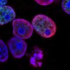
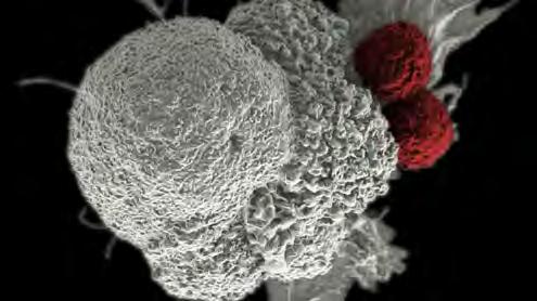
Image: Scanning electron micrograph of an oral squamous cancer cell (white) being attacked by two cytotoxic T cells (red) Credit: NIH Image Library
A blueprint for studying genes involved in immune escape
Featured team members Professor
Harnessing the microbiome to personalise colorectal cancer prevention, diagnosis and treatment
Colorectal cancer incidence is increasing worldwide, particularly in people younger than 50 years, but the reason remains a mystery. In addition, the effectiveness of treatments inexplicably varies within the same patients over time, among patients and geographically. Could the microbiome be responsible? A new cohort study, spearheaded by a global consortium of researchers and people affected by colorectal cancer, seeks to find out.
Challenge:
Microbiota: Understand how microbes inside our bodies affect cancer treatment
Team: OPTIMISTICC
The microbiome – the trillions of bacteria, viruses and fungi residing in the body – is emerging as a key player in human health, by shaping immunity, nutrition and overall well-being. The extent to which perturbations in the microbiome underpin the development and epidemiology of certain cancers is only beginning to be understood. Shedding light on this link in colorectal cancer, which is diagnosed in 1.8 million people worldwide each year, may lead to the development of new ways to detect, screen and prevent the disease. The Cancer Grand Challenges OPTIMISTICC team aims to pinpoint how the microbiome affects colorectal cancer initiation and development, as well as patient outcomes, and to translate their findings into novel preventive, diagnostic and treatment strategies.
Because the gut microbiome lies at the intersection of lifestyle and colorectal cancer development, research in this nascent area has the potential to transform understanding of this disease. “The amazing thing about the microbiome is how it’s influenced and shaped by the diet and various lifestyle factors – many of which also seem to influence colorectal cancer risk and outcomes,” explains Kimmie Ng, OPTIMISTICC co-investigator and a medical oncologist specialising in gastrointestinal cancers as the director of the Young-Onset Colorectal Cancer Center at Harvard’s Dana-Farber Cancer Institute.
A major component of the team’s programme is a first-of-its-kind clinical cohort study collecting stool and blood samples, and long-term clinical information from more than 2,500 people with colorectal cancer. The study’s ambitious design could deepen understanding of the microbiome and subsequently improve quality of life and
survival in people with colorectal cancer. “We need to identify the interactions between the microbiome and the diet and lifestyle factors that influence risk and outcomes in these patients, so we can make informed recommendations about diet and lifestyle and treatment, then move beyond observations to understand the underpinning biological mechanisms,” says Kimmie. The study has grown since it first opened and is now enrolling patients from multiple sites across the US, Spain and Brazil.
Tapping the microbiome to guide individualised treatment
Through the cohort study, the team’s first goal is investigating how lifestyle factors influence the microbiome and subsequently colorectal cancer outcomes in three patient subgroups: those with early-onset colorectal cancer (diagnosed before the age of 50), those receiving chemotherapy and those receiving immunotherapy. Serial samples from before, during and after treatment are being analysed to understand how the microbiome changes as patients’ tumours evolve or their treatments progress. Participants also fill out a comprehensive questionnaire on their diet and lifestyle so that these factors can be evaluated and incorporated.
The team hopes that these data will reveal whether the microbiome might be responsible for the dramatic increase in colorectal cancer cases in young adults – a worrying trend that has yet to be explained.
“What we are asking is: are there differences in the composition and diversity of the microbiome in younger colorectal cancer patients compared with older people?” Kimmie explains. “That would give important clues as to why early-onset colorectal cancer is on the rise, so that we can make informed recommendations about diet and lifestyle and identify high-risk individuals.”
The OPTIMSTICC team’s second major goal is to understand how the microbiome influences individuals’ responses to treatment. For example, patients across geographical areas have considerably different responses to the chemotherapy drug capecitabine: patients with colorectal cancer in North America experience significantly more severe adverse effects than those in Europe. Similarly, checkpointinhibitor immunotherapies are powerful treatment options for a small proportion of people with colorectal cancer but have very little effect in others. The team suspects that the reason for these interindividual differences may lie in the unique microbiomes resulting from differing diets and lifestyles. By tapping into the shared or
differing features between these groups, the team’s cohort study might enable clinicians to work with the microbiome to enhance the stratification of patients to treatment, alleviate chemotherapy toxicity and overcome the resistance that many people currently experience to immunotherapies.
At the time of writing, the team has enrolled more than 500 patients, collected more than 800 stool samples and 600 blood samples, and begun to analyse the preliminary data –making progress despite the setbacks of the COVID-19 pandemic, which initially hindered participant enrolment, sample collection and face-to-face meetings.
Putting patients at the heart of the research
Patient advocates – people with a personal experience of cancer – play a vital role in all Cancer Grand Challenges teams. In taking on the microbiota challenge, “the patient advocates have been absolutely critical in the design and implementation of the cohort study,” Kimmie says. This process includes ensuring that some of the study’s most important elements – such as the diet-and-lifestyle questionnaire – are relatable and understandable to participants. “A person undergoing treatment for advanced colorectal cancer will already be going through so much –the advocates have made taking part in the cohort study as accessible and the least burdensome as possible for our participants,” Kimmie elaborates.
Transcending collaborative boundaries
Taking on the Microbiota challenge and assembling a large international cohort critically relies on collaborative international efforts that transcend institutional and geographical boundaries. Researchers with diverse expertise and patient advocates from the UK, US, the Netherlands, Canada and Spain have united through the Microbiota challenge by connecting, exchanging ideas and sharing knowledge. The rich sample library collected through the cohort study is used to examine hostmicrobe interactions throughout the entire challenge, informing preclinical work through the isolation of organisms and their metabolites, and the generation of organoids and other model systems.
Chicago-based Candace Henley is one of the study’s patient advocates and a survivor of colon cancer. “We want to make sure that treatment options, clinical trials, everything about the cancer continuum for those after us is better than what we’ve experienced in our own journeys,” she says. “I’m an 18-year survivor, and I’m still experiencing challenges from my treatment. We’re advocating for early detection, the patient voice and the betterment of the patients who come after us. If our work could lead to more targeted, less harsh treatments for people, that would be amazing.”
The recruitment of patient advocates from multiple countries has facilitated enrolment of the large, diverse cohort of participants necessary for understanding the connection between colorectal cancer and the microbiome, and factors of geography or ancestry that may affect risk or outcomes. Similarly, “if we can find a common signal in the microbiome in different geographical areas that associates with early-onset colorectal cancer or poorer immunotherapy response, for example, despite otherwise different lifestyles, we can be confident that the result is robust,” explains co-investigator Josep Tabernero, director of the Vall d’Hebron Institute of Oncology in Barcelona. The team is beginning to work with more centres across Europe and North and South America. “As the cohort study continues to grow, ensuring the people recruited to take part represent the diversity of the population remains an important aim for the team going forward,” Josep says.
“The Cancer Grand Challenges mechanism is just perfect for compiling the right team and recruiting the right patients,” says Kimmie. “It’s opened my eyes to all the innovative ways that we can target the microbiome.”
Although the effects of the pandemic on the study are ongoing (for example, by limiting the supply of stool kits), this enormous collaborative effort is already fruitful. The team’s cohort study is generating encouraging data, including preliminary results on the effects of the microbiome on patients’ responses to immunotherapy and chemotherapy toxicity. Over the next year, the team hopes to perform deep sequencing of stool samples, increase patient enrolment across all study regions and begin to share their results more widely so that they can ultimately be translated to clinical interventions for patients.
The melding of disciplines is really important for advancing science – our team is a great model of that working well.
by Gege Li
Microbiome of Colorectal Cancer: Longitudinal Study of Mechanism (MICROCOSM) is a global, firstof-its-kind clinical cohort study seeking to understand the role of the microbiome in treatment response and early-onset colorectal cancer.
Image: NCI
Kimmie Ng OPTIMISTICC co-investigator
Featured team members
Dr Kimmie Ng OPTIMISTICC coinvestigator, DanaFarber Cancer Institute, US
Candace Henley OPTIMISTICC patient advocate, Blue Hat Foundation, US
Professor Josep Tabernero OPTIMISTICC coinvestigator, Vall d’Hebron Institute of Oncology, Spain
The OPTIMISTICC team is funded by Cancer Research UK with generous support from Nick and Annette Razey.
A powerful lens into tumour metabolism
The team’s pipeline could inform decision-making across the whole patient journey
In taking on our 3D Tumour Mapping challenge, the Cancer Grand Challenges Rosetta team has curated a suite of mass spectrometry imaging (MSI) techniques that can measure a broad range of metabolites and map their spatial distribution within the tumour microenvironment more holistically than ever before.
Here, team lead Josephine Bunch reflects on the obstacles the team has encountered and overcome, and looks ahead to how the pipeline could revolutionise cancer care.
Challenge:
3D Tumour Mapping: Map the molecular and cellular tumour microenvironment to define new targets for therapy and prognosis
Team: Rosetta
“Cancer
Care 2.0”
The team’s tumour mapping pipeline could inform decision-making across the whole patient journey.
I first heard about Cancer Grand Challenges on a BBC Radio 4 programme back in 2015. After discovering that one of the problems was to map tumours at a molecular and cellular level, I started discussing with other leading MSI experts which imaging modalities we could combine to tackle this question. I also talked to leading cancer biologists to find out about the types of maps they needed to help improve their understanding of tumour behaviour. Together, we formulated the Rosetta programme, setting out to apply molecular imaging to explore the secrets of tumour metabolism.
Over the past five years, we’ve successfully established a truly groundbreaking untargeted MSI-driven systems biology approach that can probe tumour metabolism in unprecedented detail. By performing measurements with a carefully compiled selection of techniques at many locations across a sample, we can achieve coverage at not only the required molecular breadth but also a range of length scales from subcellular to whole
tissues. The easiest way to conceptualise our pipeline is by likening it to Google Earth: it generates a detailed atlas of tumour metabolism – from the organ level right down to the subcellular level – with the ability to zoom in at different degrees of magnification.
The information that we are uncovering with this fundamentally new way of analysing cancer metabolism is already providing a much deeper understanding of tumour behaviour, prognosis and response to treatment. Looking ahead, we hope that the Rosetta pipeline will lead to the development of a novel suite of clinical decision-making tools that can help transform outcomes for people with cancer.
Overcoming challenges
During the first two years, we focused on developing and validating, optimising and combining the different components of the Rosetta pipeline – including matrixassisted laser desorption/ionisation (MALDI), desorption electrospray ionisation (DESI) and secondary ion mass spectrometry (SIMS).
We set out by distilling a list of around 200 highly informative metabolites – including amino acids, fatty acids, carbohydrates, vitamins and lipids –which we could use to measure our pipeline’s success in terms of our degree of coverage. But the sheer breadth of this list posed a huge analytical challenge. We needed to establish methods for a range of modalities that could monitor all these target molecules with vastly different chemical properties
1
Screening
simple, non-invasive tests to detect metabolic signatures, for example in urine and stool samples
2 Diagnostics
metabolic-signature detection of biopsies for real-time patient stratification
– including polar and non-polar, neutral, or positively or negatively charged entities with a wide range of molecular weights – in complex samples at the highest resolution, while maintaining the quality of each measurement. It was also absolutely crucial that we could review and compare data from in vitro models, including primary cell lines and patient-derived organoids, with measurements from ex vivo and in vivo samples, including whole animal organs and tissue biopsies.
A major legacy of the Rosetta pipeline will be our development of new and innovative modalities, which we did in response to our early data when we didn’t have the required sensitivity or molecular coverage. Typically, fresh frozen tissue samples give the best results for MSI, while other traditional preservation methods are less optimal or even incompatible. Developing new methods to enable the analysis of formalin-fixed paraffin-embedded material has also proven absolute dynamite – allowing us to turn our attention to beautifully curated and compiled repositories of samples from around the world that would otherwise have been impossible to analyse with our pipeline.
It’s also taken a colossal and heroic effort by many people in the team to develop a world-class computational pipeline to enable the mining of these enormous unique datasets, which we have been improving, testing, validating and extending throughout the programme. Most recently, we’ve refined our methods to run unsupervised analyses, enabling the team to make
unbiased interpretations of the data to identify novel points of interest that might otherwise have been missed.
Identifying targetable vulnerabilities
We started the project with a parallel launch of several programmes focusing on colorectal, breast and pancreatic cancers, as well as brain tumours, which have all reached different stages.
In work led by Owen Sansom at the Cancer Research UK Beatson Institute, Glasgow, we identified the antiporter SLC7A5 as a potential new therapeutic target for KRAS-driven colorectal cancers by using genetically engineered mouse models. Challenging the long-held assumption that glutamine metabolism fuels the growth of cancers, we found that SLC7A5 ejects this amino acid from cancer cells and imports other metabolites from the surrounding tissue. The results uncovered a promising new targetable vulnerability for drug discovery: targeting SLC7A5 could help starve cancer cells of metabolites necessary for cancer growth while having little effect on healthy tissues, and could
“
Over the past five years, we’ve established a truly groundbreaking pipeline that can probe tumour metabolism in unprecedented detail.
Josephine
Bunch Rosetta team lead
3 Surgical treatment using the iKnife for surgical margin control and to match patients to treatment according to metabolic changes
4 Non-surgical therapy identifying therapeutic targets and developing new drugs on the basis of metabolic weaknesses
5 Monitoring a simple test that, in addition to screening, could enable monitoring of how a patient is responding to therapy or whether their cancer is likely to return after treatment
Josephine presenting at Cancer Grand Challenges summit
Meet Margaret Grayson
MBE, chair of the Cancer Grand Challenges Advocacy Panel
As told to Alison Halliday
also help overcome drug resistance in some patients. But without our pipeline, we wouldn’t have been able to detect glutamine in the tissue surrounding the cancer cells rather than inside them.
Another study, which was led by George Poulogiannis at the Institute of Cancer Research, London, involved screening breast cancer cell lines, tumour samples and mouse models. We identified that disruptions to the PIK3CA kinase pathway cause an increase in the metabolite arachidonic acid, which helps fuel the growth of these breast tumours. We also found that drugs that interfere with the PIK3CA pathway are much more effective in slowing breast tumour growth in mice fed a diet without fatty acids than a standard diet. This is the first study showing how dietary fat restriction might play a major role in the response to treatment – and again, would not have been possible without our pipeline.
Transforming the patient journey
Developing ways to translate the Rosetta pipeline into the clinic offers unique opportunities for enabling the delivery of precision medicine. For the first time, we can start to envision an analytical pipeline that can produce data that can be compared across all stages of the cancer patient journey – from screening and diagnostics to advanced therapies and follow-up monitoring (see side figure – Cancer Care 2.0).
The intelligent surgical knife (iKnife) –which was invented by Rosetta team member Zoltan Takats at Imperial College London – provides a powerful example of a diagnostic tool with the potential to deliver benefits at different points along the patient journey. The electrosurgical device uses rapid evaporative ionisation mass spectrometry (REIMS) and is designed to deliver real-time information about tumour metabolism that can help guide margin control during surgery. In our PIK3CA programme, our results provide a proof of principle that this method could also be used to stratify cancers into subtypes according to metabolic phenotype to help guide treatment selection. Specifically, we showed that iKnife can detect elevations in arachidonic acid in cell lines, which can be mapped back to disruptions in the PIK3CA pathway and correlate with a subtype of breast cancer. We are now exploring ways to detect changes in tumour metabolism with less-invasive approaches, such as the analysis of urine and stool samples or exhaled breath.
Uniting across traditional research boundaries
Underpinning our team’s success is that we’ve united some of the most exceptional experts in the world in each of the four cancer types, tumour biology and metabolism, and each of our technologies. Although the journey has often been difficult, we’ve all thoroughly enjoyed working with one another. The long-term support for our extraordinary generation of early-career future leaders has also made a real difference – they’ve been spending time in each other’s laboratories and are the people truly driving the project.
From a personal perspective, this is the most important research I’ve ever been involved in. Analytical chemists have often been relatively agnostic to the problems brought to us and have applied very technology-focused approaches to problem-solving. But since taking on this challenge, I’ve become completely fascinated with the field of metabolism. I think my laboratory will now work in this space forever.
It’s imperative that the research we do translate into tangible impact for people with cancer. We’re committed to patient advocacy within our work, from actively involving people affected by cancer in our funded teams, to engaging our advocacy panel in the challenges we set.
How does the advocacy panel help to shape research?
We’re such a mix of people, and each of us brings our own unique experience. All of us have been impacted by cancer: some of us are patients, others of us have cared for a family member, and we’re affected by different cancer types. But our common bond is our passion for research, and through the panel, we help to craft each round of challenges and support teams to involve the voice, experience and insights of people affected with cancer in their work. When we come together, we leave behind the things that are specific to our own experiences and look at the broader picture of what’s important to many people in relation to cancer.
What’s it like being part of the Cancer Grand Challenges community?
It’s exciting. I feel a part of something that is big, bold and outside the box, helping to answer some of the big challenges in cancer.
What was your highlight from the past year?
It’s been very exciting working with the advocacy panel throughout this latest round of funding, from the expressions of interest stage through to the full applications. My highlight, as chair of the panel, has been sitting with the scientific committee and being part of the final interviews for the shortlisted teams – witnessing the passion of the teams as they present their research, and being able to ask questions on behalf of the panel around how advocacy will be embedded into their plans, has been really special.
What does global, multidisciplinary research mean to you?
To me, it’s about bringing together disciplines that have never really worked together, or might not even have worked on cancer research before. That’s what makes Cancer Grand Challenges unique: enabling the best minds from around the world to collaborate together on the greatest challenges in cancer.
Margaret Grayson MBE, chair of the Cancer Grand Challenges Advocacy Panel “
Our common bond is a passion for research.
Professor Josephine Bunch Rosetta team lead
Rosetta co-investigators at 2020 Summit
The Rosetta team
An atlas of oesophageal cancer development
A new integrated atlas of the stromal and epithelial interactions that drive cancers associated with chronic inflammation provides a new way to understand cancer types with the poorest survival.
Challenge: Cancer Causes: Understand how lifestyle factors, such as obesity, cause cancer
Team: STORMing Cancer
Inflammation – the immune system’s normal response to injury and infection – was first linked to cancer more than 150 years ago. But despite its connection to an astounding 20% to 25% of cancer deaths worldwide, very little is known about how chronic inflammation drives cancer development. The STORMing Cancer team chose to unravel this longstanding riddle when taking on our Cancer Causes challenge.
Looking up answers in the atlas of cancer progression
The atlas will map the progression of oesophageal adenocarcinoma – which Thea describes as the ‘poster child’ for chronic inflammation in cancer. This progression often starts with acid reflux, which triggers an adaptive response to inflammation, known as metaplasia, which progresses to cancer in a small percentage of people. In a protective response, oesophageal cells are replaced with gastrointestinal cells, but this shift can increase the risk of dysplasia, or abnormal growth. Between 3% and 13% of people with metaplasia later develop oesophageal adenocarcinoma.
A massive undertaking
Setting up the atlas’s novel sample collection and analysis pipeline has required vast efforts across Canada, the US, UK and Israel, originating with Lorenzo Ferri’s team and extending to more than 60 investigators plus collaborators. The side illustration depicts a single sample’s journey from its collection by surgeons to diagnosis by pathologists in Canada to its analysis in laboratories across Canada and North America.
the outputs from multiple methods is challenging but rewarding. “We went through a major exercise to find common nomenclature between these techniques, which we hope will be useful for the wider research community,” explains Philippe Gascard, the team’s programme manager. “While other studies have used these techniques side by side, this is the first time their outputs have been integrated, and a common language was necessary to do this.”
Featured team members
Our atlas is a remarkable technical achievement that shows the breadth and depth of what you can do when facilitated to work in this global, multidisciplinary way.
Thea Tlsty STORMing Cancer
team lead “
Their approach focuses not only on the tumour cells themselves but also on their interactions with its inflamed microenvironment. This milieu comprises a web of connective tissue – the stroma, including blood vessels and the extracellular matrix – that profoundly influences epithelial cells and can coax healthy cells towards malignancy (or malignant cells towards normalcy).
“Historically, the stroma’s role in cancer development has been overlooked; our hypothesis is that it’s actually the dominant driving force in the progression of chronicinflammation-associated cancers,” explains team lead Thea Tlsty. “Having our programme funded was wonderful, in part because it has finally given recognition to the importance of the stroma.”
The lack of robust models that accurately mimic this overlooked aspect of human biology has been a major obstacle to understanding the relationship between cells and their inflamed surroundings. The team is now addressing this issue by constructing an integrated atlas of human tissue that maps the progression of chronic-inflammation-associated cancer, from normal tissue through all stages of inflammation, to tumour development, assessing the multiple parts of the tissue.
“Our atlas is a remarkable technical achievement that shows the breadth and depth of what you can do when facilitated to work in this global, multidisciplinary way,” says Thea.
The atlas is based on data from 12 people with oesophageal adenocarcinoma that developed from metaplasia, who have not yet received treatment and have donated biopsy or surgical resection samples spanning the full progression from matched normal oesophageal tissue to metaplasia, dysplasia and ultimately cancer. The atlas integrates clinical data with extensive annotation of sample information, including histology and matrix stiffness data captured with atomic force microscopy, single-cell RNA sequencing, proteomic analyses of the extracellular matrix and co-detection by spatial indexing (CODEX).
Importantly, this atlas provides a much-needed tool to answer important questions about how inflammation drives malignancy. How do stromal architecture and cell-cell interactions change as the disease progresses? How do changes in the stromal transcriptome and proteome influence progression? Can probing the stroma reveal opportunities to alter the inflamed tissue, and slow, prevent or even reverse malignancy progression? “We hope the wider research community will be able to use the wealth of information provided by the atlas to develop clinical tools for risk stratification, for preventive strategies and for interventive agents,” says Thea.
This massive undertaking has not been without challenges. The COVID-19 pandemic has led to the reprioritisation of patient procedures and redeployment of clinical colleagues to the frontline, and has slowed participant recruitment and sample collection. Material transfer agreements between institutions have posed administrative barriers. In addition, although sampling pure matched normal and cancerous tissues in biopsy and surgery is fairly straightforward, the histological overlap of disease stages can make collecting and characterising distinct disease stages difficult, particularly at the metaplasia and dysplasia stages.
The data integration has perhaps been the largest barrier to unlocking the full potential of the atlas. Because multidisciplinary teams often speak different scientific languages, translating and integrating
The team is excited to share their atlas with researchers worldwide later this year. “With this sample collection and analysis pipeline, we’re providing a tool for the research community to probe the longstanding mystery of how inflammation drives cancer in human tissue, with focus on the stroma –rather than overlooking it or including it as an afterthought,” says Thea. “The more people who can be facilitated to explore the stroma’s role, the faster we’ll drive progress against chronic-inflammation-associated cancers. These are among the most lethal cancer types in the world, and this is why we are so motivated by the opportunity we have here.”
Beyond the oesophageal adenocarcinoma atlas, the team has made substantial headway in establishing a pipeline for mapping lung and stomach cancers, and hopes to achieve the same for colorectal cancer.
The journey of an atlas sample
Atlas samples are collected within two hours after surgery in Montreal, Canada, and analysed in labs across North America. The wider team of 60+ spans the US, UK, Canada and Israel, with investigators tackling different areas of the challenge.
Dr Lorenzo Ferri co-investigator, McGill University Health Centre Research Institute, Canada
Dr Sui Huang co-investigator, Institute for Systems Biology, US
Dr Birgit Schilling co-investigator, Buck Institute for Research on Aging, Novato, California, US
Dr Philippe Gascard programme manager, University of California, San Francisco, US
Dr Garry Nolan co-investigator, Stanford Medicine, US
Professor Thea Tlsty team lead, University of California, San Francisco, California, US
Single-cell RNA sequencing analysis and centralised database, Institute for Systems Biology, Seattle
Isolation of matrix and live cell derivatives, University of California, San Francisco
Proteomic analysis, Buck Institute for Research on Aging, California
Seattle
Montreal
San Francisco
CODEX, Stanford University, California
The STORMing Cancer team is funded by Cancer
Professor Thea Tlsty and professional researcher Dr Deng Pan
Sample collection and preparation, McGill University, Montreal
Single-cell RNA sequencing, McGill Genome Center, Montreal
$100m to take on four new challenges
A Cancer Grand Challenge is identified through a rigorous grassroots process, including workshops, consultation and debate, that solicits and distils ideas from the global cancer research community and people affected by cancer. The Cancer Grand Challenges scientific committee then translates these ideas into tangible challenges – intractable problems that, if solved, could enable dramatic progress in cancer research.
We were delighted to receive submissions from 169 diverse
teams, spanning more than 60 countries, outlining how they would take on our latest round of challenges. The scientific committee was tasked with narrowing these innovative ideas to a shortlist of just 11 teams, which received seed funding to develop their ideas into full proposals.
We’re now pleased to share the four challenges that will be taken on in this round of funding. The four winning teams will each receive $25m, giving them the freedom to come together, think differently and take bold new approaches to pursuing these challenges.
Cachexia
Understand and reverse cachexia and declining performance status in cancer patients
Co-led by Dr Eileen White, Rutgers Cancer Institute of New Jersey, US; Dr Marcus DaSilva Goncalves, Weill Cornell Medicine, US; and Dr Tobias Janowitz, Cold Spring Harbour Laboratory, US
Read more about the CANCAN team on page 28
Extrachromosomal DNA
Understand the biology of ecDNA generation and action, and develop approaches to target these mechanisms in cancer
Led by Paul Mischel, MD, Stanford Medicine, US
Read more about the eDyNAmiC team on page 32
Solid Tumours in Children
Develop novel therapies to target unique features in solid tumours in children
Co-led by Professor Catherine Bollard, Children’s National Hospital, US and Dr Martin Pule, University College London, UK
Read more about the NexTGen team on page 34
Normal Phenotypes
Understand how cells and tissues maintain ‘normal’ phenotypes while harbouring oncogenic mutations and how they transition to become a tumour
Co-led by Professor Allan Balmain, University of California San Francisco, US; Dr Paul Brennan, International Agency for Research on Cancer, France; and Dr Nuria Lopez-Bigas, IRB Barcelona, Spain
Read more about the PROMINENT team on page 38
Rehovot
New team funded 2022
A new virtual institute with a mission to solve cancer cachexia
Challenge: Cachexia: Understand and reverse cachexia and declining performance status in cancer patients Team: CANCAN (Cancer Cachexia Action Network)
Many patients with advanced cancer experience cachexia, a debilitating wasting syndrome characterised by extensive weight loss from both skeletal muscle and fatty tissue, which can’t be reversed with nutritional therapy. Cachexia is often accompanied by fatigue, broad organ and tissue dysfunction, and a greatly diminished quality of life. In addition, cachexia limits patients’ ability to receive systemic cancer therapies and imparts a poor prognosis.
Despite these major clinical implications, relatively little is known about cachexia, and effective therapies are lacking. Although cachexia’s major manifestation is muscular atrophy, the condition is now being
understood to be a systemic phenomenon arising from a complex set of interactions between tumours and patients, through metabolism and the immune, endocrine and central nervous systems.
A deep understanding of the mechanisms causing this syndrome would enable the development of novel interventions that could improve treatment response, quality of life and ultimately survival. This Cancer Grand Challenge invited teams to build on recent advances to expand the mechanistic understanding of cachexia, fatigue and associated poor health status, and to build a platform to develop therapeutic approaches to reversing this debilitating condition.
The Cancer Cachexia Action Network
The team taking on the Cachexia challenge is co-led by cancer biologist Eileen White, physician scientist Marcus DaSilva Goncalves, who specialises in endocrinology and metabolic disease, and biochemist and medical oncologist Tobias Janowitz. We met with the team to learn more about their programme.
Why study cachexia?
Eileen: Although cachexia research over recent decades has been fruitful, it’s been incremental and has not yet identified any upstream mediators or generated effective therapies. We’ve assembled a team with diverse and complementary expertise, which we believe can advance the field from this nascent, descriptive stage and find novel ways to remedy the condition.
Tobias: As a medical oncologist, I’ve always been very disappointed that we have so little to offer patients with cachexia in the clinic, and that the condition remains so poorly understood. A novel therapy would improve treatment response, increase quality of life and prolong survival.
Marcus: I’ve always been very interested in how different organs communicate to regulate body weight. Initially, I was interested in metabolic disease, such as diabetes, but I’m now invested in understanding how a tumour can dysregulate the entire endocrine system and drive the wasting syndrome in cachexia. We hypothesise that cancer cachexia results from a systemic metabolic imbalance driven by tumour-intrinsic factors and neuroendocrine dysregulation.
Why now?
Eileen: The timing is perfect. We’re learning more about the metabolic dysregulation at the centre of cancer cachexia, and we have a chance to connect with other researchers and combine our expertise to make a bigger impact. Cancer cachexia is a complex problem that can be tackled only by assembling a critical mass of researchers and oncologists with diverse expertise.
The CANCAN team is funded by Cancer Research UK and the National Cancer Institute.
Cachexia is one of the biggest problems we deal with in solidtumour oncology, and advances in care will only come with a deeper understanding of the mechanisms involved.
Professor Charles Swanton
Cancer Grand Challenges Scientific Committee member
Why the Cancer Grand Challenges approach?
Marcus: A strength of our team is our diversity of experience, career stage and expertise. To bring all these people together is incredibly exciting and is how I envisaged my career as a physician scientist. This is an opportunity to devote substantial time and resources to the problem of cachexia. We’ve packed many ideas into our proposal, and we really think it could revolutionise the field.
Eileen: We’re excited to join the community of teams already funded through Cancer Grand Challenges and to learn from how they’ve overcome obstacles that we might face. For example, we’re beginning to collaborate with the Rosetta team, which has experience in managing and sharing large datasets through their 3D Tumour Mapping challenge.
Tobias: Cachexia is an interconnected phenomenon that is spread across multiple systems and is beyond the reach of any individual researcher or discipline. Through Cancer Grand Challenges, we’re tackling this problem through team science, building a virtual institute of researchers from a wide range of fields, and aligning our efforts to overcome barriers that typically slow progress and therapeutic development. It’s wonderful to have this opportunity to work on a topic we’re all so passionate about.
Addressing the Cachexia challenge
Cancer imposes a metabolic stress, diverting nutrients from patients to meet its high energy needs. The team believes that this metabolic imbalance causes cancer cachexia, and that the key to finding the first successful therapy to alleviate cachexia will be targeting the metabolic imbalance itself or its upstream mediators.
To investigate this hypothesis, the team is pursuing three pillars of basic research to understand the interconnected components of cachexia biology and identify novel targets. These pillars are underpinned by a large clinical study to define clinical subtypes, towards the aim of developing individualised therapies.
Pillar 1
Metabolism. Explore cancer cachexia as a systemic metabolic imbalance
Cachexia is an amalgam of metabolic conditions that affect carbohydrate, fat, protein and energy metabolism. The team hypothesises that this metabolic imbalance is orchestrated by the tumour, which alters patients’ nutrient availability, exchange and utilisation to favour its own metabolic demands at the expense of the patient. The team will map how metabolism and nutrient distribution and utilisation change with cachexia progression in preclinical models, and will develop an experimental pipeline using in vivo isotope tracing to track how metabolic cycles change during cachexia. Additionally, they aim to identify the genes driving these changes, and explore dietary or pharmacologic strategies to rescue distinct metabolic imbalances.
Pillar 2
Tumour-secreted factors. Identify tumour-secreted factors that drive cancer cachexia
Through poorly understood mechanisms, tumour-secreted factors appear to strongly influence cancer cachexia development. Interactions between the tumour and its environment, including the microenvironment and the microbiome, remain to be explored in cachexia.
Central to this pillar is a comprehensive pipeline of target-discovery and validation models, from genetically engineered fruit fly and mouse models to the analysis of patient samples. Data and biospecimens from TRACERx, a major Cancer Research UK-funded programme profiling patients’ progression from lung cancer diagnosis to cure or relapse, will enable the team to study cachexia development in treatment-naive patients with early-stage cancer through later disease stages.
Featured team members
Dr Eileen White team lead, Rutgers Cancer Institute of New Jersey, US
Professor Janelle Ayres co-investigator, The Salk Institute for Biological Sciences, US
Pillar 3
Food regulation. Understand the how neuroendocrine dysregulation drives cancer cachexia by altering food intake and nutrient processing
Systemic metabolism and food-intake behaviour are tightly regulated by the neuroendocrine system, in which the brain receives, integrates and orchestrates myriad signals to maintain energy homeostasis. Although cachexia cannot be reversed with nutritional therapy, patients experience major changes in energy balance, metabolism, hormone levels and sickness, including loss of appetite – this paradox in the context of weight loss is highly damaging yet poorly understood.
Through this pillar, the team will develop a neuroendocrine atlas of cancer cachexia, to expand mechanistic knowledge of how hormones and cytokines drive metabolism and foodintake behaviour, and identify hormones that could be targeted to improve patients’ quality of life.
Integrating the pillars
identify distinct clinical subtypes of cancer cachexia
All patients with cancer cachexia are considered a homogeneous group, and this ignoring of differences in disease course hinders clinical intervention. The team believes that cancer cachexia is not a single disease but rather has several diverse subtypes with differing biomedical and physiological characteristics.
The team will perform the largest multi-centre, longitudinal cohort study of its kind among patients at high risk for cachexia, collaborating with two of the largest healthcare networks in the US: Kaiser Permanente Northern California and the National Cancer Institute Community Oncology Research Program (NCORP) Research Base, an expansive community-based clinicaltrial network. Through this cohort, the team hopes to identify and validate distinct cachexia subtypes and ultimately develop tailored therapeutic strategies for each disease subtype.
co-team lead, Weill Cornell Medicine, US
Dr Tobias Janowitz co-team lead, Cold Spring Harbor Laboratory, US
Dr Giulia Biffi co-investigator, University of Cambridge, UK
Dr Bette Caan co-investigator, Kaiser Permanente Medical Program of Northern California, US
Dr Steven B Heymsfield co-investigator, Louisiana State University, US
Dr Sheng (Tony) Hui co-investigator
TH Chan School of Public Health, US
Dr Mariam Jamal-Hanjani co-investigator, University College London, UK
Dr David Lewis co-investigator, Cancer Research UK Beatson Institute, UK
Professor Oliver Maddocks co-investigator, University of Glasgow, UK
Dr Karen M Mustian co-investigator, University of Rochester, US
Professor Stephen O’Rahilly co-investigator, University of Cambridge, UK
Dr Norbert Perrimon co-investigator, Harvard Medical School, US Andrea Ferris patient advocate, US
Dr Amy Moore patient advocate, US Dave Chuter patient advocate, UK
Dr Marcus DaSilva Goncalves
Cancer’s surprising circular genome: developing new ways to treat some of the most challenging forms of cancer
Challenge: Extrachromosomal DNA: Understand the biology of ecDNA generation and action, and develop approaches to target these mechanisms in cancer
Team: eDyNAmiC
New team funded 2022
The eDyNAmiC team is funded by Cancer Research UK and the National Cancer Institute.
The Cancer Grand Challenges eDyNAmiC team unites cancer biologists, geneticists, chemists, evolutionary biologists,computer scientists, mathematicians, clinicians and advocates in three countries, generating novel collaborations and fostering bold innovative solutions to one of the greatest challenges in cancer research.
Tumour evolution, driven by genetic diversity, poses a major clinical problem by enabling tumours to resist treatment. According to recent research, a major driver of tumour evolution is extrachromosomal DNA (ecDNA). These small circular DNA particles enable cells to rapidly change their genomes, and can drive adaptive evolution in diverse organisms, including bacteria, yeast and plants. Although ecDNA was first observed in cancer in 1965, we are only now appreciating its presence in up to 40% of cancers and the extent to which
it drives tumour evolution, promoting aggressive tumour behaviour and poorer patient survival.
Many questions about ecDNA remain unanswered. How does it form and function? How does it evade the immune system? Can we find its vulnerabilities and target them to benefit patients? This Cancer Grand Challenge invited teams to foster bold collaborations and innovative solutions to interrogate this fundamental aspect of cancer biology and potentially launch a new field of cancer therapeutics.
team committed to addressing the challenge from multiple interlaced angles. Creative thinking is baked into our entire programme. Inspired by concepts widely recognised to stimulate creativity, including those exemplified by the great jazz musician Miles Davis, we’ve worked hard to build a web of unconventional collaborations throughout our team, uniting mathematical modelling to predict evolution, fundamental biology studies challenging the understanding of how a cell functions, and viewing what’s happening in patients in real time.
The individual aspects of our programme, each strong in their own right, form a holistic, iterative cycle where observations in patients inform discoveries in the lab, and vice versa. I’m excited to see what new thinking these interactions will spark.
We want to bring new hope to patients and have assembled an aspirational, ambitious programme, which could happen only under the auspices of Cancer Grand Challenges. We’re thrilled to have been selected for funding and honoured to have this unique opportunity to take on the ecDNA challenge. Most of all, we’re committed to using this opportunity to make a difference; we’re poised to transform the collective understanding of many aggressive forms of cancer and provide new insight into how to diagnose, monitor and treat patients for whom current therapies do not work.
*This is being addressed by the teams taking on the 3DTumour Mapping challenge; see pages 11 and 20.
Addressing the ecDNA challenge
“We
want to bring new hope to patients”
Physician scientist Paul Mischel, MD, Stanford Medicine, leads eDyNAmiC, the team taking on the ecDNA challenge. Here, he discusses his research journey with ecDNA to date, his hopes for the programme and the team’s excitement at being selected for Cancer Grand Challenges funding.
My history with ecDNA, if I can be personal, goes back to when I was 14, when my father, aged 51, died of stomach cancer. I promised myself I was going to do something about it, so I trained as a pathologist to ‘look the enemy in the eyes’, then trained in science to better understand oncogenic signalling.
But even though our research was flourishing, our patients weren’t getting better. Our patients were receiving drugs targeting the genetic alterations that we knew were driving their cancers. Why weren’t the drugs working?
This frustration led us to look harder and to see differently, and this led us right to ecDNA. This finding was somewhat of a surprise. When we think of genes, we typically think of chromosomes – but with ecDNA, cancer-causing genes have seemingly jumped off of chromosomes onto small, circular DNA particles. Although ecDNA was actually first observed in cancer 50 years ago, it mostly flew under the radar, because the available tools weren’t advanced enough to understand it. The research community’s interest in ecDNA then faded with the advent of
imaging and sequencing technologies with much higher genomic resolution. Although these techniques allowed tumours to be viewed in greater detail, they lost the spatial resolution that older tools provided.*
By 2014, after tools had further advanced to allow the 3D structure of DNA in cancer cells to be scrutinised, there was ecDNA, staring us right in the face. From there, we learned the extent to which ecDNA confers treatment resistance, and found that it is a very common, highly important mechanism through which cancers change their genomes to develop, evolve and become resistant to conventional, targeted and even immunological treatments. It’s found in many of the most aggressive tumours, affecting children and adults with many different cancer types. The more we learn, the more we realise that ecDNA plays by a completely different rulebook from that of normal DNA – one we have yet to comprehend.
Learnings from jazz inspire scientific creativity There’s a huge unmet clinical need for people whose cancer is driven by ecDNA. Everything we’ve learned shows that we need to think about these people’s cancers in a different way.
Through Cancer Grand Challenges, we’ve been given the opportunity to do just that –to step back, think about what we really need to do to achieve transformative science in this space and unite a diverse world-class
The Cancer Grand Challenges eDyNAmiC team seeks to be a model of collaborative science, in which each element involves multiple individuals from multiple institutions. The programme aims to gain novel insights into ecDNA and translate them to new cancer treatments, and will disseminate new technologies and concepts to the broader community.
The team’s three overarching aims integrate seven areas – each aspirational but grounded in substantial preliminary data and leveraging the team members’ unique expertise:
1 Identify the mechanisms of ecDNA generation, function and maintenance Using model systems and human samples, the team aims to understand the mechanisms of ecDNA generation, function and maintenance, including underlying mutational signatures. Among many goals, they hope to better understand ecDNA hubs –precursor structures that may provide novel therapeutic targets.
2 Decipher ecDNA’s roles in tumour evolution driving cancer heterogeneity, progression and drug resistance Using multi-regional tumour sequencing, single-cell omics, live-cell imaging and computational modelling, the team hopes to understand how ecDNA subverts conventional evolution and enables tumour cells to grow, evade the immune system and resist treatments. This information could help identify patients with ecDNAdriven cancers and lead to new bloodbased diagnostics for early detection and therapeutic monitoring.
3
Identify targetable vulnerabilities of ecDNA-driven cancers B y unravelling ecDNA’s potential to trigger the immune system and relating it to features such as chromatin structure, the team hopes to enable ecDNA-targeting immunotherapies. The team will explore the vulnerabilities of ecDNAcontaining cells and attempt to target them with first-in-class chemical probes, to provide a starting point for new therapeutics targeting ecDNAdriven cancers.
Many of the Cancer Grand Challenges
eDyNAmiC team members are pioneers in the ecDNA field. Together, they hope to bring new perspectives and technologies to the ecDNA challenge, and ultimately find new ways to target highly aggressive cancers by attacking their unstable genomes and drugging currently undruggable targets. Their ambitious approach could transform understanding of this fundamental aspect of cancer biology and provide new insights into diagnosis, monitoring and treatment of patients in whom current therapies fail.
The eDyNAmiC team
Paul Mischel, MD Team lead, Stanford Medicine, US
Professor Vineet Bafna co-investigator, University of California, San Diego, US
Professor Jef Boeke co-investigator, New York University, US Howard Chang MD, PhD Stanford Medicine, US Professor Zhijian James Chen co-investigator, University of Texas Southwestern Medical Center, US Professor Benjamin Cravatt co-investigator, Scripps Research Institute, US Professor Weini Huang co-investigator, Queen Mary University of London, UK
Professor Mariam Jamal Hanjani co-investigator, University College London, UK
Professor Anton Henssen co-investigator, Max Delbrück Center for Molecular Medicine and Charité Berlin, Germany
Professor Harmit Malik co-investigator, Fred Hutchinson Cancer Research Center, US Michelle Monje, MD PhD Stanford Medicine, US Professor Serena Nik-Zainal co-investigator, University of Cambridge, UK
Professor Roel GW Verhaark co-investigator, Jackson Laboratory for Genomic Medicine, US Professor Benjamin Werner co-investigator, Queen Mary University of London, UK Pema Richeson programme manager, Stanford Medicine, US David Arons patient advocate, US Dave Chuter patient advocate, UK
Dr Britta Sommersberg patient advocate, Germany
Additional reading Nathanson DA et al. Science 2013; doi: 10.1126/science.1241328
Turner KM et al. Nature 2017; doi: 10.1038/nature21356
New team funded 2022
Next-generation cell therapies for children with solid cancers
Challenge:
Solid tumours in children. Develop novel therapies to target unique features in solid tumours in children.
Team:
NexTGen (NextGeneration T-cell therapies for childhood cancers)
Cancer is a leading cause of death by disease in children worldwide. Although survival has increased for some paediatric cancers, such as blood cancers, survival for some solid tumours has seen little improvement for more than 30 years. Standard treatments for solid tumours rely on radiotherapy and cytotoxic agents, which often cause severe long-term health complications, such as learning difficulties, abnormal growth and infertility. For children who relapse, few second-line treatments are typically available.
Many barriers exist to developing specific, effective treatments for children with solid tumours. Therapies developed to exploit cancer vulnerabilities in adults have limited
Bridging the gap: lessons learned from blood cancers applied to childhood solid tumours
Co-led by physician scientists Catherine Bollard and Martin Pule, the team taking on our Solid Tumours in Children challenge hopes to bring next-generation CAR T-cell therapies to children with sarcomas and brain tumours. Here, Catherine reflects on their hopes for their programme.
was approved for childhood B-cell malignancies in 2017. The applications of CAR T-cell therapy continue to expand, achieving durable responses without long-term toxicity in people with a range of haematologic malignancies.
efficacy in children. Moreover, the rarity of many paediatric cancer types has hindered integrated research programmes. An additional challenge in targeting brain and central-nervous-system tumours is the low permeability of the blood-brain barrier.
This Cancer Grand Challenge asks teams to deepen the understanding of childhood solid tumours; discover, develop and optimise novel therapeutics for children with solid tumours; and launch early-phase testing in small clinical trials probing the biology underlying therapeutic responses. The discoveries may improve survival in children with solid cancers and diminish the lifelong toxicities often experienced by survivors.
Our vision is that CAR T-cell therapy for solid childhood cancers will be at the front line within a decade, improving outcomes for children with the poorest prognosis and mitigating the toxicities of our current standard of care.
The NexTGen team is funded by Cancer Research UK, the National Cancer Institute and The Mark Foundation for Cancer Research.
I came to the US in 2000 to develop novel cell therapies for patients with refractory cancers. At that time, cell therapy was a very new field, and many people weren’t convinced of its efficacy in patients. Considerable efforts led to the development of the first chimeric antigen receptor (CAR) T-cell therapy to treat preB-cell acute lymphoblastic leukaemia by targeting the antigen CD19.
Witnessing and being part of this progress has been incredible. I served on the Oncologic Drugs Advisory Committee for the US Food and Drug Administration when the first CD19 CAR T-cell product
These characteristics are precisely those required for developing new treatments for children with solid cancers. We’ve identified five interconnected core barriers to producing new therapies. If we can overcome them, we believe CAR T-cell therapy has great potential for these children. Our programme builds on the lessons learned in developing cell therapies for haematological malignancies, particularly the importance of early clinical testing and the power of reverse translation. We will perform clinical and basic research together, to facilitate seamless knowledge exchange from the lab to the clinic and back again, and will continually refine our approach to the challenge.
What excites me most about this approach is the energised, passionate group of people we’ve brought together, who might not have collaborated otherwise. That’s where out-of-the-box thinking comes from – suddenly, you find you’re learning from each other and working out how you can use each other’s special expertise to tackle a complex problem. We’re also excited to unite world-renowned scientists with rising stars who will bolster our programme and who we believe will become the future leaders in the childhood oncology field.
With this Cancer Grand Challenge, we hope to bring next-generation CAR T-cell therapies to children with solid cancers. Big problems remain to be addressed, but we believe that they can be solved, and we’re the team to solve them.
Addressing the Solid Tumours in Children challenge
Surface targets
By building a deep understanding of the development of solid cancers in children, applying advanced cellular engineering technologies and performing progressive clinical studies, the team believes that they will be able to produce effective CAR T-cell therapies for children with sarcomas and brain tumours. Five interlinked aims are central to this effort:
The team taking on the Solid Tumours in Children challenge comprises experts in oncology, immunology, glycobiology, proteomics and mathematics, with extensive experience in translating scientific discoveries to clinical settings.
Featured team members
Catherine
lead, Children’s National Hospital, US
Dr Martin Pule team lead, University College London, UK
Dr Nitin Agrawal co-investigator, Children’s National Hospital, US
Carolyn Bertozzi PhD co-investigator, Stanford University, US
Professor MarcOliver Coppens co-investigator, University College London, UK
Professor Sergio Quezada co-investigator, University College London, UK
Professor Terry Rabbitts co-investigator, The Institute of Cancer Research, UK Ansuman Satpathy, MD, PhD, co-investigator, Stanford Medicine, US
1 2 3
Because cancer vulnerabilities differ between childhood and adult tumours, novel targets for cell therapy in children are urgently needed. To engineer T-cells that can recognise cancer cells, the team will explore a range of surface antigens, including traditional surface proteins, aberrant glycosylation and the “dark antigens” expressed from regions of the genome that are usually silenced.
The environment
The tumour microenvironment, a complex multicellular milieu that can enable tumours to resist treatment, is a major barrier to T-cell therapy. The team aims to design genetically encodable components that either render therapeutic T-cells resistant to the microenvironment surrounding paediatric solid tumours or modulate the microenvironment to make tumours more vulnerable.
Component engineering
Most components of immune-cell engineering are designed for adult cancers, which have well-known antigens, mutations and immuneevasion strategies, and are fundamentally different from childhood cancers. Here, the team hopes to develop novel receptors that precisely target the antigens identified in aim 1, to increase cell therapies’ potency and ability to resist inhibition by the tumour microenvironment. The most promising components will be evaluated preclinically and then in early-phase clinical trials.
4
Preclinical models
Because most existing models of paediatric cancer do not incorporate the immune environment, testing cell therapies is often difficult and largely uninformative. The team plans to use and optimise novel modelling methods, including tumour-ona-chip, patient-derived immune xenografts and mathematical models of cells’ dynamics and interactions with the microenvironment, to test different components of their cell therapies and decide which should proceed to clinical testing
5
Clinical testing
Early-stage clinical studies typically test single therapeutics after lengthy preclinical development – a process poorly suited to rapid development of complex therapies, such as engineered T-cells, in which clinical observations are unpredictable, and iterative development is often required. Early in the programme, the team will implement three innovative phase I clinical trials testing different steps in the development of T cells: a highly customisable CAR region; an engineering component that blocks a treatmentinhibiting cytokine in the microenvironment; two T-cell platforms for engineering and two administration routes. Synergy between the clinical studies and the team’s basic and preclinical research will support iterative refinement of the CAR T-cells developed through the programme
The team’s highly integrated approach will enable clinical observations to inform lab studies, whose findings will in turn be translated into novel preclinical and clinical studies.
Dr Conrad Russell Cruz co-investigator, Children’s National Hospital, US
Professor Emmanuel Donnadieu co-investigator, INSERM, France
Dr Patrick Hanley co-investigator, Children’s National Hospital, US
Dr Amy Hont co-investigator, Children’s National Hospital, US
Professor Patrick Grohar co-investigator, The Children’s Hospital of Philadelphia, US
Dr AeRang Kim co-investigator, Children’s National Hospital, US
Dr Kevin Litchfield co-investigator, University College London, UK
Robbie Majzner, MD co-investigator, Stanford Medicine, US
Professor John Maris co-investigator, The Children’s Hospital of Philadelphia, US
Dr Holly Meany co-investigator, Children’s National Hospital, US
Professor Karen Page co-investigator, University College London, UK
Professor Andrew Sewell co-investigator, Cardiff University, UK
Dr Nik Sgoukaris co-investigator, The Children’s Hospital of Philadelphia, US
Dr Karin Straathof co-investigator, University College London, UK
Irving Weissman, MD co-investigator, Stanford Medicine, US
Dr Mark Yarmarkovich co-investigator, The Children’s Hospital of Philadelphia, US
Dr Anqing Zhang co-investigator, Children’s National Hospital, US Patricia Blanc patient advocate, France Scott Crowther patient advocate, UK
Andrew Kaczynksi patient advocate, US Abbe Pannucci patient advocate, US Patrick Sullivan patient advocate, US Sara Wakeling patient advocate, UK
Professor
Bollard team
We want to stop cancer before it even starts
Challenge:
Normal Phenotypes:
Understand how cells and tissues maintain ‘normal’ phenotypes while harbouring oncogenic mutations and how they transition to become a tumour
Team: PROMINENT
“As a research community, we’re on the verge of a major leap forward in our
understanding of the factors that contribute to the risk of cancer, which could help to find new, informed ways to stop cancer before it even starts.
lead
New team funded 2022
Although the accumulation of mutations in cells has long been assumed to trigger tumorigenesis, recent studies suggest a much more complex relationship: cells often carry many known cancer-causing mutations yet remain phenotypically normal (read more on page 8). These cells, despite their remarkable genetic similarities with cancer cells, do not form tumours.
Does an intrinsic mechanism within the cell or its environment protect against tumorigenesis? How do processes such
as inflammation, ageing or exposure to certain carcinogens influence the behaviour of cells already carrying cancer-causing mutations? This Cancer Grand Challenge asked scientists to consider deep biological questions regarding what makes cells ‘normal’, the protective mechanisms that keep them that way and the steps that trigger early tumour development. This information could lead to new therapies and tools that target the critical moment of transition to malignancy.
The PROMINENT team, taking on the Normal Phenotypes challenge, is co-led by biologist and geneticist Allan Balmain, cancer epidemiologist Paul Brennan and computational biologist Nuria Lopez-Bigas. The team’s programme builds on the work of our Unusual Mutation Patterns team, on which Paul and Allan are both coinvestigators. We met with Paul and Núria to learn more about their programme.
Why take on the Normal Phenotypes challenge?
Paul: I’m very interested in understanding what causes cancer. This is what drove me to work in cancer epidemiology and genomics, and it’s been incredibly exciting to be part of this research as we’ve learned more about how carcinogens can leave their imprint on tumour DNA, or even cause cancer but leave no impression on a cell at the mutational level. With the Normal Phenotypes challenge, we have an incredible opportunity to build on current knowledge, including revisiting older hypotheses about cancer development, to make major breakthroughs in our understanding of how cancer can emerge from a seemingly normal cell.
Why the Cancer Grand Challenges approach?
Nuria: Framing the problem in the form of the Normal Phenotypes challenge has been very useful to focus our programme. It’s been exciting to think about how we’re going to bring our complementary expertise together to tackle this big question – we’ve had the opportunity to put together a great team. I’m excited we get to work together on this.
Why now?
Paul: The way we think about how cancer develops is changing – mutations are essential, but we’re learning that we need to take into account other factors within the environment that trigger a cell to become cancerous. We’re at an exciting point where both our understanding of this process and the tools we use to investigate it are so at their cutting edge that even two years ago, we wouldn’t have been able to propose a lot of this programme.
Addressing the Normal Phenotypes challenge
In the classical model of carcinogenesis, risk factors such as ultraviolet light, smoking and endogenous processes cause mutations, which accumulate over time and enable cells to independently grow and undergo malignant transformation. However, a growing body of evidence is revealing roles of other cancer risk factors –including chronic wounding, certain types of inflammation and exposure to external factors – whose relationships with oncogenic mutations are unclear. Furthermore, normal tissues are increasingly being understood to carry a much higher mutational burden than originally thought.
The PROMINENT team, taking on the Normal Phenotypes challenge, is investigating an alternative model of carcinogenesis, the promoter hypothesis, in which cells exposed to mutagenic carcinogens accumulate cancer-driving mutations but remain dormant. After exposure to a ‘promoting’ stimulus, such as chronic wounding, these ‘initiated’ cells, through an unknown mechanism, gain a selective advantage allowing them to undergo clonal expansion and progress to malignancy. “This hypothesis isn’t new – it was actually proposed in the first half of the 20th century – but at the time, the tools didn’t exist to gain mechanistic insight, so it remained purely phenomenological,” explains coteam lead Allan Balmain. “Some of our preliminary work has shown that these early speculations were spot-on.”
To probe this hypothesis further, the team’s programme is using a unique collection of resources, including a tissue bank of more than 4,000 mouse samples across all stages of carcinogenesis, and an extensively annotated collection of pairs of tumour and healthy tissue samples provided by more than 5,000 people across 20 countries, collected by the team taking on our Unusual Mutation Patterns challenge (page 8). Serial biopsies of normal tissues will also be collected from people participating in intervention studies focused on obesity
and smoking. A range of genomic and screening techniques will be used, including human organoid culture and CRISPR-Cas9 gene editing. All the data will be collected in a central repository and analysed with molecular-evolution and machine-learning models to identify the molecular signatures and mechanisms of cancer promotion.
The team hopes to answer four major questions:
1 What are the environmental, lifestyle or endogenous risk factors that promote the selection of preinitiated cells in normal tissue?
2 Which cells carry initiating mutations, where are they located, how are they selected for during ageing, and what is their relationship with normal and cancer stem cells?
3
Which mechanisms promote the first signs of neoplastic growth, and what additional changes cause transition to full malignancy?
4
How can we intervene to prevent the earliest stages of neoplastic cell selection by tumour promoters?
Answering these questions and understanding the mechanisms causing initiated cells to transition to cancer cells could offer new possibilities for intervention and cancer prevention.
“We’re very interested in investigating some compounds, which we call ‘promolytics’, which may be able to prevent or reverse the promotion step,” says co-team lead Nuria. “This, and some of our other ideas for application, feels very futuristic, but we hope to have proof of principle by the end of our programme.”
“To have this opportunity to take on the Normal Phenotypes challenge and build a deeper understanding of the cause of cancer is incredibly exciting,” adds co-team lead Paul. “What we want to do with this challenge wouldn’t even have been possible two or three years ago.”
The Cancer Grand Challenges PROMINENT team brings a new perspective to tackling fundamental questions surrounding cancer causation, by uniting investigators with broad expertise in epidemiology, genomics, animal modelling, machine learning, community engagement and cancer prevention.
The PROMINENT team
Professor Allan Balmain team lead, University of California, San Francisco, US
Dr Kim Rhoads co-investigator, University of California, San Francisco, US Dr Luke Gilbert co-investigator, University of California, San Francisco, US Professor Calvin Kuo co-investigator, Stanford University, US
Dr Chris Counter co-investigator, Duke University, US
Dr Paul Brennan team lead, International Agency for Research on Cancer, France
Professor Nuria Lopez-Bigas team lead, Institute for Research in Biomedicine Barcelona, Spain
Dr Marc Gunter co-investigator, International Agency for Research on Cancer, France Emma Lundberg, PhD co-investigator, Stanford University, US Gerald Green patient advocate, US Dale Gene O’Brien patient advocate, US Dr Ana Carolina de Carvalho programme manager, International Agency for Research on Cancer, France
Additional reading Robinson et al. Nat Genet 2021; doi: 10.1038/s41588-021-00930-y
Riva et al. Nat Genet 2020; doi: 10.1038/ s41588-020-0692-4
Allan Balmain PROMINENT co-team
The PROMINENT team is funded by Cancer Research UK, the National Cancer Institute and Asociación Española Contra el Cáncer.
Our funders and partners
Thank you to everyone who contributed to this publication.
The last word
Research thrives when investigators are empowered to transcend the traditional boundaries of geography, discipline and bureaucracy. As chair of the Cancer Grand Challenges Scientific Committee, I’ve had the privilege of witnessing firsthand the progress that our community is making against some of cancer’s toughest challenges when facilitated to work in this way. We’re beginning to see the formation of an incredibly strong international community that is working closely together to take on these very difficult problems in cancer research.
With this publication, we’re entering a new chapter for Cancer Grand Challenges as we announce the teams that will be taking on four important new challenges – something that would have been impossible without the support of our founding partners,
who bring wonderful energy, passion and commitment to what we want to achieve through Cancer Grand Challenges. Operating at such scale will always demand strong partnerships, and we’re delighted that both new and current partners have joined us in this latest round of funding.
We operate in a rapidly changing world with uncertainty on all sides. Cancer remains a global challenge that is made even greater by the pandemic. But we should take heart from our resilience and the opportunities that we can forge when we come together; the power of global team science has never felt more relevant. We hope you have enjoyed this report and come away sharing my feelings that we, as a global community, are making tangible progress against some of cancer’s toughest challenges.
Professor Sir David Lane Chair, Cancer Grand Challenges Scientific Committee