
2022/23
Discover: A
year of scientific creativity

9 new challenges announced

11 diverse global teams
A growing global community of 700+ investigators


02 | Cancer Grand Challenges | Discover: A year of scientific creativity Contents Overview 03 Welcome from David Scott and Dinah Singer 04 Addressing the obstacles that continue to impede progress in cancer research Our progress 06 The multifaceted approach of Cancer Grand Challenges research 08 IN FOCUS A new virtual reality technology available for researchers 09 Spotlight on the new Cancer Grand Challenges teams, with Charles Swanton 10 TIMELINE Highlights from the Cancer Grand Challenges research community 14 A web of interactions that drive cancer 18 COMMUNITY STORIES In conversation with Nic Jones, former member of the Cancer Grand Challenges Scientific Committee 20 IN FOCUS Targeting vulnerabilities in childhood cancers 22 Announcing nine new challenges 24 COMMUNITY STORIES Advocacy at Cancer Grand Challenges: global research requires global patient voices 26 Predictive biomarkers: how a deeper understanding of biology could predict patient outcomes and guide treatment 30 IN FOCUS Promolytics: an intriguing new avenue of prevention and treatment? 32 COMMUNITY STORIES "Being a part of Cancer Grand Challenges is truly a unique experience": reflections on the first inperson Future Leaders Conference 33 Transformative models for understanding the tumour microenvironment 37 COMMUNITY STORIES Perspectives from a Cancer Grand Challenges partner, with Ryan Schoenfeld, CEO of The Mark Foundation for Cancer Research 38 MAP Our global research community Learn more 40 Our funders and partners 42 The last word from David Lane, Cancer Grand Challenges Scientific Committee Chair
Dr David Scott Director, Cancer Grand Challenges, Cancer Research UK
 Dr Dinah Singer Deputy Director for Scientific Strategy and Development, National Cancer Institute, US

Progress against cancer calls for scientific creativity on a global scale. Cancer Grand Challenges unites the world’s brightest minds across geographical boundaries and disciplines to address the obstacles that continue to impede progress in cancer research. By funding world-class global teams, Cancer Grand Challenges aims to accelerate important and transformative discoveries that are needed to change outcomes for people with cancer.

Over the past year, the Cancer Grand Challenges community has continued to demonstrate the importance of global team science in tackling the toughest challenges in cancer research. In this edition of Discover, you’ll find out how our teams are applying new ways of thinking to develop novel model systems to study cancer (page 33); to identify biomarkers that could be used to help predict patient outcomes and inform treatment decisions (page 26); and to expand understanding of the complex webs of interactions that drive cancer (page 14).
We're proud to spotlight some of the work that our research teams are undertaking to identify new therapeutic strategies for cancer. For example, the team taking on the

Solid Tumours in Children challenge is working to develop chimeric antigen receptor T-cell therapies that target the unique vulnerabilities of solid childhood tumours (page 20). The team taking on the Normal Phenotypes challenge plans to investigate whether small molecules called ‘promolytics’ can be used to kill certain mutation-carrying cells. These cells lie dormant in tissues but are susceptible to activation by external risk factors that promote tumour growth (page 30).
We hope you enjoy reading the stories in this edition of Discover, as well as hearing from some key members of the Cancer Grand Challenges community. We're also delighted to be announcing our next round of challenges (page 22) in the second round of funding supported through our partnership between Cancer Research UK and the National Cancer Institute. We’re inviting international research teams to take on nine new and ambitious challenges. Expressions of interest will be open until 22 June 2023.
We’re excited to see what the next chapter holds for Cancer Grand Challenges, as our global community continues to grow and work together to take on some of cancer’s toughest challenges.
Overview | 03
Welcome
Welcome to Discover, Cancer Grand Challenges’ annual progress magazine that celebrates the work of our global research teams against some of cancer’s toughest challenges.
Cancer Grand Challenges: Addressing the obstacles that continue to impede progress in cancer research
Cancer Grand Challenges supports a global community of world-class research teams to come together, think differently and take on some of cancer’s toughest challenges. These challenges continue to impede research progress, and no one scientist, institution or country will be able to solve them alone. Cancer Grand Challenges teams are empowered to transcend the traditional boundaries of geography and discipline, and ultimately change outcomes for people with cancer.
Founded by the two largest funders of cancer research in the world – Cancer Research UK and the National Cancer Institute in the US – and uniting an international community of partners, Cancer Grand Challenges aims to make urgently needed progress against cancer.
The toughest challenges in cancer research
Cancer Grand Challenges works with the global research community and people affected by cancer to identify the toughest challenges in cancer research, then dares interdisciplinary, world-class teams to take them on. Our research teams are currently tackling 10 of cancer’s toughest challenges, outlined aside. We've also announced nine ambitious new challenges – read more on page 22.
10 tough challenges being tackled
11 diverse global teams

10 countries
68 research institutions
180+ research papers
700+ investigators
$270m invested to date
04 | Cancer Grand Challenges | Discover: A year of scientific creativity
3D tumour mapping
Find a way of mapping tumours at the molecular and cellular level
Cachexia
Understand and reverse cachexia and declining performance status in cancer patients
Cancer causes
Determine the mechanisms that cause cancer without known mutagenesis, such as obesity, in order to devise novel interventions
Extrachromosomal DNA
Understand the biology of ecDNA generation and action, and develop approaches to target these mechanisms in cancer
Lethal vs non-lethal cancers
Distinguish between lethal cancers that need treating and non-lethal cancers that don’t
Microbiota
Improve treatment responses by manipulating the composition and status of the microbiota
Normal phenotypes
Understand how cells and tissues maintain "normal" phenotypes whilst harbouring oncogenic mutations and how they transition to become a tumour
Solid tumours in children
Develop novel therapies to target unique features in solid tumours in children
Tissue specificity
Devise approaches to prevent or treat cancer based on mechanisms that determine tissue specificity of some cancer genes
Unusual mutation patterns
Discover how unusual patterns of mutation are induced by different cancer-causing events
Overview | 05
The multifaceted approach of Cancer Grand Challenges research

Cancer Grand Challenges research covers a broad spectrum of areas and disciplines, with teams generating novel insights at each stage of the research pipeline. Here, Gemma Balmer-Kemp, head of research at Cancer Grand Challenges, delves deeper into some of the areas of research covered by the Cancer Grand Challenges teams.
I really believe that the way we enable research through Cancer Grand Challenges will ultimately make a difference for people with cancer. One of the elements of Cancer Grand Challenges that I think is most powerful is the scope of research being undertaken by the teams, and the ability of teams to span the research pipeline, from discovery research to the clinic, and in reverse. Having this collective brainpower coalesce around a challenge area is hugely beneficial, because the problem can be viewed through multiple lenses, but everyone is focused on achieving the same goal.
For example, the CANCAN team, taking on the Cachexia challenge, is using Drosophila as a model to investigate this complex wasting syndrome that affects patients with advanced cancer and other chronic illnesses. Cachexia involves several organs and systems in the body. Using the Drosophila model developed by CANCAN co-investigator Norbert Perrimon (Harvard Medical School, US), the team can investigate tumour intrinsic factors and the tissues on which they act to induce wasting. The ability to dissect these intricate tissue-tissue interactions will be important to better understand these biological processes. This work will synergise with expertise in mouse models, metabolism and human metabolic disease, (neuro)endocrinology, immunology, clinical epidemiology and clinical research – bringing findings from fruit flies into the human context.
It is amazing that, by using a Drosophila model of organ wasting, pathways relevant to conditions as complex as cachexia can be probed. I’m excited to see the insights that the CANCAN team will generate by using this model, as well as the links to cachexia that will be discovered in humans. One exciting method that the team will be using is human chamber calorimeters, in which key metabolic and neural responses can be measured in human participants over the course of several days.
We have such a range of innovative models and approaches being utilised in Cancer Grand Challenges, and the scientists who invented them are often part of the research teams. For the Future Leaders and trainees within the programme, it’s a unique opportunity to really learn from the best.
Another great example of the scope of research and the range of models is the eDyNAmiC team, taking on the Extrachromosomal DNA (ecDNA) challenge. Team members will first use yeast as a model organism to interrogate the mechanisms of ecDNA generation, function and maintenance. They will then scale up from yeast to experiments using cancer cells, mouse models and patient tissue, to gain a comprehensive understanding of how ecDNA drives tumour evolution and therapy resistance. In addition, Ben Cravatt (Scripps Research Institute, US), eDyNAmiC coinvestigator (and 2022 awardee of the Wolf prize, alongside Nobel prize winner and NexTGen coinvestigator Carolyn Bertozzi of Stanford University, US), is already testing innovative chemical probes to pharmacologically modulate ecDNAs. The research really spans the full pipeline.
To make this range of research possible within a team, efforts on the scale of Cancer Grand Challenges are truly necessary to provide support.
06 | Cancer Grand Challenges | Discover: A year of scientific creativity
I really believe the way we enable research through Cancer Grand Challenges will ultimately make a difference for people with cancer
“
Gemma Balmer-Kemp Head of research, Cancer Grand Challenges
with Gemma Balmer-Kemp
Emerging research themes
As the teams progress through their programmes, we are starting to see some common themes emerging, as well as opportunities for crosscollaboration not only across disease types but also in other exciting areas. This means we have a multifaceted view of a problem, and this approach is immensely valuable when insights from different teams come together.
Colorectal cancer (CRC) is one example – we have different teams investigating different aspects of CRC development. The Mutographs team is looking at the mutational signatures present in CRC to understand what drives the development of this cancer type. The SPECIFICANCER team has generated interesting insights into how APC and KRAS mutations contribute to CRC, and why these mutations are more common in this cancer type than others. Complementing this work, the Rosetta team has identified a potential metabolic target – the glutamine-ejecting antiporter SLC7A5 – for treating KRAS-driven CRC. The STORMing Cancer team is also conducting in vitro work exploring the role of the stroma in CRC development, and the OPTIMISTICC team is examining elements of the microbiome
associated with CRC development and growth. This overlapping research across teams is providing a comprehensive view across organ systems.
Another emerging theme is field cancerisation, which was first described in 1953, when pathologically abnormal cells were identified in clinically normal tissue surrounding oropharyngeal carcinomas. Many Cancer Grand Challenges teams are seeing this phenomenon in their research.
The PRECISION team is examining this concept in ductal carcinoma in situ. The team’s elegant work in mouse models has shown that cells containing oncogenic mutations are spread over vast ductal areas of the breast and sensitise the epithelium to future transformation. The team is cross-validating these findings in humans by using 3D high-resolution imaging of human breasts in combination with deep sequencing to map the extent and dynamics of mutation spread, and further explore their clinical meaning.
As part of Mutographs, Peter Campbell (Wellcome Sanger Institute, UK) and his team have looked at the prevalence of oncogenic mutations in normal tissues in people with cancer. Surprisingly, normal tissue often contains cells that have oncogenic mutations yet are phenotypically normal. What happens to push these potentially ‘primed’ cells from normal to cancerous? This is the focus of the new team PROMINENT, whose work is aimed at improving understanding of cancer promotion.
Exploring the role of the tumour microenvironment (TME) is also an area of commonality among teams. Since the development of single-cell sequencing approaches, cancer cells and their intrinsic properties have become a focus, but, as many of our research teams have been showing, understanding the TME is also important.
Some teams are investigating how interactions in the TME can lead to cancer development. For example, the STORMing Cancer team is investigating how chronic inflammation affects the stroma; what happens as the tissues transition from a normal state to metaplasia, dysplasia and eventually cancer; and whether the stroma can be reprogrammed to halt or reverse cancer development.
NexTGen is working to develop chimeric antigen receptor (CAR) T-cell therapies for solid tumours in children. A key element of their programme is understanding the immunosuppressive microenvironment of paediatric solid tumours, and engineering next-generation CAR T cells that can overcome this immunosuppression to improve efficacy.
Rosetta and IMAXT – the two teams taking on the 3D Tumour Mapping challenge – have developed tools to study cancer cells in the context of their environment, in ways that were not previously possible. Using their mass spectrometry imaging platform, the Rosetta team has visualised the local distribution and metabolism of gemcitabine in tumours from mouse models of pancreatic cancer. The findings may help explain how the stroma prevents the distribution of the drug to tumour cells and leads to treatment failure.


IMAXT has built a map that gets inside tumours, providing a view of spatial information within the TME, and how TME elements affect mutational processes in the context of the actual tissue. This map has major implications for how cancer is diagnosed and treated. The IMAXT map is unique and incredibly powerful, and I’m looking forward to seeing how the research community can utilise it to drive important and impactful research.
Several themes are emerging that pull the community together, and this is great to see.
Our progress | 07
Spatial proteomics analysis of human pancreas tissue. Credit: Emma Lundberg laboratory, PROMINENT
A new virtual reality technology available for researchers

Imagine stepping inside a tumour and exploring the complex ecosystem of this mass of malignant cells, including its molecular and cellular environment; the blood vessels and molecules that nourish its growth; and the interactions among normal cells, tumour cells and other cells that drive cancer cell proliferation. That fantasy is now a reality, thanks to a new 3D tumour map released last year by the Cancer Grand Challenges IMAXT team.
Featured challenge:
3D Tumour Mapping
Featured team: IMAXT
Featured team member:
HarrisThe new technology is a detailed 3D virtual reality (VR) map, which enables researchers worldwide to explore different cell types, their genetic makeup, and how they interact with and influence their neighbours. The map, which is part of Project Theia, the world’s first VR cancer research laboratory, may enable researchers and clinicians to develop new ways to diagnose and treat cancer, or stop it from spreading or recurring.
Although biopsies play a critical role in cancer diagnosis and treatment, they provide a snapshot of individual cells and cannot reveal the intricacies of the tumour microenvironment. Now, using their new tumour map, IMAXT researchers are transcending traditional surgical biopsy.
“If you were looking for a specific tree in a forest, or a particular type of relationship that tree has within that environment, would you rather look at a photograph of that forest? Or would you rather go for a walk through it?” asks IMAXT team member Owen Harris, a video game designer and programmer who helped develop the map. “The difference between looking at a thing and being inside a thing is so viscerally and hugely different – and that is what we are trying to facilitate with this.”
A global team of experts –including physicians, astronomers, programmers, molecular biologists, statisticians, VR specialists and artists – came together to build this immersive experience. The approach requires more than 200,000 pieces of data from every cell in a tumour, including its position, shape and


Theia: virtual reality software demonstration. Credit: Suil Canada
size, and details on its genetics and the proteins found both inside and on the surfaces of its cells. To gather the data, the researchers built a high-powered microscope attached to a device that slices samples into minute layers. All layers are thoroughly imaged through mass cytometry and analysed before being recombined into a faithful 3D representation of a tumour and its microenvironment, exactly as it was in the body.
Using controllers that allow a simple mixture of pointing and grabbing, users can move through a tumour, zooming in on different cell clusters and selecting markers to visualise within the biopsy. Expression graphs for different cell types, markers, signalling pathways and protein structures can be viewed alongside one another.
By developing this 3D tumour map, the IMAXT team hopes to answer questions that have eluded cancer scientists for years, to revolutionise diagnosis and treatment, and give more people the best possible chance of surviving cancer.
08 | Cancer Grand Challenges | Discover: A year of scientific creativity
IN FOCUS
“
The difference between looking at a thing and being inside a thing is so viscerally and hugely different – and that is what we are trying to facilitate with this
Owen Harris IMAXT coinvestigatorThe IMAXT team is funded by Cancer Research UK. Owen IMAXT co-investigator, Súil Interactive Ltd
Spotlight on the new Cancer Grand Challenges teams
 with Charles Swanton
Cachexia is an incredibly complex process – one that we know very little about, and one that requires different teams’ expertise to disentangle and interpret.
Cancer Grand Challenges awarded grants to four teams –CANCAN, PROMINENT, eDyNAmiC and NexTGen – in its third round of funding in 2022. Charles Swanton, a member of the Cancer Grand Challenges Scientific Committee, shares his reflections on the work of these teams as we look towards announcing the next round of challenges.
The beauty of Cancer Grand Challenges is that they enable us to address really ambitious questions that have relevance to human health and to helping patients with cancer. These questions have the potential to meaningfully improve patients’ survival and quality of life. For example, the CANCAN team is taking on the Cachexia challenge, which addresses the critical problem of why patients lose weight during the course of cancer. The team is looking at the underlying mechanism of sarcopenia – muscle and fat loss in the body – and how and why it correlates with poor responses to therapy and poor outcomes.
I’m excited about the synergy brought about by our teams. Take Mutographs and PROMINENT as a case in point. PROMINENT spun off from findings from the Mutographs team – namely that most environmental carcinogens are not associated with DNA mutagenesis. So how are the carcinogens causing cancer if they’re not doing so through mutations? The PROMINENT team is doing spectacular work to untangle the answer to this question.
There’s also a beautiful synergy between a 2022 article by Ludmil Alexandrov (University of California, San Diego, US), showing that the anti-viral factor APOBEC recognises and mutates extrachromosomal DNA (ecDNA), and work by the Mutographs team and eDyNAmiC, led by Paul Mischel (Stanford University, US), showing that ecDNA is a potent evolutionary force that has the potential to completely change our understanding of cancer evolution. This phenomenon will be hard to control, but we’ve got the best researchers looking at it.
NexTGen is a wonderful opportunity to improve outcomes for children with cancer. It’s a clear example of where Cancer Grand Challenges funding can support fields that are under-resourced and areas of unmet need. Clearly, paediatric malignancies are a key example of that: this set of complex diseases lacks drug targets, and we’ve got the best academic groups focusing their expertise on finding new cellsurface targets for chimeric antigen receptor T-cell therapy.
In addition to these teams, what excites me more than anything about Cancer Grand Challenges is what I’ve coined ‘embracing complexity’. I come from a background in reductionist cell biology, where there’s elegance and beauty in simplicity –controlling all parameters of an experiment in more simple model systems to understand nature’s rules. The problem is that cancer and its origins, evolution and communication with the host, are hugely complex. In addition, translational research in patient cohorts is very difficult. However, the past 10 years have seen an explosion in understanding of human physiology and pathophysiology. With modern technologies and the best minds – many trained in reductionist cell biology – we can embrace this complexity and disentangle it to make major inroads into understanding this disease, and developing new therapies to improve the outcomes and quality of life of people with cancer. We’re essentially taking the elegance of reductionist cell biology and applying the same rigor and new technologies, embracing different disease areas and medical specialties, to tackle the complexity of human cancer. That, to me, is the real excitement of Cancer Grand Challenges.
Our progress | 09
The beauty of Cancer Grand Challenges is that they enable us to address really ambitious questions
“
Featured publications Bergstrom EN et al. Nature 2022; doi: 10.1038/s41586-022-04398-6
Charles Swanton Cancer Grand Challenges Scientific Committee member
Highlights from the Cancer Grand Challenges research community

NOVEMBER 2021
Demystifying oesophageal cancer incidence
Mutographs
Oesophageal squamous cell carcinoma (ESCC) affects many people in certain parts of the world but not others. To understand why, the Mutographs team looked for mutational signatures –differences in mutation patterns – unique to or elevated in countries where ESCC is common versus rare. They have reported in Nature Genetics that, although no specific mutational signature explains the discrepancies in incidence worldwide, two particular signatures involving abnormal APOBEC proteins occur in approximately 90% of ESCC cases. The findings suggest that activation of these proteins is a crucial step in ESCC tumour development, and may serve as a biomarker for the diagnosis and targeted treatment of ESCC.
Publication
Moody S et al. Nat Genet 2022; doi: 10.1038/s41588-021-00928-6
JANUARY 2022
Imaging the tumour microenvironment
IMAXT

The tumour microenvironment (TME) is a complex ecosystem of normal and immune cells, molecules and blood vessels that surround and nourish tumours. A holistic understanding of the TME and its roles in tumour development and progression requires knowledge of its molecular constituents in their original 3D context. Seeking this understanding, members of the IMAXT team have developed a 3D imaging mass cytometry technology, as described in Nature Cancer, that enables detailed visualisation of cells and molecules, and may aid in studying the effects of the TME on tumour cell biology.
Publication
Kuett L et al. Nat Cancer 2022; doi: 10.1038/s43018-021-00301-w
Tumour microenvironment concept. Credit: CIPhotos

2021 2022
10 | Cancer Grand Challenges | Discover: A year of scientific creativity
Pancreatic cancer cells grown in culture, SEM. Credit: Anne Weston, Francis Crick Institute.

Attribution-NonCommercial 4.0 International (CC BY-NC 4.0)
JANUARY 2022
Visualising the distribution of gemcitabine in pancreatic cancer
Rosetta
Gemcitabine therapy in pancreatic cancer often fails. To understand why, members of the Rosetta team have developed a multimodal imaging platform to assess the spatial distribution of gemcitabine and its metabolites in mouse models of pancreatic cancer, and to compare the drug’s effects in tumour tissue and the tumour microenvironment. They have found that phosphorylated metabolites formed through breakdown of gemcitabine have weaker effects than those of the parent drug, primarily because the stroma prevents distribution of the drug to tumour cells. The findings, published in Analytical Chemistry, suggest a possible mechanism underlying gemcitabine failure, which may enable the development of more effective treatments.
Publication
Strittmatter N et al. Anal Chem 2022; doi: 10.1021/acs.analchem.1c04579
Oesophageal squamous cell carcinoma, light micrograph, photograph under microscope. Credit: Dr Microbe

FEBRUARY 2022
Following the evolution of Barrett’s oesophagus
STORMing Cancer
Pre-malignant Barrett’s oesophagus, which can progress to oesophageal adenocarcinoma (EAC), is a model system to study the evolution of malignancy. To identify extrinsic environmental drivers that direct the progression of the condition toward malignant EAC, members of the STORMing Cancer team have explored the intrinsic genetic makeup of Barrett’s oesophagus. Their findings, published in Nature Reviews Gastroenterology and Hepatology, suggest that evolutionary dynamics in Barrett’s oesophagus lesions can help identify novel therapies and strategies to prevent the progression to EAC.
Publication
Schmidt M et al. Nat Rev Gastroenterol Hepatol 2021; doi: 10.1038/s41575-021-00531-4
page Our progress | 11
Continued on next
APRIL 2022
Exploring microcalcifications in breast cancer

PRECISION
In a recent study, members of the PRECISION team used a novel combination of techniques to reveal the chemical architecture of microcalcifications – the white specks often seen on mammograms and used to diagnose breast cancer – and how they influence whether a person’s non-invasive disease will become invasive. The findings, reported in Analyst, highlight features within ductal carcinoma in situ (DCIS) breast calcifications that may indicate disease state and the overall risk of progression. The information gained through these complementary techniques provides a robust method for understanding mineral deposits in breast tissue, which may provide a biomarker for identifying patients at risk of progression from DCIS to invasive breast cancer.
Publication
Gosling S et al. Analyst 2022; doi: 10.1039/d1an01548f
MAY 2022
Delving into the KRASmiRNA interaction
SPECIFICANCER
The KRAS gene, a master regulator of cell signalling and cell growth, is frequently mutated in human cancers. Decades of investigation have yielded clues regarding how the KRAS signalling cascade unfolds; however, much remains unknown about how this signalling promotes cell proliferation and survival. In a review in Trends in Cancer, members of the SPECIFICANCER team discuss the discovery that miRNAs – a family of small non-coding RNAs – act as translational regulators governing the expression of various oncogenes or tumoursuppressor genes, including those involved in KRAS signalling. Further studies are expected to elucidate the therapeutic potential of targeting KRAS-miRNA interactions.
Publication
Shui B et al. Trends Cancer 2022; doi: 10.1016/j.trecan.2022.01.002
Chromosomes at metaphase. Credit: Ludovic Collin. AttributionNonCommercial 4.0 International (CC BY-NC 4.0)

JUNE 2022
Mapping chromosomal changes
Mutographs
A global team of researchers, including those working on the Unusual Mutation Patterns challenge, have created a firstof-its-kind map of copy-number alterations in human cancer. Copy-number alterations alter chromosomal structure and result in large gains or losses of DNA, which can lead to cancer development and growth. With this map, the researchers have been able to assess how copynumber alterations influence outcomes for people with different types of cancer. The information, published in Nature, may help physicians better understand how individual tumours behave and select the most appropriate therapies for patients.
Publication
Steele CD et al. Nature 2022; doi: 10.1038/s41586-022-04738-6
12 | Cancer Grand Challenges | Discover: A year of scientific creativity
The SPECIFICANCER team at the 2020 summit
Our progress | 13
Colon cancer cells. Source: NCI, www.cancer.gov


JUNE
Refuting the dogma surrounding breast cancer risk
PRECISION
New findings from the PRECISION team have indicated that approximately 20% of invasive breast cancers are second primary tumours unrelated to the non-invasive breast cancers believed to be their precursors. The findings, published in Nature Genetics, now indicate that DCIS can no longer be considered merely a precursor, but instead is a risk factor, for the development of subsequent invasive breast cancer. This information may have real-world implications in the clinical management of DCIS, by helping to mitigate the burden of overtreatment.

Publication
Lips EH et al. Nat Genet 2022; doi: 10.1038/s41588-022-01082-3
OCTOBER 2022
Connecting the Western diet with colorectal cancer
OPTIMISTICC
The Western diet, high in processed foods and red meat, has been associated with an elevated risk of colorectal cancer (CRC). An analysis of two US datasets by members of the OPTIMISTICC team has revealed that a strain of Escherichia coli (E. coli), a bacterium commonly found in contaminated foods such as red meat, when present in the gut microbiota, produces a toxin associated with CRC. In addition to providing evidence of a specific bacterium linking diet and CRC, the findings, published in Gastroenterology, support the importance of diet in cancer prevention.
Publication
OPTIMISTICC
Arima K et al. Gastroenterology 2022; doi: 10.1053/j.gastro.2022.06.054 2022
Breast cancer cell. Credit: LRI EM unit
The OPTIMISTICC team at the 2022 summit
Driving colorectal cancer
Clostridioides difficile (C. diff) is a bacterium well known for causing diarrhoeal infections. Researchers on the OPTIMISTICC team have now found that it may also drive CRC. They have discovered that toxigenic C. diff, the type that causes diarrhoea, itself promotes CRC tumours in mice. Additional experiments have shown that cells exposed to toxigenic C. diff turn on genes that drive CRC and turn off protective genes. If the link between C. diff and CRC is further validated, the findings may enable screening to identify latent or previous infection as a CRC risk factor.
A web of interactions that drive cancer
The Cancer Grand Challenges community is developing tools to probe and unpick the complex network of genetic interactions, and the mutational and transcriptional landscapes across the body, to understand how cancer develops and progresses. This work promises to provide a new understanding of cancer biology and enable the development of novel treatments that benefit patients.
Unravelling the complexity of tissue specificity
DNA is organised into a tissuespecific network dictating highly specific behaviour and functions. Understanding how this DNA is programmed is key to understanding the tissue specificity of cancer.
Featured challenges:
Tissue Specificity
3D Tumour Mapping
Microbiota
Unusual Mutation Patterns
Extrachromosomal DNA
Featured teams:
SPECIFICANCER
Rosetta
OPTIMISTICC
Mutographs
eDyNAmiC
In recent years, scientists have observed that many drivers trigger cancer only in specific tissue types, despite often showing broad patterns of expression across the body. BRCA1 and BRCA2, for instance, are universally expressed essential genes, but inheriting mutations in these genes predisposes people primarily to breast and ovarian cancer, but not other tumour types.
As part of the Tissue Specificity challenge, the SPECIFICANCER team is unravelling this complexity, with the understanding that although cells in one tissue may possess the same DNA as cells in another tissue, the

In a recent study, SPECIFICANCER unlocked new information about the tissue specificity of KRAS, one of the most commonly mutated genes in cancer. Despite their ubiquity, KRAS mutations drive only a handful of cancer types, including lung, colorectal, pancreatic and endometrial cancers, and myeloma. The researchers have found that mutations at hotspots that drive tumourigenic KRAS significantly vary among tissues. KRASG12R, for example, is found almost exclusively in pancreatic cancer, whereas KRASA146T is common in colorectal cancer and myeloma, but is almost never present in pancreatic cancer.
“What we saw is that it’s not just K versus N versus HRAS that’s variable between tissues,” explains Kevin Haigis of the Dana-Farber Cancer Institute, US, and senior author of the Nature Communications article. “It’s the actual KRAS-activating mutations that vary from tissue to tissue. In lung cancer, you have a specific group of activating mutations, and another in pancreatic cancer and another in colorectal cancer.”

Each KRAS-mutant allele –including those at the same hotspot – has been found to have slightly different biochemical, structural and signalling properties. The researchers analysed nearly 13,500 samples across four tumour types (lung, pancreatic and colorectal cancers, and myeloma) to build a map of the cellular networks that permit driver mutations to initiate cancers in certain tissues and not others.
Kevin and his team believe that their findings are a first step towards a deeper understanding of the biological laws that govern tissue specificity in cancer.
14 | Cancer Grand Challenges | Discover: A year of scientific creativity
Cancer is not a single disease but instead is a complex set of malignancies driven by a web of interactions –including genetic and epigenetic alterations, aberrant signalling pathways, environmental factors and lifestyle choices – that cause cells to undergo out-ofcontrol multiplication and possibly spread to distant parts of the body.
This understanding could lead to better-informed treatment decision-making and more effectively designed clinical trials.
“Although TSGs appear to provide many routes for different tumour types to escape the immune system, our findings suggest that a finite number of escape strategies exist,” explains SPECIFICANCER team lead and senior author of the study Steve Elledge, of Harvard Medical School, US. “By learning this, we could develop whole new therapeutic approaches to prevent cancer cells from circumventing immune attack.”
Research by Steve and his team aims to identify and disrupt immune evasion routes to allow the immune system to eradicate tumours. This new understanding may help match individual patients to the therapies most likely to be effective for their particular cancer types.
Featured team members
Dr Kevin Haigis SPECIFICANCER co-investigator, Dana-Farber Cancer Institute, US
Professor Stephen Elledge SPECIFICANCER team lead, Harvard Medical School, US
Professor Wendy Garrett OPTIMISTICC coteam lead, Harvard University, US
Professor Matthew Meyerson OPTIMISTICC co-team lead, Dana-Farber Cancer Institute, US
“We’re not just trying to understand the function of cancer genes,” says Kevin. “We’re trying to understand whether the context in which that gene has mutated matters. Why does mutated gene X in the liver give you a tumour, but not in the lung? That contextual complexity is one of the major deficiencies in cancer biology.”
In another SPECIFICANCER study, researchers have found that the immune system plays a greater role in tissue specificity than previously realised. In different tissues, tumours escape immune system detection through diverse mechanisms. Despite being expressed throughout the body, many tumour-suppressor genes (TSGs) responsible for restricting tumour cell growth act in a tissuespecific manner when mutated.
Using CRISPR gene editing to reveal how genes influence the immune response, the team systematically eliminated approximately 7,500 individual genes in two mouse models with or without intact immune systems. To their surprise, they discovered that many genes that helped cancer cells evade immune system detection were TSGs. These findings add another path through which cancer cells escape immune attack.
Understanding how the microbiome influences colorectal cancer
Mounting evidence indicates that the microbiome – the bacteria, fungi and viruses that live in the body – contributes to colorectal cancer growth and development. OPTIMISTICC team members Wendy Garrett (Harvard, US), Matthew Meyerson (DanaFarber Cancer Institute, US), Emma Allen-Vercoe (University of Guelph, Canada) and Robert Holt (Genome Sciences Centre at BC Cancer, Canada) were involved in the discovery of the overabundance of the bacterium Fusobacterium nucleatum in colorectal tumours. This common oral Gram-negative bacterium travels to the colon through the bloodstream and, in tandem with other microorganisms, triggers colorectal tumourigenesis. Their work may hold the key to developing innovative ways to prevent or treat this highly prevalent cancer.
Professor Emma Allen-Vercoe OPTIMISTICC co-investigator, University of Guelph, Canada


Professor Robert Holt OPTIMISTICC coinvestigator, Genome Sciences Centre at BC Cancer, Canada


Professor Josephine Bunch Rosetta team lead, National Physical Laboratory, UK
Dr George Poulogiannis Rosetta co-investigator, The Institute of Cancer Research, UK
Dr Ludmil Alexandrov Mutographs coinvestigator, University of California, San Diego, US Continued

Dr Vineet Bafna eDyNAmiC coinvestigator, University of California, San Diego, US

Professor Paul Mischel eDyNAmiC team lead, Stanford Medicine, US

Our progress | 15
on next
page
“
We could develop whole new therapeutic approaches to prevent cancer cells from circumventing immune attack
Stephen Elledge SPECIFICANCER team lead
A team of researchers led by Wendy, OPTIMISTICC co-team lead, is seeking to improve treatment responses by understanding and potentially manipulating the composition and status of the gut microbiome. Recently, the team found that F. nucleatum colonisation in mouse models of colorectal cancer increases the expression of IL-17, a proinflammatory cytokine, and the frequency of T helper 17 (Th17) cells in the colon before tumour formation. This regulation is similar to how another microorganism, enterotoxigenic Bacteroides fragilis, promotes tumourigenesis. Additionally, the researchers have found that short-chain fatty acids, the main metabolites produced by F. nucleatum in the intestinal tract, contribute to these immunomodulatory capabilities. In colorectal cancer models, Th17 cells disrupt the IL-17 signalling that leads to either increased or decreased tumour burden, depending on the preclinical model.
“We have learned that this is a shared community of two oncomicrobes, with Th17, in early stages of disease that make T cells expand in the gut,” says Wendy, describing the findings published in Gut Microbes
Although the researchers demonstrated that F. nucleatum leads to increased IL-17 expression before tumour formation, its contribution to the development and growth of cancers associated with F. nucleatum remains to be determined. Future studies of IL-17 signalling disruption may help unravel the importance of this signalling in F. nucleatum-positive colorectal cancer in humans and offer opportunities to target this pathway in oncomicrobe-mediated colorectal cancer.
By validating ways in which the microbiome can be exploited, Wendy and her OPTIMISTICC teammates aim to maximise the effectiveness of cancer therapy according to individual patients’ microbiota.

Mapping the tumour ecosystem
The tumour ecosystem, a web of interactions between the tumour’s microenvironment and the rest of the body, evolves over time, producing a highly interconnected structure of cancer cells, normal tissues, immune cells and structural support. The Rosetta team, as part of the 3D Tumour Mapping challenge, is developing a way to visualise metabolites – fats, proteins and sugars produced by cellular processes – and map their distribution in relation to individual cells’ genetics and the overarching tumour structure. The resulting 3D representation of cancer cell metabolism will enable the visualisation of any changes that occur as a tumour develops or evolves in response to therapy, and could help lead to personalised treatment.
“The easiest way to conceptualise our pipeline is by likening it to Google Earth,” says Rosetta team lead Josephine Bunch of the National Physical Laboratory, UK. “It generates a detailed atlas of tumour metabolism – from the organ level right down to the subcellular level – with the ability to zoom in at different degrees of magnification.”
The team is using mass spectrometry imaging to map the spatial distribution of metabolites within the tumour microenvironment in unprecedented detail. The information uncovered by this fundamentally new way of analysing cancer metabolism is providing a much deeper understanding of tumour behaviour, prognosis and response to treatment.
Using this new platform in collaboration with George Poulogiannis of the Institute of Cancer Research, UK, Josephine’s team has identified that disruptions to the PIK3CA pathway cause increased production of the metabolite arachidonic acid, which is known to promote tumour growth and decrease anticancer immune responses. They have also discovered that drugs that interfere with the PIK3CA pathway are much more effective in slowing breast tumour growth in mice fed a diet without fatty acids rather than a standard diet. This is the first study indicating how dietary fat restriction might play a role in the response to treatment, and it represents a step towards understanding the complex relationships among cellular metabolism, cancer and diet.

16 | Cancer Grand Challenges | Discover: A year of scientific creativity
Wendy Garrett in the lab
Clustering mutations
Using artificial intelligence and machine learning, the Mutographs team has identified a new player in tumour evolution: clusters of mutations that aggregate at specific regions of the genome. These clusters fuel tumour evolution in approximately 10% of people with cancer, are associated with differences in survival and may be used to predict patient outcomes.
“We know that mutations cause cancer,” says Mutographs investigator Ludmil Alexandrov of the University of California, San Diego, US. “We expect to see them all over the DNA, but, in fact, we see them actually getting together, clustering in certain places. The question is why, and is that useful?”
Using SigProfiler, a suite of artificial intelligence and machine-learning tools developed by Cancer Grand Challenges researchers, the team built a comprehensive map of mutations across the cancer genomes of more than 2,500 patients, encompassing 30 different tumour types. They were able to detect clustered mutations within individuals and unravel the process that gives rise to such events.
Although multiple factors drive the formation of clustered mutations, the most notable factor identified was the activity of APOBEC3. Under normal circumstances, APOBEC3 proteins protect against viral infection by recognising the circular, double-stranded structure of certain viruses’ DNA and introducing multiple mutations that block viral function.
In tumour cells, however, small circular pieces of DNA, known as extrachromosomal DNA (ecDNA), frequently contain a plethora of cancer-causing genes. The team proposes that ecDNA’s structure and behaviour may ‘confuse’ APOBEC3 into recognising the ecDNA as a
threat to be eliminated. However, repeated mutagenic attacks to ecDNA result in clustered mutagenesis of its oncogenes and contribute to subclonal tumour evolution, aggressive behaviour and treatment resistance. The team refers to this phenomenon as ‘kyklonas’, derived from the Greek word for cyclone, which seems to be more common in difficult-to-treat cancers, including lung cancer, sarcomas and the childhood brain cancer medulloblastoma.
The research, published in Nature in 2022, was performed in collaboration with Paul Mischel of Stanford University, US, and Vineet Bafna of the University of California, San Diego, US, from the eDyNAmiC team taking on our ecDNA challenge.
The SPECIFICANCER team is funded by Cancer Research UK and the Mark Foundation for Cancer Research
The Rosetta team is funded by the Emerson Collective through Cancer Research UK
The OPTIMISTICC team is funded by Cancer Research UK with generous support from Nick and Annette Razey
The Mutographs team is funded by Cancer Research UK

The eDyNAmiC team is funded by Cancer Research UK and the National Cancer Institute
“We’ve had an exciting opportunity to explore a global dataset from an artificial intelligence perspective,” says Ludmil. “It’s fascinating to think we’ve found this completely novel route to tumour evolution.”
By examining the damaging fingerprints left on DNA by cancercausing mutations, Ludmil and his Mutographs colleagues hope to identify unknown causes of cancer and help prevent more people from developing the disease.
Featured publications
Martin T et al. Science 2021; doi: 10.1126/science.abg5784
Cook JH et al. Nat Commun 2021; doi: 10.1038/s41467-021-22125-z
Brennan CA et al. Gut Microbes 2021; doi: 10.1080/19490976.2021.1987780
Koundouros N et al. Cell 2020; doi: 10.1016/j.cell.2020.05.053
Bergstrom EN et al. Nature 2022; doi: 10.1038/s41586-022-04398-6
Our progress | 17
“
It’s fascinating to think we’ve found this completely novel route to tumour evolution
Ludmil Alexandrov Mutographs co-investigator
In

conversation with Nic Jones, former member of the Cancer Grand Challenges Scientific Committee

After three rounds of challenges, Nic Jones and Ed Harlow are stepping down from the Cancer Grand Challenges Scientific Committee, which provides input at each step of the funding process. We would like to express our gratitude to Nic and Ed for their work as members of the committee. Here, Nic –who served as Cancer Research UK’s chief scientist when Cancer Grand Challenges was established – tells us about his experience on the committee and the promise of funding global cancer research.
You were one of the first members of the Scientific Committee.
How did it all begin?
It was 2014, when I was leading a pretty big piece of work to develop a new 10-year research strategy for Cancer Research UK. In order to do that, we consulted widely with researchers in the UK and Europe about future opportunities and challenges in cancer research. These wide-ranging discussions were instrumental in shaping the new strategy.
One of the consistent messages across the board was that the way research was funded drove a very conservative approach. Scientists were reluctant to propose farout, high-risk ideas in funding applications, no matter how exciting they might be, because the feeling was they would be regarded as too risky and hence would not get funded. Therefore, we started thinking about how we could develop a funding approach that would encourage big thinking and working together internationally. That led us to a scheme that turned out to be the Cancer Grand Challenges scheme.
How important was it to approach cancer research from a global team perspective?
Through three rounds of challenges, we’ve had over 2,000 of the best cancer researchers globally, from more than 50 countries, submit preliminary applications. I think that’s phenomenal, to be quite honest.
One of the original principles was that it had to be international, and that has certainly turned out to be the case. Initially, there was some pushback from the research community in the UK, because we were spending money overseas. I think that’s gone now, because we can all see the power of this approach – we have witnessed the fantastic science that comes out of the Cancer Grand Challenges teams.
Former member of the Cancer Grand Challenges Scientific Committee18 | Cancer Grand Challenges | Discover: A year of scientific creativity Community stories
Through three rounds of challenges, we’ve had over 2,000 of the best cancer researchers globally, from more than 50 countries, submit preliminary applications. I think that’s phenomenal, to be quite honest
“
Nic Jones
What has surprised you most during your time on the committee?
I was most surprised by the almost-universal buy-in from the people we approached to be members of the scientific committee. There’s a big time commitment involved, thinking about what the challenges are, and whittling them down to three or four teams that get funded. I didn’t know whether the kind of people we wanted on the panel would be willing to put in that time. But they were excited about it because it was different, and therefore they could be involved in really shaping this kind of approach to cancer research.

How do you measure the success of the challenges?
You have to look at each individual team. Success will be different from one team to another. It depends on what the challenge is and how they’re attacking it. Obviously current teams are at different stages of their programme, depending on the round in which they were funded. But we are already seeing exceptional new insights into how cancer is initiated and progresses, how the environment is crucial and how new technological advances allow us to ask questions that we just couldn’t ask previously. It is incredibly exciting.

What’s next for you as you leave the committee?
I’m on a glidepath to retirement, but that path seems to be quite shallow right now. I am overseeing a major development in Manchester, and sit on a few boards and councils. One of those is the Cancer Research UK Governing Council, so, in that capacity, as well as obvious personal interest, I will be following the future evolution of Cancer Grand Challenges very closely. I am sure it will continue to go from strength to strength, and it will be exciting to see.
Our progress | 19
Nic Jones at the challenge-setting meeting in 2020
IN
“We’ve seen a wealth of research activity for patients with B-cell malignancies,” says NexTGen team co-lead Catherine Bollard of the Children’s National Hospital, US, “but solid tumours, and especially paediatric solid tumours, are still a massive problem, and have not yet garnered the same efficacy as they have for blood cancers.”
another and finding ways to use their collective expertise to tackle complex problems.
“NexTGen represents crucial and overdue work,” says team advocate Sara Wakeling, CEO and co-founder of the rhabdomyosarcoma network and charity Alice's Arc. “It has hope written all over it. Hopefully, one day, a family who have no idea that they will face a solid tumour diagnosis will be suitable for the innovative treatment devised by our Cancer Grand Challenges team, and their chances of a safe cure will be far better than they are today.”
Featured challenge: Solid Tumours in Children

Featured team: NexTGen
The NexTGen team is funded by Cancer Research UK, the National Cancer Institute and the Mark Foundation
Chimeric antigen receptor (CAR) T-cell therapy is a revolutionary cellular therapy that uses specially altered T cells to strengthen the inherent cancer-fighting power of these immune cells. In 2017, the first CAR T-cell therapy was approved for childhood B-cell malignancies. The NexTGen team – co-led by Martin Pule of the UCL Cancer Institute, UK, a world-leader in CAR T-cell engineering – aims to expand the use of this approach in solid tumours, in which success has been limited.
NexTGen investigators are taking a five-pronged approach to exploit the unique vulnerabilities of childhood cancers. They will identify suitable targets for T-cellbased therapies; design effective T-cell-engineering strategies to overcome the immunosuppressive tumour microenvironment in childhood cancers; generate T-cell-engineering components specifically for paediatric cancers; use novel modelling methods to test new therapies; and conduct innovative clinical trials.
The team’s multidisciplinary approach – “bringing in people who don’t always speak the same [scientific] language,” as Catherine describes it – includes immunologists, engineers, oncologists and mathematicians, who are learning from one

20 | Cancer Grand Challenges | Discover: A year of scientific creativity
Cancer is the second leading cause of death among children and adolescents worldwide. Yet, despite progress in understanding the biology of many childhood cancers, treatments, particularly for solid tumours, have scarcely advanced over the past 30 years. Taking on the Solid Tumours in Children challenge, the NexTGen team is developing novel therapies that target unique features, or vulnerabilities, in solid tumours in children.
Targeting vulnerabilities of childhood cancers FOCUS
for
CancerResearch
Sara Wakeling, NexTGen patient advocate, with daughter AliceNexTGen represents crucial and overdue work
“
Sara Wakeling CEO and co-founder of
Alice's Arc
Understanding MYC
The MYC genes – which encode a family of transcription factors that control functions such as cell growth and cell metabolism – are commonly altered in childhood cancers, including neuroblastoma, a cancer of immature nerve cells, and medulloblastoma, a common paediatric brain cancer. In a new collaboration, the Rosetta team is working with two leading childhood-cancer researchers, Louis Chesler and Alejandra Bruna, both at the Institute for Cancer Research, UK, to investigate MYC’s role in driving these aggressive childhood tumours. The researchers are using Rosetta’s mass spectrometry imaging pipeline, along with patient-derived xenografts and genetically engineered mouse models,
to evaluate how MYC interacts with other genetic drivers, the tumour microenvironment and metabolism in shaping tumour progression, the response to initial therapy and the evolution of treatment resistance. On the basis of previous knowledge of MYC-induced metabolic requirements, this work should enable the evaluation of initial and therapy-induced metabolic vulnerabilities in neuroblastoma and medulloblastoma. The work dovetails with research by Rosetta team member Mariia Yuneva, at the Francis Crick Institute, UK, who has recently identified MYC-dependent vulnerabilities in breast cancer by using vitamin B5 as a biomarker (read more on page 29). These studies may identify new therapeutic targets against MYC-driven cancers.
Featured challenge: 3D Tumour Mapping
Featured team: Rosetta
Featured team members:
The Rosetta team is funded by the Emerson Collective through Cancer Research UK


Our progress | 21
Dr Mariia Yuneva Rosetta co-investigator, Francis Crick Institute, UK
NexTGen co-team leads Martin Pule and Catherine Bollard at the Cancer Grand Challenges 2022 Summit
Announcing nine new challenges

In late 2022, we launched a global conversation to identify our next round of challenges.
After a rigorous grassroots process, involving workshops, consultation and debate with the global cancer research community and people affected by cancer, more than 300 ideas were submitted. The Cancer Grand Challenges Scientific Committee met to discuss and debate the ideas, recommending a set of complex challenges, that it believes can be solved.
We’re excited to announce nine new Cancer Grand Challenges, for which expressions of interest are now open. We're inviting international teams to apply for up to $25m of funding each to tackle them. The deadline to apply is 22 June, and shortlisted teams will be announced in August 2023.

These ambitious challenges need bold ideas, extraordinary science and the world’s best minds.
I’m excited to see how the global research community plans to take them on
“
David Lane Chair, Cancer Grand Challenges Scientific Committee
T-cell receptors
Decipher the T-cell receptor cancer-recognition code
Obesity, physical activity and cancer
Determine the mechanisms through which obesity and physical activity influence cancer risk
Early-onset cancers
Determine why the incidence of early-onset cancers in adults is rising globally
Cancer cell plasticity
Understand cancer cell plasticity and its contribution to the development of pantherapeutic resistance in cancer
Cancer inequities
Understand the mechanisms through which genetics, biology and social determinants affect cancer risk and outcomes in diverse populations, to motivate interventions to reduce cancer inequities
Retrotransposable elements

Understand the roles of retrotransposable elements in cancer
Solid tumours in children
Develop therapeutics to target oncogenic drivers of solid tumours in children
Ageing and cancer
Decipher the functional basis underlying the association between ageing somatic tissues and cancer
Chemotherapyinduced neurotoxicities
Understand and prevent chemotherapy-induced neurotoxicity and neuropathy
Our progress | 23 Fat cell Cancer cell Normal cell Ageing cell Key
Advocacy at Cancer Grand Challenges: global research requires global patient voices

The Cancer Grand Challenges Advocacy Panel is instrumental in helping embed patient advocacy within the Cancer Grand Challenges initiative. The voices, experiences and insights of people affected by cancer are central to the panel’s and the teams’ work, and shaping the challenges. Margaret Grayson, MBE, who chairs the Advocacy Panel, shares her experiences regarding the panel’s beginnings, the global nature of advocacy work and what she has learned from her work as an advocate.

The first time I heard about Cancer Grand Challenges (formerly Cancer Research UK Grand Challenges) was in 2015. I had done quite a lot of advocacy work with Cancer Research UK, so I was invited to something called the Edinburgh Big Think. I was at a table with an oncologist, haematologist, natural chemist, discovery chemist, biologist, physicist and an engineer. These were people who had obviously never spoken to each other before about cancer research, because they didn’t move in the same circles. And there was me. Cancer Research UK didn’t have to invite advocates to be there, but they did, and we had a place at the table.
The Advocacy Panel, with the late Peter Rainey as Chair, met for the first time in London to discuss the role of the advocacy voice, what the Cancer Grand Challenges team's thoughts were and what the panel members thought we should be doing. We had a meeting with Cancer Grand Challenges Director David Scott, who listened to all of our thoughts, and we listened to his ideas, and we moved forward in partnership.
Since its start, this partnership between Cancer Research UK and the National Cancer Institute has grown so much, with around 700 researchers and advocates from 10 countries now involved. We have people from the UK, the US, France, Spain and around the globe. I can see why the efforts are global: cancer is a global problem that’s not just here in Belfast, where I live, or in the UK. These challenges are critical for answering the types of global
questions that would not get answered with any other sort of funding.
If you sit in a room with a group of people diagnosed with cancer, you can see that people are affected regardless of their background: cancer is a great leveller. That’s why the global aspect of the research and advocacy is so important. Because global research requires global patient voices, advocacy is central to Cancer Grand Challenges. What’s exciting about being on the Advocacy Panel is that all the members come from different backgrounds, and have different ways of thinking and different skills. Those of us appointed to the panel had different experiences outside of Cancer Grand Challenges, but we knew one thing: cancer research is important. That knowledge bound us together. The Cancer Grand Challenges teams are the best in their specific areas of research, and they involve advocates who are experts in living with the disease or caring for someone with the disease. Over the years, I’ve learned that the partnership between researchers and advocates is a trust exercise: it’s about building relationships and having mutual respect for one another. It is important to understand the roles and expectations of both researchers and advocates. It is essential that the research within Cancer Grand Challenges translates into impact for people affected by cancer around the globe.
I admired the researchers before I became involved as an advocate 12 years ago. I’ve come to respect them even more, after seeing that they are as passionate as I am about answering the important questions in cancer. Since I joined the Advocacy Panel, I’ve learned so much. I am constantly learning from each round of funding and from the time I’ve spent with advocates on the Cancer Grand Challenges teams and the panel.
24 | Cancer Grand Challenges | Discover: A year of scientific creativity
Community stories
I’ve also learned to stop and listen to the researchers and advocates. Respecting others’ viewpoints and having the ability to learn about cancers beyond your particular cancer type is important. My type is breast cancer, but I’ve been able to ‘leave breast cancer at the door’, so to speak, because the scope of Cancer Grand Challenges is so much broader than that. We’re looking at all types of cancer. No one is attached to only a certain cancer diagnosis. As advocates, we were people before the patient label of a cancer diagnosis, so we bring in many different skills and life experiences – that’s the richness within the Advocacy Panel.
I was passionate about research when I first became an advocate, and I’m still as passionate about it now. I feel I have gotten far more back from being an advocate than I have ever given over these years.

Our progress | 25
As advocates, we were people before the patient label of a cancer diagnosis, so we bring in many different skills and life experiences – that’s the richness within the Advocacy Panel
“
Margaret Grayson Chair of the Cancer Grand Challenges Advocacy PanelMargaret Grayson at the challenge-setting meeting in 2020.
Predictive biomarkers: how a deeper understanding of biology could predict patient outcomes and guide treatment
The use of biomarkers – biological molecules found in tissues, blood or other body fluids that signify a normal or abnormal condition, or disease – has revolutionised the treatment of many types of cancers and promoted more personalised approaches to care. By understanding the characteristics of a patient’s tumour, physicians can customise treatments, guided by the specific biology of each patient’s cancer.
Featured challenges:
Extrachromosomal DNA
Microbiota
Lethal versus Non-Lethal
3D Tumour Mapping
Featured teams:
eDyNAmiC OPTIMISTICC PRECISION
Rosetta
The different types of cancer biomarkers include diagnostic biomarkers used to confirm the presence of cancer, prognostic biomarkers indicating the likelihood of disease recurrence or progression and predictive biomarkers used to measure an individual’s likelihood of responding or not responding to a particular therapy.
Several Cancer Grand Challenges teams are investigating how biomarkers, particularly predictive biomarkers, coupled with a deeper understanding of biology, can be used to predict patient outcomes, and potentially guide patient management and treatment decision-making.
Subverting conventional tumour evolution
When we think about human genes, we think about chromosomes. However, in cancer, some of the most important genes that cause tumour growth are actually not on chromosomes but on circular extrachromosomal DNA (ecDNA) particles. Scientists first observed ecDNA in 1965, but the scientific community has only recently come to appreciate the importance of these small circular DNA particles, which are found in many of the most aggressive types of cancer, and the extent to which they drive tumour evolution. ecDNA enables tumour cells to rapidly change their genomes and can drive adaptive evolution in diverse organisms.
Funded in 2022, the eDyNAmiC team, led by Paul Mischel of Stanford University, US, seeks to gain novel insights into ecDNA and translate their findings into new cancer treatments. Using multiple technologies, the team is investigating how ecDNA subverts conventional evolution and enables tumour cells to grow, evade the immune system and resist treatments. Furthermore, the team is exploring the vulnerabilities of ecDNA-containing cells and attempting to target them with first-in-class therapies to treat ecDNA-driven cancers.
“What we’re trying to do is to bring new hope to patients,” says Paul, who showed in 2017 that ecDNA is prevalent in many human cancers. “These are patients who have ecDNA and have done the worst: generally, they are the least responsive to treatments and have the poorest survival. It’s a huge unmet clinical need.”
Despite a long absence of interest in ecDNA, primarily because of a lack of tools to understand these molecules, many scientists now believe that elevated ecDNA levels can serve as a biomarker of cancer pathogenesis. To better understand how ecDNA can be used as both a prognostic and predictive biomarker, and how these biomarkers may be used to deploy more effective treatments, the eDyNAmiC team is using multiple approaches to decipher the roles of ecDNA in tumour evolution, and in driving cancer heterogeneity, progression and drug resistance. Information from these studies should help identify patients with cancers with prevalent ecDNA and lead to new blood-based diagnostics for early detection and therapeutic monitoring. Furthermore, the team is using high ecDNA levels as a biomarker to identify vulnerabilities in cancers, and to understand the relationship between ecDNA and the immune system, with the goal of developing ecDNAtargeting immunotherapies.
26 | Cancer Grand Challenges | Discover: A year of scientific creativity
Identifying ‘bugs’ that drive cancer development
The human body is home to trillions of microorganisms, including bacteria, fungi and viruses. Collectively, these microorganisms form the microbiome – a community of ‘bugs’ that differs from organ to organ, and from person to person. The relationship between the microbiome and the body is mostly beneficial, particularly in the gut, where the microbiome plays critical roles in digesting fibre and proteins from food, regulating the immune system, providing nutrients to the host and helping the body respond to disease-causing pathogens. However, this system can go awry. The microbiota is increasingly being understood to influence cancer initiation and development, and to affect people’s responses to therapy. Nowhere is this influence more evident than in colorectal cancer, the third most common cancer worldwide. The OPTIMISTICC team has identified several biomarkers from the gut microbiome that have helped explain why this cancer develops and how it may be treated more effectively.

Two studies led by OPTIMISTICC’s Philip Quirke of the University of Leeds, UK, have shed light on a unique bacterial signature associated with colorectal cancer. The studies, published in Clinical Cancer Research and Genome Medicine in 2021, identified a disease-associated blueprint in stool samples from patients in four countries. This signature can be reliably detected in stool samples collected through an inexpensive card-based method, called a guaiac faecal occult blood test, and analysed through ribosomal-RNA sequencing. The team has recently been working to determine whether microbiome analysis of this bacterial signature can be integrated into the faecal immunochemical test currently used for colorectal cancer screening.

The findings not only hint at the existence of microbiome biomarkers for colorectal cancer but also may translate to a simple, effective screening tool that could be used in many countries. In a puzzling development, early-onset colorectal cancer (diagnosed in people under 50 years of age) is becoming more frequent.
Continued on next page
Featured team members
Professor Paul Mischel eDyNAmiC team lead, Stanford Medicine, US
Professor Philip Quirke OPTIMISTICC coinvestigator, University of Leeds, UK
Professor Cynthia Sears OPTIMISTICC co-investigator, Johns Hopkins University School of Medicine, US

Sarah Gosling PRECISION team member, PhD candidate, Cranfield University, UK

Professor Jelle Wesseling PRECISION team lead, Netherlands Cancer Institute, the Netherlands


Doriana Calabrese PRECISION team member, PhD candidate, University of Exeter, UK

Jayakrupakar Nallala PRECISION team member, Postdoctoral Fellow, University of Exeter, UK
Professor Josephine Bunch Rosetta team lead, National Physical Laboratory, UK
Professor
Human colorectal cancer cells. Source: NCI, www.cancer.gov

Our progress | 27
Dr Mariia Yuneva Rosetta coinvestigator, Francis Crick Institute, UK
Owen Sansom Rosetta co-investigator, Cancer Research UK Beatson Institute, UK
This trend indicates a distinct change, because older people are generally more prone to developing cancer. In a 2022 study published in Cancer Discovery, OPTIMISTICC investigator Cynthia Sears of Johns Hopkins University, US, has suggested that a common diarrhoea-causing infection, Clostridioides difficile (commonly called C. diff), may be a possible culprit.

Cynthia’s team has found that more than half of patients with colorectal cancer have dense collections of bacteria, called bacterial biofilms, on the colon surface. In her recent study, these bacterial collections have been found to induce tumours in most mice infected with biofilm samples derived from humans with colorectal cancer. Most recently, a specific strain of the bacterium, toxigenic C. diff, has been found to be a biomarker present only in samples causing tumours in mice. These findings suggest that decreasing the risk of primary infection with C. diff might potentially spare people from the immediate consequences of severe diarrhoea and limit later colorectal cancer risk.
Lessening the burden of breast cancer progression
Most cases of ductal carcinoma in situ (DCIS), in which abnormal but non-invasive cells are present in the breast milk ducts, never progress to invasive breast cancer. However, almost all people diagnosed with DCIS undergo aggressive and often unnecessary treatment.
As part of the Lethal versus NonLethal challenge, the PRECISION team, led by Jelle Wesseling of the Netherlands Cancer Institute, the Netherlands, has identified biomarkers of progression that may help prevent overtreatment of DCIS. Although no single biomarker is likely to predict DCIS progression, the team’s research is uncovering novel means through which DCIS does or does not evolve into invasive breast cancer. “Annually, our work could ultimately save tens of thousands of people across the globe the burden of invasive treatment that has little benefit,” says Jelle.
In a recent study in Analyst, PRECISION team members have reported that the physicochemical properties of microcalcifications, the white flecks of calcium deposits often seen on mammograms, may serve as a biomarker of whether a person’s DCIS will become invasive. Two key factors – the distribution of calcium phosphate minerals across the calcifications, and the association of these minerals with proteins in and around the calcifications – may influence progression. “Our findings pinpoint novel features within DCIS breast calcifications, such as gradient mineralisation and collagen formation, which may be indicative of the DCIS state and overall risk of progression to invasive breast cancer,” says Sarah Gosling of Cranfield University, UK, who is co-lead author of the study, together with Doriana Calabrese and Jayakrupakar Nallala of the University of Exeter, UK.
Another study by the PRECISION team has found that features of breast adipose (fatty) tissue are linked to the risk of progression to invasive breast cancer. The researchers have used digital pathology to analyse breast adipose tissue of patients diagnosed with primary DCIS. They have found that a combination of large breast adipocytes (specialised cells for the storage of fat) and high expression of the COX-2 protein, which drives inflammation, is associated with a 12-fold higher risk of subsequent invasive breast cancer than smaller adipocytes and low COX2 expression. These findings suggest that breast adipocyte size and COX-2 are promising biomarkers for predicting invasive breast cancer risk in patients with primary DCIS. The findings, published in npj Breast Cancer, could be used to improve the clinical management of patients at DCIS diagnosis.

28 | Cancer Grand Challenges | Discover: A year of scientific creativity
Mapping metabolic biomarkers
The Rosetta team, led by Josephine Bunch of the National Physical Laboratory, UK, has created a suite of mass spectrometry imaging (MSI) techniques that can measure a broad range of metabolites and map their spatial distribution within the tumour microenvironment more holistically than ever before. Their work has identified several metabolic biomarkers that may serve as targets for cancer therapy.

“Metabolism gives us insight into how a particular tumour or patient might respond to different therapies,” says Josephine. “If we have an idea of different pathways which might be altered, we can think about what in that pathway provides the basis for therapeutic targeting.”
The group of Rosetta team member Mariia Yuneva at the Francis Crick Institute, UK, has revealed new MYC-dependent metabolic vulnerabilities in breast tumours. Through using MSI to explore the metabolic heterogeneity of tumours, Mariia’s team has identified vitamin B5, or pantothenic acid, in animal models of breast cancer and human samples as a strong biomarker of oxidative metabolism associated with high MYC expression. MYC, encoding a protein involved in multiple cellular functions, is one of the most highly amplified genes in human cancers. Their findings suggest that vitamin B5 may be applied to predict sensitivity to therapeutic interventions for MYCassociated cancers.
Part of Rosetta’s challenge is to develop new ways to visualise metabolites – fats, proteins and sugars produced by cellular processes – and to map their distribution in relation to individual cells’ genetics and the overarching structure of tumours. Using MSI to better understand tumour metabolism, the team, led by Owen Sansom of the Cancer Research UK Beatson Institute, UK, has
identified a potential metabolic biomarker, called SLC7A5, for treating KRAS-driven colorectal cancers. The findings, published in Nature Genetics, may help patients whose tumours have become resistant to other therapies.
SLC7A5 is an antiporter that ejects glutamine, a major source of energy for rapidly dividing cells, while importing other essential amino acids into cells. This transport allows cells to balance their demands for amino acids, and protein synthesis and various other metabolic reactions. On the basis of these findings, the researchers are developing potential therapeutic antibodies against SLC7A5, and smallmolecule drugs targeting the antiporter are currently in phase I clinical trials led by other groups. In recent years, vast amounts of genomic, transcriptomic, proteomic and metabolomic data have helped researchers identify novel biomarkers required for the diagnosis and prognosis of cancer, to identify the most appropriate and most effective treatments for individual patients, and to predict which patients might respond to certain therapies. These biomarkers, including those discovered by Cancer Grand Challenges teams, provide new tools enabling clinicians to chart the right course of treatment for the right patient at the right time.
Featured publications
Drewes JL et al. Cancer Discov 2022; doi: 10.1158/2159-8290.CD-21-1273
Najumdeen AK et al. Nat Genet 2021; doi: 10.1038/s41588-020-00753-3
Gosling S et al. Analyst 2022; doi: 10.1039/D1AN01548F
Young C et al. Clin Cancer Res 2021; doi: 10.1158/1078-0432.CCR-20-3807
Young C et al. Genome Med 2021; doi: 10.1186/s13073-021-00844-8
Almekinders MMM et al. npj Breast Cancer 2021; doi: 10.1038/s41523-021-00232-w
Our progress | 29
The eDyNAmiC team is funded by Cancer Research UK and the National Cancer Institute
The OPTIMISTICC team is funded by Cancer Research UK with generous support from Nick and Annette Razey
The Rosetta team is funded by the Emerson Collective through Cancer Research UK
The PRECISION team is funded by Cancer Research UK and the Dutch Cancer Society
Metabolism gives us insight into how a particular tumour or patient might respond to different therapies
“
Josephine Bunch Rosetta team lead
Promolytics: an intriguing new avenue of prevention and treatment?
Featured challenges:
Normal Phenotypes
Unusual Mutation Patterns
Featured teams: PROMINENT Mutographs
The PROMINENT team is funded by Cancer Research UK, the National Cancer Institute, Asociación Española Contra el Cáncer
The Mutographs team is funded by Cancer Research UK
To solve the mystery of what leads these normal tissues to become cancerous, through the Normal Phenotypes challenge, the PROMINENT team – co-led by Allan Balmain (University of California, San Francisco, US), Paul Brennan (International Agency for Research on Cancer, France) and Nuria Lopez-Bigas (Institute for Research in Biomedicine, Spain) – is investigating the ‘promoter’ hypothesis. In this alternative model of cancer formation, cells exposed to carcinogens accumulate cancer-driving mutations but remain dormant. After exposure to a ‘promoting’ stimulus, these cells, through an unknown mechanism, gain a selective advantage that allows them to undergo clonal expansion and progress to malignancy. One example of this promotion effect comes from a recent study by Charlie Swanton of the Cancer Grand Challenges Scientific Committee, UK, showing that air pollution can lead to lung cancer in people who have never smoked (data presented at the ESMO Congress, 2022). Air pollution causes inflammation that does not
directly damage DNA but stimulates lung cells carrying cancer-causing mutations to proliferate and form tumours. In another study, published in Nature Genetics, the Mutographs team has shown that no clear mutational signature can explain the profound geographic differences in the incidence rates of oesophageal squamous cell carcinoma worldwide. These findings suggest that factors beyond mutations alone contribute to driving cancer development.
“We’ve been working for many years on identifying driver mutations, which we see as necessary to drive cancer,” says Nuria. “The question is, how can we have all these driver mutations in normal tissues, and what is it about these cells with driver mutations that promotes tumourigenic pathways?”
The answer may be that external risk factors, such as obesity, smoking, alcohol use, exposure to ultraviolet light or, in some cases, drinking extremely hot beverages, activate dormant mutations and promote tumour formation.

Nuria and colleagues plan to study whether this promotion step could be reversed through treatment with ‘promolytics’ – small molecules that kill mutant cells or activate their removal by the immune system. The term is based on the concept of ‘senolytics’, which kill senescent cells that cause inflammation and may promote tumours. The PROMINENT team believes that the promotion step is reversible, and this provides an opportunity to develop promolytics, which could kill the mutationcarrying cells or stop their proliferation after activation by tumour promoters.
“We have this idea of promolytics and trying to understand candidates that can inhibit proliferation,” says Paul. “We’re using our genomics and genomic epidemiology studies to come up with strong candidates for proteins that might protect against a particular cancer.”

30 | Cancer Grand Challenges | Discover: A year of scientific creativity
The accumulation of mutations in cells has long been assumed to trigger tumour development. Yet recent studies on normal tissues from different anatomical sites have revealed that normal cells can carry cancer mutations, such as RAS alterations or p53 loss, without becoming cancerous.
IN FOCUS
The researchers are quick to point out that their work on the promoter theory does not negate any of the work that has been done on driver mutations. However, their studies are shifting the debate on tumourigenesis in a new direction, by suggesting that non-mutagenic agents may play a greater role in tumour promotion than originally thought. Their findings promise to bring new perspectives on early tumour development, and to identify new routes to prevention and treatment.
Featured publications
Moody S et al. Nat Genet 2021; doi: 10.1038/s41588-021-00928-6
Shimshoni E et al. Matrix Biology 2021; doi: 10.1016/j.matbio.2020.11.001
Featured team members
Reprogramming the stroma to quell inflammation
Inflammation, one of the most powerful tools of the human immune system, is normally tightly regulated but can spiral out of control and become chronic. Nearly 20% of cancers worldwide are linked to chronic inflammation; however, little is known about how inflammation drives cancer development.
To address this lack of knowledge, the STORMing Cancer team, led by Thea Tlsty of the University of California, San Francisco, US, is studying inflamed tissues and how they become cancer. These tissues include the stroma – the connective tissue that makes up a major part of organs and also forms the tumour’s neighbourhood. The stroma influences the cell types into which stem cells differentiate and can cause non-cancerous cells to become cancerous. Thea’s team is trying to harness this knowledge to reprogram the stromal microenvironment, which constantly changes and can become fertile ground for tumourigenesis. Scientists had assumed that these changes, including the accumulation of immune cells and alterations in cell architecture, occur after
inflammation develops and progresses. However, studies conducted by the STORMing Cancer team, as well as other laboratories, have indicated that many of these changes actually occur early in the inflammatory process.
By focusing on these very early structural changes, researchers may be able to achieve early targeting of the stroma –possibly before symptoms start – and develop new strategies for cancer prevention, detection and treatment that can improve outcomes for patients.
Featured challenges: Cancer Causes
Featured teams: STORMing Cancer
Featured team member:
Professor Thea Tlsty STORMing Cancer team lead, University of California, San Francisco, US

The STORMing Cancer team is funded by Cancer Research UK
Professor Allan Balmain PROMINENT coteam lead, University of California, San Francisco, US

Dr Paul Brennan PROMINENT co-team lead, International Agency for Research on Cancer, France

Professor Nuria Lopez-Bigas PROMINENT coteam lead, Institute for Research in Biomedicine, Spain
Our progress | 31
“
We’re using our genomics and genomic epidemiology studies to come up with strong candidates for proteins that might protect against a particular cancer
Paul Brennan PROMINENT co-team lead
Being a part of Cancer
Grand Challenges is truly a unique experience
Reflections on the first in-person Future Leaders Conference


In November 2022, Cancer Grand Challenges held its first in-person Future Leaders Conference in Barcelona, providing an opportunity for early-career researchers worldwide to connect, network and spark new scientific collaborations. The conference was organised by a steering committee of Cancer Grand Challenges postdoctoral fellows and PhD students, including the SPECIFICANCER team’s Amy Schade of Brigham and Women’s Hospital, US, who shared her thoughts on the event.
The Future Leaders Conference gave us many opportunities to meet and engage with our peers on other Cancer Grand Challenges teams. For some of the new teams, this was their first time to understand the global context of Cancer Grand Challenges and to learn what the previously funded teams were working on. There was a great balance of scientific talks, poster sessions and social networking events, including one at the National Art Museum of Catalonia.
The conference also gave us the opportunity to think about the future of Cancer Grand Challenges and team science, as we worked as teams to pitch what we thought would be the next round of challenges. These ideas were added to the list discussed by the Cancer Grand Challenges Scientific Committee to decide on the new set of challenges announced at the March 2023 Cancer Grand Challenges Summit in London.
For me, the conference was a great time to get to know other earlycareer professionals across all the Cancer Grand Challenges teams. I heard from many other Future Leaders that they really valued having the time to get to know their peers and learn more about what the other Cancer Grand Challenges teams were tackling. There was a lot of excitement about what the future of team science will look like during our careers, and how we have the opportunity to try to influence it. Having such an exciting and permissive
environment, as well as meeting so many like-minded people, was very encouraging and motivating.
My biggest takeaway from the conference is that being a part of Cancer Grand Challenges is truly a unique experience. Throughout the conference, I had the opportunity to engage with my peers working on some of the biggest questions in cancer research. It was particularly exciting for me to hear from the newly funded teams about what they plan to do.
Given the positive feedback that we received, the conference organisers are starting to think about next year’s conference to make it an even bigger success. Our goals are to further develop the programme of activities beyond the conference to support Cancer Grand Challenges
Future Leaders and capitalise on the unique opportunities that this global initiative offers – for example, activities to encourage collaboration or lab exchanges, the launch of a virtual collaboration tool to easily access the expertise of our peers, and more to come. I’m really excited about the activities that we are planning to support the Future Leaders community.
32 | Cancer Grand Challenges | Discover: A year of scientific creativity
Community stories
“
Future Leaders Conference 2022.
Transformative models for understanding the tumour microenvironment
Cancer models are naturally existing or artificially induced systems that scientists use to study cancer biochemical or genetic pathways, and disease pathology. Cancer Grand Challenges teams are pioneering novel model systems – including patientderived xenografts, genetically engineered mice and cell coculture models – to build a detailed picture of the tumour microenvironment in humans and develop new, more effective therapies for patients with cancer.
Featured challenges:
Microbiota
Lethal versus Non-lethal Cancers
Cancer Causes
Cachexia
Featured teams:
OPTIMISTICC PRECISION
STORMing Cancer CANCAN
In the early 1950s, HeLa cells – derived from cervical cancer cells taken from a woman named Henrietta Lacks – became the first cultured immortal cell line for cancer research. Since then, hundreds of cell lines have been established to study the biology of cancer and the efficacy of anti-cancer drugs. Fruit flies (Drosophila melanogaster) and mice are also commonly used cancer models, because they share many genes with humans and therefore are excellent systems for studying human diseases, including cancer.
Because human cancers are vastly more complex than malignancies in either Drosophila or mice, Cancer Grand Challenges teams are developing and using next-generation models to delve deeper into the biology, genetics and molecular processes that drive cancer development and progression.
Co-culturing the microbiome with organoids
Colorectal cancer – the third most common cancer worldwide – is increasingly understood to be linked to microorganisms in the gut. The OPTIMISTICC team, as part of the Microbiota challenge, is exploring how the microbiome − the trillion-member community of microorganisms (bacteria, fungi and viruses) living in the human body – contributes to the development of colorectal cancer. To do so, the team is using a range of models, including organoid co-cultures, to discern how these microorganisms promote tumour development and influence a person’s response to treatment.
Organoids are artificially grown masses of cells that form selforganised 3D tissue cultures. Because they are derived from stem cells, they replicate the complexity of human organs. By culturing tissues in this way, researchers can obtain a comprehensive view of the organ and a detailed understanding of how tumours develop, progress and respond to treatment.
Led by Hans Clevers at Utrecht University, the Netherlands, OPTIMISTICC researchers are generating organoids from fresh tumour biopsies collected in the MICROCOSM study, a first-of-itskind clinical cohort study collecting stool and blood samples, and corresponding clinical information, from more than 2,500 people with colorectal cancer. Wendy Garrett, OPTIMISTICC’s co-team lead, at Harvard, US, says that the group has learned a great deal about the disease from these models, including the roles played by Escherichia coli and Fusobacterium nucleatum – highly diverse species commonly found in the tumour microenvironment and stool samples of people with colorectal cancer.
“The OPTIMISTICC team is using organoid models to study how oncomicrobes change cancer cells and immune cells, which can promote colorectal cancer growth and spread,” says Wendy. “We’ve learned about T cells and F. nucleatum, and myeloid cells and F. nucleatum in mice and organoids; now we need to bring [these findings] to humans.”
Using these models to explore how this microorganism, and possibly others, drives cancer and influences a person’s response to treatment has the potential to transform colorectal cancer outcomes for people around the world.
Our progress | 33
Continued on next page
MINDing non-invasive breast cancer

Ductal carcinoma in situ (DCIS) is a non-invasive breast cancer, in which cells that line the milk ducts have become cancerous but have not spread through the duct wall into nearby breast tissue. Studies have indicated that four out of six DCIS lesions will never become invasive; however, most patients diagnosed with DCIS undergo aggressive and unnecessary treatment, because whether an individual will go on to develop invasive breast cancer is impossible to predict. To address this problem, members of the PRECISION team, part of the Lethal versus Non-Lethal Cancers challenge, utilised the 3D MouseINtraDuctal (MIND) model, the first in vivo model for studying patient-derived DCIS, which was developed at the Baylor College of Medicine, US.
MIND, a patient-derived xenograft model in which samples from patients with DCIS are injected into mice, mimics patient histology and biomarker expression. This model provides a valuable tool for studying the natural evolution of DCIS, and the molecular and cellular mechanisms through which some DCIS undergo invasive progression and others remain indolent.
“If we understand the molecular and cellular mechanisms by which some DCIS advance and some don’t,” says PRECISION investigator Fariba Behbod of the University of Kansas Medical Center, US, who played a key role in developing MIND, “we can come up with a set of biomarkers – a set of gene signatures – that can be used to spare patients from harsh treatments like mastectomy and radiation therapy.”
Using the MIND model, Fariba and her colleagues have observed two distinct DCIS growth patterns:
an expansive pattern associated almost exclusively with a very high risk of developing invasive breast cancer and one that is associated almost exclusively with indolent DCIS. The researchers’ molecular analyses of the outgrown lesions have revealed the protein HER2 as a risk factor for progressive disease. The role of HER2 is now being investigated with MIND. Because the model is immunocompromised, the PRECISION team is now developing a humanised version of MIND to examine the microenvironment of DCIS tumours. Information from the humanised model will be used to identify ligands or receptors that can be targeted for breast cancer prevention. Ultimately, the MIND model may have real-world implications for how physicians manage DCIS in the clinic, as they weigh the benefits of a watch-and-wait approach versus conventional treatment.
34 | Cancer Grand Challenges | Discover: A year of scientific creativity
Breast cancer cells. Credit: Anne Weston, Francis Crick Institute. Attribution-NonCommercial 4.0 International (CC BY-NC 4.0)
Investigating stromalepithelial interactions to study inflammation
Nearly 160 years ago, scientists discovered a link between inflammation – a carefully choreographed biological response of body tissues to harmful stimuli – and cancer. Inflammation is normally tightly regulated but can spiral out of control and become chronic. As many as 1 in 4 cancer deaths worldwide is influenced by chronic inflammation, yet scientists are still uncertain as to how inflammation transforms healthy cells into cancer cells. In her laboratory at the University of California, San Francisco, US, STORMing Cancer team lead Thea Tlsty studies how chronic inflammation and other forms of damage – such as smoking or air pollution – can cause changes in healthy lung tissues. The team is focusing on the interaction between epithelial cells, which line the lungs’ airways, and the stromal cells beneath them. Accurately mimicking the human physiology of the lung has been difficult; however, Deng Pan in Thea’s laboratory has been utilising a model, dubbed an ‘air-liquid interface’, that recapitulates bronchiole structure and behaviour, including hair-like cilia that move microorganisms and debris up and out of the airways.

This co-culture model uses an airliquid interface – a two-sided well filled with lung cells from human donors. Stromal cells line the well’s bottom, and bronchial epithelial cells sit on the top. A partially permeable membrane separates the cells from the media and allows signalling factors and other molecules to pass through. This is a powerful co-culture model that pairs healthy lung epithelial cells with inflamed stromal cells from people with lung cancer.
The air-liquid interface technology has revealed that, when paired with fibroblasts from tumours, lung cells once fringed with cilia can
be induced to be replaced by cells resembling those more commonly found in the oesophagus, skin or cervix – a phenomenon known as squamous metaplasia. When the same cells are paired with noncancerous stromal cells, they do not go through metaplasia. This finding provides further evidence that the stroma plays a key role in inflammation-induced lung cancers. By unravelling the mystery of chronic inflammation, Thea and her colleagues aim to reprogramme the inflamed tumour microenvironment to stop or slow the cascade of inflammation and progression to malignancy, and potentially provide a route to preventing 1 in 4 cancer deaths worldwide.
“The co-culture experiments described illustrate the remarkable power of the stromal components in modulating important cellular changes that occur when tissue is injured,” says Thea.
Continued on next page
Featured team members
Professor Hans Clevers OPTIMISTICC co-investigator, Utrecht University, the Netherlands

Professor Wendy Garrett OPTIMISTICC coteam lead, Harvard University, US
Professor Fariba Behbod PRECISION co-investigator, University of Kansas Medical Center, US

Professor Thea Tlsty STORMing Cancer team lead, University of California, San Francisco, US
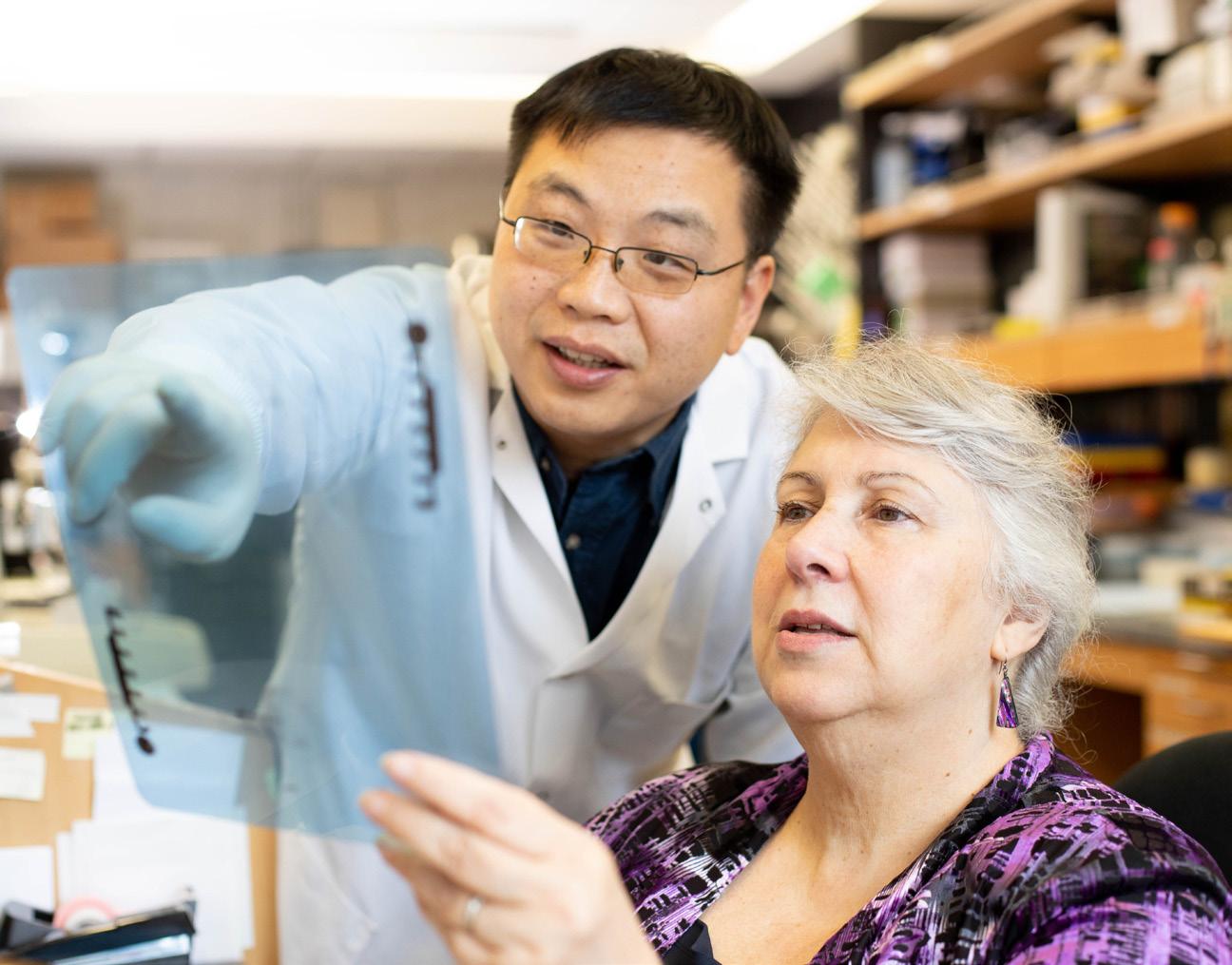

Professor Eileen White CANCAN co-team lead, Rutgers Cancer Institute of New Jersey, US

Our progress | 35
Thea Tlsty and professional researcher Deng Pan
Taking aim at cachexia
Cachexia, a wasting syndrome that often accompanies advanced cancers, is characterised by extensive weight loss from both skeletal muscle and fatty tissue, and cannot be reversed by nutritional therapy. Cachexia is often accompanied by fatigue, broad organ and tissue dysfunction, and greatly diminished quality of life. Additionally, cachexia limits patients’ ability to receive systemic cancer therapies and is associated with poor prognosis. Despite these major clinical implications, relatively little is known about cachexia, and no effective therapies are available to treat the condition.
To understand and treat this currently intractable condition, as part of the Cachexia challenge, the CANCAN team is identifying tumour-secreted factors that strongly influence cachexia development. Central to this effort is a comprehensive pipeline of validation models of lung, pancreatic and colorectal cancers, including genetically engineered mouse models, organoids and Drosophila models. These models are paired with clinical studies profiling cancer cachexia in patients.
“Describing cancer cachexia is not sufficient to identify the cause,” says CANCAN co-team lead Eileen White of the Rutgers Cancer Institute of New Jersey, US. “One needs to identify the mechanisms driving cachexia and target that for therapy, because treating the symptoms has not been successful. If we can inhibit the core mechanisms driving cachexia – either with targeted drugs or diet – in these models, then that would be evidence and justification to try the approach in humans.”
As part of their approach, CANCAN researchers are using data from TRACERx, a major programme funded by Cancer Research UK that profiles patients’ progression
from lung cancer diagnosis to cure or relapse, and PEACE, a study in which patients donate samples posthumously to support the study of cancer cachexia development. The team is mining data from these studies to identify patients who lose total body weight, skeletal muscle mass and white adipose tissue – the predominant type of fat in the human body – and to discover factors that predict subsequent cachexia. The CANCAN team has also incorporated large clinical studies to characterise cachexia in people with cancer in detail to uncover clinical subtypes critical for directing the right treatment to the right patient, and to complement the TRACERx work.
The OPTIMISTICC team is funded by Cancer Research UK with generous support from Nick and Annette Razey
The PRECISION team is funded by Cancer Research UK and the Dutch Cancer Society
The STORMing Cancer team is funded by Cancer Research UK
The CANCAN team is funded by Cancer Research UK and the National Cancer Institute

“Together, these programmes make an incredible resource of tissue, following patient samples from diagnosis through death, which is hugely valuable for our team, as it looks at cachexia in the later stages of disease,” says Eileen. Importantly, a deeper understanding of cachexia would enable the development of novel interventions that improve patients’ responses to treatment, quality of life and ultimately survival.
Featured publications
Hong Y et al. J Pathol 2022; doi: 10.1002/path.5820
Almekinders MMM et al. npj Breast Cancer 2022; doi: 10.1038/s41523-021-00232-w
36 | Cancer Grand Challenges | Discover: A year of scientific creativity
Together, these programmes make an incredible resource of tissue
“
Eileen White CANCAN co-team lead
Perspectives from a Cancer Grand Challenges partner, with Ryan Schoenfeld, CEO
of The Mark Foundation for Cancer Research
We’re proud to have a global network of partners who share our vision to fund research that tackles cancer's toughest challenges, so that we can ultimately change outcomes for patients. The Mark Foundation for Cancer Research supports two Cancer Grand Challenges teams – SPECIFICANCER and NexTGen.

Although the medical community has made great progress, cancer continues to plague humankind. The stealth and devastation of cancer is daunting, and the end goals often feel beyond reach. Fortunately, The Mark Foundation for Cancer Research is dedicated to tackling those challenges. Recognising the obstacles that prevent scientific advances from improving patient outcomes, The Mark Foundation maintains a nimble, high-impact approach to funding cancer research that bridges the gap between bench and bedside. To fulfil their mission of accelerating research that will transform the prevention, diagnosis and treatment of cancer, the Foundation looks to support groundbreaking science around the world.
“The Cancer Grand Challenges program appealed to us on several levels,” says Ryan Schoenfeld, CEO of The Mark Foundation for Cancer Research. “First and foremost is the quality of the science –every cycle brings forward teams comprised of highly accomplished investigators with exciting new ideas to address some of the most important questions in cancer research. Second is the ambitious (grand) scale and scope of the program, taking on the big questions that require a large team approach to address. Third is the value we place on collaboration between funding organisations such as The Mark Foundation, Cancer Research UK and the National Cancer Institute; we enjoy working with, and learning so much from, our partners.”
After joining The Mark Foundation in 2018, Ryan was named CEO in 2022. During his time with the organisation, he has worked to establish The Mark Foundation as a leader in accelerating innovative cancer research across diverse areas of science, including molecular biology, chemistry, immunology, genetics and data science. The Mark Foundation for Cancer Research has been a critical partner to the Cancer Grand Challenges initiative since 2019, first supporting the SPECIFICANCER team in 2019 and then a second Cancer Grand Challenge team, NexTGen, in 2022. “We hope to see both projects lead to better treatments for cancer patients,” Ryan says. “For the SPECIFICANCER project, we are already seeing impact in the form of new hypotheses about patient responses to therapies that were enabled by the grant and now are being tested in the clinic. For the NexTGen project, our vision is that chimeric antigen receptor T-cell therapy for solid childhood cancers will be at the front line of treatment options within a decade, improving outcomes for children with the poorest prognosis and mitigating the toxicities of the current standard of care.”
Cancer Grand Challenges believes that to drive a steep change in cancer research, we need to be ambitious. When asked what he thinks is the key for advancing cancer research, Ryan cites “a combination of hard work, innovation, collaboration, fresh ideas and a boldness to take risks.” He explains, “Cancer Grand Challenges projects hold the potential to embody all those qualities in the drive to make a large impact in cancer research, ultimately for the benefit of cancer patients.”

Community stories
We hope to see both projects lead to better treatments for cancer patients
“
Our progress | 37
Ryan Schoenfeld CEO, The Mark Foundation for Cancer Research


38 | Cancer Grand Challenges | Discover: A year of scientific creativity Our global research community Baltimore San Diego Durham Houston Kansas Baton Rouge Dallas Montreal Seattle Vancouver San Francisco New New Brunswick Farmington Chicago Boston Washington DC Philadelphia Stanford Guelph
Learn more | 39 Dublin Birmingham Oxford Cardiff Glasgow Leeds Cambridge Manchester Zurich Barcelona Utrecht Berlin Paris Lyon Madrid Lausanne Amsterdam London York Rehovot
Our funders and partners


We
Founded
In

And a special thanks to Cancer Research UK's donors

The Kamini and Vindi Banga Family Trust
Nick and Annette Razey
Bjorn and Inger Saven
Please
Funding
Be
40 | Cancer Grand Challenges | Discover: A year of scientific creativity Get in touch
get in touch to discuss specific areas of work, partnering opportunities, publicising your work
opportunities to work together. info@cancergrandchallenges.org
or any
opportunities
the first
know
Join the conversation CancerGrandChallenges.org @CancerGrand @CancerGrandChallenges
to
about future rounds of challenges: sign up to our newsletter. cancergrandchallenges.org
our
to our global
would like to express
gratitude
network of partners who share our ambitions to make radical progress against cancer’s toughest challenges.
partnership with
by Partners

Learn more | 41
The last word
Cancer is a global challenge that no single scientist, institution or country can tackle alone. Cancer Grand Challenges breaks down the barriers that stand between scientists across the world and across disciplines, to drive the progress that we urgently need in cancer research.
I have found it inspiring to see the advancements that the Cancer Grand Challenges community has continued to make against some of cancer’s toughest challenges over the past year, highlighted by the stories in this edition of Discover. This progress would not be possible without the hard work of our dedicated international community of partners, donors, researchers, patient advocates and Scientific Committee
members – some of whom you have heard from in this publication.
The release of this edition of Discover coincides with the announcement of our brand-new set of challenges. It has been a privilege for me, as chair of the Cancer Grand Challenges Scientific Committee, to work with the global research community and people affected by cancer to identify the obstacles that continue to stand in the way of progress.
We hope you are as excited as we are to see what the future holds for Cancer Grand Challenges as we move into our next round of funding and dare new global teams to think differently and tackle more of cancer’s toughest challenges.
 Professor Sir David Lane Chair, Cancer Grand Challenges Scientific Committee

42 | Cancer Grand Challenges | Discover: A year of scientific creativity
Thank you to everyone who contributed to this publication.
Acknowledgements
Writing, editing and designing
Scott Edwards
Maya Shani
Bethan Warman
Rebecca Martinho
Contributors
Ludmil Alexandrov
Gemma Balmer-Kemp
Fariba Behbod
Catherine Bollard
Paul Brennan
Josephine Bunch
Lorenzo de la Rica
Steve Elledge
Emily Farthing
Rosalyn Flower
Wendy Garrett
Sarah Gosling
Margaret Grayson
Kevin Haigis
Owen Harris
Nic Jones
Nuria Lopez-Bigas
Paul Mischel
Philip Quirke
Owen Sansom
Sheona Scales
Amy Schade
Ryan Schoenfeld
Cynthia Sears
Charles Swanton
Thea Tlsty
Sara Wakeling
Jelle Wesseling
Eileen White
Jack Woodcraft
Mariia Yuneva
Image credits
Where required, image credits have been provided alongside the relevant images.
Learn more | 43
Cancer Research UK 2 Redman Place London E20 1JQ cancergrandchallenges.org
Registered charity number
England and Wales: 1089464
Scotland: SCO41666
Isle of Man: 1103

© Cancer Grand Challenges 2023 Cancer Grand Challenges

