

Meet the teams driving progress against cancer’s toughest challenges
A growing global community of 600+ investigators
9 bold new challenges – each with the potential to transform outcomes for people with cancer


Welcome to the first edition of Discover, our annual progress publication for Cancer Grand Challenges, celebrating how our teams are coming together, thinking differently and working towards overcoming cancer’s toughest challenges.
The 12 months covered in this report, April 2020 to March 2021, against the backdrop of a pandemic, were a year like no other. Yet despite the many difficulties posed by the pandemic – including lab closures, delayed trials and some of our clinical scientists returning to the healthcare frontlines – our teams determinedly continued to forge ahead. With this publication, we’re proud to celebrate our researchers’ continued advances, including improving breast cancer risk prediction; refining tumour metabolic maps to identify novel therapeutic targets; identifying microbiological signatures of colorectal cancer; sparking new debate about the events leading to cancer; elucidating the role of matrix remodelling in the chronic inflammation linked to cancer; producing detailed 3D maps of the entire tumour landscape; and unravelling the complex networks driving the tissue specificity of cancer.
Dr
David Scott Director, Cancer Grand Challenges, Cancer Research UK
Dr Dinah Singer
Deputy
Director
Development,
for Scientific Strategy and
National Cancer Institute, US
The teams’ collective efforts represent important steps forward in solving their Cancer Grand Challenges – six of the toughest problems in cancer research that, if solved, could radically change how we think about the disease.
The past year has also seen major milestones for Cancer Grand Challenges. Our partnership between Cancer Research UK and the US National Cancer Institute, launched in August 2020, has elevated the initiative to a new level. With the National Cancer Institute on board, we’ve been able to broaden our ambitions and increase our funding capacity in each round of challenges to four teams, each receiving £20 million.
In October, we challenged the research community to consider a new set of nine tough obstacles in cancer research. We were delighted that nearly 170 teams from 60 countries submitted bold ideas outlining how they would take these challenges on. A shortlist of 11 teams have received seed funding, and we’re now eagerly awaiting their full applications. Winning teams will be announced in March of 2022 – you can read more on page 22.
Through Cancer Grand Challenges, our community of diverse global teams continues to transcend national boundaries and disciplines in applying new thinking to problems that have long hindered progress. We hope you enjoy the stories we’ve picked for this edition of Discover, showcasing the power of global collaboration and how our teams continue to drive high-impact science and make important strides in dismantling research barriers.

Solving cancer’s toughest challenges demands tenacity, creativity and worldwide collaboration. No single researcher, institution or scientific discipline can solve these problems alone.
Cancer Grand Challenges unites a global community of researchers, patient advocates, funders and partners to make radical progress against cancer.
Cancer Grand Challenges is a global funding initiative that supports diverse world-class teams coming together and thinking differently to overcome cancer’s toughest challenges. Addressing these challenges – such as longstanding gaps in the understanding of complex biological phenomena and a lack of evidence to guide improvements in global public health – may provide the key to achieving radically different outcomes for people with cancer.
Co-founded in August 2020 by Cancer Research UK (CRUK) and the National Cancer Institute in the US, Cancer Grand Challenges builds on the success of CRUK’s Grand Challenge and currently supports seven worldclass teams in nine countries. These teams are already making impressive strides, bolstered by the support of funders and partners who believe in the power of global scientific collaboration to tackle large problems in science.

Global teams transcending boundaries

New findings define clinical criteria that may distinguish between patients with harmless breast carcinoma and those with a higher risk of invasive cancer.
Team: PRECISION (PREvent ductal Carcinoma In Situ Invasive Overtreatment Now)
Challenge:
Lethal vs nonlethal cancers: distinguish between lethal cancers that need treating and non-lethal cancers that don’t
To identify the factors associated with DCIS and IBC risk, the team focused not on the lesions themselves but on the surrounding adipose tissue. Obesity and enlarged adipocytes (fat cells) in the breast have been associated with DCIS. However, the role of adipose tissue in the development of IBC in people with DCIS had not previously been explored.
We’re moving closer to defining the very specific criteria that could change the way we treat
DCIS in the clinic.
Jelle Wesseling PRECISION team lead, Netherlands Cancer Institute
Although breast cancer screening has been crucial in diagnosing more people in early disease stages, when treatment is more than three times more likely to be successful, it has also greatly increased the diagnosis of ductal carcinoma in situ (DCIS). DCIS was rarely diagnosed before the era of screening but now accounts for approximately onequarter of the 2.3 million breast cancers diagnosed each year. In most people, the lesions will remain harmless, but some people will develop ipsilateral invasive breast cancer (IBC).
Because of the inability to identify which people with DCIS will develop IBC, treatment is recommended for all cases. Each year, many thousands of people undergo needless surgery, radiation and hormone therapy, accompanied by unnecessary stress, adverse effects and anxiety. To avoid overdiagnosis and overtreatment of DCIS, there is an urgent need for prognostic markers to distinguish between DCIS that will remain indolent and DCIS that may progress to breast cancer.
Meeting this need is the central goal of the PRECISION team, which is working to identify the features and molecular players involved in the progression of DCIS to IBC, and to develop tools that can distinguish harmless from potentially hazardous DCIS and ultimately prevent overtreatment. The team uses a multidisciplinary approach to solve the highly complex puzzle of DCIS progression.
The study’s lead author, Mathilde Almekinders, then a PhD student on the PRECISION team, had the idea of studying the adipocytes surrounding the breast ducts. “Despite being able to influence the behaviour of their neighbouring cells, adipocytes are not typically the focus of breast cancer studies,” Mathilde explains. “But fat tissue is a major component of the breast. It’s not just a storage organ – it has many functions, including in the endocrine and immune systems.”
As reported in npj Breast Cancer, the team used digital pathology to analyse breast adipocyte features in a nested case-control study of patients diagnosed with primary DCIS who had different health outcomes. A combination of both large breast adipocytes and high expression of the COX-2 protein, rather than smaller adipocytes and low COX2 expression, was associated with a 12-fold-higher risk of subsequent invasive IBC.
“Findings like these bring us closer to solving our Cancer Grand Challenge and our vision of understanding harmless versus hazardous disease,” says Jelle Wesseling, PRECISION team lead, based at the Netherlands Cancer Institute. Next, the team will crossvalidate these findings in cohorts, including their own and those from the STORMing Cancer team.
Breast adipocyte size is associated with ipsilateral invasive breast cancer (iIBC) risk after DCIS. Left: DCIS surrounded by large breast adipocytes from a 68-year-old woman who developed subsequent iIBC. Right: DCIS surrounded by small adipocytes from a 52-yearold woman. No iIBC occurred during follow-up. Image credit: Mathlide Almekinders
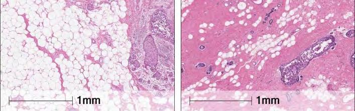

A major focus of the PRECISION team has been uncovering the poorly understood, complex biology of DCIS by developing new models to uncover the mechanisms associating DCIS and cancer risk. Adipocyte size and COX-2 expression are just one piece of the complex DCIS puzzle. The team has also demonstrated that expression of the biomarkers HER2 and ER can help pathologists classify DCIS grade more accurately. In addition, the team is developing and validating artificial-intelligence algorithms to assess the calcifications detected in mammography screens and determine which are associated with low-risk versus high-risk DCIS.
The team’s efforts collectively represent a major step towards solving the challenge of DCIS overtreatment. The findings may eventually help doctors provide science-based recommendations, and empower patients and clinicians to make informed decisions regarding care.
“For people with DCIS, there has to be a way to avoid such heavy medical interventions without compromising on an excellent health outcome,” says Jelle. “I hope we’ve found it.”
“
We hope to communicate a new way to identify people with low-risk DCIS who may be able to avoid treatment and people who may need to be watched more carefully.
The PRECISION team ultimately aims to develop the first risk-prediction model for ductal carcinoma in situ (DCIS). If validated, this model could eventually enable the de-escalation of treatment for people with low-risk DCIS –allowing thousands of people to safely adopt a ‘watch and wait’ approach to monitor their condition, while ensuring that highrisk individuals get the treatment they need.


Hidden treasure: do the clues to preventing and detecting colorectal cancer lie in the microbiome?
The identification and easy detection of microbiological signatures may make colorectal cancer screening available for all.
Team: OPTIMISTICC
(Opportunity To Investigate the Microbiome’s Impact on Science and Treatment In Colorectal Cancer)
Challenge: Microbiota: understand how microbes inside our bodies affect cancer treatment
Colorectal cancer is the third most common cancer worldwide: more than 1.8 million people are diagnosed each year, and cases continue to rise. Mounting evidence clearly shows that the disease is intricately linked to the microbiome: the trillions of bacteria, viruses and fungi that reside in the body. Clarifying this association would open a host of new opportunities for prevention, diagnosis and treatment.
The OPTIMISTICC team is taking on this immense challenge.
In two studies released in 2021 in Clinical Cancer Research and Genome Medicine, the team identified a unique bacterial signature associated with colorectal cancer, which was consistently found in stool samples from patients in four countries. This microbiome signature can be reliably detected in stool samples collected through an inexpensive cardbased method (guaiac faecal occult blood test, or gFOBT) and analysed through ribosomal-RNA sequencing.

Collectively, the findings not only hint at the existence of microbiome biomarkers for colorectal cancer but also may translate to a simple, effective and scalable screening test that could be used worldwide, including in underresourced settings. Such testing could be used to decrease the incidence of the disease and help more people get diagnosed earlier, when treatment is more likely to be successful.
Multidisciplinary perspectives enable breakthroughs
The OPTIMISTICC team has made breakthroughs by integrating the complementary perspectives of the quantitative and clinical sides of the team, led by co-investigators Curtis Huttenhower, a bioinformatician based at the Harvard TH Chan School of Public Health, US, and Phil Quirke, a pathologist based at the University of Leeds School of Medicine, UK.
“I tend to take a basic approach, using computational techniques to answer questions about microbial ecology,” says Curtis. “Phil’s side of the team brings a translational perspective – I wouldn’t necessarily have considered the route to screening without their input.”
Accordingly, the results reflect these dual perspectives: the team both identified bacteria associated with colorectal cancer and devised a simpler, less expensive approach to microbial testing that could enable broad initial screening of patients worldwide.
“There are two arms to these findings –one technical and one biological,” explains Curtis. “From a technical perspective, in the stool of people with early-stage colorectal cancer, this very broad gFOBT-card-based approach can detect microbial signals that are similar to much more detailed, deep shotgun metagenomic studies of people with late-stage disease. I’m actually kind of amazed that these signals are comparable. They’re not as rich – you’re not measuring the whole microbial community, or individual strains, or all the chemical and functional activities that you could with a higher-quality sample. But it’s a good first-pass screen to test whether someone is at an elevated risk of colorectal cancer.”
From a biological perspective, the findings reveal a shift in the microbiome early in disease development. This shift is seen in samples collected from a range of countries: the UK and US (with high incidence of colorectal cancer), Argentina

and Chile (with intermediate incidence), and Southern India and Vietnam (with low incidence but increasing cases).
“What’s interesting is that ‘normal’ microbiomes vary dramatically between these countries,” says Phil. “But a consistent story is emerging, independent of the microbiome’s starting point, highlighting a group of bacteria associated with colorectal cancer. We don’t know yet if there’s a causative link between the bacteria and the disease. But the association is good enough when we’re looking to improve screening and trying to identify people at risk.”
An adaptable tool for different national healthcare contexts
The team members operate in national healthcare contexts with different screening policies. The UK’s populationbased National Health Service Bowel Cancer Screening Programme decides who should be referred for colonoscopy by looking for traces of blood in stool samples. Although stool tests are available in the US, screening in the private healthcare system relies heavily on colonoscopy.
As Phil notes, “It’s less of a concern in the US, but colonoscopy is one of the biggest bottlenecks in the UK’s screening programme. Here, we’ve shown that microbiome analysis could provide a more effective screening approach if combined with looking for blood, which could help reduce the number of unnecessary colonoscopies while ensuring those who need one are referred.”
The findings also hold promise for use in under-resourced healthcare settings worldwide, such as in developing

“
It’s a fascinating story that may lead us to the causes and prevention of colorectal cancer.
Philip Quirke
OPTIMISTICC co-investigator, University of Leeds, UK
countries where colorectal cancer incidence is increasing and the screening infrastructure is limited.
“Freezing stool samples takes significant amounts of storage space; shipping on dry ice is expensive,” explains the studies’ first author, Caroline Young, a histopathologist based at the University of Leeds. “For these studies, we kept everything at room temperature – from sample collection in the pilot nations (Vietnam, Chile, Argentina and India) to shipment to and storage in the UK. The microbiome on the card remains stable enough for screening throughout. Our findings indicate microbiome analysis of gFOBT cards could offer a resource-light protocol, even in very hot countries with limited infrastructure.”
Looking forward, the team has clear plans to explore how their findings could translate to a wider screening programme that might make care accessible to all. The microbial biomarker pilot study will be expanded to examine whether detection of the signature is reproducible across additional populations. The UK screening programme recently moved from gFOBT to the faecal immunochemical test (FIT), a similar stool test with greater compliance and usage by the general population. Will the findings be consistent when this collection method is used? And could probing the microbiome signature reveal more information about the causes and early development of colorectal cancer?
“The question now is how we tie it all together,” Phil muses. “It’s a fascinating story that may lead us to the causes and prevention of colorectal cancer.”
Featured team members

Professor Philip Quirke OPTIMISTICC co-investigator, University of Leeds School of Medicine, UK

Dr Caroline Young OPTIMISTICC team member, University of Leeds School of Medicine, UK

Professor Curtis Huttenhower
OPTIMISTICC coinvestigator, Harvard TH Chan School of Public Health, Boston, US

Chronic inflammation is directly linked to up to 25% of cancer deaths worldwide. Could reprogramming of the inflamed microenvironment prevent, slow or even revert the cascade of inflammation to malignancy?
Team:
STORMing Cancer
(STrOmal ReprograMing provides new directions to prevent and revert chronic inflammationassociated cancers)
Challenge: Cancer causes: understand how lifestyle factors, such as inflammation, cause cancer
Certain factors – such as obesity, physical inactivity and chronic inflammation – are strongly associated with increased cancer incidence, but the underlying mechanisms remain to be clearly defined. For example, inflammation is usually a finely orchestrated reaction driving tissue repair after injury, but when it becomes chronic, it has long been known to create an environment conducive to many diseases, including cancer. Elucidating the mechanisms underlying this complex association would reveal precisely how inflammation influences disease development and recurrence. More importantly, this understanding could lead to new and improved approaches to prevent and treat inflammationassociated cancers, which are particularly lethal.
The STORMing Cancer team aims to solve this longstanding mystery by focusing on an overlooked aspect of chronic-inflammation-associated cancers (CIACs): how inflammation affects the stroma – the connective tissue that surrounds organs. The rationale for this focus is that stromal characteristics can influence the cell types into which stem cells differentiate and can cause non-cancerous cells to transform into cancerous cells. Importantly, placing a tumour cell on healthy stroma can slow tumour progression or even guide the cell back to a non-tumour state.
The team’s extensive work centres on the premise of reprogramming the stromal microenvironment, which evolves throughout the course of inflammation, providing increasingly fertile ground for tumorigenesis. Could reprogramming the stroma help treat
CIACs by guiding them back to a noncancerous or benign state? And could cancer be prevented in people with chronic inflammatory conditions, such as Barrett’s oesophagus or inflammatory bowel disease (IBD), by reversing the inflammation effects in the stroma?
“It seemed to me that reprogramming the stroma to control adjacent cancer cells could provide a more effective option than targeting the cancer cells themselves,” says Thea Tlsty, leader of the STORMing Cancer team, based at the University of California, San Francisco. “It’s a bold idea, but that’s exactly what Cancer Grand Challenges urges: a bold leap forward.”
Shifting perspectives: an early, active role of the ECM in inflammation
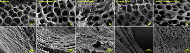
Chronic inflammatory conditions are associated with many changes in tissues, including the accumulation of immune cells and alterations in cell architecture, such as the degradation of the extracellular matrix (ECM), the network that surrounds and supports cells. These changes had been assumed to occur after inflammation develops and progresses. But members of the STORMing Cancer team have recently shown that the ECM undergoes distinct remodelling changes much earlier than originally thought – even before the effects of inflammation appear.
These findings came from a study coconceptualised by former PhD student Elee Shimshoni and group leader Irit Sagi at the Weizmann Institute of Science, Israel, and conducted in collaboration with STORMing Cancer co-investigator Uri Alon. The results, published in Matrix Biology, show how pioneer immune cells infiltrate the areas affected by IBD very early in the inflammatory process, bringing with them the remodelling enzymes MMP2 and MMP9, which degrade the basement membrane, a dense type of ECM important for structural support. Damage to this membrane exposes the immune system to resident bacteria in the colon, which can further drive inflammation.
“This correlates with a number of studies that have found ECM remodelling enzymes upregulated in human IBD,” explains Elee, the first author of the study. “But the surprising factor is that this occurs pre-symptomatically.”
Clinical and conceptual importance
The findings were observed in mouse models of two types of inflammation – a rapid, acute model of colitis (induced by the chemical dextran sodium sulfate) and a chronic inflammation model (based on interleukin-10 gene deficiency) that closely mimics IBD in humans – and were also validated in human samples. “This was surprising, as chronic and acute inflammation are very different processes,” Elee notes. “From an ECM standpoint, however, it doesn’t seem to matter if it will be chronic or acute –there’s something very similar happening to prepare the tissue for inflammation.”
Collectively, the findings have biological importance. “Conceptually, changes to the ECM are often seen as by-products of inflammation. Here, we show these changes are actively contributing to its progression,” Elee adds.
Clinically, early intervention is key for preventing the cascade of chronic inflammation into irreparable tissue damage. “The pre-symptomatic ECM has a unique signature distinguishing it from a healthy one: it’s less stiff, with a different composition of proteins and an overall altered structure. This could support early detection of IBD and lead to new, improved therapies.”

Cancer
“At the crux of what we’re exploring”
“Elee, Uri and Irit’s study is seminal, really quite beautiful and at the crux of what we’re exploring in the STORMing Cancer team,” says Thea.
STORMing Cancer is studying CIACs in four tissues: the oesophagus, stomach, colon and lung. The team’s unique experimental design enables the development of pioneering preclinical models as well as the ability to test potential therapeutics. As part of this effort, Elee, now a Cancer Grand Challenges postdoctoral researcher in the laboratory of STORMing Cancer co-investigator Don Ingber at the Wyss Institute for Biologically Inspired Engineering, US, is exploring the roles of stromal components in maintaining a healthy epithelium versus promoting neoplastic transformation and cancer progression, by using 3D co-cultures of samples from healthy, abnormal and cancerous regions of the same patient.
Given that up to 25% of all cancer deaths worldwide are a direct consequence of chronic inflammatory disease, a better understanding of the underlying mechanisms and changes triggered throughout the inflammatory process will be key to improving outcomes, leading to novel strategies for prevention, detection and treatment. By broadening the focus to include these very early structural changes, this study further highlights the potential for early stromal intervention, possibly before symptoms start.
“
Inflammationassociated cancers are among the most lethal cancer types in the world. This is what keeps me up at night, and why I’m so excited by the opportunity we have with Cancer Grand Challenges.
Thea Tlsty STORMing Cancer team lead, University of California San Francisco


US
At the core of Cancer Grand Challenges is a cohort of more than 200 early-career researchers helping drive progress against cancer’s toughest challenges. These researchers are based in nine countries and have expertise in areas as diverse as physics, bioinformatics and clinical research.
By empowering these researchers to transcend the boundaries of institutions, disciplines and geographies, Cancer Grand Challenges aims to develop a community of brave, daring future leaders who are willing to unite and challenge the status quo.
Bringing new perspectives to cancer genomics
Sergey Senkin, Mutographs team member, International Agency for Research on Cancer, France
After graduating from the Moscow Institute of Physics and Technology, I took part in CMS and ATLAS, two major experiments at CERN’s large hadron collider. During my PhD studies at the University of Bristol and later my postdoctoral work, my research focused on precision measurements of the topquark properties, as well as the search for the dark-matter particles.

While I have always considered fundamental science extremely important, I also wondered if I could use my skills to contribute to resolving global issues, like cancer. Cancer Grand Challenges presented a perfect opportunity. Cancer research seems a far cry from my background in high-energy and particle physics, but many of the computational methods of data analysis are similar. With cancer genomics in particular, we’re faced with computational tasks of ever-growing complexity. By bringing new perspectives to the field and combining them with the huge amounts of data produced, we can often find new patterns and results to help inform and answer pertinent questions.
Being part of this community has been extremely rewarding – the 2019 Mutographs team meeting in Lyon and the 2020 Cancer Grand Challenges summit in Windsor have been real highlights, bringing together hundreds of researchers for incredibly fruitful scientific discussions. Global collaboration like this is crucial for Mutographs and wider research. While it’s harder in a remote world, we continue to strive to achieve the best from our ongoing collaboration.
Sergey Senkin Mutographs team member “
Being part of this community has been incredibly rewarding.

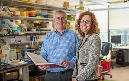
The value of good mentorship Amy Schade joined Karen Cichowski’s lab (Brigham and Women’s Hospital, Harvard Medical School, US) as a postdoctoral research fellow in 2019. Karen is Amy’s mentor, and both are part ofthe Cancer Grand Challenges SPECIFICANCER team.
Amy: Karen is a fantastic mentor who encourages me to pursue tough scientific questions and challenges me to fully delve into the complexity of the biology. Importantly, Karen gives me many opportunities to present my project at meetings and collaborate with other scientists. It has been great to learn a new scientific perspective from Karen, and I think it is helping me become a more well-rounded scientist.
Karen: Mentorship is one of the most enjoyable aspects of my job, providing robust multidisciplinary training in cancer research and helping my trainees achieve their professional goals. I firmly believe that one of our most important goals as a field is to provide equivalent opportunities for everyone. As a more senior member of the field, I work hard to support my junior colleagues in this way and, when necessary, remind my contemporary peers of our shared mission.
Featured team members

Dr Sergey Senkin Mutographs team member, International Agency for Research on Cancer, France

Dr Amy Schade
SPECIFICANCER team member, Harvard Medical School, US

Professor Karen Cichowski SPECIFICANCER co-investigator, Harvard Medical School, US
A deeper understanding of the metabolic changes during tumour development could radically change clinical decision-making and unlock opportunities to intervene across the entire patient journey.
to better understand tumour metabolism and make discoveries that could potentially improve all stages of the patient journey. In findings published in Nature Genetics in early 2021, the team identified a new therapeutic target for colorectal cancer, shedding light on the profound rewiring of tumour metabolism that occurs when APC and KRAS are concomitantly mutated.
Enabling unexpected insights: rethinking glutamine addiction in KRAS-driven colorectal cancer
Challenge:
3D tumour mapping: map the molecular and cellular tumour microenvironment to define new targets for therapy and prognosis
“We believe our pipeline could provide ‘Cancer Care 2.0’ –a novel suite of decisionmaking tools that could ultimately change outcomes for people with cancer.
Josephine Bunch
Rosetta
team lead, National Physical Laboratory, UK
Cancer cells exist in complex communities: each cell in a tumour is different from its neighbour and relies on its microenvironment to support its existence. These interactions are likely to contain new information on tumour behaviour, prognosis and treatment responses, and therapeutic targets waiting to be uncovered.
The 3D tumour-mapping challenge seeks to unravel this complexity by taking a holistic view of tumours as an organised population of cells. Rather than examining individual aspects in isolation, this view integrates genetic, molecular and biochemical information alongside the intricate intra-tumour architecture.
Taking on this challenge, the Rosetta team is establishing an organ-tosubcellular tumour map by using a range of mass spectrometry imaging (MSI) techniques, including matrix-assisted laser desorption/ionisation (MALDI), desorption electrospray ionisation (DESI) and rapid evaporative ionisation (REIMS). This pipeline of techniques can be used to image and correlate the distribution of molecules, genetic information and metabolic signatures in single cells, to produce a ‘Rosetta stone’ for cancer metabolism.
Over the past four years, the Rosetta team has advanced the pipeline beyond the original expectations. The team has made important technological breakthroughs and produced a sophisticated suite of tools that have elevated MSI from a promising research method to a fundamentally new way of analysing cancer.
Rosetta has now turned its collective attention towards studies with greater clinical focus, by using the MSI pipeline
Mutant KRAS is a pressing clinical problem that is found in as many as 45% of colorectal cancers and 95% of pancreatic cancers, and is often associated with poorer patient outcomes. Although this mutation has long been known to alter tumour metabolism by rendering cells ‘addicted’ to the amino acid glutamine to support their proliferation, no clinically relevant targets have emerged to date.
Using its unique approach for imaging metabolic heterogeneity, the Rosetta team has identified a potential metabolic target: the antiporter SLC7A5. The findings challenge the long-held assumption that the breakdown of glutamine fuels growth in these cells. Instead, the increased demand for bulk protein synthesis is supported by SLC7A5’s ejection of glutamine from the cell and the importation of other products of metabolism from the surrounding tissue. The team has also demonstrated that Slc7a5 is critical for tumour development in both early and late stages of Kras-driven colorectal cancer in mouse models.
Rosetta’s study advances the field beyond simply documenting the effects of KRAS mutations on tumour metabolism, through beginning to reveal the complex mechanisms underlying these changes. By pinpointing SLC7A5 as a promising target for treating KRAS-driven colorectal cancer, Rosetta’s findings could be transformative for people whose tumours have become resistant to other treatments. The team’s fundamentally new way of analysing cancer could be valuable throughout the course of detection and treatment, from screening and diagnostics to advanced therapies and follow-up monitoring.
The discovery, only one of many clinically relevant findings emerging from the Rosetta team, highlights the power of the 3D tumour-mapping platform and the novel insights that can be gained when tumour metabolism is viewed in this holistic way.

Rosetta team members Arafath Najumudeen (study first author; cancer biologist and postdoctoral researcher) and Chelsea Nikula (chemist and higher research scientist) discuss how multidisciplinary collaboration facilitated the discovery of SLC7A5 as a promising target for treating KRAS-driven colorectal cancer.
How has Rosetta’s collaborative nature contributed to these findings?
Arafath: Studying metabolism in vivo has been a challenge for the community, and we were excited to address this in the context of KRAS using the Rosetta imaging pipeline. Having close collaborations with the Rosetta team and expertise from our colleagues at NPL (National Physical Laboratory, London) meant that we could visualise glutamine in tissues and understand its role in KRAS-mutant tumour metabolism.
Chelsea: Having close collaborations with cancer biologists, data scientists, chemists and physicists within our Cancer Grand Challenges team brings unique perspectives to the complex datasets we produce and enhances the interpretation of our results. This means we gain a better understanding of our MSI data in relation to biological impact – which is crucial when forming hypotheses and conclusions.
How have you found being part of a Cancer Grand Challenges team?
Arafath: Challenging, rewarding, confusing and exciting all at the same time, but a wonderful opportunity to be part of a unique and international team. The diversity of the team helps us embed our different disciplines together for a strong partnership.
Chelsea: There was definitely a learning curve at first – of each other’s models, analytical techniques and methods of data processing and how to present data to each other to facilitate understanding and discussion. I find it very rewarding, both intellectually and personally. I enjoy engaging with experts from a variety of areas, learning from them while sharing my passion for MSI.
Without Rosetta’s imaging pipeline, we would never have been able to visualise the exchange of these metabolites or identified SLC7A5 as a potential therapeutic target.
Owen Sansom
Rosetta co-investigator, Cancer Research UK Beatson Institute, UK
Featured team members


Professor Owen Sansom
Rosetta coinvestigator, Cancer Research UK Beatson Institute, Glasgow, UK

Dr
Najumudeen Rosetta team member, Cancer Research UK Beatson Institute, Glasgow, UK
UK

UK
In addition, multiplexed error-robust fluorescence in situ hybridisation (MerFISH) can analyse the number of copies and the distribution of RNAs in single cells, and imaging mass cytometry can image the microenvironment by defining the spatial architecture of different cells.
A technological toolbox with broad biological applications
Team:
IMAXT (Imaging and Molecular Annotation of Xenografts and Tumours)
Challenge:
3D tumour mapping: map the molecular and cellular tumour microenvironment in order to define new targets for therapy and prognosis
When the 3D tumour mapping challenge was set in 2015, emerging single-cellanalysis technologies were beginning to reveal important information about the diverse cells within tumours. But in isolation, those methods miss important contextual information about the whole tumour landscape.
The IMAXT team, like Rosetta (page 14), is now advancing the challenge by evolving existing and novel analytical techniques, integrating their outputs and placing them within a 3D context to capture this missing whole-tumour information. The result is a detailed view of the community of cells that make up a tumour, including their precise locations, characteristics and behaviours –information that should enable important questions about tumour biology and evolution to be answered.
In January 2021, the IMAXT team added a new component, expansion sequencing (ExSeq), to this suite of tools and reported the methods in Science. ExSeq creates accurate, nanoscale-resolution maps of biological samples, annotated with localised expression information about thousands of genes, by combining two powerful techniques: expansion microscopy, which expands a biological sample to make it physically larger, and fluorescence in situ sequencing, which enables profiles of RNA expression to be determined in intact cells and tissues. Untargeted ExSeq enables unrestricted exploration of the entire landscape of RNA expression, uncovering unexpected information about gene expression and localisation, whereas targeted ExSeq enables a smaller, pre-defined set of genes to be examined at high resolution.
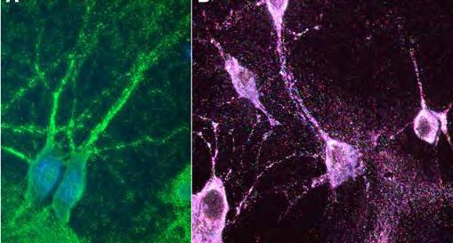
Despite having a major role in tumour mapping in the IMAXT programme, ExSeq was originally developed for use in neuroscience. The locations of RNAs within a neuron’s cell body or its thousands of dendrites and synapses can dramatically influence learning, memory and development. “Using untargeted ExSeq, we’ve revealed surprising information about neuron physiology,” says Dan Goodwin, IMAXT PhD student and co-lead author of the ExSeq study at MIT. “We’ve found elements in the dendrite that are only ‘supposed’ to be found in the cell body, like unspliced introns and long noncoding RNAs. This sort of data would normally be dismissed as an artefact, if it were from ground tissue put through a flow cell machine. But because we’re physically able to see their spatial distribution, we’re then able to use targeted ExSeq to explore it further.”
These regions are a characteristic of aggressive tumours that are rapidly outgrowing their surrounding vasculature, and their presence can influence treatment decisions. “Just by looking at gene expression in neighbouring cells, we can now identify potential hypoxic microenvironments,” says IMAXT PhD student Anu Sinha, co-lead author of the ExSeq study. “Seeing this tissue organisation was very exciting and something that would not have been possible without the ability to look at many genes in space.”
When integrated, these technologies appear to capture even more information than when used alone. But with this integration comes another major challenge: such rich data need to be presented in an understandable, interactive form that enables further investigation.


of biology involves spatial gene expression. Our aim was always that ExSeq could be used as a powerful tool, anywhere in biology.
Ed Boyden IMAXT co-investigator, MIT
Central to this approach is a suite of cutting-edge tumour mapping tools. For example, the team is optimising techniques such as serial two-photon tomography, which produces faithful 3D images of a biological sample.
“We’ve made much progress using multiplexed RNA imaging,” explains Shahar Alon, co-lead author of the study and group leader at Bar-Ilan University, Israel. “But these methods are limited in their spatial precision. With ExSeq, we bridge two worlds, achieving the precision of single-cell sequencing, with the added dimension of spatial coordinates.”
“Our aim was always that ExSeq could be used as a powerful tool anywhere in biology,” Ed Boyden, IMAXT coinvestigator at MIT and senior author of the study, adds. “All of biology – be that neuroscience, cancer, development, plant biology, viral transmission – involves spatial gene expression.”
After careful optimisation, ExSeq can now be used in diverse types of tissue, including brain specimens and breast tumours.
With the development and refinement of techniques such as ExSeq, the IMAXT team is already identifying and investigating unexpected features of the 3D tumour environment.
For example, pairing imaging mass cytometry with genomic data has revealed how the genome influences the physical tumour landscape. Applying Clonealign – a statistical method integrating DNA and RNA sequencing in single cells – to breast and ovarian cancer samples has revealed information not visible through either method alone, including dysregulated pathways in specific cell clones.
With ExSeq, the team has identified several novel avenues to explore, including oxygen-depleted (hypoxic) regions in human breast cancer tissue.
As technologies such as ExSeq mature, the team is exploring virtual reality as a new way to interact with and manipulate such rich data. The virtual lab, named Project Theia, can visualise both imaging and molecular data, and scientists can now not only fly through their data, virtually soaring through and between tumour cells, but also launch a series of analyses directly from the virtual environment. The interface is also designed for a multi-user mode, so that geographically distant collaborators can meet in the virtual lab to explore and analyse their data together. Theia v1.0 was released for testing in March 2021, and an updated version is due to be released later in the year.
Ultimately, through the integration of these techniques, explored in virtual reality, the team is developing an entirely new way to explore tumour biology, providing an unprecedented view of the architecture of human cancer and unlocking information that could lead to more effective preventive, diagnostic and therapeutic strategies.
“I liken our vision to putting a man on Mars,” muses IMAXT team lead, Greg Hannon. “There’s nothing that violates the laws of physics, but there’s so much technology you have to develop to do it. I’m proud of how far the team has progressed our aims over the past four years.”


The rules (not the exceptions) of cancer tissue specificity
The SPECIFICANCER team delves deeper into the biological laws that govern tissue specificity in cancer and reveals intricate webs of genetic interactions.
Team: SPECIFICANCER
Challenge:
Tissue specificity: understand why mistakes in certain genes cause cancer in only specific parts of the body
A cornucopia of cancer genetics data in recent years has helped identify the genes and mutations that propel malignancy. But many of these drivers, despite being broadly expressed throughout the body, often trigger cancer only in specific tissue types. The mechanisms underlying this tissue specificity in cancer remain mostly elusive.
The SPECIFICANCER team aims to clarify the complex mechanisms of tissue specificity, guided by an overarching philosophy that the tissue’s pre-existing characteristics – including its epigenetics, microenvironment and cell-cell interactions – dictate whether a mutated gene can drive tumorigenesis. If true, cancer drivers would be able to exert their effects only if these characteristics permit.

Central to the team’s aims is the development of the first functional atlas of tissue specificity, a comprehensive map of the cellular networks that allow drivers to initiate cancers in certain tissues but not others. By unlocking the molecular mechanisms underpinning tissue specificity in tumours, the team would provide a foundation for rational interventions and therapeutic choices made by physicians and people with cancer throughout the patient journey.
In a study recently published in Nature Communications, SPECIFICANCER unlocks new information about the tissue specificity of KRAS – which, despite being one of the most commonly mutated cancer-driving genes (oncogenes), drives only a few cancer types: lung, colorectal, pancreatic and endometrial cancers, and myelomas. KRAS is located at a critical signalling junction and, when its gene is mutated at one of four hotspots (codons G12, G13, Q61 and A146), it hyperactivates many downstream pathways, leading to changes in cellular behaviour.
Intriguingly, the activating alleles (copies of the genes carrying mutations at these hotspots that drive tumorigenic KRAS) vary significantly among these tissues. For example, the KRASG12R allele is almost exclusively found in pancreatic cancer, whereas the KRASA146T allele is rarely found in pancreatic cancer, but is fairly common in colorectal cancer and myeloma. Distinct alleles are also associated with varied responses to treatment and clinical outcomes in patients.
“How can we explain these epidemiological observations?” asks Kevin Haigis, SPECIFICANCER coinvestigator at Dana-Farber Cancer Institute, who co-led the study along with SPECIFICANCER colleague Peter Park.
“Our first hypothesis was that different alleles are found in different tissues, simply due to location-specific mutational processes.” For example, damage from reactive oxygen species is linked to the SBS18 signature in the colon and pancreas, which is strongly associated with the KRASG12C allele.
However, in bioinformatics explorations, the team found that this association did not hold true for most KRAS alleles.
“This told us two things,” explains Kevin. “Yes, mutational processes can contribute to tissue-specific alleles –but in most cases, there’s something else in the tissue that is acting as a biological driver.”

Co-mutation networks: a new layer of complexity
Each KRAS mutant allele, including those at the same hotspot, has slightly different biochemical, structural and signalling properties. Reasoning that these properties should cause each allele to behave differently, the team hypothesised that each must also interact with a distinct tissue-specific genetic network to drive cancer.
Analysing nearly 13,500 samples, across lung, colorectal and pancreatic cancers as well as myelomas, the team built a large map of genes with co-mutations, which help drive cancer when they are mutated alongside certain KRAS alleles but not others.
“Cancer is a cooperative process,” Kevin notes. “Each KRAS mutant has a slightly different function and acts within a distinct genetic environment, so it needs a different collection of mutational partners to drive tumour development.”
Intricate webs: the rule, not the exception
The concept of co-mutations, beyond extending the basic understanding of cancer genetics and tissue specificity, could enable prediction of how individual people might respond to certain drugs.
For example, BRAF is another oncogene with distinct patterns of tissue specificity. Although BRAFdriven melanoma often responds well to the drug vemurafenib, no response is usually seen in people with BRAF-driven colorectal cancer. Kevin and the team reason that this differential response is due to BRAF’s tissue-specific comutation networks. Could a deeper understanding of the networks at play help guide treatment decisions for all oncogene-driven cancers? Similarly, could this information be harnessed to stratify patients into subgroups with cancers driven by different alleles and co-mutation networks, to design more effective clinical trials?
Our findings highlight that the complexity of tissue specificity is even greater than we thought.
Kevin
Haigis
SPECIFICANCER co-investigator, Dana Faber Cancer Institute
By beginning to pick apart these intricate tissue-specific webs, the SPECIFICANCER team reinforces their overarching hypothesis and moves a step closer to solving their Cancer Grand Challenge. “We’re not just trying to understand how oncogenes drive cancer, we’re trying to understand the context in which they function,” says Kevin. “This contextual complexity is one of the major deficiencies in our understanding of cancer biology, and it’ll be the key to effectively treating people with KRAS-driven and other types of cancers.”
The concept of comutations not only extends our basic understanding of tissue specificity, it could also transform our ability to predict how a person would respond to certain drugs.
Kevin Haigis SPECIFICANCER co-investigator, Dana Faber Cancer Institute
SPECIFICANCER


Professor Kevin Haigis SPECIFICANCER co-investigator, Dana-Farber Cancer Institute, US
Professor Peter Park SPECIFICANCER co-investigator, Harvard Medical School, US
New findings, old ideas: reviving a decades-old view on the causes of cancer
Surprising discoveries from the Mutographs team challenge the classical view that all carcinogens directly cause mutations and suggest that non-mutagenic agents play a greater role in tumour promotion than originally thought.
Team: Mutographs
Challenge:
Unusual mutation patterns: discover how unusual patterns of mutation are induced by different cancercausing events
Dangerous levels of exposure to a carcinogen can elicit specific patterns of DNA damage, both genetic and epigenetic. If a tumour eventually develops, these mutational signatures can be used to work backwards to identify exposure to both known and previously unknown carcinogenic agents. Many mutational signatures exist whose origins are not yet known. Understanding the mechanisms that lead from an initial injury to mutation might hold promise in uncovering new preventive targets to stop, delay or weaken carcinogenic effects.
The Cancer Grand Challenges Mutographs team is tackling this challenge, uniting a global, multidisciplinary team doing transformative work at the intersection of epidemiology and genomics. By studying the mutational signatures in the genomes of cancer cells versus normal cells, the team aims to comprehensively catalogue the mutational processes that cause cancers in humans, understand their causes and apply this knowledge to cancer prevention.
“We’ve only very recently entered the era where all this is possible,” says Mike Stratton, Mutographs team lead at Wellcome Sanger Institute, UK. “The first human genome was only sequenced in 2009 – but to do epidemiological studies, tens or hundreds or thousands of genomes are required. Outstanding recent technological advances in sequencing methodology mean it is now possible to sequence tens of thousands of genomes in one study – and that is what makes Mutographs possible.”
Validating approaches and challenging classical hypotheses
One arm of the Mutographs team aims to identify, characterise and understand the biological processes underlying mutational signatures by using animal models. In late 2020, in Nature Genetics, the team published their results, which unexpectedly suggest an alternative view challenging the classical view that carcinogens directly cause mutations.
Led by Allan Balmain with colleagues David Adams and Laura Riva, the team worked with the US National Toxicology Program (NTP) to conduct the first survey of a broad range of chemicals that were known or suspected environmental carcinogens in mouse models that accurately represented exposure. Most of the agents in the NTP sample collection were expected to be mutagens – one such chemical was trichloropropane (TCP), which is often present in drinking water near toxic-waste sites. Some people living in areas where the groundwater has been contaminated have high TCP exposure.
However, what the team discovered was surprising. Only three of the 20 compounds tested had clear evidence of mutagenic activity, inducing tumours with more mutations and distinct mutational signatures. These three compounds included TCP. “This validates the animalmodel component of the Mutographs programme,” explains Allan. “We’ve shown you can use genome sequencing to link carcinogens to a molecular signature of exposure.”
Interestingly, even though most of the chemicals did not have a mutational signature specific to exposure, they did accelerate tumour development. The chemicals appeared to promote the transition of spontaneously mutated cells in mice from a dormant state to actively growing lesions, and consequently increase tumour incidence. These results align with findings from a study of oesophageal cancer in humans (to be published later this year) from another part of the Mutographs programme.
Mutations in cancer: revisiting an alternative side in the debate
“The concept of non-mutagenic promoting agents is not new –researchers were conducting experiments 70 years ago, which suggested some chemicals acted as promoters,” says Allan. “But it was phenomenology then, as they didn’t have the tools to interrogate mechanisms as we do now. We’ve repeated some of these experiments and applied our newer tools, and their interpretations were spoton – without even knowing what a gene was at the time.”
In the literature from the past decades, the debate regarding the roles of mutations in cancer has focused predominantly on mutations that either spontaneously occur in DNA or are induced by environmental agents. In 2015, a seminal article by Tomasetti and Vogelstein gave rise to the badluck hypothesis, which suggests that mutations are mainly caused by spontaneous errors in DNA replication. The debate since then has centred on whether these mutations are spontaneous replication errors or are induced by the environment.
But Allan, Laura, David and their team believe an alternative view is missing from the debate: the promoter
hypothesis. Perhaps another risk factor exists that is unrelated to the induction of mutations – for example, tumour-promoting factors might activate spontaneously or chemically mutated cells as the rate-limiting step in tumour formation. We know from epidemiological data that factors such as inflammation and obesity contribute to cancer in humans, but this relationship remains to be quantified and explored at a mechanistic level.* Could this contribution occur via tumour promotion?
The team’s data suggest that, in mice, the rate-limiting factor is the promotion, not the mutation. Now how this finding in mice translates to the human setting must be understood. “It certainly doesn’t negate any of the work that’s been done on mutational analysis, as mutations are clearly essential for tumours to develop,” Allan notes. But these findings could shift the debate towards exploring how other risk factors affect cancer incidence in humans, taking us back to basic cell biology to understand how these agents work and how they can cause single mutated cells to become active growing lesions.
*Note: Research exploring this relationship is ongoing and includes the STORMing Cancer team, which is studying how chronic inflammation drives cancer (see page 10).

“
Our data demonstrate carcinogenesis is a complex process not fully explained by mutagenic processes alone.


A new round of challenges posed to the global research community
Identifying Cancer Grand Challenges is a rigorous grassroots process that uses workshops, consultation and debate to solicit ideas from researchers, the public and people affected by cancer. The Cancer Grand Challenges scientific committee then distils these thoughts into tangible challenges –intractable problems that, if solved, could enable radical progress in cancer research.
In October, we launched a new round of nine challenges and were delighted to receive so many submissions in pursuit of solving them, from nearly 170 diverse global teams spanning more than 60 countries. These teams unite more than 1,960 investigators bringing broad perspectives including oncology, cardiology, chronobiology, evolutionary biology, mathematical modelling and bioengineering.
The scientific committee was tasked with narrowing the 169 innovative ideas down to a shortlist of just 11 teams, which have now received seed funding to develop their ideas into full proposals. The winners will each receive up to £20 million, an amount giving researchers the freedom to come together, think differently and unleash their scientific creativity against some of cancer’s toughest challenges.
11 global teams competing for a share of £80m
of the toughest challenges in


E-cigarettes
Dinah Singer Deputy Director for Scientific Strategy and Development, US National Cancer Institute “
The 11 shortlisted teams now have the opportunity to further develop their vision to advance bold cancer research. I’m looking forward to seeing their full applications.
teams Early 2022 winning teams announced 147 investigators 18 countries
Understand and reverse cachexia and declining performance status in cancer patients

Extrachromosomal DNA
Understand the biology of ecDNA generation and action and develop approaches to target these mechanisms in cancer
Identify and target dormant cancer cells
Determine the potential benefits and risks of e-cigarette use

Inflammation
Determine how inflammation causes cancer

Macromolecules
Systemically deliver macromolecules to intracellular targets for therapeutic benefit in cancer

Normal phenotypes
Understand how cells and tissues maintain ‘normal’ phenotypes whilst harbouring oncogenic mutations and how they transition to become a tumour

Senescence
Understand and exploit senescence to improve cancer treatment

Solid tumours in children
Develop novel therapies to target unique features in solid tumours in children


I had the pleasure of stepping into the role of chair of the Cancer Grand Challenges scientific committee this year, having sat as a committee member since the initiative’s early origins. And what a year it has been, with new donors and partners coming on board, a new set of challenges for the global research community to consider and nearly 170 applications submitted in pursuit of solving them.
The most exciting thing for me has been the extraordinary science that has continued to pour from the teams – vital new information about the biology of cancer, new leads for novel therapies and discoveries that challenge the current dogma. We have a community of very dedicated, hardworking people uniting across disciplines and countries,
Chair, Cancer Grand Challenges Scientific Committee