A
B
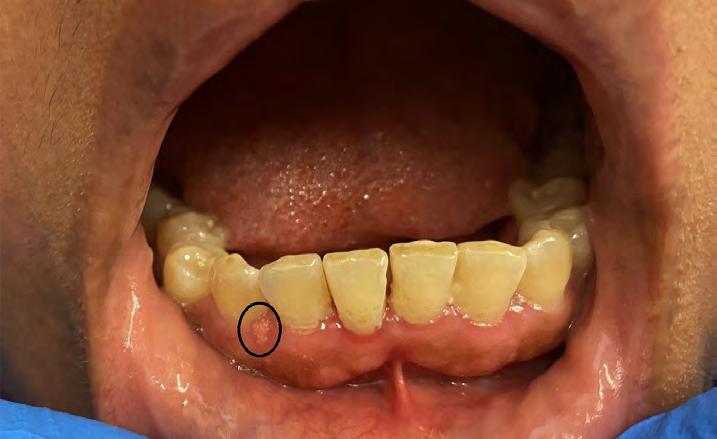
Figure 1: Faintly yellow lesion with corrugated surface on facial gingiva of anterior mandible.
The color ranges from pink, yellowish-red, to red, which prompts a wide spectrum of differential diagnoses, such as papilloma, leukoplakic lesions or Fordyce granules.[6] A biopsy is thus warranted for definitive diagnosis. While the majority of verruciform xanthomas occur as isolated lesions, concurrent association with a compromised immune system or a number of inflammatory disorders, such as lichen planus, discoid lupus erythematosus, graft-versus-host disease, or pemphigus vulgaris, has also been reported.[7] Microscopically, verruciform xanthoma is characterized by a hyperkeratotic verrucous or papillomatous surface with elongated, uniform rete pegs.[8-9] Parakeratin, often described on routine microscopy as characteristically orange in color, can be seen filling in the clefts between the epithelium projections.[3] Within the papillae of the connective tissue, foamy macrophages (or xanthoma cells) are characteristically noted.[1] Treatment for verruciform xanthoma consists of simple excision, with excellent prognosis and rare recurrence.[1,2] Conclusion The gingiva is a site that can be affected by a variety of oral pathologies, and chairside diagnosis frequently presents a challenge for the clinician. A biopsy is often necessary in rendering a definitive diagnosis for gingival lesions. p The authors deny any conflicts of interest. Nor did their study receive any commercial funding. Queries about this article can be sent to Dr. Peters at smpeters1@geisinger.edu.
Figure 2. (A) Papillomatous proliferation of epithelium, 100x (hematoxylin-eosin); (B) Focally acanthotic squamous epithelium and numerous foamy histiocytes in connective tissue papillae, 200x (hematoxylin-eosin).
REFERENCES 1.
2.
3.
4. 5.
6.
7. 8.
9.
Belknap AN, Islam MN, Bhattacharyya I, Cohen DM, Fitzpatrick SG. Oral verruciform xanthoma: a series of 212 cases and review of the literature. Head and Neck Pathology 2020;14(3):742–748. https://doi.org/10.1007/s12105-019-01123-0. Philipsen HP, Reichart PA, Takata T, Ogawa I. Verruciform xanthoma—biological profile of 282 oral lesions based on a literature survey with nine new cases from Japan. Oral Oncology 2003;39(4):325–336. https://doi.org/10.1016/s1368-8375(02)00088-x. Baig FAH, Luqman M, Vij H, Ibrahim M. Oral verruciform xanthoma of lateral border of tongue – a sheep in wolf’s clothing. Journal of Stomatology, Oral and Maxillofacial Surgery 2019;120(5):480–482. https://doi.org/10.1016/j.jormas.2018.12.002. Harris L, Staines K, Pring M. Oral Verruciform Xanthoma. Case Reports 2015(mar27 1). https://doi.org/10.1136/bcr-2014-209216. Hegde U, Doddawad VG, Sreeshyla HS, Patil R. Verruciform xanthoma: a view of the concepts of its etiopathogenesis. Journal of Oral and Maxillofacial Pathology 2013;17(3):392. https://doi.org/10.4103/0973-029x.125205. Gannepalli A, Appala A, Reddy L, Babu DBG. Insight into verruciform xanthoma with oral submucous fibrosis: case report and review of literature. Journal of Oral and Maxillofacial Pathology 2019;23(4):43. https://doi.org/10.4103/jomfp.jomfp_210_18. Singh HP.. Verruciform xanthoma of oral cavity: a case report. Journal of Clinical and Diagnostic Research 2014. https://doi.org/10.7860/jcdr/2014/8822.4590. Byakodi S, Kumar B, Patil S, Shinde S. Verruciform xanthoma of the tongue: case report and review of literature. Journal of Experimental Pathology 2021;2(2). https://doi.org/10.33696/ pathology.2.017. Tamiolakis P, Theofilou VI, Tosios KI., Sklavounou-Andrikopoulou A. Oral verruciform xanthoma: report of 13 new cases and review of the literature. Medicina Oral Patología Oral y Cirugia Bucal 2018. https://doi.org/10.4317/medoral.22342.
Khanh Trinh, D.M.D., is a resident, Division of Oral and Maxillofacial Pathology, Columbia University Medical Center, New York, NY. Daniel Nassimi, D.D.S., is a resident, Department of Oral and Maxillofacial Surgery, Woodhull Medical Center, Brooklyn, NY. Daria Vasilyeva, D.D.S., is a resident, Division of Oral and Maxillofacial Pathology, Columbia University Medical Center, New York, NY. Scott M. Peters, D.D.S., is associate professor, Oral and Maxillofacial Pathology, Geisinger Health System, Danville, PA.
The New York State Dental Journal . NOVEMBER 2023
21