6 minute read
Verruciform Xanthoma


Verruciform Xanthoma
Case Report and Literature Review
Khanh Trinh, D.M.D.; Daniel Nassimi, D.D.S.; Daria Vasilyeva, D.D.S.; Scott M. Peters, D.D.S.
ABSTRACT
Verruciform xanthoma (VX) is a benign mucosal or cutaneous proliferation typically found on the masticatory mucosa. VX usually affects patients in their fifth to sixth decade of life. VX is treated with simple excision and has a low chance of recurrence after surgery.
Verruciform xanthoma is an uncommon benign mucosal or cutaneous proliferation seen most frequently in adults in the fifth to sixth decade of life. It has a slight male predilection in patients under 50 years old and a slight female predilection in patients over 50 years old. It most often occurs on the masticatory mucosa, where it presents as a sessile or pedunculated lesion with a granular or pebbled surface. VX is treated with simple excision and has a low rate of recurrence. Herein, we present a case of a VX occurring in a 41-year-old female.
Case Report
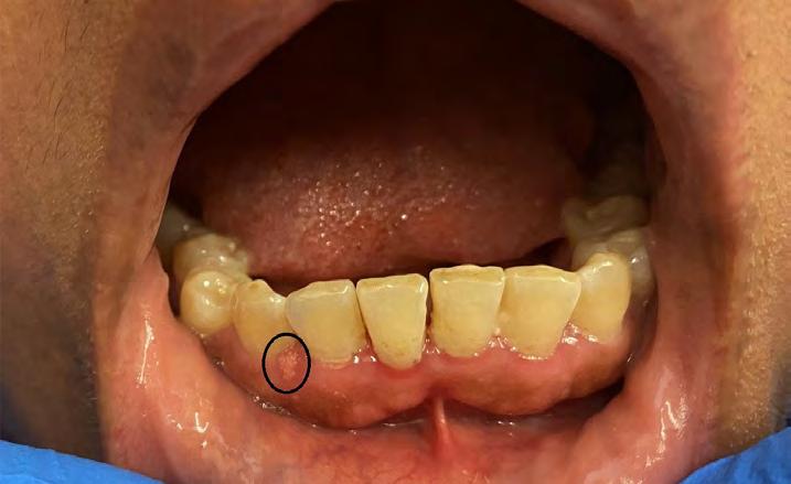
A 41-year-old female was referred to an oral surgeon at Woodhull Medical Center for evaluation of a painless pink lesion on the facial gingiva of the anterior mandible. Her medical history was significant for anemia and several orthopedic surgeries following a motorcycle accident two years prior. The review of systems was otherwise noncontributory, and the extraoral examination was within normal limits. Intraoral examination revealed a discrete, faintly yellow, pebbly lesion on the facial gingiva of the anterior mandible (Figure 1). The area was nontender and nonindurated. A periapical radiograph taken of the site was unremarkable.
For diagnostic purposes, the lesion was completely excised. Histologic examination revealed a papillomatous proliferation of acanthotic epithelium (Figure 2a), with overlying keratin and numerous foamy histiocytes in the papillae of underlying connective tissue (Figure 2b). Based on these findings, a diagnosis of verruciform xanthoma was rendered.
Discussion
Verruciform xanthoma is an uncommon, benign surface lesion with a poorly understood etiology. It is thought to be a benign reactive process, with proposed triggers including local trauma, irritation and altered immunological response.[1] The lesion most commonly occurs in the fifth to sixth decade of life. While in patients under the age of 50 years it is slightly more common in males, there is a slight female predilection in patients over 50 years old.[2]
The majority of verruciform xanthomas occur intraorally, with the most common sites of occurrence including masticatory mucosa (particularly gingival margin), hard palate and buccal mucosa.[1] Verruciform xanthomas have also been reported, albeit less commonly, at such oral sites as ventrolateral tongue or floor of the mouth.[3-5] Extraorally, the majority of cases involve the anogenital skin of the vulva, penis and scrotum.[2]
Clinically, verruciform xanthomas have a sessile or pedunculated base, and granular or pebbled surface.[1]


The color ranges from pink, yellowish-red, to red, which prompts a wide spectrum of differential diagnoses, such as papilloma, leukoplakic lesions or Fordyce granules.[6] A biopsy is thus warranted for definitive diagnosis. While the majority of verruciform xanthomas occur as isolated lesions, concurrent association with a compromised immune system or a number of inflammatory disorders, such as lichen planus, discoid lupus erythematosus, graft-versus-host disease, or pemphigus vulgaris, has also been reported.[7]
Microscopically, verruciform xanthoma is characterized by a hyperkeratotic verrucous or papillomatous surface with elongated, uniform rete pegs.[8-9] Parakeratin, often described on routine microscopy as characteristically orange in color, can be seen filling in the clefts between the epithelium projections.[3] Within the papillae of the connective tissue, foamy macrophages (or xanthoma cells) are characteristically noted.[1] Treatment for verruciform xanthoma consists of simple excision, with excellent prognosis and rare recurrence.[1,2]
Conclusion
The gingiva is a site that can be affected by a variety of oral pathologies, and chairside diagnosis frequently presents a challenge for the clinician. A biopsy is often necessary in rendering a definitive diagnosis for gingival lesions. p
The authors deny any conflicts of interest. Nor did their study receive any commercial funding. Queries about this article can be sent to Dr. Peters at smpeters1@geisinger.edu.
REFERENCES
1. Belknap AN, Islam MN, Bhattacharyya I, Cohen DM, Fitzpatrick SG. Oral verruciform xanthoma: a series of 212 cases and review of the literature. Head and Neck Pathology 2020;14(3):742–748. https://doi.org/10.1007/s12105-019-01123-0.
2. Philipsen HP, Reichart PA, Takata T, Ogawa I. Verruciform xanthoma—biological profile of 282 oral lesions based on a literature survey with nine new cases from Japan. Oral Oncology 2003;39(4):325–336. https://doi.org/10.1016/s1368-8375(02)00088-x.
3. Baig FAH, Luqman M, Vij H, Ibrahim M. Oral verruciform xanthoma of lateral border of tongue – a sheep in wolf’s clothing. Journal of Stomatology, Oral and Maxillofacial Surgery 2019;120(5):480–482. https://doi.org/10.1016/j.jormas.2018.12.002.
4. Harris L, Staines K, Pring M. Oral Verruciform Xanthoma. Case Reports 2015(mar27 1). https://doi.org/10.1136/bcr-2014-209216.
5. Hegde U, Doddawad VG, Sreeshyla HS, Patil R. Verruciform xanthoma: a view of the concepts of its etiopathogenesis. Journal of Oral and Maxillofacial Pathology 2013;17(3):392. https://doi.org/10.4103/0973-029x.125205.
6. Gannepalli A, Appala A, Reddy L, Babu DBG. Insight into verruciform xanthoma with oral submucous fibrosis: case report and review of literature. Journal of Oral and Maxillofacial Pathology 2019;23(4):43. https://doi.org/10.4103/jomfp.jomfp_210_18.
7. Singh HP.. Verruciform xanthoma of oral cavity: a case report. Journal of Clinical and Diagnostic Research 2014. https://doi.org/10.7860/jcdr/2014/8822.4590.
8. Byakodi S, Kumar B, Patil S, Shinde S. Verruciform xanthoma of the tongue: case report and review of literature. Journal of Experimental Pathology 2021;2(2). https://doi.org/10.33696/ pathology.2.017.
9. Tamiolakis P, Theofilou VI, Tosios KI., Sklavounou-Andrikopoulou A. Oral verruciform xanthoma: report of 13 new cases and review of the literature. Medicina Oral Patología Oral y Cirugia Bucal 2018. https://doi.org/10.4317/medoral.22342.
Khanh Trinh, D.M.D., is a resident, Division of Oral and Maxillofacial Pathology, Columbia University Medical Center, New York, NY.
Daniel Nassimi, D.D.S., is a resident, Department of Oral and Maxillofacial Surgery, Woodhull Medical Center, Brooklyn, NY.
Daria Vasilyeva, D.D.S., is a resident, Division of Oral and Maxillofacial Pathology, Columbia University Medical Center, New York, NY.
Scott M. Peters, D.D.S., is associate professor, Oral and Maxillofacial Pathology, Geisinger Health System, Danville, PA.