12 minute read
Use of a 3D Intraoral Scanner for Prosthetic Rehabilitation of Cocaine-induced Oronasal Fistula

Use of a 3D Intraoral Scanner for Prosthetic Rehabilitation of Cocaine-induced Oronasal Fistula
A Clinical Report
Anita Agarwal, B.D.S., D.M.D., M.P.H.; Timothy Levine, D.M.D.; Victor Badner, D.M.D., M.P.H.
ABSTRACT
A novel method using a 3D intraoral scanner (IOS) to overcome difficulties in obtaining conventional impressions to fabricate an obturator for a posteriorly located oronasal fistula secondary to chronic nasal cocaine use is described.
Cocaine abuse remains a constant challenge in the United States involving approximately 30% of drug abuse-related hospital emergency visits. Cocaine is an addiction promoting obstructive psychostimulant, and management is often difficult due to drug dependence and noncompliance. Intranasal use results in rapid absorption into the bloodstream, causing immediate euphoria, making snorting an extremely common route of intake. This may lead to severe ischemia and tissue destruction in the nasopharyngeal area, resulting in the formation of oral, nasal and palatal communications.[1-3]
Midline destructive lesions have a wide range of etiologies and clinical diagnosis is critical.[4-6] Although surgical treatment is the desired approach to close such defects, patient commitment to rehabilitation is imperative to achieve successful results. Often, there are contemporaneous medical and psychiatric comorbidities that complicate treatment and successful outcomes are dependent upon perioperative considerations.[7,8]
Traditionally, conventional dental impression materials have been used as the standard to capture defect architecture for fabrication of prostheses. Often, this is a difficult and unpleasant procedure for the patient, especially in the presence of a gag reflex. It can be a technique-sensitive procedure, entailing an exceptional degree of skill from the provider depending upon the extent and location of the defect. In addition, conventional impressions utilize a number of materials and processes, which makes them prone to distortion.[9]
Intraoral scanners (IOSs) have emerged as an alternative to conventional impression material for dentulous reconstructions. IOS systems eliminate the need for multistep procedures, minimize materials used and allow for immediate visualization and planning. Images can also be stored indefinitely and accessed at any time.[9,10]
In vitro and in vivo studies comparing various scanners have shown that intraoral scanning systems have a high degree of accuracy, trueness and precision of scan patterns. [9,11-14] In a comparative study of the accuracy of digital models obtained from conventional impressions versus intraoral scans, IOSs had acceptable clinical accuracy. The study suggests that intraoral scans may prove to be more accurate than conventional methods.[15]
Patients have reported a preference to scanning versus conventional impressions due to increased comfort and efficiency, thus supporting replacement of conventional impressions with intraoral 3D scans.[16-18]
Our report is based on the use of an IOS to obtain accurate impressions for fabrication of a prosthetic appliance.
Clinical Report
A 36-year-old patient was referred to the dental department for fabrication of an obturator due to oronasal perforation as a sequelae of chronic cocaine abuse for over 21 years. Surgical reconstruction was not an option because of continued cocaine use via nasal inhalation. Social history included current use of cocaine, marijuana, tobacco and alcohol. The past medical history included intermittent rhinorrhea, recurrent nasal infection, chronic migraines, psychiatric care for anxiety, osteoarthritis, back surgery, drug abuse, hip pain and steroid injections. Medication history included cyclobenzaprine, Xanax, Zyrtec and an extensive history of Percocet and oxycodone use. The patient presented with a surgical history of multiple sinus debridement, most recently six months prior to presentation. The nasal septum was collapsed, and there was a shift of the left eye. Patient wore a patch on the left eye during the clinical visit.


One week prior to presentation, the patient noticed a blister on the palate while eating and then proceeded to remove the overlying skin, which resulted in an oral-nasal communication. They denied history of any other maxillofacial trauma.
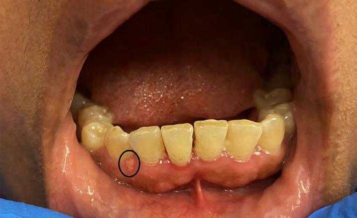
On initial presentation (Figures 1,2), the lesion was centrally located on the posterior soft palate in front of uvula and posterior to the junction of hard and soft palate. The fistula measured 6.7 mm anteroposteriorly and 4.0 mm transversely. The periphery of the lesion appeared red, soft and nonhealing. The patient was missing an upper right molar tooth, but the remaining dentition was intact. Patient reported no pain per Wong-Baker FACES Pain Rating Scale. However, they reported severe discomfort of 10/10 due to difficulty in eating, drinking and speech.
A collaborative effort between prosthodontics and orthodontics was used to determine the most comfortable and least invasive method for resolving the condition. Rehabilitation with the use of an obturator device as a definitive treatment was planned. The goal of the treatment was to improve the patient’s quality of life by providing comfort and restoring functions of speech, swallowing and mastication.[19,20,21]


Due to the posterior location of the defect and presence of adequate dentition, an IOS was used to obtain a digital imprint of the defect and surrounding areas. A scan of the maxillary arch (Figure 3) was obtained, and soft tissue was captured to approximately 1 cm beyond the fistula. The intraoral scan was then uploaded directly to the dental laboratory and the STL data was imported into the computer-assisted design (CAD) software.
Since no teeth were affected, a lower scan or bite registration scan was not required. The appliance was designed as a modified Hawley retainer with C-clasps at the second molars, ball clasps between the first and second premolars and full palatal acrylic coverage to obturate the lesion (Figure 4). The model was printed, and some soft-tissue inconsistencies were noted. These were manually corrected in the laboratory and a conventional obturator was fabricated.[22,23]
Six weeks later, on the day of insertion, it was noted that the lesion was larger (Figure 5), measuring 10.8 mm anterposteriorly and 5.2 mm transversely. The obturator was inserted; it covered the entire defect. Minimal adjustments were needed, with the fistula completely obturated (Figure 6). The patient was advised to return two weeks later for follow-up and adjustments.
The patient did not present for follow-up despite multiple contact attempts for over a year. The patient was unable to return due to COVID-19 and multiple personal tragedies. They reported not seeing another healthcare provider during that time. Upon return, the patient detailed wearing the obturator consistently throughout the past year and stopped using cocaine completely. Pain level was 0 on the Wong-Baker FACES Pain Rating Scale, and the patient reported a discomfort level of 3/10 due to food impacting between the prosthesis and lesion.
The patient stated: “This has been a big help to me. I am able to speak better, and people understand me better. I had trouble articulating B, Z and M sounds, and this has helped me retrain my speech.” The patient referenced a sentence they coined to evaluate their speech: “‘My dog’s name is bison, and he has babies.” The patient also felt the lesion had become larger, having noticed a gap between the appliance and defect, which resulted in food impaction in the space leading to difficulties during eating. Otherwise, the obturator was reported to function well.

At the re-evaluation visit (13 months post initial insertion), it was noted that the lesion size had increased considerably. A new scan (Figure 7) was obtained to accurately measure and document the lesion, which was found to be almost three-times the original size and measuring 18.8 mm anteroposteriorly and 7.7 mm transversely. The periphery of the lesion was non-erythematous. Although the appliance covered the posterior extent of the lesion, it did not fully obturate the depth of the defect. A soft-tissue reline material was used to reline the obturator and capture the depth of the defect (Figure 8) at the time of the patient’s return to the clinic. The patient reported much improvement in fit, function and plosive sounds (consonant sounds like /p/, /t/, and /k/, or voiced, like /b/, /d/, and /g/) were much clearer.

Discussion
The use of 3D IOSs can greatly increase patient comfort, decrease the number of visits, reduce tissue irritation and greatly increase ease of procedures.
Limitations include difficulty in capturing depth of the lesion accurately. This was remedied by clinical reline in the office, which is a procedure usually warranted at follow-up visits with conventional fabrication.

Summary
Intranasal use of recreational drugs can result in large, difficult-to-manage oronasal openings, which create functional difficulties requiring prosthetic rehabilitation when surgery is not an option. Uncorrected, these defects can severely affect a person’s quality of life.
With digital technology, a well-fitting device can be fabricated easily, and records of defects and treatment can be stored indefinitely for future reference. Further studies utilizing IOSs for scanning these types of defects are indicated for use in mainstream prosthetic dentistry.
The authors of this case report state they received no financial support and/or sponsorship and that they possess no conflicts of interest. Due to the case report nature of this manuscript Institutional Review Board approval or waiver was not required. Queries about this article can be sent to Dr. Agarwal at Anita.Agarwal@nychhc.org.
REFERENCES
1. NIDA. What is the scope of cocaine use in the United States? National Institute on Drug Abuse website. https://www.drugabuse.gov/publications/research-reports/cocaine/whatscope-cocaine-use-in-united-states. December 22, 2021. Accessed January 4, 2022.
2. Goodger NM, Wang J, Pogrel MA. Palatal and nasal necrosis resulting from cocaine misuse. Br Dent J 2005 Mar 26;198(6):333-4.
3. Nord GA, Rock A, Murphy FJ, Miloslavskiy I, Miller DJ, Wasserman BS. Prosthetic and surgical management of oronasal communications secondary to cocaine abuse. N Y State Dent J 2012 Jan;8(1):22-5.
4. Hofstede TM, Jacob RF. Diagnostic considerations and prosthetic rehabilitation of a cocaine-induced midline destructive lesion: a clinical report. Journal of Prosthetic Dentistry 2010-01-01;103(1):1-5
5. Westreich RW, Lawson W. Midline necrotizing nasal lesions: analysis of 18 cases emphasizing radiological and serological findings with algorithms for diagnosis and management. Am J Rhinol 2004;18:209-219.
6. Cregler LL, Mark H. Medical complications of cocaine abuse. N Engl J Med 1986; 315:14951500.
7. Tartaro G, Rauso R, Bux A, Santagata M, Colella G. An unusual oronasal fistula induced by prolonged cocaine snort. Case report and literature review. Minerva Stomatol 2008;57(4):20310.
8. Walker A, Joshi A, D’Souza A. Care of the cocaine user with nasal deformity. Facial Plast Surg 2017;33(4):411-18.
9. Abduo J, Elseyoufi M. Accuracy of intraoral scanners: a systematic review of influencing factors. Eur J Prosthodont Restor Dent 2018-08-30;26(3):101-121.
10. Kravitz ND, Groth C, Jones PE, Graham JW, Redmond WR. Intraoral digital scanners. J Clin Orthod 2014;48:337-347.
11. Nedelcu R, Olsson P, Nyström I, Rydén J, Thor A. Accuracy and precision of 3 intraoral scanners and accuracy of conventional impressions: a novel in vivo analysis method. J Dent 2018;69:110-118.
12. Ender A, Zimmermann M, Mehl A. Accuracy of complete- and partial-arch impressions of actual intraoral scanning systems in vitro. Int J Comput Dent 2019; 22(1):11-19.
13. Latham J, Ludlow M, Mennito A, Kelly A, Evans Z, Renne W. Effect of scan pattern on complete-arch scans with 4 digital scanners. J Prosthet Dent 2020;123(1): 85-95.
14. Cao Y, Chen JK, Deng KH, Wang Y, Sun YC, Zhao YJ. Accuracy of three intraoral scans for primary impressions of edentulous jaws. Beijing Da Xue Bao Yi Xue Ban 2020;52(1):129-137.
15. Tomita Y, Uechi J, Konno M, Sasamoto S, Iijima M, Mizoguchi I. Accuracy of digital models generated by conventional impression/plaster-model methods and intraoral scanning. Dent Mater J 2018;37(4):628-633.
16. Chalmers EV, McIntyre GT, Wang W, Gillgrass T, Martin CB, Mossey PA. Intraoral 3D scanning or dental impressions for the assessment of dental arch relationships in cleft care: which is superior? Cleft Palate Craniofac J 2016;53(5): 568-577.
17. Yuzbasioglu E, Kurt H, Turunc R, Bilir H. Comparison of digital and conventional impression techniques: evaluation of patients’ perception, treatment comfort, effectiveness and clinical outcomes. BMC Oral Health 2014 Jan 30;14:10.
18. Carneiro Pereira AL, Martins de Aquino LM, Carvalho Porto de Freitas RF, Soares Paiva Tôrres AC, da Fonte Porto Carreiro A. CAD/CAM-fabricated removable partial dentures: a case report. Int J Comput Dent 2019;22(4):371-379.
19. Goiato MC, dos Santos DM, Moreno A, et al. Prosthetic treatments for patients with oronasal communication. J Craniofac Surg 2011;22(4):1445-1447.
20. Ali MM, Khalifa N, Alhajj MN. Quality of life and problems associated with obturators of patients with maxillectomies. Head Face Med 2018 Jan 5;14(1):2.
21. Irish J, Sandhu N, Simpson C, et al. Quality of life in patients with maxillectomy prostheses. Head Neck 2009;31(6):813-821.
22. Devlin H, Barker GR. Prosthetic rehabilitation of the edentulous patient requiring a partial maxillectomy. J Prosthet Dent 1992 Feb;67(2):223-7.
23. Gay WD, King GE. Applying basic prosthodontic principles in the dentulous maxillectomy patient. J Prosthet Dent 1980 Apr;43(4):433-5.


Anita Agarwal, B.D.S., D.M.D., M.P.H., is faculty attending, Department of Dentistry/OMFS, NYC Health + Hospitals/Jacobi, and clinical assistant professor, Albert Einstein College of Medicine, Bronx, NY.
Timothy Levine, D.M.D., is director, orthodontics, and director, Congenital Craniofacial Care Center, Department of Dentistry/OMFS, NYC Health + Hospitals/Jacobi, and instructor, Albert Einstein College of Medicine, Bronx, NY.
Victor Badner, D.M.D., M.P.H., is chair, Department of Dentistry/OMFS, NYC Health + Hospitals/ Jacobi, and professor, Albert Einstein College of Medicine, Bronx, NY.